Embed Size (px)
Citation preview
American Journal of Advanced Drug Delivery
www.ajadd.co.uk
American Journal of Advanced Drug Delivery www.ajadd.co.uk
Original Article
Antioxidant and Antimicrobial Activities of Arnebia hispidissima
Bharat Singh*1 and Ram Avtar Sharma2
1Institute of Biotechnology, Amity University Rajasthan, Jaipur, India. 2Department of Botany, University of Rajasthan, Jaipur, India.
ABSTRACT
Objective: The present study aimed to the isolation and characterization of phytochemicals from tissue cultures of Arnebia hispidissima. The isolated compounds were evaluated for their antioxidant and antimicrobial activities. Materials and Methods: Six derivatives of alkannin and shikonin were isolated and characterized by using various physical and spectroscopic techniques from callus tissue of A. hispidissima. The callus tissue was generated by using Murashige and Skoog culture medium. The isolated compounds were assayed for DPPH-radical, reducing power, superoxide anion and metal chelating scavenging and antimicrobial activities by adopting well established methods. Results: The observed results revealed that deoxyshikonin showed maximum inhibition (290.20±0.440%) to DPPH-radical scavenging assay and shikonin demonstrated higher reducing ability (498.32±0.431%). Similarly, the propionylalkannin and acetylshikonin possessed moderate to maximum superoxide (345.45±0.632%) and metal chelating abilities (264.76±0,531%). Moreover, effective antimicrobial activity was also demonstrated by all the isolated derivatives of alkannin and shikonin. Conclusion: The results of present study suggested that the isolated compounds demonstrated modern antioxidant and high antimicrobial activities. Therefore, further investigations need to be carried out to isolate new phytochemicals from tissues cultures and screening for other different biological and pharmacological activities.
Keywords: Arnebia hispidissima, Antioxidant activity, Antimicrobial activity, Alkannin, Shikonin derivatives.
Date of Receipt- 07/03/2014 Date of Revision- 13/03/2014 Date of Acceptance- 21/03/2014
Address for Correspondence Institute of Biotechnology, Amity University Rajasthan, Jaipur 303002, India.
Tel. +91-1426 212139.
E-mail: bharatsingh217 @gmail.com
Singh et al______________________________________________________ ISSN 2321-547X
AJADD[2][2][2014]224-237
INTRODUCTION
Nature has been a rich source of phytomedicinal agents for thousands of years and an impressive number of modern drugs have been isolated from plants, many of them based on their use in traditional medicine. Plants are the basic source of knowledge of Ayurvedic and modern medicine.1,2 A. hispidissima occurs in northern tropical Africa, Egypt, Pakistan and drier part of Rajasthan, India. The roots of A. hispidissima used in treatment of boils, heart ailments, headache, and fever. The crude extract of floral parts traditionally known for tonic, diuretic and expectorant.3,4
The roots of Arnebia species contain mixture of naphthaquinones including derivatives of alkannin and shikonin.5-9 Shikonin and its derivatives were also investigated from tissue cultures in Arnebia species.10,11 These phytochemicals are potent pharmaceutical substances that showed significant biological activities including antioxidant12-14 and antimicrobial15,16. The main focus of our study was to isolation, characterization of phytochemicals from callus cultures of A. hispidissima and evaluation of their antioxidant and antimicrobial activities.
MATERIALS AND METHODS Plant materials
Arnebia hispidissima (Lehm.) DC. (Boraginaceae) was collected (February, 2011) from the local fields of Jaipur and botanical identification confirmed by Professor R.S. Mishra, Department of Botany, University of Rajasthan, Jaipur, India (Herbarium sheet no. RUBL 19441). The plants were grown in greenhouse of the Amity University Rajasthan, Jaipur and waited for maturity of seeds. The seeds were germinated in greenhouse to obtain the seedlings for tissue culture studies.
General experimental conditions The spectral data were obtained on the
following instruments: MS, Hewlett Packard HP 5972 A spectrometer equipped with 30 m long x 0.25 mm i.d. and HP5-MS capillary column; GC–MS, equipped with a HP 5933 data system, direct inlet at 70 eV; UV, Perkin-Elmer, model-200; 1H-NMR and 13C-NMR, Bruker AM 400 system recorded at 400, 200 and 50 MHz FT-NMR spectrometer; ESI-MS spectra at 3200 QTRAP LC/MS/MS mass spectrometer, an ion spray voltage +4, 500 V, declustering potential-50 V, entrance potential 10V. [α]D values were measured by Perkin-Elmer 341 polarimeter. Silica gel 60 (230–400 mesh; Merck) was used for column chromatography and silica gel G used for TLC and preparative TLC (Merck).
Tissue culture studies
The seedlings of A. hispidissima were surface sterilized with 0.1% (w/v) HgCl2 solution for 1.5-2 min and then rinsed 3-4 times with sterilized distilled water. These sterilized seedlings (3-4 mm in size) were then aseptically inoculated onto Murashige and Skoog17 culture medium and divided into two groups. In first group, the MS culture medium was supplemented with 0.5 mg/L indole-3-acetic acid (IAA) and 1.20 mg/L benzyl amino purine (BAP), 3.0% sugar and 150 mg/L activated charcoal for the isolation of alkannin derivatives while in the second group, the explants were cultured onto MS medium supplemented with 5.0 mg/L 2, 4-dichlorophenoxyacetic acid (2, 4-D) + 2.50 mg/L BAP and 3.0 % sugar + 100 mg/L activated charcoal used for the isolation of shikonin and its derivatives. The seedlings thus started to differentiated tissue formation after 25-28 days of inoculation. These cultures were incubated at 25±1 °C with 60% relative humidity in the dark. The callus tissue sample was transferred onto the fresh MS
Singh et al______________________________________________________ ISSN 2321-547X
AJADD[2][2][2014]224-237
medium after 4-5 weeks intervals. The callus tissue was harvested separately of both groups (first and second) at the transfer age of 5 weeks.
Extraction and characterization
Lyophilized callus tissue (188.975 g) of A. hispidissima was Soxhlet extracted (first group) with n-hexane for 4 days, filtered and concentrated by removing n-hexane (3.116 g). The red semi-solid n-hexane extract (2.115 g) was subjected to column chromatography for separation of fractions using the gradients of n-hexane: EtOAc (100-00, 75-25, 50-50, 25-75, 00-100, v/v) and 08 fractions were collected (A-H). These separated fractions were further examined by preparative TLC: fraction A (565 mg) developed with cyclohexane - EtOAc - AcOH (80: 19: 1.0, v/v) yielded compound I ( yield-178 mg); fractions B-D pooled (647 mg) and developed with the solvent as cyclohexane: Et2O: AcOH (85 – 14 – 1.0, v/v) afforded the compound II (217 mg); similarly, remaining fractions E-H pooled (578 mg) together and developed with PhMe – EtOAc - AcOH (93: 6.0: 1.0, v/v) gave the compound III (228 mg).
For the isolation of shikonin and its derivatives, the lyophilized callus tissue (164.975 g) of A. hispidissima was Soxhlet extracted (second group) with n-hexane – dichloromethane (1: 1, v/v) for 4 days, filtered and concentrated by removing n-hexane - dichloromethane (2.342 g). The red semi-solid n-hexane - dichloromethane extract (2.115 g) was subjected to column chromatography for separation of fractions using the gradients of n-hexane: dichloromethane (100-00, 75-25, 50-50, 25-75, 00-100, v/v) and 08 fractions were collected (A-H). The shikonin and its derivatives were isolated by preparative TLC as follows: fractions A-C pooled together (653 mg) and developed with n-hexane: MeOH: AcOH (62 – 20 – 18, v/v) afforded the compound as IV (167 mg); fractions D-F
pooled (589 mg) and developed with the solvent as acetonitrile – MeOH – AcOH (60: 20: 20, v/v) yielded the compound V (237 mg); the remaining fractions as G-H pooled together (469 mg) and developed with acetonitrile: MeOH: AcOH (60 – 20 – 20, v/v) gave the compound as VI (317 mg).
Propionylalkannin (I)
Red semi-solid; [α]25D: − 221 (c =
0.00136); 1H-NMR (400 MHz, CDCl3) δ 1.17 (3H, t, J = 7.6 Hz, H-3''), 1.58 and 1.69 (2 × 3H, 2 × s, H-5' and H-6'), 2.43 (2H, q, J = 7.6 Hz, H-2''), 2.47 and 2.62 (2 × 1H, 2 × m, H-2'), 5.12 (1H, m, H-3'), 6.03 (1H, m, H-1'), 6.98 (1H, d, J = 1.0 Hz, H-3), 7.19 (2H, s, H-6 and H-7), 12.44 and 12.60 (2 × 1H, 2 × s, OH). 13C-NMR (50 MHz, CDCl3) δ 9.0 (C-3''), 17.9 (C-6'), 25.8 (C-5'), 27.6 (C-2''), 32.8 (C-2'), 69.2 (C-1'), 111.6 and 111.8 (C-9 and C-10), 117.7 (C-3'), 131.4 (C-3), 132.7 and 132.8 (C-6 and C-7), 136.0 (C-4'), 148.4 (C-2), 166.8 and 167.3 (C-5 and C-8), 173.2 (C-1''), 176.8 and 178.3 (C-1 and C-4); ESI-MS m/z 345 [M+H]+. 1H-NMR and assignments of carbon are an identical with data reported in literature.16,18
β-hydroxyisovalerylalkannin (II)
Red semi-solid; [α]25D : − 147 (c =
0.00136); 1H-NMR (400 MHz, CDCl3) δ 1.30 and 1.31 (2 × 3H, 2 × s, H-4'' and H-5''), 1.59 (3H, s, H-6'), 1.69 (3H, s, H-5'), 2.51 and 2.62 (2 × 1H, 2 × m, H-2'), 2.59 (2H, s, H-2''), 5.12 (1H, t, J = 6.7 Hz, H-3'), 6.09 (1H, dd, J = 4.6 and 7.2 Hz, H-1'), 7.02 (1H, s, H-3), 7.18 (2H, s, H-6 and H-7), 12.42 and 12.60 (2 × 1H, 2 × s, OH). 13C-NMR (50 MHz, CDCl3) δ 17.9 (C-6'), 25.7 (C-5'), 29.1 and 29.2 (C-4'' and C-5''), 32.9 (C-2'), 46.5 (C-2''), 69.1 (C-3''), 69.8 (C-1'), 111.6 and 111.8 (C-9 and C-10), 117.6 (C-3'), 131.3 (C-3), 133.1 and 133.3 (C-6 and C-7), 136.4 (C-4'), 147.5 (C-2), 168.2 and 168.7 (C-5 and C-8), 171.7 (C-1''), 175.3 and 176.9 (C-1 and C-4); ESI-MS m/z 389 [M+
H]+. 1H-NMR and assignments of carbon are
Singh et al______________________________________________________ ISSN 2321-547X
AJADD[2][2][2014]224-237
an identical with data reported in literature.16,18
Teracrylalkannin (III)
Red semi-solid; [α]25D: −521 (c =
0.00192); 1H-NMR (400 MHz, CDCl3) δ 1.53 and 1.56 (2 × 3H, 2 × s, H-6'' and H-7''), 1.57 and 1.68 (2 × 3H, 2 × s, H-5' and H-6'), 2.00 (3H, s, H-5''), 2.47 and 2.61 (2 × 1H, 2 × m, H-2'), 2.94 and 3.02 (2 × 1H, 2 × d, J = 14.4 Hz, H-2''), 5.12 (1H, t, J = 7.0 Hz, H-3'), 6.03 (1H, dd, J = 4.4 and 7.2 Hz, H-1'), 7.01 (1H, s, H-3), 7.18 (2H, s, H-6 and H-7), 12.42 and 12.59 (2 × 1H, 2 × s, OH); ESI-MS, m/z 399 [M+ H]+. 1H-NMR is an identical with data reported in literature.16,19
Shikonin (IV)
Red semi-solid; 1H-NMR (CDCl3 - 500 MHz ) δ 12.50 (2H, s, H4, 5), 7-7.20 (3H, s, H6, 7, 3), 7.00 (2H, s, H1, 8), 5.10 (1H, s, t, H10), 2.60 (2H, m, H11, ), 2.10 (2H, m, H2, 9), 1.68 (2H, s, H15,13 ), 1.60 (2H, s, H16, 12). 13C-NMR (CDCl3 - 500 MHz) δ 181.00 (C-1), 169.76 (C-4), 167.00 (C-5), 166.94 (C-8), 148.49 (C-2), 131.40 (C-6, C-7), 136.10 (C-3), 111.84 (C-10), 111.59 (C-9), 132.86 (C-14), 31.93 (C-12), 117.69 (C-13), 26.63 (C-11), 22.69 (C-15), 22.00 (C-6). 1H-NMR and assignments of carbon are an identical with data reported in literature.15
Deoxyshikonin (V)
Red oil, 1H-NMR (200 MHz, CDCl3), δ 12.65 (s, 1H, phenolic OH), 12.48 (s, 1H, phenolic OH), 7.21 (s, 2H, and H-7), 6.85 (t, 1H, H-3, J = 1.2 Hz), 5.14 (tm, 1H, H-3, J = 7.1 Hz), 2.64 (dt, 2H, H-1’, J = 7.2, 1.2Hz), 2.30 (q, 2H, H-2’, J = 7.2 Hz), 1.70 (d, 3H, H-5’, J = 1.1 Hz), 1.58 (s, 3H, H-6’). ESI-MS m/z 272.1 (M+, 30), 229.0 (15), 216.0 (18), 204.0 (54), 69.0 (100). 1H-NMR is an agreement with data given in available literature. 20,21
Acetylshikonin (VI) Dark red solid; 1H-NMR (200 MHz,
CDCl3) δ 12.56 (s, 1H, phenolic OH), 12.40 (s, 1H, phenolic OH), 7.16 (s, 2H, H-6 and H-7), 6.98 (d, 1H, H-3, J = 1.0 Hz), 6.01 (ddd, 1H, H-1’, J = 7.0, 4.6, 1.0 Hz), 5.11 (tm, 1H, H-3’, J = 6.0 Hz), 2.65-2.42 (m, 2H, H-2’), 2.14 (s, 3H, H-2”), 1.68 (s, 3H, H-5’), 1.56 (s, 3H, H-6’). 13C-NMR (50 Hz, CDCl3) δ 178.0 (C-4), 176.6 (C-1), 169.6 (C-1”), 167.5 (C-8), 167.0 (C-5), 148.2 (C-2), 136.0 (C-4), 132.8 (C-6), 132.6 (C-7), 131.5 (C-3), 117.7 (C-3’), 111.8 (C-9), 111.6 (C-10), 69.5 (C-1’), 32.9 (C-2’), 25.6 (C-5’), 20.8 (C-2”), 17.9 (C-6’). ESI-MS (m/z) 330.1 (M+, 3), 270.3 (100), 255.2 (55), 219.2 (88), 191.2 (12), 83 (30). 1H-NMR is an identical with data reported in literature20 and assignments of carbon was written as available in literature.5
Antioxidant activity assays DPPH-radical scavenging activity
For the evaluation of DPPH-free radical scavenging activity, the protocol of Yokozawa et al.22 was used and performed by preparing the reaction mixture of DPPH in methanol (150 µM). Then 0.5 mL of DPPH solution was added to 0.5 mL of isolated compounds at different concentrations (10, 20, 30 µM). The reaction mixture was then stirred, incubated at room temperature for 1.5 h; absorbance values were measured at 517 nm in a UV spectrophotometer against methanol as blank (negative control) and (±)–α–tocopherol as positive control. The inhibition percentage was calculated of DPPH–radical scavenging activity as follows:
Inhibition (%) = (Absorbance control – Absorbance sample)/Absorbance control × 100.
Reducing power
Reducing power of n-hexane fraction and isolated compounds was assessed as per the established method of Shimada et al.23 Three different concentrations of isolated
Singh et al______________________________________________________ ISSN 2321-547X
AJADD[2][2][2014]224-237
compounds (10, 20, 30 µM) were mixed with potassium ferric cyanide and phosphate buffer (pH 6.8). The reaction mixture was incubated at 52 °C for 20 min. Trichloroacetic acid (TCA) was added (3-4 mL) to the reaction mixture and centrifuged for 20 min at 1000 g. After centrifugation, the upper layer of reaction mixture (1.0 mL) was added in the mixture of distilled water (1.0 mL) and FeCl3
(1.0 mL), incubated the mixture for 20 min and finally the absorbance was determined at 700 nm in an UV spectrophotometer. The inhibition percentage was calculated of reducing power activity as follows:
Inhibition (%) = (Absorbance control – Absorbance sample)/Absorbance control × 100.
Superoxide anion scavenging activity
Superoxide scavenging activity of n-hexane fraction and isolated compounds was assayed by adopting the protocol of Robak and Gryglewski.24 The superoxide radicals were generated in phenazine methosulfate – nicotinamide adenine dinucleotide systems by nicotinamide adenine dinucleotide oxidation and assayed by the reduction process of nitro blue tetrazolium. Superoxide radicals were incubated with three different concentrations of isolated compounds and (±)-α-tocopherol (10, 20, 30 µM). The process of chemical reaction was started with mixing of 1.0 mL of phenazine methosulfate (110 µM) solution to the reaction mixture. The reaction mixture was kept stable at room temperature for 15 min and the absorbance was measured at 560 nm against blank sample by using UV spectrophotometer. The inhibition of superoxide anion scavenging activity was calculated as follows:
Inhibition (%) = (Absorbance control – Absorbance sample)/Absorbance control × 100.
Metal chelating scavenging activity
The metal chelating activity of n-hexane fraction and isolated compounds was determined according to the method of Dinis
et al.25 The different concentrations of isolated compounds (10, 20, 30 µM) in MeOH (1.0 ml) were added to 3.7 mL of MeOH and 0.1 mL of 2.0 µM/L ferrous chloride. The reaction was started by the mixing of 0.2 mL of 5.0 µM/L ferrozine. Reaction mixture was incubated for 15 min at room temperature; the absorbance was measured at 562 nm against blank as MeOH. The inhibition percentage of the ferrozine-Fe2+ complex formation by the isolated compounds and the synthetic chelator EDTA calculated by using the following formula:
Metal chelating scavenging ability (%) = [Acontrol – Asample/ Acontrol] × 100.
Sources of microorganisms
Bacterial pure cultures, Enterobacter cloacae (ATCC-25294), Escherichia coli (TACC-5922), Staphylococcus aureus (ATCC-25923), Klebsiella pneumonia (ATCC-59008), Bacillus subtilis (ATCC-10031), and Streptococcus pneumoniae (ATCC-10032) obtained from S. M. S. Medical College, Jaipur and were cultured on nutrient growth agar culture medium at 37 °C for 24 h. The fungi, Aspergillus niger, A. flavus, Rhizoctonia phaseoli, Penicillium chrysogenum (from Seed Pathology Laboratory, Department of Botany, University of Rajasthan, Jaipur), were grown on potato dextrose agar medium at 27 °C for 48 h and Candida albicans (obtained from Superior Diagnostic Centre, Jaipur) was grown in Sabouraud dextrose broth medium at 30 °C for 5 days.26
Antimicrobial assay
The minimum inhibitory concentration (MIC) of n-hexane fraction and isolated compounds was evaluated against selected bacteria and fungi by microdilution method.27 The test microorganisms were cultured in nutrient broth (Difco Co.) at 37 °C for bacteria and Sabouraud dextrose broth medium (Difco Co.) at 30 °C for C. albicans,
Singh et al______________________________________________________ ISSN 2321-547X
AJADD[2][2][2014]224-237
respectively. After 24 h, 1.0 mL of culture broth from culture was transferred to 10 mL of the same medium and further incubated for 6 h and each culture was adjusted with nutrient broth or Sabouraud dextrose broth to obtain 0.1 mL cell culture was inoculated in tubes with 0.9 mL of nutrient broth or Sabouraud dextrose broth supplemented with different concentrations of the n-hexane fraction, isolated compounds and reference compounds which were dissolved in dimethyl sulphoxide. Culture with dimethyl sulphoxide (0.5%) was used as solvent control, and culture supplemented with ciprofloxacin, tobramycin (for bacteria) and amphotericin-B (for fungi) was used as positive control, respectively. The MIC was expressed as the lowest concentration that able to inhibit and visible microbe growth was calculated of cell growth OD after 48 h cultivation. All data are presented as mean values of triplicate readings of each microorganism.
Statistical analysis
The data were expressed for antioxidant activity as mean, SD and statistically assessed by one analysis of variance (ANOVA). P < 0.05 was considered significant for antioxidant activity. RESULTS AND DISCUSSION Establishment of callus
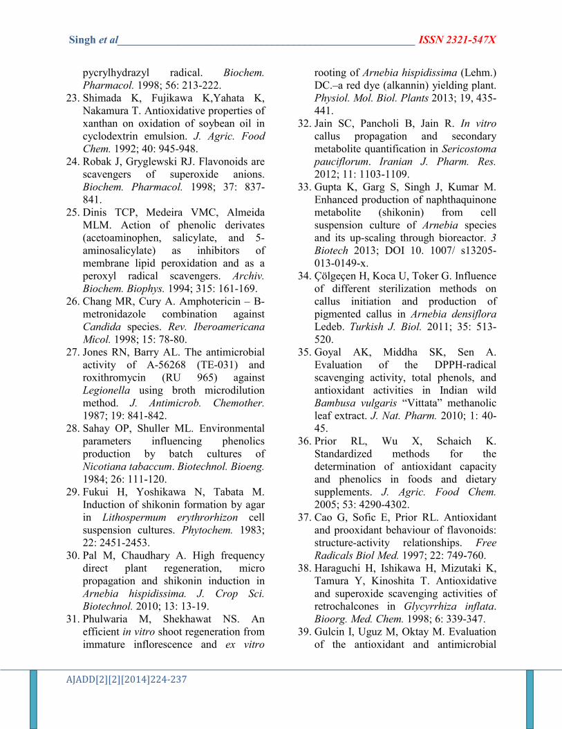
The seedlings of A. hispidissima were inoculated in two different combinations of MS culture medium containing 0.5 mg/L IAA and 1.20 mg/L BAP (first group) and another combination was supplemented with 5.0 mg/L 2, 4-D and 2.50 mg/L BAP (second group) for induction of callus. The highest frequency of growth of callus was optimized by using different concentrations of growth hormones of two combinations of MS culture medium (Fig. 1). After successful attempts, we observed the occurrence of different compounds in MS culture medium. Cultures
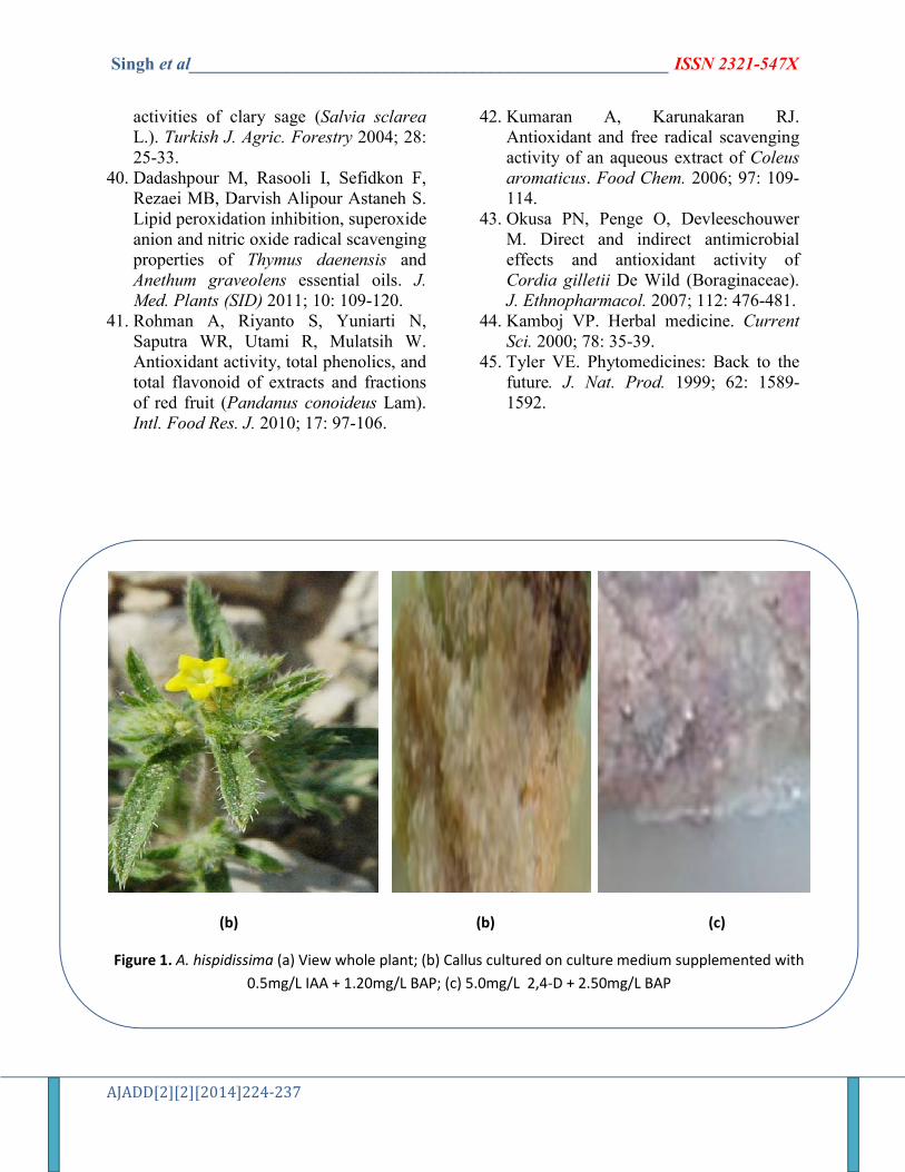
of both groups were incubated in dark. Cultures of both groups were harvested separately after 5 weeks of incubation. Harvested callus of both groups was used for isolation and characterization of phytochemicals. After chemical characterization, we investigated three alkannin and three shikonin derivatives from callus tissues of A. hispidissima, viz, (I) propionylalkannin (178 mg; first group), (II) β-hydroxyisovaleryl alkannin (217 mg; first group), (III) teracrylalkannin (228 mg; first group), (IV) shikonin (167 mg; second group), (V) deoxyshikonin (237 mg; second group), and (VI) acetylshikonin (317 mg; second group) (Fig. 2).
The observed results showed that the callus was dark red and morphologically similar but the accumulation of phytochemicals was different. To promote continuous accumulation of phytochemicals, the activated charcoal was supplemented in culture medium at two different concentrations in cultures of group first (150 mg/L) and second (100 mg/L). After comparison, it was found that 100 mg/L concentration promoted more accumulation of phytochemicals than 150 mg/L in cultures. The concentration of growth regulators and adsorbents individually or in combination significantly promotes the growth and accumulation of secondary metabolites in cell cultures e.g., naphthalene acetic acid (NAA) or indole-3-acetic acid (IAA) increased the production of nicotine in suspension cultures of Nicotiana tabaccum, shikonin production in Lithospermum erythrorhizon28,29 and A. hispidissima,30,31 phenol production in Sericostoma pauciflorum.32 Previous studies have proved that photoperiod incubation reduced the accumulation of pigments in cell cultures while dark period incubation promoted two times higher accumulation of pigments in cultures of Arnebia species. 33,34
Singh et al______________________________________________________ ISSN 2321-547X
AJADD[2][2][2014]224-237
Antioxidant activity The antioxidant activity of n-hexane
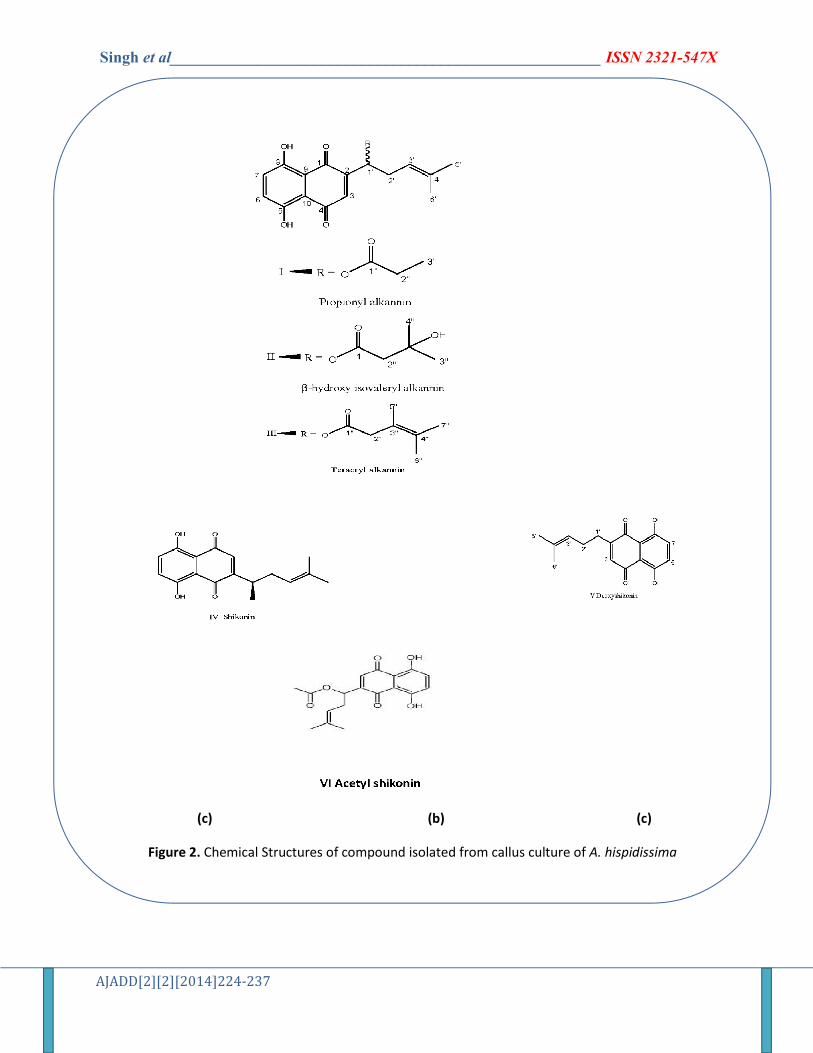
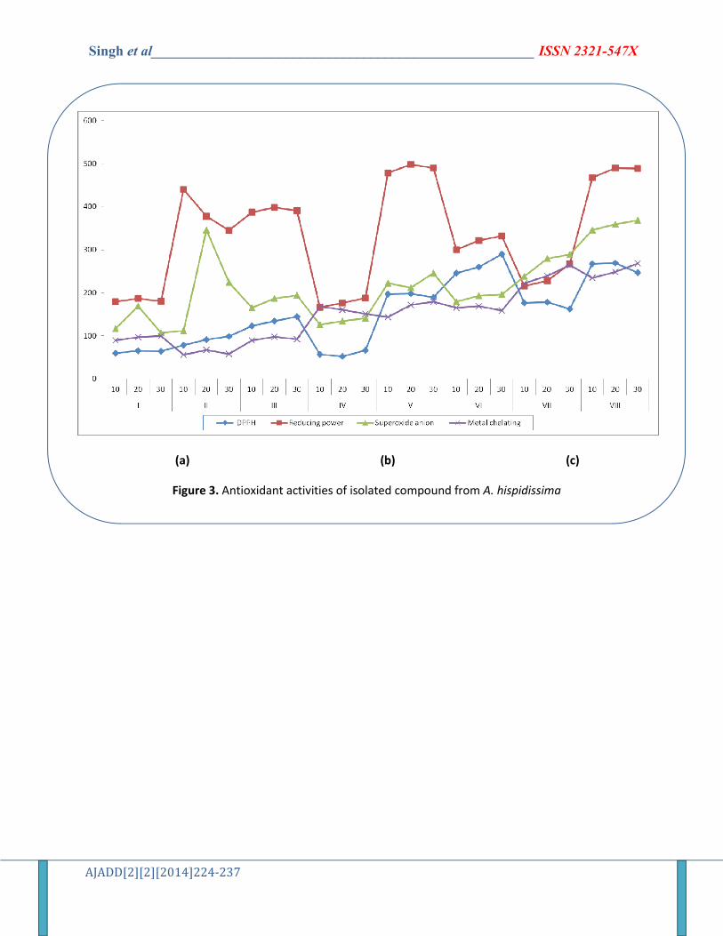
fraction and isolated compounds was assessed by using different well established protocols. The DPPH-free radical scavenging activity of isolated compounds was measured in terms of donating the hydrogen or free radical scavenging ability. Among these isolated compounds, deoxyshikonin showed maximum activity (290.20±0.440%) at 30 μM as compared with (±)-α-tocopherol (reference compound) which demonstrated 269.44 ± 0.852% inhibition at 20 μM (Fig. 3) concentrations. The DPPH-free radicals’ scavenging activity is based on the ability of DPPH, to decolorize in the presence of antioxidative compounds. The DPPH-radical contains an odd electron that is responsible for the absorbance at 540 nm and also visible deep purple color. When DPPH accepts an electron donated by an antioxidant compound, the DPPH is decolorized, which can quantitatively measure from changes in absorbance.35 Several antioxidative agents react so quickly with peroxyl radicals may react so slowly or may even be inert to DPPH due to steric inaccessibility. The DPPH is decolorized by the reducing agents and transfer of H, which is significant in inaccurate interpretation of assessing antioxidant capacity.36
In case of the reducing power ability, the n-hexane fraction and isolated compounds cause the reaction of reduction of the ferric/ferricyanide complex to ferrous complex. Figure 3 shows the sequence of the reducing power of isolated compounds as shikonin > propionylalkannin > β-hydroxyisovaleryl alkannin > acetylshikonin > teracrylalkannin. In reducing power activity, it has been argued that the capacity to reduce iron has very little relationship to the radical quenching processes mediated by most of the antioxidant agents. However, reduction of radical ions still stops radical chains, and reducing power reflects the ability
of compounds to modulate redox tone in plasma and tissues.37
Superoxide anion scavenging activity profiles of isolated compounds showed that propionylalkannin had higher inhibition percentage (345.45 ± 0.632%) at 20 μM concentration (Fig. 3). Interestingly, the lower concentration (20 μM) of propionylalkannin displayed higher superoxide scavenging activity than higher concentration as 30 μM (224.78 ± 0.390%). Superoxide radicals are produced in human body by various oxidative enzymes in form of one electron reduction of molecular oxygen. Xanthine oxidase is one of the major oxidative enzyme to produce superoxide radical as a result in tissue injury.38 In vitro superoxide radical was generated by xanthine oxidase during the reaction; nitro blue tetrazolium undergoes oxidation and leads to water soluble blue formazone (Gulcin et al., 2004).39 The decrease in blue color formation after adding the solvent fractions of isolated compounds in the reaction mixture was measured as superoxide radical scavenging. Many phenolic compounds have been reported to be potent inhibitors of xanthine oxidase.40
In metal chelating scavenging study, the chelation of ferrous ions by isolated compounds was monitored by the method of Dinis et al.25 The metal chelating compounds may stop lipid peroxidation by stabilizing transition metals. Formation of Fe2+ and ferrozine complex was completed in the presence of isolated compounds indicating that acetylshikonin showed higher chelating ability (264.76 ± 0.531%) than other isolated compounds and capture ferrous ions (Fig. 3). Ferrozine can quantitatively form Fe2+ complexes, the complex formation can be checked by the other complexing agents which cause a decrease in the intensity of red color of complexes. It has been established that chelating agents, that form σ-bonds with the heavy metal, are effective antioxidants because they reduce the redox potential, that’s
Singh et al______________________________________________________ ISSN 2321-547X
AJADD[2][2][2014]224-237
by stabilizing the oxidized form of metal ion.41,42
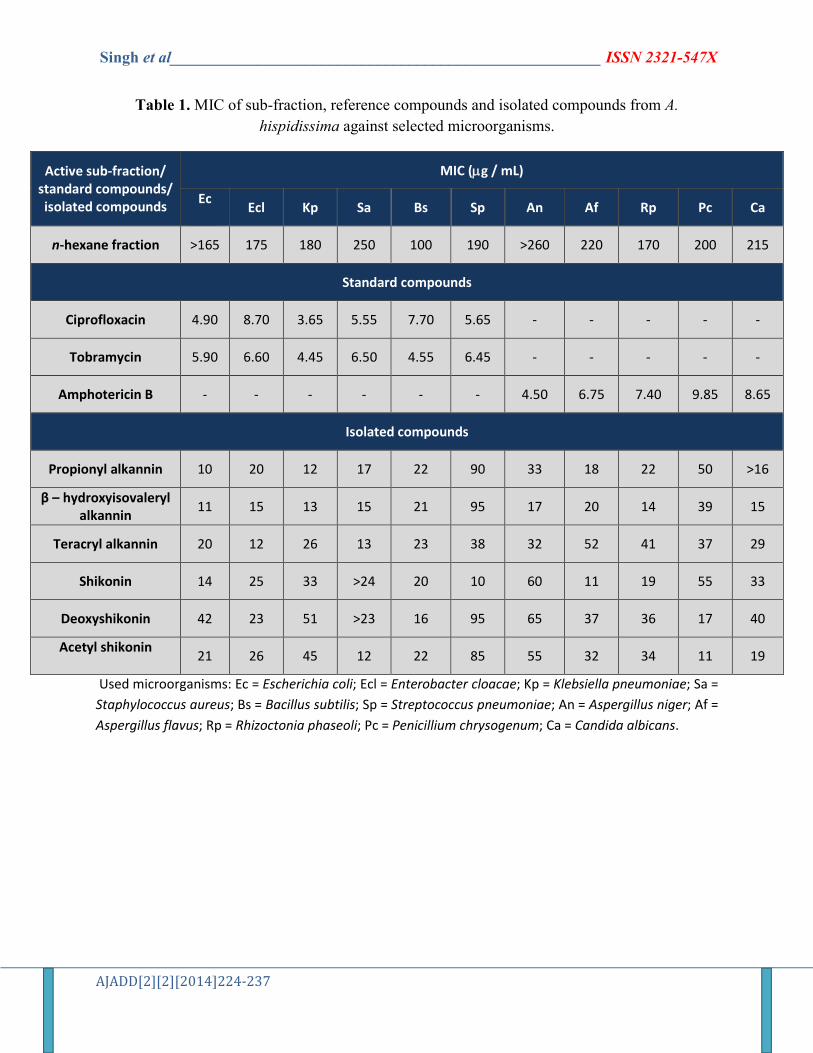
Antimicrobial activity
The antimicrobial profiles of the n-hexane and isolated compounds are presented in the Table 1. The n-hexane fraction, isolated compounds of A. hispidissima and reference compounds showed fairly well antimicrobial activity against selected microorganisms. Almost all the isolated compounds demonstrated varying degrees of antimicrobial potential. Propionylalkannin showed maximum antibacterial activity against E. coli (10 µg/ mL) and shikonin demonstrated similar activity to Streptococcus pneumoniae (10 µg/ mL). The acetylshikonin exhibited potent antifungal activity against P. chrysogenum at the dose of 11 µg/ mL. The ciprofloxacin and tobramycin possessed antibacterial activity at all the tested concentrations (1.0 – 10 µg/ mL) and amphotericin-B was assessed to active at 1.0 – 10 µg/ mL. The MIC values of isolated compounds varied from 10 – 95 µg/ mL. In the available literature, majority of reports says that 5 - 100 µg/ mL are considered as effective antimicrobial agents.43
CONCLUSION
The market demand for plant derived drugs and dyestuffs in the world are increasing day by day. As per available data, there has been shift in the preference of consumers from synthetic chemicals to plant derived medicinal agent,44 and in this line, pharmaceutical companies seeking continuously new compounds for drugs to improve their efficacy.45 The present study describes about the application of plant tissue cultures for the optimization of growth and accumulation of phytochemicals in cell cultures. In the present study, the six compounds were isolated from tissue cultures of A. hispidissima and evaluated for
antioxidant and antimicrobial activities. This has evidenced from the results, that alkannin and shikonin derivatives demonstrate modern antioxidant and high antimicrobial activities. Therefore, further investigations need to be carried out to isolate new phytochemicals from tissues cultures and screening for other different biological and pharmacological activities. REFERENCES 1. Aswal MA, Nahar A, Hossain MS, Bari
MA, Rahman M, Haque ME. Brain shrimp toxicity of leaf and seed extracts of Cassia alata Linn. and their antibacterial activity. J. Med. Sci. 2004; 4: 188-193.
2. Jiang C, Chang M, Wen C, Lin Y, Hsu F, Lee M. Natural product of cosmetics: analysis of extracts of plant endemic to Taiwan for the presence of tyrosinase inhibitory melanin reducing and free radical scavenging activities. J. Food Drug Analysis 2006; 14: 346-352.
3. Chopra RN, Nayar SL, Chopra IC. Glossary of Indian Medicinal Plants. New Delhi: CSIR, 1956.
4. Jain SK, Defilipps RA. Medicinal Plants of India. Michigan, USA: Reference Publications Inc, 1991.
5. Shen CC, Syu WJ, Li SY, Lin CH, Lee GH, Sun CM. Antimicrobial activities of naphthazarins from Arnebia euchroma. J. Nat. Prod. 2002; 65: 1857-1862.
6. Graikou K, Damianakos H, Syklowska-Baranek K, Pietrosiuk A, Jeziorek M, Chinou I. Isohexenylnaphthazarins from Lithospermum canescens (Michx.) Lehm. And Arnebia euchroma (Royle) Jonst. in vitro culture. Planta Med. 2010; 76: 196-204.
7. Wang R, Yin R, Zhou W, Xu D, Li S. Shikonin and its derivatives: a patent review. Expert Opin. Ther. Patents 2012; 22, 977-997.
Singh et al______________________________________________________ ISSN 2321-547X
AJADD[2][2][2014]224-237
8. Cui X. A novel extraction method of shikonin and the determination of contact angle. J. Med. Plants Res. 2013; 7: 1429-1433.
9. Liu T, Ma C, Yang L, Wang W, Sui X, Zhao C, Zu Y. Optimization of shikonin homogenate extraction from Arnebia euchroma using response surface methodology. Molecules 2013; 18: 466-481.
10. Sharma N, Sharma UK, Malik S, Bhushan, Kumar V, Verma SC, Sharma N, Sharma M, Sinha AK. Isolation and purification of acetylshikonin and β-acetoxyisovaleryl shikonin from cell suspension cultures of Arnebia euchroma (Royle) Johnston using rapid preparative HPLC. J. Separation Sci. 2008; 31: 629-635.
11. Malik S, Bhushan S, Sharma S, Ahuja PS. Physico-chemical factors influencing the shikonin derivatives production in cell suspension cultures of Arnebia euchroma (Royle) Johnston, a medicinally important plant species. Cell Biol. Intl. 2011; 35: 153-158.
12. Chang M-J, Huang G-J, Ho Y-L, Lin I-H, Huang S-S, Chang T-N, Chang H-Y, Chang Y-S. Study on the antioxidant activities of crude extracts from the roots of Arnebia euchroma and Lithospermum erythrorhizon. Mid-Taiwan J. Med. 2008; 13: 113-121.
13. Fazal H, Ahmad N, Khan MA. Physico-chemical, phytochemical evaluation and DPPH-scavenging antioxidant potential in medicinal plants used for herbal formulation in Pakistan. Pakistan J. Bot. 2011; 43: 63-67.
14. Ganie SA, Jan A, Muzaffar S, Zargar BA, Hamid R, Zargar MA. Radical scavenging and antibacterial activity of Arnebia benthamii methanol extract. Asian Pacific J. Tropical Med. 2012; 5: 766-772.
15. Al-Mussawi AA. Isolation and identification of shikonin from Arnebia decumbens L. and its antimicrobial activity. J. Appl. Sci. Res. 2010; 6: 1452-1456.
16. Damianakos H, Kretschmer N, Syklowska-Baranek K, Pietrosiuk A, Bauer R, Chinou I. Antimicrobial and cytotoxic isohexenylnaphthazarins from Arnebia euchroma (Royle) Johnst. (Boraginaceae) callus and cell suspension culture. Molecules 2012; 17: 14310-14322.
17. Murashige T, Skoog F. A revised medium for rapid growth and bioassays with tobacco tissue cultures. Physiol. Planta. 1962; 15: 473-497.
18. Papageorgiou VP, Assimopoulou AN, Couladouros EA, Hepworth D, Nicolaou KC. The chemistry and biology of alkannin, shikonin and related naphthazarin natural products. Angewandte Chem. Intl. Edition 1999; 38: 270-300.
19. Khan HA, Chandrashekharan I, Ghanim A. Naphthazarins from Arnebia hispidissima. Phytochem. 1983; 22: 614-615.
20. Papageorgiou VP, Winkler A, Sagredos AN, Digenis GA. Studies on the relationship of structure to antimicrobial properties of naphthaquinones and other constituents of Alkanna tinctoria. Planta Med. 1979; 35: 56-60.
21. Bozan B, Baser KHC, Kara S. Quantitative determination of naphthaquinones of Arnebia densiflora (Nordm.) Ledeb. by an improved high performance liquid chromatographic method. J. Chromatogr. A 1997; 782: 133-136.
22. Yokozawa T, Chen CP, Dong E, Tanaka T, Nonaka GL, Nishioka I. Study on the inhibitory effect of tannins, flavonoids against the 1, 1-diphenyl-2-
Singh et al______________________________________________________ ISSN 2321-547X
AJADD[2][2][2014]224-237
pycrylhydrazyl radical. Biochem. Pharmacol. 1998; 56: 213-222.
23. Shimada K, Fujikawa K,Yahata K, Nakamura T. Antioxidative properties of xanthan on oxidation of soybean oil in cyclodextrin emulsion. J. Agric. Food Chem. 1992; 40: 945-948.
24. Robak J, Gryglewski RJ. Flavonoids are scavengers of superoxide anions. Biochem. Pharmacol. 1998; 37: 837-841.
25. Dinis TCP, Medeira VMC, Almeida MLM. Action of phenolic derivates (acetoaminophen, salicylate, and 5-aminosalicylate) as inhibitors of membrane lipid peroxidation and as a peroxyl radical scavengers. Archiv. Biochem. Biophys. 1994; 315: 161-169.
26. Chang MR, Cury A. Amphotericin – B- metronidazole combination against Candida species. Rev. Iberoamericana Micol. 1998; 15: 78-80.
27. Jones RN, Barry AL. The antimicrobial activity of A-56268 (TE-031) and roxithromycin (RU 965) against Legionella using broth microdilution method. J. Antimicrob. Chemother. 1987; 19: 841-842.
28. Sahay OP, Shuller ML. Environmental parameters influencing phenolics production by batch cultures of Nicotiana tabaccum. Biotechnol. Bioeng. 1984; 26: 111-120.
29. Fukui H, Yoshikawa N, Tabata M. Induction of shikonin formation by agar in Lithospermum erythrorhizon cell suspension cultures. Phytochem. 1983; 22: 2451-2453.
30. Pal M, Chaudhary A. High frequency direct plant regeneration, micro propagation and shikonin induction in Arnebia hispidissima. J. Crop Sci. Biotechnol. 2010; 13: 13-19.
31. Phulwaria M, Shekhawat NS. An efficient in vitro shoot regeneration from immature inflorescence and ex vitro
rooting of Arnebia hispidissima (Lehm.) DC.–a red dye (alkannin) yielding plant. Physiol. Mol. Biol. Plants 2013; 19, 435-441.
32. Jain SC, Pancholi B, Jain R. In vitro callus propagation and secondary metabolite quantification in Sericostoma pauciflorum. Iranian J. Pharm. Res. 2012; 11: 1103-1109.
33. Gupta K, Garg S, Singh J, Kumar M. Enhanced production of naphthaquinone metabolite (shikonin) from cell suspension culture of Arnebia species and its up-scaling through bioreactor. 3 Biotech 2013; DOI 10. 1007/ s13205-013-0149-x.
34. Çölgeçen H, Koca U, Toker G. Influence of different sterilization methods on callus initiation and production of pigmented callus in Arnebia densiflora Ledeb. Turkish J. Biol. 2011; 35: 513-520.
35. Goyal AK, Middha SK, Sen A. Evaluation of the DPPH-radical scavenging activity, total phenols, and antioxidant activities in Indian wild Bambusa vulgaris “Vittata” methanolic leaf extract. J. Nat. Pharm. 2010; 1: 40-45.
36. Prior RL, Wu X, Schaich K. Standardized methods for the determination of antioxidant capacity and phenolics in foods and dietary supplements. J. Agric. Food Chem. 2005; 53: 4290-4302.
37. Cao G, Sofic E, Prior RL. Antioxidant and prooxidant behaviour of flavonoids: structure-activity relationships. Free Radicals Biol Med. 1997; 22: 749-760.
38. Haraguchi H, Ishikawa H, Mizutaki K, Tamura Y, Kinoshita T. Antioxidative and superoxide scavenging activities of retrochalcones in Glycyrrhiza inflata. Bioorg. Med. Chem. 1998; 6: 339-347.
39. Gulcin I, Uguz M, Oktay M. Evaluation of the antioxidant and antimicrobial
Singh et al______________________________________________________ ISSN 2321-547X
AJADD[2][2][2014]224-237
activities of clary sage (Salvia sclarea L.). Turkish J. Agric. Forestry 2004; 28: 25-33.
40. Dadashpour M, Rasooli I, Sefidkon F, Rezaei MB, Darvish Alipour Astaneh S. Lipid peroxidation inhibition, superoxide anion and nitric oxide radical scavenging properties of Thymus daenensis and Anethum graveolens essential oils. J. Med. Plants (SID) 2011; 10: 109-120.
41. Rohman A, Riyanto S, Yuniarti N, Saputra WR, Utami R, Mulatsih W. Antioxidant activity, total phenolics, and total flavonoid of extracts and fractions of red fruit (Pandanus conoideus Lam). Intl. Food Res. J. 2010; 17: 97-106.
42. Kumaran A, Karunakaran RJ. Antioxidant and free radical scavenging activity of an aqueous extract of Coleus aromaticus. Food Chem. 2006; 97: 109-114.
43. Okusa PN, Penge O, Devleeschouwer M. Direct and indirect antimicrobial effects and antioxidant activity of Cordia gilletii De Wild (Boraginaceae). J. Ethnopharmacol. 2007; 112: 476-481.
44. Kamboj VP. Herbal medicine. Current Sci. 2000; 78: 35-39.
45. Tyler VE. Phytomedicines: Back to the future. J. Nat. Prod. 1999; 62: 1589-1592.
(b) (b) (c)
Figure 1. A. hispidissima (a) View whole plant; (b) Callus cultured on culture medium supplemented with
0.5mg/L IAA + 1.20mg/L BAP; (c) 5.0mg/L 2,4-D + 2.50mg/L BAP
Singh et al______________________________________________________ ISSN 2321-547X
AJADD[2][2][2014]224-237
(c) (b) (c)
Figure 2. Chemical Structures of compound isolated from callus culture of A. hispidissima
Singh et al______________________________________________________ ISSN 2321-547X
AJADD[2][2][2014]224-237
(a) (b) (c)
Figure 3. Antioxidant activities of isolated compound from A. hispidissima
Singh et al______________________________________________________ ISSN 2321-547X
AJADD[2][2][2014]224-237
Table 1. MIC of sub-fraction, reference compounds and isolated compounds from A.
hispidissima against selected microorganisms.
Active sub-fraction/ standard compounds/ isolated compounds
MIC (g / mL)
Ec
Ecl Kp Sa Bs Sp An Af Rp Pc Ca
n-hexane fraction >165 175 180 250 100 190 >260 220 170 200 215
Standard compounds
Ciprofloxacin 4.90 8.70 3.65 5.55 7.70 5.65 - - - - -
Tobramycin 5.90 6.60 4.45 6.50 4.55 6.45 - - - - -
Amphotericin B - - - - - - 4.50 6.75 7.40 9.85 8.65
Isolated compounds
Propionyl alkannin 10 20 12 17 22 90 33 18 22 50 >16
β – hydroxyisovaleryl alkannin
11 15 13 15 21 95 17 20 14 39 15
Teracryl alkannin 20 12 26 13 23 38 32 52 41 37 29
Shikonin 14 25 33 >24 20 10 60 11 19 55 33
Deoxyshikonin 42 23 51 >23 16 95 65 37 36 17 40
Acetyl shikonin
21 26 45 12 22 85 55 32 34 11 19
Used microorganisms: Ec = Escherichia coli; Ecl = Enterobacter cloacae; Kp = Klebsiella pneumoniae; Sa =
Staphylococcus aureus; Bs = Bacillus subtilis; Sp = Streptococcus pneumoniae; An = Aspergillus niger; Af =
Aspergillus flavus; Rp = Rhizoctonia phaseoli; Pc = Penicillium chrysogenum; Ca = Candida albicans.