Embed Size (px)
Citation preview

Published Ahead of Print 11 June 2012. 10.1128/AAC.00407-12.
2012, 56(9):4594. DOI:Antimicrob. Agents Chemother. Chinniah, Anirban Pal, Sudip K. Kar and Pratap K. DasSuchandra Goswami, Rajendra S. Bhakuni, Annalakshmi Artemisinin and Its DerivativesAnti-Helicobacter pylori Potential of
http://aac.asm.org/content/56/9/4594Updated information and services can be found at:
These include:
REFERENCEShttp://aac.asm.org/content/56/9/4594#ref-list-1at:
This article cites 42 articles, 10 of which can be accessed free
CONTENT ALERTS more»articles cite this article),
Receive: RSS Feeds, eTOCs, free email alerts (when new
http://journals.asm.org/site/misc/reprints.xhtmlInformation about commercial reprint orders: http://journals.asm.org/site/subscriptions/To subscribe to to another ASM Journal go to:
on August 20, 2012 by Indian Inst of C
hem B
iologyhttp://aac.asm
.org/D
ownloaded from
Anti-Helicobacter pylori Potential of Artemisinin and Its Derivatives
Suchandra Goswami,a Rajendra S. Bhakuni,b Annalakshmi Chinniah,a Anirban Pal,b Sudip K. Kar,a and Pratap K. Dasa
CSIR–Indian Institute of Chemical Biology, Kolkata, India,a and CSIR–Central Institute of Medicinal and Aromatic Plants, Lucknow, Indiab
The antimalarial drug artemisinin from Artemisia annua demonstrated remarkably strong activity against Helicobacter pylori,the pathogen responsible for peptic ulcer diseases. In an effort to develop a novel antimicrobial chemotherapeutic agent contain-ing such a sesquiterpene lactone endoperoxide, a series of analogues (2 natural and 15 semisynthetic molecules), including eightnewly synthesized compounds, were investigated against clinical and standard strains of H. pylori. The antimicrobial spectrumagainst 10 H. pylori strains and a few other bacterial and fungal strains indicated specificity against the ulcer causing organism.Of five promising molecules, a newly synthesized ether derivative �-artecyclopropylmether was found to be the most potentcompound, which exhibited MIC range, MIC90, and minimum bactericidal concentration range values of 0.25 to 1.0 �g/ml, 1.0�g/ml, and 1 to 16 �g/ml, respectively, against both resistant and sensitive strains of H. pylori. The molecule demonstratedstrong bactericidal kinetics with extensive morphological degeneration, retained functional efficacy at stomach acidic pH unlikeclarithromycin, did not elicit drug resistance unlike metronidazole, and imparted sensitivity to resistant strains. It is not cyto-toxic and exhibits in vivo potentiality to reduce the H. pylori burden in a chronic infection model. Thus, �-artecyclopropyl-mether could be a lead candidate for anti-H. pylori therapeutics. Since the recurrence of gastroduodenal ulcers is believed to bemainly due to antibiotic resistance of the commensal organism H. pylori, development of a candidate drug from this finding iswarranted.
Helicobacter pylori is the major cause of gastroduodenal infec-tions and has been implicated in the pathogenesis of active
and chronic gastritis, peptic ulcer, and gastric carcinoma (29, 30).The organism changes the gastric epithelium directly throughbacterial toxicity and indirectly via inflammation-mediated dam-age (1). Antibiotic therapy has been used successfully with a com-bination of two or three drugs to eradicate H. pylori infections (12,32) and also to cure gastroduodenal ulcers (30). However, resis-tance to amoxicillin (AMX), clarithromycin (CLR), and metroni-dazole (MNZ) in particular is increasingly becoming widespreaddue to constant use of these drugs against such infection (7, 33,34). Development of drugs derived from natural sources is receiv-ing increasing attention particularly because of their potential tokeep pathogenic strains sensitive. Sustained efforts worldwide insearching anti-H. pylori leads from natural products have alreadyled to the elaboration of many plant-derived molecules with in-teresting prospects (10, 23, 36, 48).
Many unanswered questions on how a plant or its product(s)can be successfully used to cure a disease in traditional and/oralternate modes of treatment are now being addressed. Examplesinclude delineation of the stimulating property of tea, the anti-inflammatory and antiseptic properties of turmeric, the analgesicand anti-inflammatory use of willow bark, the antidiabetic poten-tial of Madagascar Periwinkle, the antimalarial property of theplant Artemisia annua, etc. However, recent additional findingsregarding the anticancer activity of tea catechins, the antitumorpotential of curcumin, the blood-thinning property of the well-known analgesic molecule aspirin, or the therapeutic effect ofvinca alkaloids in Hodgkin’s lymphoma (see, for example, refer-ence 41) not only brought forth a few unquestioned answers butalso opened up innovative spin-offs to revisit the wealth of tradi-tional knowledge through the eyes of modern-day understandingabout the pathophysiological targets and therapeutic principles.In a nationwide networked program in India (19), large-scalescreening of natural product based molecules is being carried out
in appropriate preclinical experimental models, wherein we havebeen searching for anti-H. pylori principle(s). The rationale hasbeen that many plant materials are being successfully used to curestomach ailments for centuries; thus, some active principles couldact via the anti-H. pylori mechanism as well. Such random screen-ing demonstrated the remarkably high and hitherto-unknownanti-H. pylori activity of artemisinin.
Use of Artemisia annua to treat malaria has been known for atleast 1,600 years. Artemisinin, a sesquiterpene lactone endoperox-ide, is a secondary metabolite from this plant that is widely dis-persed throughout the temperate region (43). The plant has tra-ditionally been grown in China as a medicinal plant and, morerecently, in Europe for its aromatic leaves, which are used in fla-voring beverages. Besides antimalarial property, artemisinin andits analogues were shown to possess immunomodulatory and an-titumor effects (20, 27, 45). Many natural and semisynthetic de-rivatives of artemisinin and other compounds containing an en-doperoxide bridge have been described as biologically active (37,40). The generation of semisynthetic derivatives such as artesu-nate, dihydroartemisinin, and arteether, etc., has helped in deci-phering the mechanism of action as an antimalarial, antitumor,anti-inflammatory, or immunosuppressive agent (25, 45, 47). Theendoperoxide moiety in the chemical structure of artemisinin isthought to be responsible for the bioactivity (25).
In an effort to look into the details of anti-H. pylori profiles of
Received 22 February 2012 Returned for modification 23 March 2012Accepted 2 June 2012
Published ahead of print 11 June 2012
Address correspondence to Rajendra S. Bhakuni, [email protected], orPratap K. Das, [email protected].
Copyright © 2012, American Society for Microbiology. All Rights Reserved.
doi:10.1128/AAC.00407-12
4594 aac.asm.org Antimicrobial Agents and Chemotherapy p. 4594–4607 September 2012 Volume 56 Number 9
on August 20, 2012 by Indian Inst of C
hem B
iologyhttp://aac.asm
.org/D
ownloaded from
such endoperoxide molecules, a series of artemisinin analogueswere investigated that included two isolated molecules, sevenknown semisynthetic derivatives, and eight newly synthesizedcompounds. Because artemisinin is thought to be useful againstresistant strains of malaria (20, 25), it was of additional interest tosee whether antibiotic-resistant strains of H. pylori can also bekilled by such natural-product-based molecules. Based on pri-mary screening through a disc diffusion sensitivity assay and MIC/minimum bactericidal concentration (MBC) studies using bothstandard strains and clinical isolates and also based on investiga-tions of the general antimicrobial features, including both anti-bacterial and antifungal activities, we selected five molecules aspotential anti-H. pylori candidates. Such molecules were furtherevaluated for their acid stability (because these are supposed to actunder stomach acidic pH), bactericidal kinetics, and sensitivity/resistance profile. The most potent compound was further exam-ined for its capacity to induce morphological deformity, showsynergism with anti-H. pylori antibiotics, and reduce the H. pyloriburden in vivo. Typically, the objective and the approach planhave been to synthesize newer derivatives upon observing anti-H.pylori activity profile of some molecules, so as to be able to bringout compounds endowed with desired attributes. It is concludedthat �-artecyclopropylmether could be a lead candidate foranti-H. pylori therapeutics.
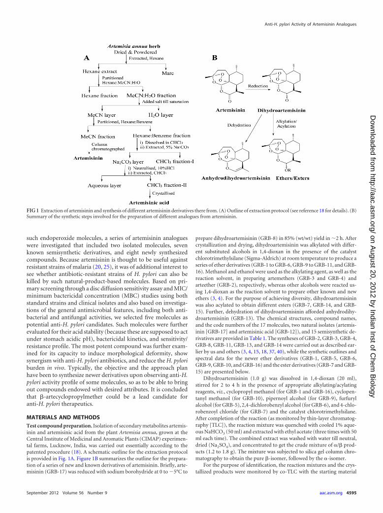
MATERIALS AND METHODSTest compound preparation. Isolation of secondary metabolites artemis-inin and artemisinic acid from the plant Artemisia annua, grown at theCentral Institute of Medicinal and Aromatic Plants (CIMAP) experimen-tal farms, Lucknow, India, was carried out essentially according to thepatented procedure (18). A schematic outline for the extraction protocolis provided in Fig. 1A. Figure 1B summarizes the outline for the prepara-tion of a series of new and known derivatives of artemisinin. Briefly, arte-misinin (GRB-17) was reduced with sodium borohydride at 0 to �5°C to
prepare dihydroartemisinin (GRB-8) in 85% (wt/wt) yield in �2 h. Aftercrystallization and drying, dihydroartemisinin was alkylated with differ-ent substituted alcohols in 1,4-dioxan in the presence of the catalystchlorotrimethylsilane (Sigma-Aldrich) at room temperature to produce aseries of ether derivatives (GRB-1 to GRB-6, GRB-9 to GRB-11, and GRB-16). Methanol and ethanol were used as the alkylating agent, as well as thereaction solvent, in preparing artemethers (GRB-3 and GRB-4) andarteether (GRB-2), respectively, whereas other alcohols were reacted us-ing 1,4-dioxan as the reaction solvent to prepare other known and newethers (3, 4). For the purpose of achieving diversity, dihydroartemisininwas also acylated to obtain different esters (GRB-7, GRB-14, and GRB-15). Further, dehydration of dihydroartemisinin afforded anhydrodihy-droartemisinin (GRB-13). The chemical structures, compound names,and the code numbers of the 17 molecules, two natural isolates (artemis-inin [GRB-17] and artemisinic acid [GRB-12]), and 15 semisynthetic de-rivatives are provided in Table 1. The syntheses of GRB-2, GRB-3, GRB-4,GRB-8, GRB-11, GRB-13, and GRB-14 were carried out as described ear-lier by us and others (3, 4, 15, 18, 37, 40), while the synthetic outlines andspectral data for the newer ether derivatives (GRB-1, GRB-5, GRB-6,GRB-9, GRB-10, and GRB-16) and the ester derivatives (GRB-7 and GRB-15) are presented below.
Dihydroartemisinin (1.0 g) was dissolved in 1,4-dioxan (20 ml),stirred for 2 to 4 h in the presence of appropriate alkylating/acylatingreagents, viz., cyclopropyl methanol (for GRB-1 and GRB-16), cyclopen-tanyl methanol (for GRB-10), pipernoyl alcohol (for GRB-9), furfurylalcohol (for GRB-5), 2,4-dichlorobenzyl alcohol (for GRB-6), and 4-chlo-robenzoyl chloride (for GRB-7) and the catalyst chlorotrimethylsilane.After completion of the reaction (as monitored by thin-layer chromatog-raphy [TLC]), the reaction mixture was quenched with cooled 1% aque-ous NaHCO3 (50 ml) and extracted with ethyl acetate (three times with 50ml each time). The combined extract was washed with water till neutral,dried (Na2SO4), and concentrated to get the crude mixture of �/� prod-ucts (1.2 to 1.8 g). The mixture was subjected to silica gel column chro-matography to obtain the pure �-isomer, followed by the �-isomer.
For the purpose of identification, the reaction mixtures and the crys-tallized products were monitored by co-TLC with the starting material
FIG 1 Extraction of artemisinin and synthesis of different artemisinin derivatives there from. (A) Outline of extraction protocol (see reference 18 for details). (B)Summary of the synthetic steps involved for the preparation of different analogues from artemisinin.
Anti-H. pylori Activity of Artemisinin Analogues
September 2012 Volume 56 Number 9 aac.asm.org 4595
on August 20, 2012 by Indian Inst of C
hem B
iologyhttp://aac.asm
.org/D
ownloaded from
artemisinin or dihydroartemisinin in the solvent system n-hexane– ethylacetate (70:30). Purity of the compounds was assumed if a single spotdeveloped on TLC. Infrared (IR) spectra were recorded using a Perkin-Elmer Bx infrared spectrophotometer with a KBr window. 1H and 13Cnuclear magnetic resonance (NMR) spectra were determined in CDCl3 onBruker Avance spectrometer operating at 300 and 75 MHz, respectively.The chemical shifts are reported in ppm. Spectra were interpreted fromDEPT-90 and DEPT-35 (distortionless enhancement by polarizationtransfer) and COSY (correlation spectroscopy) results. Electrospray ion-ization (ESI) mass spectra were recorded on a JEOL-AccuTOF JMS-T100LC mass spectrometer and using the Shimadzu LC-MS (liquid chro-matography-mass spectrometry) system, respectively.
For �-artecyclopropylmether (GRB-1), the column was eluted withn-hexane– ethyl acetate (98:2) to yield GRB-1, 76% (wt/wt), Rf � 0.54 (7:3n-hexane– ethyl acetate), colorless plates (n-hexane), melting point (mp)
59 to 60°C; IR �max � 3,089, 3,010 (cyclopropane), 2,870, 1,597, 1,453,1,377, 1,102, and 1,025 cm�1; 1H NMR � � 0.18 and 0.46 (2H each, m,H2-3=/H2-4=), 0.89 (3H, d, J � 7.2 Hz, H3-13), 0.93 (3H, d, J � 6.3 Hz,H3-14), 1.02 (1H, m, H-2=), 1.40 (3H, s, H3-15), 3.32 (1H, dd, J � 10.5, 6.9Hz, Ha-1=), 3.55 (1H, dd, J � 10.5, 3.9 Hz, Hb-1=), 4.80 (1H, d, J � 3.3 Hz,�H-12), and 5.38 (1H, m, H-5); 13C NMR � � 2.7 and 3.2 (C-3= andC-4=), 10.9 (C-2=), 13.2 (C-13), 20.6 (C-14), 24.8 (C-2), 25.1 (C-8), 26.5(C-15), 31.3 (C-11), 35.2 (C-9), 36.9 (C-3), 37.9 (C-10), 45.0 (C-7), 53.1(C-1), 72.7 (C-1=), 81.4 (C-6), 88.2 (C-5), 101.9 (C-12), and 104.3 (C-4).ESI-MS (m/z) � 339 [M�H]�, 361[M�Na]� (C19H30O5).
For �-artecyclopropylmether (GRB-16), column elution withn-hexane– ethyl acetate (98:2), followed by preparative TLC in n-hexane–ethyl acetate (95:5), provided the viscous compound GRB-16, 14% (wt/wt), Rf � 0.49 (7:3 n-hexane– ethyl acetate); IR �max � 3083 (cyclopro-pane), 2,873, 1,596, 1,454, 1,378, 1,102, and 1,027 cm�1; 1H NMR � �
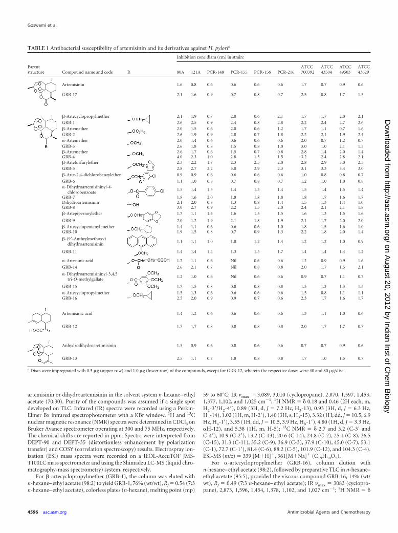
TABLE 1 Antibacterial susceptibility of artemisinin and its derivatives against H. pyloria
Parentstructure Compound name and code R
Inhibition zone diam (cm) in strain:
80A 121A PCR-148 PCR-155 PCR-156 PCR-216ATCC700392
ATCC43504
ATCC49503
ATCC43629
Artemisinin 1.6 0.8 0.6 0.6 0.6 0.6 1.7 0.7 0.9 0.6
GRB-17 2.1 1.6 0.9 0.7 0.8 0.7 2.5 0.8 1.7 1.5
�-Artecyclopropylmether 2.1 1.9 0.7 2.0 0.6 2.1 1.7 1.7 2.0 2.1
GRB-1 2.6 2.5 0.9 2.4 0.8 2.8 2.2 2.4 2.7 2.6�-Artemether 2.0 1.5 0.6 2.0 0.6 1.2 1.7 1.1 0.7 1.6GRB-2 2.6 1.9 0.9 2.8 0.7 1.8 2.2 2.1 1.9 2.4�-Artemether 2.0 1.4 0.6 0.6 0.6 0.6 2.0 0.7 1.2 0.7GRB-3 2.6 1.8 0.8 1.5 0.8 1.0 3.0 1.0 2.1 1.5�-Artemether 2.6 1.7 0.6 1.5 0.7 0.8 2.8 1.4 2.0 1.4GRB-4 4.0 2.3 1.0 2.8 1.5 1.5 3.2 2.4 2.8 2.1�-Artefurfurylether 2.3 2.2 1.7 2.3 2.5 2.0 2.8 2.9 3.0 2.5GRB-5 2.8 2.7 2.2 3.0 2.9 2.3 3.1 3.3 3.4 3.0
�-Arte-2,4-dichlorobenzylether 0.9 0.9 0.6 0.6 0.6 0.6 1.0 0.8 0.8 0.7
GRB-6 1.1 1.0 0.8 0.7 0.8 0.7 1.2 1.0 1.0 0.8�-Dihydroartemisininyl-4-
chlorobenzoate1.5 1.4 1.5 1.4 1.3 1.4 1.5 1.4 1.5 1.4
GRB-7 1.8 1.6 2.0 1.8 1.8 1.8 1.8 1.7 1.6 1.7Dihydroartemisinin 2.1 2.0 0.8 1.3 0.8 1.4 1.5 1.3 1.4 1.0GRB-8 3.0 2.7 0.9 2.2 1.5 2.0 2.4 2.1 2.1 1.8�-Artepipernoylether 1.7 1.1 1.4 1.6 1.5 1.5 1.6 1.3 1.5 1.6
GRB-9 2.0 1.2 1.9 2.1 1.8 1.9 2.1 1.7 2.0 2.0�-Artecyclopentanyl mether 1.4 1.1 0.6 0.6 0.6 1.0 1.8 1.5 1.6 1.0GRB-10 1.9 1.5 0.8 0.7 0.9 1.3 2.2 1.8 2.0 1.4
�-(9=-Anthrylmethoxy)dihydroartemisinin
1.1 1.1 1.0 1.0 1.2 1.4 1.2 1.2 1.0 0.9
GRB-11 1.4 1.4 1.4 1.3 1.5 1.7 1.4 1.4 1.4 1.2
�-Artesunic acid 1.7 1.1 0.6 Nil 0.6 0.6 1.2 0.9 0.9 1.6
GRB-14 2.6 2.1 0.7 Nil 0.8 0.8 2.0 1.7 1.5 2.1
�-Dihydroartemisininyl-3,4,5tri-O-methylgallate
1.2 1.0 0.6 Nil 0.6 0.6 0.9 0.7 1.1 0.7
GRB-15 1.7 1.5 0.8 0.8 0.8 0.8 1.5 1.3 1.3 1.5
�-Artecyclopropylmether 1.5 1.3 0.6 0.6 0.6 0.6 1.5 0.8 1.1 1.1GRB-16 2.5 2.0 0.9 0.9 0.7 0.6 2.3 1.7 1.6 1.7
Artemisinic acid 1.4 1.2 0.6 0.6 0.6 0.6 1.3 1.1 1.0 0.6
GRB-12 1.7 1.7 0.8 0.8 0.8 0.8 2.0 1.7 1.7 0.7
Anhydrodihydroaretimisinin 1.5 0.9 0.6 0.8 0.6 0.6 0.7 0.7 0.9 0.6
GRB-13 2.5 1.1 0.7 1.8 0.8 0.8 1.7 1.0 1.5 0.7
a Discs were impregnated with 0.5 g (upper row) and 1.0 g (lower row) of the compounds, except for GRB-12, wherein the respective doses were 40 and 80 g/disc.
Goswami et al.
4596 aac.asm.org Antimicrobial Agents and Chemotherapy
on August 20, 2012 by Indian Inst of C
hem B
iologyhttp://aac.asm
.org/D
ownloaded from

0.22 and 0.48 (2H each, m, H2-3=/H2-4=), 0.90 (3H, d, J � 7.5 Hz, H3-13),0.95 (3H, d, J � 5.7 Hz, H3-14), 1.03 (1H, m, H-2=), 1.43 (3H, s, H3-15),3.33 (1H, dd, J � 10.5, 6.9 Hz, Ha-1=), 3.74 (1H, dd, J � 10.5, 3.6 Hz,Hb-1=), 4.80 (1H, d, J � 9.3 Hz, �H-12), and 5.32 (1H, m, H-5); 13CNMR � � 2.6 and 3.2 (C-3= and C-4=), 10.4 (C-2=), 12.6 (C-13), 20.2(C-14), 22.1 (C-2), 24.7 (C-8), 26.0 (C-15), 32.4 (C-11), 34.2 (C-9), 36.3(C-3), 37.3 (C-10), 45.3 (C-7), 51.6 (C-1), 73.1 (C-1=), 80.3 (C-6), 91.2(C-5), 99.4 (C-12), and 104.2 (C-4). ESI-MS (m/z) � 339 [M�H]�
(C19H30O5).For �-artecyclopentanylmether (GRB-10), the column was eluted
with n-hexane– ethyl acetate (98:2) to yield GRB-10, 84% (wt/wt), vis-cous, Rf � 0.57 (7:3 n-hexane– ethyl acetate); IR �max � 2,950, 2,869,2,873, 1,595, 1,457, 1,380, 1,103, and 1,027 cm�1; 1H NMR � � 0.90 (3H,d, J � 7.5 Hz, H3-13), 0.95 (3H, d, J � 6.0 Hz, H3-14), 1.24 (4H, m, H2-4=,H2-5=), 1.44 (3H, s, H3-15), 1.72 (4H, m, H2-3=, H2-6=), 2.15 (1H, t, J � 7.5Hz, H-2=), 3.25 (1H, dd, J � 9.3, 9.3 Hz, Ha-1=), 3.72 (1H, dd, J � 9.3, 7.5Hz, Hb-1=), 4.77 (1H, d, J � 3.0 Hz, �H-12), and 5.39 (1H, s, H-5); 13CNMR � 13.0 (C-13), 20.4 (C-14), 24.4/24.7 (C-2/C-8), 26.2 (C-15), 26.6(C-4= and C-5=), 29.4/29.8 (C-3=/C-6=), 31.0 (C-11), 34.7 (C-9), 36.4 (C-3), 37.5 (C-10), 39.5 (C-2=), 44.5 (C-7), 52.6 (C-1), 72.9 (C-1=), 81.2(C-6), 87.9 (C-5), 102.0 (C-12), and 104.0 (C-4); COSY (1H-1H correla-tions): correlations of Ha-1=, Hb-1= (3.25, 3.72) with H-2= (2.15),H-2=with H2-3=, H2-6= (1.72) and H2-3=, H2-6= with H2-4=, H2-5= (1.24).ESI-MS (m/z) � 389 [M�Na]�, 267 [M�C6H11O]� (C21H34O5).
For �-artepipernoylether (GRB-9), the column was eluted withn-hexane–ethyl acetate (95:5) to yield derivative GRB-9, 68% (wt/wt), Rf �0.47 (7:3 n-hexane–ethyl acetate), colorless needles (n-hexane), mp � 118 to120°C; IR �max � 1,595 and 927 (pipernoyl group) cm�1;1H NMR � � 0.90(3H, d, J � 7.2 Hz, H3-13), 0.95 (3H, d, J � 5.7 Hz, H3-14), 1.46 (3H, s,H3-15), 4.42 (1H, d, J � 12 Hz, Ha-1=), 4.78 (1H, d, J � 12 Hz, Hb-1=), 4.89(1H, d, J � 3.3 Hz, �H-12), 5.46 (1H, s, H-5), 5.96 (2H, s, H2-8=), 6.77 (1H, s,H-3=), and 6.81 (2H, d, J � 8.1 Hz, H-6= and H-7=); 13C NMR � 13.4 (C-13),20.6 (C-14), 24.9 (C-8), 25.1 (C-2), 26.5 (C-15), 31.3 (C-11), 35.0 (C-9), 36.8(C-3), 37.8 (C-10), 44.8 (C-7), 53.0 (C-1), 70.01 (C-1=), 81.5 (C-6), 88.3(C-5), 101.2, 101.5 (C-8=, C-12), 104.4 (C-4), 108.3 (C-3=), 108.5 (C-6=),121.2 (C-7=), 132.6 (C-2=), 147.3 (C-5=), and 148.0 (C-4=). ESI-MS (m/z) �441 [M�Na]� (C23H30O7), 267 [M�C8H7O3]�.
�-Artefurfurylether (GRB-5) was obtained from the column with n-hexane– ethyl acetate (95:5), yield � 61% (wt/wt), Rf � 0.51 (7:3n-hexane– ethyl acetate), colorless amorphous solid (n-hexane– ethyl ac-etate), mp � 145 to 147°C; IR �max � 1,595, 874, and 747 (furan skeleton)cm�1; 1H NMR � � 0.84 (3H, d, J � 7.5 Hz, H3-13), 0.94 (3H, d, J � 6.0Hz, H3-14), 1.45 (3H, s, H3-15), 4.52 (1H, d, J � 12.9 Hz, Ha-1=), 4.75(1H, d, J � 12.9 Hz, Hb-1=), 4.89 (1H, d, J � 3.3 Hz, �H-12), 5.46 (1H, s,H-5), 6.31 (1H, dd, J � 6.3, 1.2 Hz, H-3=), 6.33 (1H, d, J � 6.3Hz, H-4=),and 7.39 (1H, brs, H-5=); 13C NMR � 13.0 (C-13), 20.5 (C-14), 24.7(C-8), 24.9 (C-2), 26.4 (C-15), 31.0 (C-11), 34.8 (C-9), 36.6 (C-3), 37.6(C-10), 44.6 (C-7), 52.8 (C-1), 61.8 (C-1=), 81.4 (C-6), 88.2 (C-5), 100.9(C-12), 104.3 (C-4), 109.1 (C-4=), 110.3 (C-3=), 142.8 (C-5=), and 151.9(C-2=). FABMS (positive) � 365 [M�H]� (C20H28O6).
For �-arte-2,4-dichlorobenzylether (GRB-6), elution of the columnwith n-hexane– ethyl acetate (95:5) yielded the oily compound GRB-6,93% (wt/wt), Rf � 0.55 (7:3 n-hexane– ethyl acetate); IR �max � 1,592,1,102, and 753 (Cl groups) cm�1; 1H NMR � � 0.92 (3H, d, J � 7.2 Hz,H3-13), 0.95 (3H, d, J � 6.0 Hz, H3-14), 1.45 (3H, s, H3-15), 4.48 (1H, d,J � 13.2 Hz, Ha-1=), 4.96 (1H, d, J � 13.2 Hz, Hb-1=), 4.94 (1H, d, J � 3.3Hz, �H-12), 5.47 (1H, s, H-5), 7.24 (1H, dd, J � 8.4, 1.8 Hz, H-6=), 7.35(1H, d, J � 8.4 Hz, H-7=), and 7.37 (1H, brs, H-4=); 13C NMR � � 13.3(C-13), 20.5 (C-14), 24.7, 24.9 (C-2, C-8), 26.2 (C-15), 31.1 (C-11), 34.8(C-9), 36.5 (C-3), 37.6 (C-10), 44.5 (C-7), 52.8 (C-1), 67.4 (C-1=), 81.2(C-6), 88.3 (C-5), 100.2 (C-12), 104.4 (C-12), 127.2 (C-7=), 129.3 (C-4=),129.8 (C-6=), 130.2 (C-3=), 133.8 (C-5=), and 135.0 (C-2=). FABMS (pos-itive) � 443 [M�H]�, 465 [M�Na]� (C22H28O5Cl2).
�-Dihydroartemisininyl-4-chlorobenzoate (GRB-7) was obtainedfrom the column with n-hexane– ethyl acetate (95:5), yield � 91% (wt/
wt), Rf � 0.52 (7:3 n-hexane– ethyl acetate), colorless plates (n-hexane),mp � 98 to 100°C; IR �max � 1733 (ester CO), 1,592, 1,090, and 758 (Clgroup) cm�1; 1H NMR � � 0.95 (3H, d, J � 7.2 Hz, H3-13), 0.98 (3H, d,J � 5.7 Hz, H3-14), 1.42 (3H, s, H3-15), 5.52 (1H, s, H-5), 5.98 (1H, d, J �9.9 Hz, �H-12), 7.41 (2H, d, J � 8.4 Hz, H-4=, H-6=), 8.01 (1H, d, J � 8.4Hz, H-7=), and 8.08 (1H, d, J � 8.4 Hz, H-3=); 13C NMR � � 12.9 (C-13),20.2 (C-14), 22.1 (C-8), 24.5 (C-2), 25.9 (C-15), 31.9 (C-11), 34.1 (C-9),36.2 (C-3), 37.3 (C-10), 45.3 (C-7), 51.6 (C-1), 80.1 (C-6), 91.6 (C-5),92.7 (C-12), 104.4 (C-4), 128.7 (C-4=, C-6=), 131.5 (C-3=, C-7=), 131.9(C-2=), 139.8 (C-5=), and 164.4 (C-1=). FABMS (positive) � 423[M�H]�, 445 [M�Na]� (C22H27O6Cl).
For �-dihydroartemisininyl 3,4,5 tri-O-methylgallate (GRB-15), di-hydroartemisinin (1.0 g), trimethyl ether of gallic acid (1 g), N,N=-dicyc-lohexylcarbodiimide (1 g), and the catalyst 4-dimethylaminopyridine(160 mg) were stirred in dichloromethane (40 ml) for 4 h at room tem-perature. The reaction mixture was prepared as described above. Elutionof the column with n-hexane– ethyl acetate (95:5), followed by crystalli-zation from n-hexane–acetone (8:2), yielded GRB-15 76% (wt/wt), Rf �0.36 (7:3 n-hexane– ethyl acetate), colorless plates (ethyl acetate), mp �102 to 104°C; IR �max �1,727 (ester CO), 1,591, and 1,128 cm�1; 1HNMR � � 0.91 (3H, d, J � 6.9 Hz, H3-13), 0.99 (3H, d, J � 5.7 Hz, H3-14),1.44 (3H, s, H3-15), 3.88, 3.91, 3.92 (3H each, s, 3OMe), 5.53 (1H, s,H-5), 5.99 (1H, d, J � 9.6 Hz, �H-12), and 7.37 (2H, s, H-3=, and H-7=);13C NMR � � 12.2 (C-13), 20.2 (C-14), 22.0 (C-8), 24.5 (C-2), 25.9(C-15), 32.0 (C-11), 34.0 (C-9), 36.2 (C-3), 37.2 (C-10), 45.3 (C-7), 51.6(C-1), 56.3 (2OMe), 56.9 (OMe), 80.2 (C-6), 91.6 (C-5), 92.6 (C-12),104.4 (C-4), 107.3 (C-3=, C-7=), 124.5 (C-2=), 142.5 (C-5=), 152.8 (C-4=,C-6=), and 164.9 (C-1=). FABMS (positive) � 479 [M�H]� (C25H34O9).
Culture of H. pylori. Four ATCC standard strains (700392, 43504,43629, and 49503) and six clinical isolates (80A, 121A, PCR-148, PCR-155, PCR-156, and PCR-216) were used for in vitro studies and wereroutinely cultured either in brain heart infusion (BHI) agar plates con-taining 7% fetal calf serum (FCS), 0.5% IsoVitaleX (Becton Dickinson,USA) and 0.0025% Dent (Oxoid, England) or in brucella broth (BB)medium containing 0.0025% Dent and 5% FCS under a microaerophilicenvironment (10% CO2, 85% N2, and 5% O2 and �95% relative humid-ity) at 37°C for 48 to 72 h (11) in a double-gas CO2 incubator (Heraeus,model HERAcell 240 or BB6020). For working purposes, either a 3-day-old culture in BB containing 5% FCS and 0.0025% Dent (CFU at ca. 5 107/ml) or freshly grown cells in BHI agar plates, scraped, and diluted inBB (CFU at ca. 107 to 108/ml) was used. The strains were routinely storedin BHI medium containing 20% glycerol (vol/vol) in �70°C for furtheruse. The Sydney strain SS1, used for in vivo studies, was grown and main-tained as described above; however, for large-scale production, we used ashake-culture method (detailed below). One ATCC standard strain,700392, and all of the clinical strains were kindly provided by A. K. Muk-hopadhyay, National Institute of Cholera and Enteric Diseases (NICED),Kolkata, India. The Sydney strain SS1 was procured from J. O’Rourke,University of New South Wales, Sydney, Australia. Standard strain ATCC49503 was kindly provided by M. Sitaram Kumar, Dr. Reddy’s Labora-tory, Hyderabad, India.
Disc diffusion sensitivity assay. For H. pylori susceptibility testing,0.5-ml portions of the inoculum (�108 CFU/ml) for each of the strainstested were flooded on freshly prepared BHI agar plates. Sterilized discs (5mm in diameter), each containing either a test compound or a standardantibiotic, were placed on the agar surface. The plates were incubated for3 days in a microaerophilic atmosphere at 37°C. At the end of the incuba-tion period, the inhibition zone diameter (IZD) was measured in centi-meters (31). General antibacterial and antifungal activities were deter-mined using 12 bacterial strains, including seven Gram-positive and fiveGram-negative aerobic bacteria and eight fungal species. A petri dish con-taining 20 ml of either Mueller-Hinton (MH) agar (for bacteria) or Sab-ouraud dextrose agar (for fungi) was spread plated with 100 l of a 0.5McFarland standard inoculum of the strain. Sterile discs (6 mm in diam-eter) impregnated with the test compounds were placed on the plate,
Anti-H. pylori Activity of Artemisinin Analogues
September 2012 Volume 56 Number 9 aac.asm.org 4597
on August 20, 2012 by Indian Inst of C
hem B
iologyhttp://aac.asm
.org/D
ownloaded from

followed by incubation at 37°C for 24 h for bacteria or at 28°C for 48 h forfungi (2). The IZDs were measured, and samples were graded accordingly.
Broth microdilution assay. Twofold serial dilutions of the test com-pounds were prepared in a 96-well microtiter plate containing 100 l ofMH broth supplemented with 5% FCS and 0.0025% Dent. A 3-day-old H.pylori liquid culture was diluted 10 times in MH broth and 100 l of thiswas inoculated into each well to give a final concentration of ca. 5 105 to1 106 CFU/well. The plates were incubated for 3 days in a microaero-philic atmosphere at 37°C. After incubation, the plates were examinedvisually, and the lowest concentration showing complete inhibition ofgrowth was recorded as the MIC of the respective material (13). Aliquots(10 l) of a 72-h-old culture, in which no growth had been detected, weretaken from the wells of the above microtiter plates and streaked onto freshBHI agar plates. The MBCs were determined by visual inspection of suchplates after further incubation for 72 h at 37°C. The titer point at which nogrowth (fewer than 10 colonies) appeared was considered the MBC. Sam-ples pretreated in acidic pH were also examined for MIC and MBC valuesvis-à-vis the chemical stability. Briefly, a 1-ml stock (4 to 8 mg/ml) of anysample was treated with 200 to 300 l of 0.12 N HCl to bring the pH of thesolution to �2.0 and then kept for 24 h at 25°C. After 2 h, a part of suchacid-treated samples was serially diluted (2-fold) with medium in 96-wellmicrotiter plates to determine the MIC and MBC values (10), while theother part was analyzed for acid stability at different time points (0, 1, 2,and 24 h) by reversed-phase high-performance liquid chromatography(HPLC) using the following operating conditions: a X Bridge C18 column(250 by 4.6 mm [inner diameter], 5 m), acetonitrile–water–1,4-dioxane(65:32:3 [vol/vol/vol]), a flow rate of 1 ml/min, and a detector wavelengthof 235 nm (44).
Bactericidal kinetics. The rate and extent of killing of H. pylori by thetest compounds were assessed in liquid culture with constant shaking(22). In 50-ml flasks, 5 ml of BB containing 5% FCS in the presence orabsence (control) of samples at 0 to 32 times the MIC was inoculated withbacteria obtained from an overnight culture to yield an initial cell densityof �106 CFU/ml. The culture was shaken at 150 rpm inside the incubatorin an otherwise identical growth condition as stated before. At differenttime points (0, 3, 6, 9, 12, 24, and 36 h), aliquots (100 l) of culture weretaken out to monitor the growth by direct microscopic examination, andthe viable cells were counted by inoculating 10-fold serially diluted sus-pensions onto BHI agar plates. Colonies were counted after 4 days ofincubation, and the bactericidal activities were assessed in terms of de-crease in cell count (CFU/ml). The rate and extent of killing were deter-mined by plotting viable counts (log10 CFU/ml) against time (h).
Drug resistance study. Samples were prepared as 2-fold serial dilu-tions in MH broth containing 5% FCS and 0.0025% Dent in 96-wellmicrotiter plates. H. pylori strains (ATCC 43504 and clinical isolate 80A)were inoculated into each dilution series at an inoculum size of ca. 106 to107 CFU/ml. After 3 days of incubation, the culture from each series withthe highest concentration of test compound or antibiotic and also show-ing turbidity was subcultured in a fresh series of the same sample. Thisprocedure was repeated for up to 10 cycles, and alterations in MICs duringthe course of continued exposure were determined (17).
Combination effect of antibiotics with test compounds. Checker-board titration was carried out using test samples and standard antibiotics(CLR, AMX, and MNZ), alone or in combination, to determine fractionalinhibitory concentration (FIC) index (24, 49). The inoculum size and themedia and culture conditions were the same as those described for theMIC determination by broth microdilution assay. The FIC indices wereinterpreted as follows: �0.5 � synergy, �0.5 to �4.0 � nonsynergistic,and �4.0 � antagonistic.
Ultrastructural alteration study. Sample processing was performedessentially according to a published procedure (38). Briefly, H. pylori cells(ATCC 43504), after exposure to test compound GRB-1 (1 g/ml) for 24h under microaerophilic condition at 37°C, were harvested by centrifuga-tion, resuspended, washed in 0.1 M potassium phosphate buffer (pH 7.0),and fixed at 4°C for 24 h by the addition of glutaraldehyde to a final
concentration of 2.5%. The samples were then rinsed with 0.1 M potas-sium phosphate buffer, centrifuged at 9,000 rpm for 20 min (twice), andpostfixed with 1.0% osmium tetroxide in 0.1 M potassium phosphatebuffer overnight at room temperature. After a wash with the same buffer,the samples were dehydrated multiple times in increasing concentrationsof ethanol and embedded in Spurr resin. Sections (30 to 50 nm thick) werecut with a diamond knife on an ultramicrotome (Leica EM FC6) andapplied to copper grids. The grids were contrasted with uranyl acetate andlead citrate. The sections were examined with a transmission electronmicroscope (FEI Tecnai Spirit; FEI, Eindhoven, Netherlands) at an accel-erating voltage of 60 kV.
Infection of mice with H. pylori strain SS1. Female C57BL/6J mice(procured from National Institute of Nutrition, Hyderabad, India)were used as the host, and the H. pylori SS1 strain was used as theinfecting agent. Experiments approved by the Animal Ethics commit-tee of the Institute were carried out according to the guidelines pro-vided to use the minimum number of animals for valid evaluation.Each mouse (body weight, 20 to 24 g) was acclimatized in laboratoryconditions under a 12-h light-dark schedule for at least 2 weeks priorto the experiment in a clean setup and fed standard pellets and water adlibitum. Freshly cultured H. pylori SS1 from about four BHI agar plateswas scraped, inoculated into four 100-ml flasks containing 20 ml ofBHI broth supplemented with 5% FCS, 0.5% IsoVitaleX, and 0.0025%Dent, and then incubated at 37°C in a microaerophilic environmentfor 36 to 48 h with shaking at 60 rpm. After incubation, the cultureswere centrifuged at 5,000 rpm for 5 min, and the cell pellets werepooled and resuspended in 2 ml of sterile BHI (�109 CFU/ml). Priorto infection, the cultures were examined microscopically to confirmpurity and motility (mostly spiral rather than coccoid form). Animalswere inoculated orogastrically three times over a 5-day period with a0.1 ml of suspension containing �108 organisms. At 2 weeks afterinoculation, the mice were randomly divided into four groups andtreated as follows: group 1 (eight animals) was the infection controlwith no treatment, group 2 (six animals) received treatment with astandard drug combination (triple-therapy omeprazole-CLR-MNZ[OCM]), group 3 (six animals) received treatment with dual therapy(CLR-MNZ [CM]), and group 4 (six animals) received treatment withthe test compound. The standard triple therapy was administered per-orally twice daily for 7 days: omeprazole (400 mol/kg/day) was fed 30to 60 min before the administration of combined CLR (7.15 mg/kg/day) and MNZ (14.2 mg/kg/day). The test sample (50 mg/kg/day) andthe antibiotics were suspended in 0.5% hydroxypropylmethylcellu-lose. Each animal received the corresponding treatment twice daily for7 days (46). Animals were sacrificed 36 h and 29 days, respectively,after the cessation of treatment for the assessment of H. pylori coloni-zation for clearance and eradication profiles. Assessment of remissionof infection was carried out as follows. One-half of each stomach wasdipped in urease reagent containing 10% urea and 0.05% phenol red inphosphate-buffered saline (PBS) for qualitative assessment of rapidurease activity. The other half of the stomach was used for the deter-mination of viable counts. Weighed stomach halves were placed in 1ml of BHI broth and homogenized gently in a hand homogenizer usinga Teflon pestle (10 strokes). Serial dilutions (both 10- and 100-fold)were prepared in PBS. Aliquots containing various dilutions werestreaked onto fresh BHI agar plates and incubated for 3 to 5 days undermicroaerophilic conditions at 37°C. Viable counts were expressed asthe log10 CFU/g of stomach tissue (wet weight).
Cytotoxicity assay using the MTT [3-(4,5-dimethylthiazol-2-yl)-2,5-diphenyltetrazolium bromide] method. HepG2 (human hepatocel-lular carcinoma), MCF-7 (human breast adenocarcinoma), AGS (humangastric adenocarcinoma), and RAW 264.7 (mouse leukemic monocyticmacrophage) cells, all procured from Infectious Diseases and Immunol-ogy division of the Indian Institute of Chemical Biology (IICB), weregrown as monolayer cultures in Dulbecco modified Eagle medium sup-plemented with 10% FCS and 1% penicillin-streptomycin by incubation
Goswami et al.
4598 aac.asm.org Antimicrobial Agents and Chemotherapy
on August 20, 2012 by Indian Inst of C
hem B
iologyhttp://aac.asm
.org/D
ownloaded from
in a humidified atmosphere of 5% CO2 and 95% air at 37°C. Mouseperitoneal macrophages (MPM) were isolated from 6- to 8-week-old fe-male BALB/c mice by peritoneal lavage with ice-cold PBS at 48 h afterintraperitoneal injection of 2.5 ml of sterile 4% starch suspension (9). Thecells were washed in cold PBS, resuspended in the medium describedabove, and incubated at 37°C for 24 h. The nonadhering cells were thenremoved by washing with PBS and prewarmed to 37°C. More than 95% ofthe adherent cells were macrophages.
For evaluation of the cytotoxicity, cells were seeded in a 96-well mi-croplate at 104 cells per well (105 cells/well for MPM) and cultured at 37°Cfor 24 h for all of the cell lines except for MPM, which were incubated for48 h. The test compounds were then added at the indicated concentra-tions, using the solvent dimethyl sulfoxide (DMSO) as an untreated con-trol, and further incubated for 48 h (24 h for MPM). At the end of thetreatment, MTT was added into each well at 0.2 mg/ml, followed by incu-bation at 37°C for 4 h in dark (35). The culture medium containing MTTwas aspirated off, and dye crystals were dissolved in 100 l of DMSO. Theviable cells were detected by reading the absorbance of formazan at 570nm using a Bio-Rad microplate reader (model 680 XR; Bio-Rad Labora-tories, CA). The results are expressed as the 50% inhibitory concentration(IC50), the dose capable of killing 50% of the cells compared to the con-trol.
Predictive values of biological properties. Physicochemical valuessuch as lipophilicity (log P) and plasma protein binding (PPB) values werecalculated by predicted mathematical methods using Cerius2 software,version 4.10 (Accelrys, Inc., San Diego, CA), wherein the PPB levels aredefined as 0 (binding � 90%), 1 (binding � 90%), and 2 (binding �95%). The log S values were calculated based on atom contribution ap-proach (16).
RESULTSSynthesis and screening of artemisinin derivatives against H.pylori. During large-scale random screening of natural productbased molecules for anti-H. pylori potential, we had observedartemisinin to exhibit a ca. 1.5- to 2.5-cm inhibition zone diame-ter (IZD) at a dose of 1 to 5 g/disc during antibacterial suscepti-bility testing against both clinical and standard strains. The com-pound also exhibited both potent bacteriostatic and bactericidalactivity (MIC � 1 g/ml, MBC � 4 g/ml). It did not show anyantibacterial activity against a few Gram-positive and Gram-neg-ative bacteria (MIC � 8 g/ml). All such primary observations,made for the first time, tempted us to investigate further about thetherapeutic prospect of such sesquiterpene lactone endoperox-ides. Since many synthetic analogues of artemisinin have beenprepared for the sake of designing an appropriate antimalarialdrug (25, 27, 42), it was of interest to examine such compoundsfor the putative potential of anti-H. pylori therapeutics. Further,looking at the primary results with known artemisinin analogues,we attempted to synthesize a few more derivatives with a view togenerating more potent and useful anti-H. pylori molecules.
Artemisinin (GRB-17) was reduced with sodium borohydrideto prepare dihydroartemisinin (GRB-8). In this reduction step,the lactone or C-12 carbonyl function of artemisinin was con-verted to lactol (hemiacetal) function. Since presence of the lactolfunction in dihydroartemisinin was reported to make the com-pound a more potent antimalarial than artemisinin, we synthe-sized a number of dihydroartemisinin ether/ester/12-dehydratedderivatives focusing changes at the C-12 position (Fig. 1B). Alky-lation of dihydroartemisinin with different alcohols gave rise to 10new or known ether derivatives, among which artecyclopropyl-methers (GRB-1 and GRB-16), artepipernoylether (GRB-9), arte-cyclopentanylmether (GRB-10), artefurfurylether (GRB-5), and
arte-2,4-dichlorobenzylether (GRB-6) are new compounds. Next,dihydroartemisinin was acylated to obtain three C-12 esters, ofwhich dihydroartemisininyl-4-chlorobenzoate (GRB-7) and di-hydroartemisininyl-3,4,5-tri-O-methylgallate (GRB-15) are new.The alkylated products obtained were predominantly �-isomers,whereas the �-isomers were found to be the major products dur-ing acylation process. The minor �-ether (except �-artemether)and �-ester derivatives could not, however, be isolated. Dehydra-tion of dihydroartemisinin afforded anhydrodihydroartemisinin(GRB-13). All of the semisynthetic compounds were identified byNMR (1H, 13C, DEPT, and COSY) and MS and by comparisonwith data in the literature. The 1H and 13C NMR data of the eightnewly synthesized compounds (GRB-1, GRB-5, GRB-6, GRB-7,GRB-9, GRB-10, GRB-15, and GRB-16) are reported here for thefirst time.
The 17 test molecules (abbreviated as GRB-1 to GRB-17),which included the two naturally isolated compounds artemisinin(GRB-17) and artemisinic acid (GRB-12, not an endoperoxide,however), were examined against six clinical isolates and fourATCC standard strains using disc diffusion susceptibility assay.Based on the experimental outcome with majority of the strains ata 1-g/disc dose, the samples were initially grouped as “stronglyactive” with an IZD � 2.0 cm (GRB-1, GRB-2, GRB-4, and GRB-5), “moderately active” with an IZD � 1.5 to 2.0 cm (GRB-3,GRB-7, GRB-8, GRB-9, and GRB-10), and “active” with an IZD �1.0 to 1.5 cm (GRB-6, GRB-11, GRB-12, GRB-13, GRB-14, GRB-15, GRB-16, and GRB-17). Under identical conditions, clarithro-mycin showed IZD values of 1.8 cm for the strains 80A and 121Aat 0.01 g/disc, 1.0 to 1.5 cm for PCR-148, PCR-155, PCR-156,and PCR-216 at 0.02 g/disc, and 2.8, 2.0, 1.7, and 1.5 cm, respec-tively, with strains 700392 (0.4 g/disc), 43504 (0.04 g/disc),43629 (0.1 g/disc), and 49503 (0.005 g/disc). Evaluation of theresults in terms of range and average values revealed the rankorder to remain similar at both doses of 0.5 and 1.0 g/ml (Table1). Interestingly, we observed standard strains to exhibit bettersusceptibility compared to clinical strains, particularly in the caseof less active molecules. The newly synthesized molecules GRB-1and GRB-5 appeared more promising.
The anti-H. pylori potential of all of the compounds was fur-ther assessed in terms of bacteriostatic and bactericidal potentialusing a broth microdilution assay against all 10 strains (Table 2).The molecules showing MIC50 values ranging from ca. 0.5 to 1.0g/ml and MBC50 values ranging from ca. 1 to 2 g/ml wereseparated out (GRB-1 to GRB-5 and GRB-14 to GRB-16). Thenext best group comprised GRB-8 and GRB-13, which exhibitedMIC50 values from ca. 1 to 2 g/ml and MBC50 values from ca. 2 to4 g/ml. A third group comprising GRB-7, GRB-9, GRB-11, andGRB-17 showed MIC50s of 2 to 8 g/ml and MBC50s of 4 to 8g/ml. Among the 17 compounds, GRB-6 and GRB-12 appearedto be the least active molecules (both MIC50 and MBC50 � 8g/ml). However, if one considers the MIC range, the ester deriv-atives GRB-14 and GRB-15 appear to be as potent as the etherderivatives (GRB-1 to GRB-5). An assessment of anti-H. pylorispectrum in terms of MIC90 values tends to indicate that etherderivatives GRB-1 to GRB-5 have significant potential (MIC90 �0.5 to 2 g/ml). However, based on evaluation of the MBC rangeand mean MBC values, GRB-5 appeared to be the most potent,followed by GRB-1, GRB-3, GRB-4, and GRB-14. The mean MBCvalue (in g/ml), if extrapolated in terms of the micromolar con-centration, exhibited a remarkable similarity in rank order for all
Anti-H. pylori Activity of Artemisinin Analogues
September 2012 Volume 56 Number 9 aac.asm.org 4599
on August 20, 2012 by Indian Inst of C
hem B
iologyhttp://aac.asm
.org/D
ownloaded from

17 molecules. The relatively higher sensitivity of the less activemolecules toward standard strains, particularly ATCC 43629, waspronounced in terms of the MIC and MBC also. Further, the etherderivatives were found to be more active than esters. Summariz-ing, we were successful in synthesizing few new compounds thatproved to be highly potent, such as GRB-1 and GRB-5.
Antimicrobial spectrum of artemisinin derivatives. With aview to understanding the nature of specificity of such com-pounds against H. pylori, a general antimicrobial pattern of all 17molecules was evaluated using 12 bacterial strains and 8 fungalspecies (Table 3). Between the two natural product molecules,artemisinin (GRB-17) did not exhibit any significant zone of in-hibition with the bacterial strains used, whereas artemisinic acid(GRB-12) showed a somewhat strong inhibition of S. mutans, S.aureus, methicillin-resistant S. aureus (MRSA), and S. epidermidis,besides exerting mild inhibitory effect on three other bacterialstrains. Of the most potent anti-H. pylori derivatives (GRB-1 toGRB-5), GRB-2 and GRB-4 did not show sensitivity to any of thebacterial strains and therefore may be considered as specific anti-H.pylori molecules. The compound GRB-3, however, showed mildactivity against E. coli, E. aerogenes, and S. aureus (J) (IZD � 1.2 to1.6 cm). While GRB-5 exhibited high activity against S. aureus(IZD � 1.8 cm) and mild activity against E. coli, S. mutans, S.aureus (J), MRSA, S. epidermidis, and B. subtilis (IZD � 1.2 to 1.6cm), GRB-1 showed relatively strong inhibition of S. aureus (J)(IZD � 1.8 cm) and mild activity against E. coli, S. aureus andMRSA (IZD � 1.6 cm). Standard antibiotics amikacin (30 g/disc) and ampicillin (10 g/disc) showed IZDs of 1.2 to 2.4 cm and1.0 to 1.6 cm, respectively, against all of the bacterial strains. Fur-ther, when GRB-1, GRB-3, and GRB-5 were examined for MICvalues using a broth microdilution assay, none of the samplesshowed activity below 100 g/ml, where standard antibiotics ami-
kacin and ampicillin exhibited MIC ranges of 1 to 4 and 2 to 4g/ml, respectively.
Against a panel of eight fungal species, artemisinin and some ofits synthetic analogues (GRB-6, GRB-7, GRB-9 to GRB-11, GRB-14, and GRB-15) were found to be inactive against all of the fungi.However, potent anti-H. pylori derivatives such as GRB-1 toGRB-5 exhibited mild activity against one strain, C. parapsilosis.Standard antifungal fluconazole at 25 g/disc exhibited IZDs of1.0 to 2.0 cm against all of the fungal strains. An assessment ofMIC values of such five compounds against C. parapsilosis indi-cated values of 62.5 to 125 g/ml, where fluconazole exhibited anMIC of 0.976 g/ml. In contrast, the least anti-H. pylori-specificcompound, GRB-12, showed strong inhibition of Microsporumgypseum and Epidermophyton floccosum and mild activity againstCandida albicans and C. parapsilosis. Therefore, the five activeanti-H. pylori analogues (GRB-1 to GRB-5) besides the parentmolecule (GRB-17) may be considered to have insignificant activ-ity against such fungi.
Acid stability of artemisinin and some analogues. To be aneffective anti-H. pylori agent, the molecule is required to be stablein a stomach acidic pH environment, because inactivation by lowpH may be a factor that contributes to the limited clinical effica-cies of antimicrobial agents active in vitro against H. pylori. This isparticularly important in view of the report (21) that the acetal(C-O) type artemisinin derivatives have shorter half-life in simu-
TABLE 2 Anti-H. pylori spectrum of artemisinin and its derivatives asdetermined by broth microdilution assaya
Compound
MIC (g/ml) MBC (g/ml)
Range MIC50 MIC90 Range MBC50 Meanb
GRB-1 0.25–1 1 1 1–16 2 3.7 (10.95)GRB-2 0.5–4 1 2 0.5–�16 4 5.25 (16.83)GRB-3 0.5–1 0.5 0.5 1–�16 1 4.1 (13.76)GRB-4 0.5–2 0.5 1 0.5–�16 1 3.0 (10.07)GRB-5 0.5–1 0.5 1 0.5–4 1 1.85 (5.08)GRB-6 4–�128 �8 �128 �8–�128 �32 37.6 (84.90)GRB-7 2–8 2 4 2–16 8 6.8 (16.08)GRB-8 0.5–8 2 4 1–�8 4 4.5 (15.85)GRB-9 1–8 4 8 2–�8 8 7.0 (16.75)GRB-10 2–�128 �8 �128 4–�128 �16 33.2 (90.70)GRB-11 4–�128 8 �128 8–�128 �8 36.8 (77.64)GRB-12 32–�128 64 �128 128–�128 �128 128 (547)GRB-13 1–8 2 8 1–�8 4 4.7 (17.60)GRB-14 0.25–4 0.5 4 1–16 2 4.1 (10.68)GRB-15 0.5–4 1 4 1–32 4 8.8 (18.41)GRB-16 0.5–8 1 8 0.5–32 2 8.05 (23.82)GRB-17 0.5–�8 2 8 1–�32 8 13.3 (47.16)CLR 0.016–0.064 0.032 0.064 0.032–0.128 0.064 0.080 (0.107)AMX 0.016–0.064 0.032 0.064 0.032–1 0.128 0.225 (0.616)MNZ 1.56–100 25 100 6.25–�100 50 50.375 (294.6)
a For the clinical strains 80A and 121A and the reference strains 700392, 43504, and49503, the compounds were examined in the range from 8 to 0.125 g/ml (except forGRB-12, for which a dose range of 128 to 2 g/ml was used), and for strains PCR-148,PCR-155, PCR-156, PCR-216, and ATCC 43629, for which a dose range of 128 to 0.125g/ml was used.b Data in parentheses represent mean MBCs in micromolar (M) concentrations.
TABLE 3 Antibacterial and antifungal activities of artemisinin and itsderivativesa
a Discs were impregnated with 80 g and 160 g of the compounds to assessantibacterial and antifungal activities, respectively. The values for IZD were graded, andthe results have been presented in a spectrum format as follows: IZD � 1.2 cm , notactive (white); IZD � 1.2–1.6 cm, intermediate active (grey); and IZD � 1.6 cm, active(black).
Goswami et al.
4600 aac.asm.org Antimicrobial Agents and Chemotherapy
on August 20, 2012 by Indian Inst of C
hem B
iologyhttp://aac.asm
.org/D
ownloaded from
lated stomach acidic environment. Therefore, the acid stability ofa few potent compounds such as GRB-1 to GRB-5 and also arte-misinin (GRB-17) was assessed in terms of functional competency(MIC/MBC determination) vis-à-vis chemical degradation(HPLC analysis). Upon acid treatment, there was no perceptibleincrease in the MIC and MBC values of such molecules in generalwhen examined against both the ATCC standard strain 43504 andthe clinical isolate 80A (Table 4). Only a 2-fold increase in MICvalue was observed with GRB-4, GRB-5, and artemisinin againstboth strains, whereas the molecule GRB-1 showed a 2-fold in-crease in MIC against ATCC 43504 alone. The bactericidal activ-ity, on the other hand, did not vary with any of the tested com-pounds when the standard strain was used. However, a 2-foldincrease in MBC value against clinical strain was noted in the caseof GRB-1, GRB-2, and GRB-4.
HPLC analysis of the molecules, as determined before and afterthe addition of HCl at different time points (i.e., after 0, 1, 2, and24 h of exposure), indicated ca. 1 to 10% decomposition at up to 2h of exposure at pH 2.0 in 0.1 N HCl at 25°C (Table 4). However,some of these compounds do get degraded to higher extents after24 h of exposure under identical acidic conditions (GRB-2 exhib-ited ca. 80% degradation; GRB-1, GRB-4, and GRB-5 showed ca.20 to 30%), as was observed by others (21). It seems that thedecomposition of the molecules under simulated acidic condi-tions is negligible up to 2 h, and this corroborates well with theinsignificant changes in MICs and MBCs observed upon acidtreatment. This implies that such potent molecules are expected tobe functionally active in a stomach acidic environment except forGRB-2. It should be remembered that under acidic conditions,acetal-type derivatives are expected to generate dihydroartemis-inin (GRB-8), which is an intermediate-type anti-H. pylori mole-cule. The equilibrium mixture of the putative drug and the com-pound GRB-8 could thus act as a functionally effective molecule asthe pH of the stomach oscillates between 2.0 and 6.8.
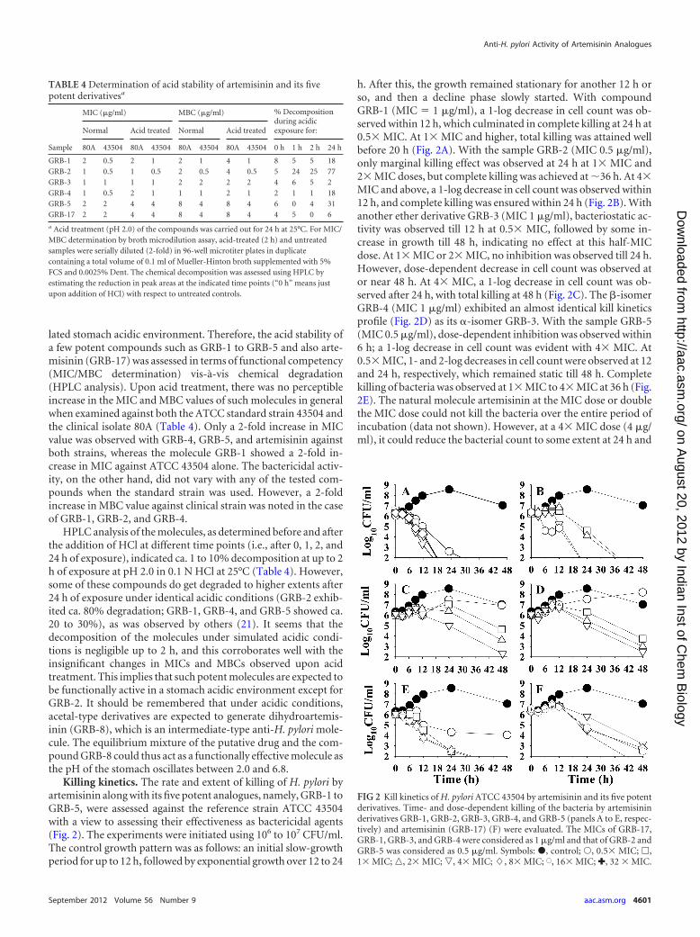
Killing kinetics. The rate and extent of killing of H. pylori byartemisinin along with its five potent analogues, namely, GRB-1 toGRB-5, were assessed against the reference strain ATCC 43504with a view to assessing their effectiveness as bactericidal agents(Fig. 2). The experiments were initiated using 106 to 107 CFU/ml.The control growth pattern was as follows: an initial slow-growthperiod for up to 12 h, followed by exponential growth over 12 to 24
h. After this, the growth remained stationary for another 12 h orso, and then a decline phase slowly started. With compoundGRB-1 (MIC � 1 g/ml), a 1-log decrease in cell count was ob-served within 12 h, which culminated in complete killing at 24 h at0.5 MIC. At 1 MIC and higher, total killing was attained wellbefore 20 h (Fig. 2A). With the sample GRB-2 (MIC 0.5 g/ml),only marginal killing effect was observed at 24 h at 1 MIC and2 MIC doses, but complete killing was achieved at �36 h. At 4MIC and above, a 1-log decrease in cell count was observed within12 h, and complete killing was ensured within 24 h (Fig. 2B). Withanother ether derivative GRB-3 (MIC 1 g/ml), bacteriostatic ac-tivity was observed till 12 h at 0.5 MIC, followed by some in-crease in growth till 48 h, indicating no effect at this half-MICdose. At 1 MIC or 2 MIC, no inhibition was observed till 24 h.However, dose-dependent decrease in cell count was observed ator near 48 h. At 4 MIC, a 1-log decrease in cell count was ob-served after 24 h, with total killing at 48 h (Fig. 2C). The �-isomerGRB-4 (MIC 1 g/ml) exhibited an almost identical kill kineticsprofile (Fig. 2D) as its �-isomer GRB-3. With the sample GRB-5(MIC 0.5 g/ml), dose-dependent inhibition was observed within6 h; a 1-log decrease in cell count was evident with 4 MIC. At0.5 MIC, 1- and 2-log decreases in cell count were observed at 12and 24 h, respectively, which remained static till 48 h. Completekilling of bacteria was observed at 1 MIC to 4 MIC at 36 h (Fig.2E). The natural molecule artemisinin at the MIC dose or doublethe MIC dose could not kill the bacteria over the entire period ofincubation (data not shown). However, at a 4 MIC dose (4 g/ml), it could reduce the bacterial count to some extent at 24 h and
FIG 2 Kill kinetics of H. pylori ATCC 43504 by artemisinin and its five potentderivatives. Time- and dose-dependent killing of the bacteria by artemisininderivatives GRB-1, GRB-2, GRB-3, GRB-4, and GRB-5 (panels A to E, respec-tively) and artemisinin (GRB-17) (F) were evaluated. The MICs of GRB-17,GRB-1, GRB-3, and GRB-4 were considered as 1 g/ml and that of GRB-2 andGRB-5 was considered as 0.5 g/ml. Symbols: �, control; Œ, 0.5 MIC; �,1 MIC; o, 2 MIC; p, 4 MIC; �, 8 MIC; �, 16 MIC; ✚, 32 MIC.
TABLE 4 Determination of acid stability of artemisinin and its fivepotent derivativesa
Sample
MIC (g/ml) MBC (g/ml) % Decompositionduring acidicexposure for:Normal Acid treated Normal Acid treated
80A 43504 80A 43504 80A 43504 80A 43504 0 h 1 h 2 h 24 h
GRB-1 2 0.5 2 1 2 1 4 1 8 5 5 18GRB-2 1 0.5 1 0.5 2 0.5 4 0.5 5 24 25 77GRB-3 1 1 1 1 2 2 2 2 4 6 5 2GRB-4 1 0.5 2 1 1 1 2 1 2 1 1 18GRB-5 2 2 4 4 8 4 8 4 6 0 4 31GRB-17 2 2 4 4 8 4 8 4 4 5 0 6
a Acid treatment (pH 2.0) of the compounds was carried out for 24 h at 25°C. For MIC/MBC determination by broth microdilution assay, acid-treated (2 h) and untreatedsamples were serially diluted (2-fold) in 96-well microtiter plates in duplicatecontaining a total volume of 0.1 ml of Mueller-Hinton broth supplemented with 5%FCS and 0.0025% Dent. The chemical decomposition was assessed using HPLC byestimating the reduction in peak areas at the indicated time points (“0 h” means justupon addition of HCl) with respect to untreated controls.
Anti-H. pylori Activity of Artemisinin Analogues
September 2012 Volume 56 Number 9 aac.asm.org 4601
on August 20, 2012 by Indian Inst of C
hem B
iologyhttp://aac.asm
.org/D
ownloaded from
drastically kill the bacteria at the end of 48 h. Using still higherdoses of 8 MIC, 16 MIC, and 32 MIC, only a 2-log decreasein cell count was observed after 24 h, although total killing wasattained only at 48 h (Fig. 2F). Summarizing, GRB-1 appeared tobe the strongest anti-H. pylori molecule among the potent com-pounds tested, as far as the killing efficacy is concerned. At a doserange of 1 to 4 g/ml, it induced complete killing of the bacteriawithin 20 h, whereas at 0.5 g/ml complete killing was observedonly after 24 h. GRB-5 appeared to be the next most effective,exhibiting complete killing at 0.5 to 2 g/ml after 30 h. The thirdpromising candidate is GRB-2, which at 0.5 to 1 g/ml killed thebacteria within 36 h, while at the higher dose of 2 to 8 g/ml thesame was achieved at �24 h. The compounds GRB-3 and GRB-4,on the other hand, appeared to be less active among these potentmolecules in exhibiting their bactericidal potential. To summa-rize, all of the compounds exhibited true bactericidal activity (�3-log decrease in viable cell count) after 24 h and are more effectivethan the natural molecule artemisinin (GRB-17) as far as killingefficacy is concerned.
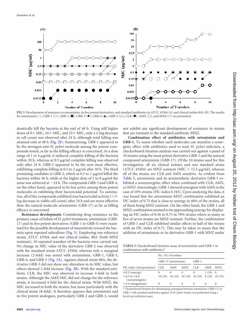
Resistance development. Considering drug resistance as theprimary cause of failure of H. pylori treatment, artemisinin (GRB-17) and its five potent derivatives (GRB-1 to GRB-5) were exam-ined for the possible development of insensitivity toward the bac-teria upon repeated subculture (Fig. 3). Employing one referencestrain, ATCC 43504, and one clinical isolate, 80A (both MNZresistant), 10 repeated transfers of the bacteria were carried out.No change in MIC value of the derivative GRB-2 was observedwith the standard strain ATCC 43504, whereas only a marginalincrease (2-fold) was noted with artemisinin, GRB-1, GRB-3,GRB-4, and GRB-5 (Fig. 3A). Against clinical strain 80A, the de-rivative GRB-3 did not show any alteration in its MIC value, butothers showed 2-fold increase (Fig. 3B). With the standard anti-biotic CLR, the MIC was observed to increase 4-fold in bothstrains. Although the AMX MIC did not change for the referencestrain, it increased 4-fold for the clinical strain. With MNZ, theMIC increased in both the strains, but more particularly with theclinical strain (8-fold). It therefore appears that artemisinin andits five potent analogues, particularly GRB-2 and GRB-3, would
not exhibit any significant development of resistance in strainsthat are resistant to the standard antibiotic MNZ.
Combination effect of antibiotics with artemisinin andGRB-1. To assess whether such molecules can manifest a syner-gistic effect with antibiotics used to treat H. pylori infection, acheckerboard titration analysis was carried out against a panel of10 strains using the most potent derivative GRB-1 and the naturalcompound artemisinin (GRB-17). Of the 10 strains used for thisinvestigation, all six clinical isolates and one standard strain(ATCC 43504) are MNZ resistant (MIC � 12.5 g/ml), whereasall of the strains are CLR and AMX sensitive. As evident fromTable 5, artemisinin and its semisynthetic derivative GRB-1 ex-hibited a nonsynergistic effect when combined with CLR, AMX,or MNZ. Interestingly, GRB-1 showed synergism with AMX in thecase of 10% strains (FIC index 0.345). Upon analyzing the data, itwas found that the artemisinin-MNZ combination exhibited anFIC index of 0.75 that is close to synergy in 40% of the strains, allof them being MNZ resistant. On the other hand, the GRB-1 andMNZ combination seemed to be approaching synergy for display-ing an FIC index of 0.56 to 0.75 in 70% strains where as many asfive of seven strains are MNZ resistant. Further, the combinationof GRB-1 and CLR exhibited similar effects in half of the strainswith an FIC index of 0.75. This may be taken to mean that theaddition of artemisinin or its derivative GRB-1 with MNZ under
FIG 3 Development of resistance to artemisinin, its five potent derivatives, and standard antibiotics in ATCC 43504 (A) and clinical isolate 80A (B). The resultsfor artemisinin (�), GRB-1 (Œ), GRB-2 (�), GRB-3 (}), GRB-4 (Œ), GRB-5 (o), CLR (�), AMX (�), and MNZ (�) are presented.
TABLE 5 Checkerboard titration assay of artemisinin and GRB-1 incombination with antibioticsa
FIC index (interpretation)
No. (%) of isolates
GRB-17 (artemisinin) GRB-1
CLR AMX MNZ CLR AMX MNZ
�0.5 (synergy) 0 0 0 0 1 (10) 0�0.5 to �4.0
(nonsynergistic)10 (10) 10 (10) 10 (10) 10 (10) 9 (10) 10 (10)
�4.0 (antagonism) 0 0 0 0 0 0a Checkerboard titration for determining synergism between artemisinin (GRB-17) orGRB-1 with standard antibiotics against 10 H. pylori strains was performed using abroth microdilution assay.
Goswami et al.
4602 aac.asm.org Antimicrobial Agents and Chemotherapy
on August 20, 2012 by Indian Inst of C
hem B
iologyhttp://aac.asm
.org/D
ownloaded from
in vitro conditions has resulted in the development of sensitivity inMNZ-resistant strains. For example, the standard strain ATCC43504 is MNZ resistant (MIC 50 g/ml) but, in combination with1 g of GRB-1/ml, the MIC value of MNZ was reduced to 6.25g/ml, resulting in an FIC index of 0.625. The MIC value of MNZthus decreased 8-fold, indicating that GRB-1 in combination withMNZ can enhance the activity of the antibiotic against resistantstrains. The same observation was noted against clinical strain80A, where MNZ alone showed an MIC of 50 g/ml, while incombination with GRB-1 (1 g/ml), the MIC value decreased to6.25 g/ml. In the case of MNZ-sensitive H. pylori, the MIC valuesof two of three strains were found to decrease 8- to 16-fold,whereas in all 10 CLR-sensitive strains a 4-fold decrease in MICvalues of CLR was observed with half of the strains. Taking all ofthese results into account, it is evident that both GRB-1 andGRB-17 lack the potential to exhibit synergism with the threeantibiotics tested.
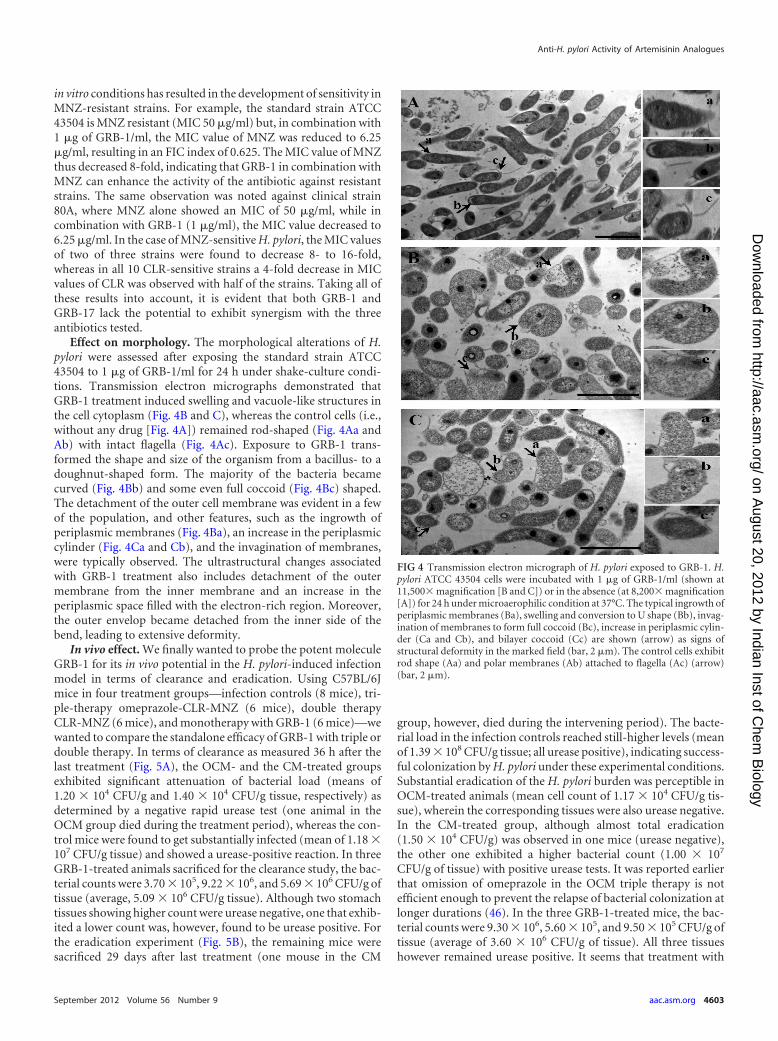
Effect on morphology. The morphological alterations of H.pylori were assessed after exposing the standard strain ATCC43504 to 1 g of GRB-1/ml for 24 h under shake-culture condi-tions. Transmission electron micrographs demonstrated thatGRB-1 treatment induced swelling and vacuole-like structures inthe cell cytoplasm (Fig. 4B and C), whereas the control cells (i.e.,without any drug [Fig. 4A]) remained rod-shaped (Fig. 4Aa andAb) with intact flagella (Fig. 4Ac). Exposure to GRB-1 trans-formed the shape and size of the organism from a bacillus- to adoughnut-shaped form. The majority of the bacteria becamecurved (Fig. 4Bb) and some even full coccoid (Fig. 4Bc) shaped.The detachment of the outer cell membrane was evident in a fewof the population, and other features, such as the ingrowth ofperiplasmic membranes (Fig. 4Ba), an increase in the periplasmiccylinder (Fig. 4Ca and Cb), and the invagination of membranes,were typically observed. The ultrastructural changes associatedwith GRB-1 treatment also includes detachment of the outermembrane from the inner membrane and an increase in theperiplasmic space filled with the electron-rich region. Moreover,the outer envelop became detached from the inner side of thebend, leading to extensive deformity.
In vivo effect. We finally wanted to probe the potent moleculeGRB-1 for its in vivo potential in the H. pylori-induced infectionmodel in terms of clearance and eradication. Using C57BL/6Jmice in four treatment groups—infection controls (8 mice), tri-ple-therapy omeprazole-CLR-MNZ (6 mice), double therapyCLR-MNZ (6 mice), and monotherapy with GRB-1 (6 mice)—wewanted to compare the standalone efficacy of GRB-1 with triple ordouble therapy. In terms of clearance as measured 36 h after thelast treatment (Fig. 5A), the OCM- and the CM-treated groupsexhibited significant attenuation of bacterial load (means of1.20 104 CFU/g and 1.40 104 CFU/g tissue, respectively) asdetermined by a negative rapid urease test (one animal in theOCM group died during the treatment period), whereas the con-trol mice were found to get substantially infected (mean of 1.18 107 CFU/g tissue) and showed a urease-positive reaction. In threeGRB-1-treated animals sacrificed for the clearance study, the bac-terial counts were 3.70 105, 9.22 106, and 5.69 106 CFU/g oftissue (average, 5.09 106 CFU/g tissue). Although two stomachtissues showing higher count were urease negative, one that exhib-ited a lower count was, however, found to be urease positive. Forthe eradication experiment (Fig. 5B), the remaining mice weresacrificed 29 days after last treatment (one mouse in the CM
group, however, died during the intervening period). The bacte-rial load in the infection controls reached still-higher levels (meanof 1.39 108 CFU/g tissue; all urease positive), indicating success-ful colonization by H. pylori under these experimental conditions.Substantial eradication of the H. pylori burden was perceptible inOCM-treated animals (mean cell count of 1.17 104 CFU/g tis-sue), wherein the corresponding tissues were also urease negative.In the CM-treated group, although almost total eradication(1.50 104 CFU/g) was observed in one mice (urease negative),the other one exhibited a higher bacterial count (1.00 107
CFU/g of tissue) with positive urease tests. It was reported earlierthat omission of omeprazole in the OCM triple therapy is notefficient enough to prevent the relapse of bacterial colonization atlonger durations (46). In the three GRB-1-treated mice, the bac-terial counts were 9.30 106, 5.60 105, and 9.50 105 CFU/g oftissue (average of 3.60 106 CFU/g of tissue). All three tissueshowever remained urease positive. It seems that treatment with
FIG 4 Transmission electron micrograph of H. pylori exposed to GRB-1. H.pylori ATCC 43504 cells were incubated with 1 g of GRB-1/ml (shown at11,500 magnification [B and C]) or in the absence (at 8,200 magnification[A]) for 24 h under microaerophilic condition at 37°C. The typical ingrowth ofperiplasmic membranes (Ba), swelling and conversion to U shape (Bb), invag-ination of membranes to form full coccoid (Bc), increase in periplasmic cylin-der (Ca and Cb), and bilayer coccoid (Cc) are shown (arrow) as signs ofstructural deformity in the marked field (bar, 2 m). The control cells exhibitrod shape (Aa) and polar membranes (Ab) attached to flagella (Ac) (arrow)(bar, 2 m).
Anti-H. pylori Activity of Artemisinin Analogues
September 2012 Volume 56 Number 9 aac.asm.org 4603
on August 20, 2012 by Indian Inst of C
hem B
iologyhttp://aac.asm
.org/D
ownloaded from

GRB-1 alone at 50 mg/kg/day (single therapy) could reduce the H.pylori load in vivo to some extent during clearance stage and per-haps a little more at the eradication stage. Such indications usingmonotherapy are encouraging, especially since this novel artemis-inin derivative did not elicit any toxicity at this dose, whereas wetypically observed diarrhea-like symptom in most of the animalstreated with OCM and CM.
In vitro cytotoxicity evaluation. In order to assess the cytotox-icity, all 17 molecules were tested against five different cell lines.Except for GRB-10 and GRB-11, most of the compounds, such asGRB-1 to GRB-5, GRB-12, GRB-13, GRB-16, and GRB-17 (arte-misinin), appeared to be safe since none of them showed anypotential cytotoxic effect around their MBC50 values against H.pylori (Table 6). The cytotoxicity values obtained with GRB-8,GRB-14, and GRB-17 correlate well with the experimental find-ings of other groups (see, for example, reference 5), and hence the
activity of eight new molecules, including GRB-1 to GRB-5, couldbe accepted as valid. The compounds GRB-6 to GRB-9, GRB-14,and GRB-15 can be considered intermediate cytotoxic. Neverthe-less, in the tumor macrophage cell line (RAW 264.7), but not inthe normal macrophage primary culture, the majority of the mol-ecules exhibited cytotoxicity. The potential lethal effects of mostof the compounds against RAW 264.7 cell lines indicate that all ofthe compounds probably have dramatic effect on tumor-associ-ated macrophages where the cells automatically develop anti-in-flammatory cytokines such as interleukin-10 and show signs oftissue repair functions (14). In contrast, none of the compoundsexerted cytotoxic effects against normal mouse macrophage celllines (which generate huge amounts of proinflammatory cyto-kines, express elevated levels of major histocompatibility complexmolecules, and are powerful killers of pathogens and tumor cells).
Pharmacokinetic property predictions. The compounds
FIG 5 In vivo effect of GRB-1, dual therapy (CM [clarithromycin and metronidazole]), and triple therapy (OCM [omeprazole, clarithromycin, and metroni-dazole]) on H. pylori infection in C57BL/6J mice. Clearance (A) indicates reduction in bacterial load 36 h after the cessation of treatment, whereas eradication (B)indicates the same 29 days after the treatment (reduction in bacterial load in terms of 8 mice for the infection control, five each for OCM and CM, and six forGRB-1, 50 mg/kg/day). The animals were sacrificed after 36 h (4 mice for the infection control, 2 mice for OCM, 3 mice with CM and GRB-1 each) and 29 days(4 mice for the infection control, 3 mice for OCM, 2 mice for CM, and 3 mice GRB-1) after the last treatment. Each point represents the viable count obtainedfrom each animal in the particular group.
TABLE 6 Cytotoxic activity and pharmacokinetic parameters of the test compoundsa
Sample code(mol wt)
H. pylori activity(MBC50 [g/ml])
Cytotoxicity (IC50 [g/ml]) Pharmacokinetic parameters
HepG2 MCF-7 AGS RAW 264.7 MPM log P log S PPBb (%)
GRB-1 (338) 2 �100 50 50 10 �100 4.0407 –4.17808 �90GRB-2 (312) 4 �100 75 75 10 100 3.6733 –3.87658 �90GRB-3 (298) 1 �100 �100 �100 75 �100 3.3308 –3.54937 �90GRB-4 (298) 1 �100 100 100 75 �100 3.3308 –3.54937 �90GRB-5 (364) 1 �100 50 75 10 75 4.0558 –5.06884 �90GRB-6 (443) �32 75 100 75 5 50 6.1434 –6.78585 �95GRB-7 (423) 8 �50 100 100 5 75 5.6125 –6.31709 �95GRB-8 (284) 4 �50 50 100 5 100 3.0525 –3.20419 �90GRB-9 (418) 8 75 50 50 5 �100 4.4419 –5.27237 �95GRB-10 (366) �16 75 50 30 0.5 75 4.8333 –5.20852 �90GRB-11 (474) �8 �100 0.5 0.5 0.5 75 7.1118 –9.07303 �95GRB-12 (234) �128 100 �100 �100 100 �100 3.4474 –4.21376 �90GRB-13 (267) 4 �100 100 100 100 �100 3.2401 –2.97035 �90GRB-14 (384) 2 50 100 100 1 10 2.9735 –3.57458 �90GRB-15 (478) 4 50 100 100 1 50 4.3364 –5.73394 �95GRB-16 (338) 2 �100 100 100 10 100 4.0407 –4.17808 �90GRB-17 (282) 8 �100 �100 �100 �100 �100 3.0853 –3.47168 �90Mitomycin C 1.1 6.8 2.5 3.3a For evaluation of cytotoxicity by an MTT assay, test compounds and the standard mitomycin C were examined over dose ranges of 100 to 0.5 g/ml and 16.7 to 0.17 g/ml,respectively. The pharmacokinetic parameters were calculated by using Cerius2 software, version 4.10.b PPB, plasma protein binding.
Goswami et al.
4604 aac.asm.org Antimicrobial Agents and Chemotherapy
on August 20, 2012 by Indian Inst of C
hem B
iologyhttp://aac.asm
.org/D
ownloaded from
showed lipophilicity, with log P values in the range of 2.9735 to7.1118, and aqueous solubility, with log S values in the range of�2.97035 to �6.78585. GRB-6, GRB-7, and GRB-11 were themost lipophilic compounds of the series and thus considered notappropriate for drug therapy. Interestingly, the five potent anti-H.pylori molecules (GRB-1 to GRB-5), along with artemisinin(GRB-17) and dihydroartemisinin (GRB-8), exhibited log P val-ues in the range of 3.0853 to 4.0558 and log S values in the range of�3.20419 to �5.06884, indicating their potential as candidatedrugs (Table 6). Plasma protein binding (PPB) levels determinethe extent to which a compound is likely to be bound with theprotein fraction of blood. This influences the half-life of the com-pound in the body and hence is an important parameter. Most ofthe molecules exhibited acceptable PPB levels with a bindingefficiency of �90% (PPB level 0), including artemisinin, dihydro-artemisinin, and the five potent compounds. GRB-10 had an effi-ciency of �90% (PPB level 1) and GRB-6, GRB-7, GRB-9, GRB-11, and GRB-15 had an efficiency of �95% (PPB level 2),indicating a lower probability of free drug concentration at the siteof action and hence were considered less suitable for drug therapy.
DISCUSSION
Considering the widespread and commensal nature of H. pyloriand the rate at which resistance to some of the more commonantibiotics used in H. pylori treatments manifests jeopardizingtheir effectiveness in managing other infections (7, 26, 32, 34), it isnecessary to look for chemotherapeutic agents that should be botheffective and safe in eradicating both antibiotic-susceptible and-resistant strains. A novel anti-H. pylori drug should (i) exert po-tent and selective antibacterial action against both clinical andstandard H. pylori strains, (ii) be stable at the acidic pH of thestomach, (iii) show antibacterial action against CLR-resistant andMNZ-resistant strains, (iv) not lead to the development of drugresistance, (v) possess wide safety, and (vi) be easily affordable.The present investigation was therefore designed to find potentanti-H. pylori molecules and also to consider their functional use-fulness in identifying prospective candidate leads.
Based on our serendipitous observation that artemisinin couldbe effective against H. pylori, we evaluated a series of semisyntheticanalogues, including a few newly synthesized molecules, with aview to profiling their anti-H. pylori features. When screened fortheir potential in terms of disc diffusion sensitivity, the MIC andMBC values against a panel of clinical and standard strains of H.pylori, 5 of 17 compounds, namely, �-artecyclopropylmether,�-arteether, �-artemether, �-artemether, and �-artefurfurylether(GRB-1 to GRB-5) proved to be reasonably prospective (Tables 1and 2). Such strongly active compounds can be considered spe-cific anti-H. pylori agents because all of them demonstrated a lackof sensitivity against the majority of other bacterial and fungalstrains tested, barring perhaps C. parapsilosis; �-artefurfurylether,however, exhibited the highest antimicrobial activity of the fivemolecules (Table 3). Looking at the structural features of the com-pounds, it seems that a small, nonpolar, �-oriented C-12 substit-uent besides the endoperoxide linkage confers higher activity. Pre-sumably, the �-substituent, having the same orientation as theperoxo bridge, offers steric hindrance and interferes with the ac-tivity.
We performed a series of experiments with these five alkoxyderivatives to probe their functional efficacy. The derivativesshowed no significant changes in their MIC and MBC values even
after exposure to acidic pH (Table 4). This finding is in contrast toCLR, for which the MIC/MBC increases with a decrease in pH (8),necessitating the enhancement of the stomach pH by gastric anti-secretory agents such as H2 receptor blockers or proton pumpinhibitors for the effective clearance and eradication of H. pylori-induced gastroduodenal ulcers by the antibiotics (46). Thus, func-tional competency at a stomach acidic pH would augur well forsuch smaller alkoxy derivatives of artemisinin. Second, all fivemolecules showed potential activity even against resistant strains,a desirable attribute for any putative anti-H. pylori agent. This ismore important because of the indiscriminate use of antibioticssuch as CLR, AMX, tetracycline, and particularly MNZ for curingH. pylori-induced gastric ulcer, leading to the development of re-sistance to this organism (7, 12, 26, 32). Third, the killing efficacyof artemisinin and its five derivatives indicated �-artecyclopropy-lmether as the most potent molecule, which killed bacteria within24 h at 0.5 g/ml and within 20 h at �1 g/ml (Fig. 2A). The nexteffective molecule is �-artefurfurylether, which showed killingwithin 36 h at doses of 0.5 to 2 g/ml (Fig. 2E). Interestingly,�-artecyclopropylmether was found to be much less active than its�-isomer (data not shown). Fourth, the five potent compoundsdid not show any tendency to develop resistance against either theclinical isolate or standard strain (Fig. 3). Thus, such moleculeswould not be expected to develop any resistance and, therefore,could prove useful in treating H. pylori infection successfully, par-ticularly in low socioeconomic settings where majority of the iso-lated strains were found to be antibiotic resistant (7, 33, 34). It wasof further interest to see whether such activity is due to the man-ifestation of urease inhibition as H. pylori surface is richly coatedwith urease and is considered as a putative target in designingpotential anti-H. pylori principles (28). However, artemisinin andthe potent five analogues did not inhibit H. pylori urease in vitro(data not shown).
A comparative viewing of these five derivatives in terms of IZD,MIC, and MBC values, as well as the four experimental outcomesdescribed above, indicated that in majority of the experiments,both �-artecyclopropylmether and �-artefurfurylether proved tobe superior compared to �-arteether, �-artemether, and �-arte-mether. However, in terms of killing efficacy, �-artecyclopropyl-mether proved to be the most potent. We therefore further exam-ined this lead molecule for its capacity to induce morphologicaldeformity, show synergism with anti-H. pylori antibiotics, andreduce the H. pylori burden in vivo. The phenotypic changes of thebacteria in terms of morphological transformation from rod tospiral to coccoid form, and the cytological alteration, leading tothe formation of swelling and vacuole-like structures in the cyto-plasm with detachment of outer cell membranes (Fig. 4) have beendiscernible in the presence of �-artecyclopropylmether. Next, acheckerboard titration assay indicated that �-artecyclopropyl-mether in combination with MNZ under in vitro condition exhib-ited the development of sensitivity in MNZ-resistant strains, sug-gesting its potential as a therapeutic agent against resistant strains,although the compound could not qualify as synergistic with anyof the antibiotics tested (Table 5). The molecule, however, exhibitsa somewhat mild in vivo potential as a single therapeutic agent inthe chronic H. pylori infection model at a dose of 50 mg/kg/day(Fig. 5). It remains to be seen whether it can substitute one or bothantibiotics in triple OCM therapy or at least reduce the dose re-quirement of such gastric irritant antibiotics. Since artemisininand other derivatives, including �-artecyclopropylmether, did
Anti-H. pylori Activity of Artemisinin Analogues
September 2012 Volume 56 Number 9 aac.asm.org 4605
on August 20, 2012 by Indian Inst of C
hem B
iologyhttp://aac.asm
.org/D
ownloaded from
not exhibit gastric anti-H�,K�-ATPase activity (data not shown),we do not anticipate the molecule to replace omeprazole in tripletherapy. Further experiments are required to be carried out with�-artecyclopropylmether and CLR-MNZ in a combination for-mat and also as a function of dosage. Although the majority of thein vivo validation experiments with newly discovered anti-H. py-lori principle(s) tend to indicate the necessity of simultaneous useof one or two conventional antibiotics, albeit with lower doses(24), success with monotherapy has also been reported (22).
In an attempt to see the role of antimalarial drugs devoid ofperoxide linkage, we have examined seven commonly used anti-malarials, namely, quinine, chloroquine, primaquine, pyrimeth-amine, lumefantrine, piperaquine, and mefloquine, against oneclinical strain (80A) and one standard strain (ATCC 49503) of H.pylori. Barring mefloquine (MIC � 16 to 32 g/ml) and pyri-methamine (MIC � 32 to 64 g/ml), none could exert any signif-icant anti-H. pylori potential. It may not be out of place to men-tion here that a recent epidemiological study in child refugees inWestern Australia indicated that premigration antimalarial treat-ment significantly reduced the odds of H. pylori infection afteradjusting for age and sex and therefore predicted a protective ef-fect of antimalarials against H. pylori infection (6). An earlier invitro study with backpackers from developed countries after travelto tropical countries indicated that mefloquine, a traveler’s choiceantimalarial, has anti-H. pylori activity (39). Whereas the latterstudy corroborates the findings described above, the former studyindirectly supports our serendipitous observation that artemis-inin has strong anti-H. pylori effect. We therefore anticipate ob-serving the absence of H. pylori seroprevalence among malariapatients treated with artemisinin combination drugs as opposedto antimalarials devoid of artemisinin.
Whatever may be the mechanism(s) of action of artemisininanalogues in manifesting strong and potentially useful anti-H. py-lori features, it is worth reiterating that artemisinin is a cheap andrelatively safe drug with few and minor side effects even at highdoses. Because much of the pharmacokinetic and pharmacody-namic information is available, it can very well take a faster routein developing an appropriate candidate drug against H. pylori in-fections. Our efforts in evaluating physicochemical parameters(log P, aqueous solubility, and PPB) and the cytotoxicity profiles,particularly of the eight newly synthesized molecules, revealedthat the majority of the compounds, including GRB-1 to GRB-5,lie well within acceptable ranges to qualify as therapeutic candi-date (Table 6). Further investigation using quantitative structure-activity relationship studies would help predict which among suchpotent molecules would be best for the development of a candi-date drug for H. pylori treatment. Such preclinical findings must,however, be validated in a clinical setting to judge the efficacy ofthe molecule as a useful anti-H. pylori agent and hence as a gastricanti-ulcer principle.
ACKNOWLEDGMENTS
This study was supported by an interlaboratory network project (COR-0023) of the Council of Scientific and Industrial Research (CSIR). We alsothank the CSIR for the award of Senior Research Fellowships to S.G., A.C.,and S.K.K. during the course of this investigation.
We thank A. K. Mukhopadhyay, NICED, Kolkata, India, for providingclinical strains and one standard strain ATCC 700392 and M. SitaramKumar of Dr. Reddy’s Laboratory, Hyderabad, India, for the standardstrain ATCC 49503 of H. pylori. The other bacterial strains were kindly
provided by Anil Tyagi, University of Delhi South Campus, Delhi, India(all MTCC strains); Amita Jain, King George Medical University, Luc-know, India (MRSA strain); and A. K. Saxena, Indian Institute of Integra-tive Medicine, Jammu, India [S. aureus (J)]. Uma Banerjee of the Depart-ment of Microbiology, All India Institute of Medical Sciences, New Delhi,India, kindly provided all of the clinical fungal strains. We also thank S. K.Puri of Central Drug Research Institute (CSIR-CDRI), Lucknow, India,for kindly providing the seven antimalarial compounds. We acknowledgethe interpretation of EM pictures by J. Sengupta, the calculation of phys-icochemical values (log P, log S, and PPB) by M. Prabu, and the HPLCstudy by B. Chakraborty of CSIR-IICB. We are indebted to B. Achari(CSIR-IICB) for valuable comments and critical review of the manuscript.
REFERENCES1. Andersen LP. 2007. Colonization and infection by Helicobacter pylori in
humans. Helicobacter 12(Suppl 2):12–15.2. Bauer AW, Kirby WM, Sherris JC, Turck M. 1966. Antibiotic suscepti-
bility testing by a standardized single disk method. Am. J. Clin. Pathol.45:493– 496.
3. Bhakuni RS, Jain DC, Sharma RP. 1995. An improved procedure for thesynthesis of ether of dihydroartemisinin. Ind. J. Chem. 34B:529 –530.
4. Bhakuni RS, et al. January 2004. Single pot conversion of artemisinin intoartemether. U.S. patent 6,683,193, MY-129031-A(2007).
5. Chaijaroenkul W, Viyanant V, Mahavorasirikul W, Na-Bangchang K.2011. Cytotoxic activity of artemisinin derivatives against cholangiocarci-noma (CL-6) and hepatocarcinoma (Hep-G2) cell lines. Asian Pac. J. Can-cer Prev. 12:55–59.
6. Cherian S, Forbes D, Sanfilippo F, Cook A, Burgner D. 2008. Theepidemiology of Helicobacter pylori infection in African refugee childrenresettled in Australia. Med. J. Aust. 189:438 – 441.
7. Datta S, et al. 2005. Most Helicobacter pylori strains of Kolkata in India areresistant to metronidazole but susceptible to other drugs commonly usedfor eradication and ulcer therapy. Aliment. Pharmacol. Ther. 22:51–57.
8. Erah PO, Goddard AF, Barrett DA, Shaw PN, Spiller RC. 1997. Thestability of amoxicillin, clarithromycin, and metronidazole in gastric juice:relevance to the treatment of Helicobacter pylori infection. J. Antimicrob.Chemother. 39:5–12.
9. Fahey TJ, et al. 1992. Macrophage inflammatory protein 1 modulatesmacrophage function. J. Immunol. 148:2764 –2769.
10. Funatogawa K, et al. 2004. Antibacterial activity of hydrolyzable tanninsderived from medicinal plants against Helicobacter pylori. Microbiol. Im-munol. 48:251–261.
11. Glupczynski Y. 1996. Culture of Helicobacter pylori from gastric biopsiesand antimicrobial susceptibility testing, p 17–28. In Lee A, Megraud F(ed), Helicobacter pylori: techniques for clinical diagnosis and basic re-search. WB Saunders Co, Ltd, London, England.
12. Graham DY, Qureshi WA. 2000. Antibiotic-resistant Helicobacter pyloriinfection and its treatment. Curr. Pharm. Des. 6:1537–1544.
13. Hachem CY, et al. 1996. Antimicrobial susceptibility testing of Helico-bacter pylori: comparison of E-test, broth microdilution, and disc diffu-sion for ampicillin, clarithromycin, and metronidazole. Diagn. Microbiol.Infect. Dis. 24:37– 41.
14. Hagemann T, et al. 2008. “Re-educating” tumor-associated macrophagesby targeting NF- B. J. Exp. Med. 205:1261–1268.
15. Haynes RK, et al. 2002. C-10 ester and ether derivatives of dihydroarte-misinin–10-� artesunate, preparation of authentic 10-� artesunate, and ofother ester and ether derivatives bearing potential aromatic intercalatinggroups at C-10. Eur. J. Org. Chem. 2002:113–132.
16. Hou TJ, Xia K, Zhang W, Xu XJ. 2004. ADME evaluation in drugdiscovery. 4. prediction of aqueous solubility based on atom contributionapproach. J. Chem. Infect. Comput. Sci. 44:266 –275.
17. Iwao E, Yamamoto K, Yokoyama Y, Hirayama F, Haga K. 2004. Potentantibacterial activity of Y-754, a novel benzimidazole compound withselective action against Helicobacter pylori. J. Infect. Chemother. 10:90 –96.
18. Jain DC, et al. September 1999. Process for the simultaneous productionof artemisinin and essential oil from plant Artemisia annua. U.S. patent5,955,084.
19. Jayaraman KS. 2003. Technology, tradition unite in India’s drug discov-ery scheme. Nat. Med. 9:982.
20. Jung M, Lee K, Kim H, Park M. 2004. Recent advances in artemisinin
Goswami et al.
4606 aac.asm.org Antimicrobial Agents and Chemotherapy
on August 20, 2012 by Indian Inst of C
hem B
iologyhttp://aac.asm
.org/D
ownloaded from

and its derivatives as antimalarial and antitumor agents. Curr. Med.Chem. 11:1265–1284.
21. Jung M, Lee S. 1998. Stability of acetal and non-acetal-type analogs ofartemisinin in simulated stomach acid. Bioorg. Med. Chem. Lett. 8:1003–1006.
22. Kanamaru T, et al. 2001. In vitro and in vivo antibacterial activities ofTAK-083, an agent for treatment of Helicobacter pylori infection. Antimi-crob. Agents Chemother. 45:2455–2459.
23. Kawase M, Motohashi N. 2004. Plant-derived leading compound foreradication of Helicobacter pylori. Curr. Med. Chem. 3:89 –100.
24. Koga T, et al. 2002. Effect of plaunotol in combination with clarithromy-cin or amoxicillin on Helicobacter pylori in vitro and in vivo. J. Antimicrob.Chemother. 50:133–136.
25. Kuhn T, Wang Y. 2008. Artemisinin: an innovative cornerstone for anti-malaria therapy. Prog. Drug Res. 383:385– 422.
26. Kwon DH, et al. 2000. Isolation and characterization of tetracycline-resistant clinical isolates of Helicobacter pylori. Antimicrob. Agents Che-mother. 44:3203–3205.
27. Li J, Zhou B. 2010. Biological actions of artemisinin: insights from me-dicinal chemistry studies. Molecules 15:1378 –1397.
28. Lin YT, Kwon YI, Labbe RG, Shetty K. 2005. Inhibition of Helicobacterpylori and associated urease by oregano and cranberry phytochemical syn-ergies. Appl. Environ. Microbiol. 71:8558 – 8564.
29. Malfertheiner P, et al. 2005. Helicobacter pylori eradication has the po-tential to prevent gastric cancer: a state-of-the-art critique. Am. J. Gastro-enterol. 100:2100 –2115.
30. Marshall BJ. 2001. One hundred years of discovery and rediscovery ofHelicobacter pylori and its association with peptic ulcer disease, p 19 –24. InMobley HLT, Mendz GL, Hazell SL (ed), Helicobacter pylori: physiologyand genetics. ASM Press, Washington, DC.
31. McNulty C, et al. 2002. Helicobacter pylori susceptibility testing by discdiffusion. J. Antimicrob. Chemother. 49:601– 609.
32. Megraud F. 2004. Basis for the management of drug-resistant Helicobacterpylori infection. Drugs 64:1893–1904.
33. Megraud F, Lehn N, Lind T. 1997. The MACH 2 Study: Helicobacterpylori resistance to antimicrobial agents and its influence on clinical out-come. Gastroenterology 112:A216.
34. Mendonça S, et al. 2000. Prevalence of Helicobacter pylori resistance tometronidazole, clarithromycin, amoxicillin, tetracycline, and furazoli-done in Brazil. Helicobacter 5:79 – 83.
35. Mosmann T. 1983. Rapid colorimetric assay for cellular growth and sur-vival: application to proliferation and cytotoxicity assays. J. Immunol.Methods 65:55– 63.
36. Nostro A, et al. 2005. Antibacterial effect of plant extracts against Heli-cobacter pylori. Phytother. Res. 19:198 –202.
37. O’Neill PM, et al. 2001. Optimization of the allylsilane approach to C-10deoxo-carba analogues of dihydroartemisinin: synthesis and in vitro anti-malarial activity of new, metabolically stable C-10 analogues. J. Chem.Soc. Perkin Trans. 1:2682–2689.
38. O’Rourke J, Bode G. 2001. Morphology and ultrastructure, p 53– 67. InMobley HLT, Mendz GL, Hazell SL (ed), Helicobacter pylori: physiologyand genetics. ASM Press, Washington, DC.
39. Potasman I, Yitzhak A. 1998. Helicobacter pylori serostatus in backpack-ers following travel to tropical countries. Am. J. Trop. Med. Hyg. 58:305–308.
40. Posner GH, et al. 1999. Antimalarial, antiproliferative, and antitumoractivities of artemisinin-derived, chemically robust, trioxane dimers. J.Med. Chem. 42:4275– 4280.
41. Schuster BG. 2001. Demonstrating the validity of natural products asanti-infective drugs. J. Altern. Complement. Med. 7(Suppl 1):73– 82.
42. Shrimali M, Bhattacharya AK, Bhakuni RS, Jain DC, Sharma RP. 2001.A process for the preparation of sodium artelinate. Indian patent 185198.
43. Simon JE, Chadwick AF, Craker LE. 1984. Herbs: an indexed bibliogra-phy, p 1971–1980. In The scientific literature on selected herbs and aro-matic and medicinal plants of the temperate zone. Archon Books, Ham-den, CT.
44. Singh BL, et al. 2001. Simultaneous determination of antimalarial arte-misinin, dihydroartemisinin and arteether using reversed phase high per-formance liquid chromatography. J. Ind. Chem. Soc. 78:489 – 491.
45. Tawfik AF, Bishop SJ, Ayalp A, el-Feraly FS. 1990. Effects of artemisinin,dihydroartemisinin and arteether on immune responses of normal mice.Int. J. Immunopharmacol. 12:385–389.
46. van Zanten SJOV, Kolesnikow T, Leung V, O’Rourke JL, Lee A. 2003.Gastric transitional zones, areas where Helicobacter treatment fails: resultsof a treatment trial using the Sydney strain mouse model. Antimicrob.Agents Chemother. 47:2249 –2255.
47. Wang J, Guo Y, Zhang BC, Chen ZT, Gao JF. 2007. Induction ofapoptosis and inhibition of cell migration and tube-like formation bydihydortemisinin in murine lymphatic endothelial cells. Pharmacology80:207–218.
48. Williamson JS. 2001. Helicobacter pylori: current chemotherapy and newtargets for drug design. Curr. Pharm. Des. 7:355–392.
49. Yanagawa Y, Yamamoto Y, Hara Y, Shimamura T. 2003. A combinationeffect of epigallocatechin gallate, a major compound of green tea cat-echins, with antibiotics on Helicobacter pylori growth in vitro. Curr. Mi-crobiol. 47:244 –249.
Anti-H. pylori Activity of Artemisinin Analogues
September 2012 Volume 56 Number 9 aac.asm.org 4607
on August 20, 2012 by Indian Inst of C
hem B
iologyhttp://aac.asm
.org/D
ownloaded from