Embed Size (px)
Citation preview
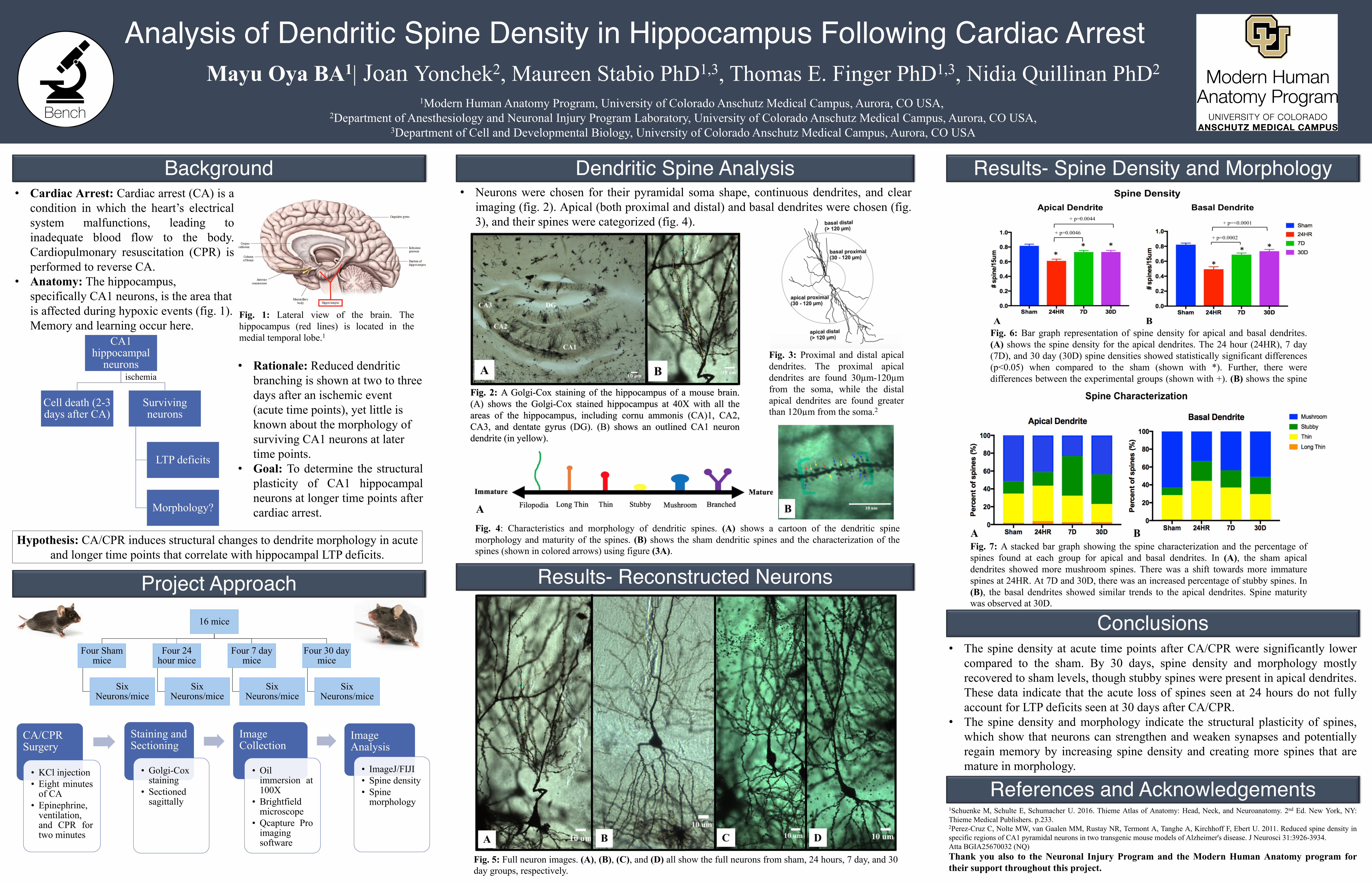
• Cardiac Arrest: Cardiac arrest (CA) is acondition in which the heart’s electricalsystem malfunctions, leading toinadequate blood flow to the body.Cardiopulmonary resuscitation (CPR) isperformed to reverse CA.
• Anatomy: The hippocampus, specifically CA1 neurons, is the area that is affected during hypoxic events (fig. 1). Memory and learning occur here.
Analysis of Dendritic Spine Density in Hippocampus Following Cardiac Arrest
• The spine density at acute time points after CA/CPR were significantly lowercompared to the sham. By 30 days, spine density and morphology mostlyrecovered to sham levels, though stubby spines were present in apical dendrites.These data indicate that the acute loss of spines seen at 24 hours do not fullyaccount for LTP deficits seen at 30 days after CA/CPR.
• The spine density and morphology indicate the structural plasticity of spines,which show that neurons can strengthen and weaken synapses and potentiallyregain memory by increasing spine density and creating more spines that aremature in morphology.
Mayu Oya BA1| Joan Yonchek2, Maureen Stabio PhD1,3, Thomas E. Finger PhD1,3, Nidia Quillinan PhD2
1Modern Human Anatomy Program, University of Colorado Anschutz Medical Campus, Aurora, CO USA,2Department of Anesthesiology and Neuronal Injury Program Laboratory, University of Colorado Anschutz Medical Campus, Aurora, CO USA,
3Department of Cell and Developmental Biology, University of Colorado Anschutz Medical Campus, Aurora, CO USA
1Schuenke M, Schulte E, Schumacher U. 2016. Thieme Atlas of Anatomy: Head, Neck, and Neuroanatomy. 2nd Ed. New York, NY:Thieme Medical Publishers. p.233.2Perez-Cruz C, Nolte MW, van Gaalen MM, Rustay NR, Termont A, Tanghe A, Kirchhoff F, Ebert U. 2011. Reduced spine density inspecific regions of CA1 pyramidal neurons in two transgenic mouse models of Alzheimer's disease. J Neurosci 31:3926-3934.Atta BGIA25670032 (NQ)
• Neurons were chosen for their pyramidal soma shape, continuous dendrites, and clearimaging (fig. 2). Apical (both proximal and distal) and basal dendrites were chosen (fig.3), and their spines were categorized (fig. 4).
Dendritic Spine Analysis
References and Acknowledgements
CA/CPR Surgery
• KCl injection• Eight minutes
of CA• Epinephrine,
ventilation,and CPR fortwo minutes
Staining and Sectioning
• Golgi-Coxstaining
• Sectionedsagittally
Image Collection
• Oilimmersion at100X
• Brightfieldmicroscope
• Qcapture Proimagingsoftware
Image Analysis
• ImageJ/FIJI• Spine density• Spine
morphology
Hypothesis: CA/CPR induces structural changes to dendrite morphology in acute and longer time points that correlate with hippocampal LTP deficits.
Background
16 mice
Four Sham mice
Six Neurons/mice
Four 24 hour mice
Six Neurons/mice
Four 7 day mice
Six Neurons/mice
Four 30 day mice
Six Neurons/mice
• Rationale: Reduced dendritic branching is shown at two to three days after an ischemic event (acute time points), yet little is known about the morphology of surviving CA1 neurons at later time points.
• Goal: To determine the structuralplasticity of CA1 hippocampalneurons at longer time points aftercardiac arrest.
Project Approach
Conclusions
Thank you also to the Neuronal Injury Program and the Modern Human Anatomy program fortheir support throughout this project.
Results- Reconstructed Neurons
CA1 hippocampal
neurons
Cell death (2-3 days after CA)
Surviving neurons
LTP deficits
Morphology?
ischemia
Fig. 1: Lateral view of the brain. Thehippocampus (red lines) is located in themedial temporal lobe.1
Fig. 3: Proximal and distal apicaldendrites. The proximal apicaldendrites are found 30µm-120µmfrom the soma, while the distalapical dendrites are found greaterthan 120µm from the soma.2
Fig. 4: Characteristics and morphology of dendritic spines. (A) shows a cartoon of the dendritic spinemorphology and maturity of the spines. (B) shows the sham dendritic spines and the characterization of thespines (shown in colored arrows) using figure (3A).
Results- Spine Density and Morphology
Fig. 2: A Golgi-Cox staining of the hippocampus of a mouse brain.(A) shows the Golgi-Cox stained hippocampus at 40X with all theareas of the hippocampus, including cornu ammonis (CA)1, CA2,CA3, and dentate gyrus (DG). (B) shows an outlined CA1 neurondendrite (in yellow).
Fig. 5: Full neuron images. (A), (B), (C), and (D) all show the full neurons from sham, 24 hours, 7 day, and 30day groups, respectively.
Fig. 6: Bar graph representation of spine density for apical and basal dendrites.(A) shows the spine density for the apical dendrites. The 24 hour (24HR), 7 day(7D), and 30 day (30D) spine densities showed statistically significant differences(p<0.05) when compared to the sham (shown with *). Further, there weredifferences between the experimental groups (shown with +). (B) shows the spinedensities for basal dendrites. Same trend was seen for basal dendrites.
Fig. 7: A stacked bar graph showing the spine characterization and the percentage ofspines found at each group for apical and basal dendrites. In (A), the sham apicaldendrites showed more mushroom spines. There was a shift towards more immaturespines at 24HR. At 7D and 30D, there was an increased percentage of stubby spines. In(B), the basal dendrites showed similar trends to the apical dendrites. Spine maturitywas observed at 30D.


![Dendritic spine geometry and spine apparatus organization ...Spine apparatus fluxes: In this model, the spine apparatus acts as a Ca2+ sink in the 10-100 ms timescale [18,36,37]. The](https://img.pdfslide.us/doc/110x75/5f8ecf9bc051fa64d806da8b/dendritic-spine-geometry-and-spine-apparatus-organization-spine-apparatus-fluxes.jpg)


![Modulation of dendritic spine development and plasticity ... · [24, 26, 53]. Dendritic pathologies in neurodevelopmental disorders associated with mental retardation Neurodevelopmental](https://img.pdfslide.us/doc/110x75/5fb97ad8bd70563f8e7e4027/modulation-of-dendritic-spine-development-and-plasticity-24-26-53-dendritic.jpg)


![The Effect of Glucocorticoid and Glucocorticoid Receptor ...downloads.hindawi.com/journals/np/2017/8640970.pdf · Decreased dendritic arborization, CA3 hippocampus [50] Reduced astrocyte](https://img.pdfslide.us/doc/110x75/5f23a53665628f1b883f8364/the-effect-of-glucocorticoid-and-glucocorticoid-receptor-decreased-dendritic.jpg)