Embed Size (px)
Citation preview

Influence of matrix metalloproteinase MMP-9 ondendritic spine morphology
Piotr Michaluk1,2,*, Marcin Wawrzyniak1, Przemyslaw Alot1, Marcin Szczot3, Paulina Wyrembek3,Katarzyna Mercik3, Nikolay Medvedev4, Ewa Wilczek5, Mathias De Roo6, Werner Zuschratter7,Dominique Muller6, Grzegorz M. Wilczynski5, Jerzy W. Mozrzymas3, Michael G. Stewart4, Leszek Kaczmarek1,*and Jakub Wlodarczyk1,*1Department of Molecular and Cellular Neurobiology, The Nencki Institute, Pasteura 3, 02-093 Warsaw, Poland2Department of Physiological Chemistry and Centre for Biomedical Genetics, University Medical Center Utrecht, Universiteitsweg 100, 3584 CGUtrecht, The Netherlands3Laboratory of Neuroscience, Department of Biophysics, Wroclaw Medical University, Chalubinskiego 3, 50-367 Wroclaw, Poland4Department of Life Sciences, The Open University, Milton Keynes MK7 6AA, UK5Department of Neurophysiology, The Nencki Institute, Pasteura 3, 02-093 Warsaw, Poland6Department of Neuroscience, Faculty of Medicine, University of Geneva, 1 rue Michel-Servet, CH-1211 Geneva 4, Switzerland7Laboratory for Electron- and Laserscanning Microscopy, Leibniz Institute for Neurobiology, Brenneckestraße 6, 39118 Magdeburg, Germany
*Authors for correspondence ([email protected]; [email protected]; [email protected])
Accepted 28 May 2011Journal of Cell Science 124, 3369–3380� 2011. Published by The Company of Biologists Ltddoi: 10.1242/jcs.090852
SummaryAn increasing body of data has shown that matrix metalloproteinase-9 (MMP-9), an extracellularly acting, Zn2+-dependentendopeptidase, is important not only for pathologies of the central nervous system but also for neuronal plasticity. Here, we use three
independent experimental models to show that enzymatic activity of MMP-9 causes elongation and thinning of dendritic spines in thehippocampal neurons. These models are: a recently developed transgenic rat overexpressing autoactivating MMP-9, dissociatedneuronal cultures, and organotypic neuronal cultures treated with recombinant autoactivating MMP-9. This dendritic effect is mediated
by integrin b1 signalling. MMP-9 treatment also produces a change in the decay time of miniature synaptic currents; however, it doesnot change the abundance and localization of synaptic markers in dendritic protrusions. Our results, considered together with severalrecent studies, strongly imply that MMP-9 is functionally involved in synaptic remodelling.
Key words: Brain, Extracellular matrix, Plasticity, Proteases, MMP-9
IntroductionIt has been clearly established that experience modifies
functional circuits in the brain (Chklovskii et al., 2004).
Furthermore, it has been shown that changes in the morphology
of dendritic spines, carrying postsynaptic domains of excitatory
synapses, might be involved in synaptic plasticity, as well as in
learning and memory (Holtmaat and Svoboda, 2009; Holtmaat et
al., 2006; Moser et al., 1994; Xu et al., 2009). Moreover,
induction of long-term potentiation (LTP) is associated with
spine growth (De Roo et al., 2008b; Matsuzaki et al., 2004; Yang
et al., 2008), whereas induction of long-term depression (LTD) is
associated with spine shrinkage (Zhou et al., 2004).
Recent studies have indicated that spine structure can be
regulated by extracellular matrix (ECM) proteins, such as reelin
(Niu et al., 2008), as well as cell surface proteins [e.g. N-cadherin
(Mysore et al., 2007), ephrin receptors (Moeller et al., 2006) and
integrins (Shi and Ethell, 2006)]. Whereas the exact mechanism
of this regulation is poorly understood, extracellularly acting
proteases targeting ECM and/or surface proteins have recently
been implicated in different forms of neuronal plasticity (Brown
et al., 2009; Dityatev et al., 2010; Mizoguchi et al., 2007;
Pizzorusso et al., 2002; Rivera et al., 2010).
Matrix metalloproteinases (MMPs) are predominantly secreted
extracellular endopeptidases that can modify ECM components
and control cell behavior (Mott and Werb, 2004; Sternlicht and
Werb, 2001). Their expression and activity are tightly regulated;
they are expressed (often in response to a cell activation) in an
inactive form and require enzymatic processing in order to reveal
the catalytic site. Once activated, they can be inhibited by tissue
inhibitors of metalloproteinases (TIMPs) but their activity is also
regulated by glycosylation and internalization (Yong, 2005).
Previously MMP-9 was believed to be associated mainly with
pathologies of the brain, such as ischemia, gliomas or epilepsy
(Asahi et al., 2000; Gu et al., 2002; Wilczynski et al., 2008;
Yong, 2005); however, recently its involvement in brain
physiology has been partially elucidated (Nagy et al., 2006;
Szklarczyk et al., 2002).
An involvement of MMPs in modulation of morphology of
dendritic spines has recently been observed. Bilousova et al.
(Bilousova et al., 2006) showed that MMP-7, in an N-methyl-D-
aspartate (NMDA)-dependent manner, appears to cause
transformation of mature mushroom-shaped spines into long
filopodia-like structures in cultures of dissociated neuronal cells.
Tian et al. (Tian et al., 2007), also in dissociated cultures, showed
that either MMP-2 or MMP-9, through cleavage of the
intercellular adhesion molecule-5 (ICAM-5), can also cause
elongation of dendritic filopodia. Furthermore, Wang et al. (Wang
et al., 2008) showed that in acute hippocampal slices MMP-9 was
Research Article 3369
Jour
nal o
f Cel
l Sci
ence

necessary for the enlargement of spines associated with LTP
induction. Notably, Bilousova et al. (Bilousova et al., 2009)
showed that in the fragile X mouse model (Fmr1-knockout mice)
there was an increase in the ratio of filopodia to mature spines; this
effect could be reversed by minocycline, whose pleiotropic effects
include the ability to inhibit MMP-9 expression. Furthermore, an
incubation of a dissociated neuronal cell culture with recombinant
MMP-9 caused transformation of dendritic spines from
mushroom- into filopodia-like protrusions (Bilousova et al., 2009).
Here, we set out to verify directly the effects of MMP-9 on spine
morphology. Recombinant autoactivating MMP-9 (Fisher et al.,
2002) was either introduced into the rat brain in the form of a
neuronally overexpressed transgene, or produced in a heterologous
baculoviral expression system as a recombinant protein. Because
MMP-9 has been shown to exert some effects independently of its
enzymatic activity (e.g. through specific protein–protein
interactions) (Ezhilarasan et al., 2009; Redondo-Munoz et al.,
2010), we also produced a non-enzymatically-active form of the
MMP-9 and tested its effects on spine morphology.
ResultsTransgenic rats overexpressing an autoactivating mutant
of MMP-9 display longer and thinner dendritic spines
In order to observe the influence of enzymatic activity of MMP-9
on dendritic spines in vivo, we compared spine shapes in transgenic
rats overexpressing autoactivating mutants of MMP-9 under the
control of the synapsin I promoter (MMP-9 transgenic rats; MMP-9
TR) with spines in wild-type (WT) rats (Wilczynski et al., 2008).
Morphometric analysis of spines in neurons stained with a
lipophilic dye, DiI was carried out in the CA1 area of the
hippocampus (Fig. 1A). In our studies, we used a scale-free
parameter, the length-to-width ratio (i.e. the length divided by the
width), which reflects the spine shape (see Discussion) and thus
effectively describes the spine form. MMP-9 TR rats displayed an
larger average length-to-width ratio (2.845¡0.119, n54 rats) than
did WT rats (2.057¡0.112, n54 rats) (Student’s t-test revealed that
there was a significant difference in the spine shape parameter,
length:width, of MMP-9 TR compared with WT rats; t53.914,
P50.0021; Fig. 1B). Interestingly, the spine density in MMP-9 TR
was unchanged in comparison with WT rats (Student’s t-test did not
show a significant difference; t50.4178, P50.6907; Fig. 1C). We
did not observe differences in spine length between WT rats
(1.782¡0.0434 mm) and MMP-9 TR (1.826¡0.0258 mm)
(t50.8843, P50.4106); however, there was a significant
difference (t52.497, P50.0467) in the width of the spine head
between WT rats (0.7292¡0.0280 mm) and MMP-9 TR
(0.6443¡0.0193 mm).
In addition, spine morphology was analysed using transmission
electron microscopy and subsequent three-dimensional
reconstruction of series of ultrathin sections, and we also
performed analyses of synaptic densities and the categories of
synapses. There were no differences in synaptic densities between
WT rats (294.61¡19.883 synapses per 100 mm3) and MMP-9 TR
(323.56¡27.53 synapses per 100 mm3) (F1,850.72632;
P50.4421). To determine whether there were changes in the
morphology of mushroom and thin dendritic spines, we
reconstructed 75 mushroom and 75 thin spines and their
postsynaptic densities (PSDs) from WT and TR rats. Fig. 1D
shows examples of these two categories of spines (the left-hand
panel of Fig. 1D shows four electron micrographs, and the right-
hand panel reconstructions of two mushroom and two thin spines).
Fig. 1E shows that there was a statistically significant decrease in
the proportion of mushroom spines in CA1 area of MPP-9 TR
(10.62¡0.92) compared with WT rats (15.1¡1.17) (F1,859.0588;
P50.0395) with a corresponding increase in the proportion of thin
spines in MPP-9 TR (86.69¡1.46) compared with WT rats
Fig. 1. Transgenic rats overexpressing
autoactivating mutant of MMP-9 have longer and
thinner dendritic spines. (A) Examples of DiI-stained
neurons in the CA1 area of rat hippocampus of wild-
type (WT) and transgenic rats overexpressing the
autoactivating mutant of MMP-9 under the control of
the synapsin I promoter (MMP-9 TR). Pictures represent
secondary apical dendrites. (B) Cumulative frequency of
the shape parameter (length/width) of spines in WT and
MMP-9 TR rats. Student’s t-test revealed a significant
difference in the spine shape parameter of WT
compared with MMP-9 TR rats (t53.914; P50.0021).
(C) Mean (¡s.e.m) spine density in WT and MMP-9
TR rats. Student’s t-test did not show significant
differences (t50.5225; P50.6154). (D) Examples of
mushroom and thin spines in the CA1 area: the left-hand
panel shows four electron microscopic images of two
mushroom (sp1, sp2) and two thin (sp3, sp4) dendritic
spines; the right-hand panel shows a three-dimensional
reconstruction of these four spines. PSD, post-synaptic
density; head, spine head; neck, spine neck; den,
dendrite; axon, presynaptic varicosity; SA, spine
apparatus. (E) Redistribution of dendritic spines
between the four main categories of spines and synapses
(mean ¡ s.e.m.). One-way ANOVA showed that there
is a significant decrease in the proportion of mushroom
spines in MPP-9 TR compared with WT (F59.0588,
P50.0395), whereas the proportion of thin spines
increases (F511.2548; P50.0284).
Journal of Cell Science 124 (19)3370
Jour
nal o
f Cel
l Sci
ence

(81.39¡0.66) (F1,8511.2549; P50.02844). However, no changes
were found in the proportion of stubby spines and shaft synapses
(Fig. 1E). No changes were found in the size of mushroom and thin
spines and their PSDs in MPP-9 TR compared with WT rats. To
describe possible changes in spine shape in the active zone area,
we analysed the curvature of the PSD area of mushroom and thin
spines [as described in the Materials and Methods and in Popov et
al. (Popov et al., 2008)] but no significant changes were found in
spine curvature. The curvature of mushroom spines in the PSD
area was (–4.66¡1.40˚ for WT rats and –0.19¡1.33˚ for MMP-9
TR (F1,850.6148; P50.4768); the curvature of thin spines in the
PSD area was, respectively, –6.25¡0.53˚ for WT and –
1.34¡0.62˚ for MMP-9 TR rats (F1,851.1313; P50.3474).
Production of autoactivating MMP-9 and its non-active
analogue
In order to examine the influence of MMP-9 on the morphology
of dendritic spines, we applied a recombinant autoactivating
mutant of MMP-9 (Fisher et al., 2002). For protein expression we
used the Bac-to-Bac baculovirus expression system.
Recombinant baculovirus was used for infection of High-Five
cells and conditioned medium was collected for analysis of
expression by gel zymography. At 24, 48 and 72 hours after
infection, medium was collected and equal volumes were
subjected to gel zymography. The largest expression level was
observed at 48 hours post infection (Fig. 2A). We then used the
cell medium at 48 hours after infection with baculovirus for
purification of the recombinant autoactivating mutant of MMP-9,
using affinity chromatography on gelatine–Sepharose resin. The
left-hand panel of Fig. 2B shows an SDS-PAGE gel of the
recombinant MMP-9 after purification, and the protein identity
was confirmed by western blotting and gel zymography (Fig. 2B
middle and right-hand panel respectively). In order to obtain the
non-enzymatically active form of MMP-9, glutamate 402 in the
catalytic centre of the autoactivating mutant was replaced by
alanine, so that enzyme activity was lost. To produce the
recombinant inactive mutant (E402A) of autoactivating MMP-9
we again used the Bac-to-Bac baculovirus expression system.
The expression and purification procedure was the same as for
autoactivating MMP-9. We obtained protein with the same
molecular mass as autoactivating MMP-9 and with the same
immunoreactivity with anti-human-MMP-9 antibody, but without
enzymatic activity, as determined by gel zymography (Fig. 2B).
We then checked the enzymatic activity of the obtained
recombinant MMP-9 and MMP-9 E402A in solution using DQ-
gelatine, a standard fluorescence substrate for gelatinases. As
expected, MMP-9 displayed strong enzymatic activity in
solution, whereas MMP-9 E402A did not (Fig. 2C). We also
tested the potency of commercially available inhibitors of MMPs
towards recombinant MMP-9 at standard concentration employed
in our experiments (i.e. 400 ng/ml). Indeed the broad-spectrum
MMP inhibitor GM6001 and the more specific Inhibitor-I were
able to effectively decrease the activity of MMP-9. Importantly,
0.1% DMSO, which was a solvent for both inhibitors, in solution
affected MMP-9 activity only slightly (Fig. 2C).
Live imaging of neurons in organotypic hippocampal
cultures reveals the influence of active MMP-9 on dendritic
spines
To observe the influence of MMP-9 on spines in a simpler model
than live animals, and one which allows easier manipulation and
temporal observations, we employed an organotypic hippocampal
culture and live imaging of the dendrites. After transfection with a
pcDNA3-eGFP plasmid using a biolistic method, hippocampal
pyramidal neurons of CA1 area, which expressed eGFP, were
visualized in a confocal microscope. Slices were treated with either
recombinant MMP-9 or MMP-9 E402A, and images were taken
following 30 and 90 minutes of incubation (Fig. 3A–C). The
length-to-width parameter was used to evaluate the spine shape
and a one-way ANOVA showed a significant difference between
groups in cultures incubated with the MMP-9 (F58.206,
P50.0023; n58 cells) and post-hoc Tukey test reached
significance for t50 (2.930¡0.100) compared with the MMP-9
incubation for 30 minutes (3.433¡0.1564) (P50.05) and for
Fig. 2. Expression and analysis of purified recombinant autoactivating mutant of MMP-9 and its inactive mutant MMP-9 E402A. (A) Gel zymography
of samples of culture medium from High-Five cells infected with recombinant baculovirus carrying autoactivating MMP-9. The biggest activity, and thus protein
concentration, is observed 48 hours post infection (h.p.i.). (B) Left-hand panel: SDS-PAGE gel of purified MMP-9 and MMP-9 E402A confirms the molecular
mass of the purified protein; middle panel: western blot for human MMP-9 confirms that the purified protein is human MMP-9; right-hand panel: gel zymography
shows the enzymatic activity of MMP-9 and the lack of this activity in the mutated protein MMP-9 E402A. (C) Enzymatic assay using DQ-gelatine (a fluorescent
substrate of gelatinases, including MMP-2 and -9). 400 ng/ml of purified protein was incubated with DQ-gelatine in 37 C̊ and fluorescence was measured
every minute. MMP-9 activity (red line) can be inhibited by the general MMP inhibitor GM6001 (25 mM; pink line) or the more specific Inhibitor I of MMP-9
and -13 (5 mM; black line). The inactive mutant MMP-9 E402A does not show enzymatic activity in this assay (green line). 0.1% DMSO, the diluent of the
inhibitors slightly decreases the activity of autoactivating MMP-9 (blue line).
MMP-9 influences dendritic spines 3371
Jour
nal o
f Cel
l Sci
ence

90 minutes (3.713¡0.152) (P50.001). We observed no changes
in the shape parameter (length:width, L:W) in cultures treated with
inactive protease MMP-9 E402A (one-way ANOVA failed to
reach significance; F50.04065, P50.9602; n59 cells), which was
found to be: L:Wt50, 3.214¡0.196; L:Wt530, 3.197¡0.124 and
L:Wt590, 3.264¡0.188.
The enzymatic activity of MMP-9 causes changes indendritic spine morphology producing longer and thinnerspines in dissociated cultures
To investigate further the influence of MMP-9 on morphology of
dendritic spines, and to extend the results obtained with the
aforementioned models, we used dissociated hippocampal cultures.
Neurons were transfected with plasmid vector carrying eGFP under
the control of the b-actin promoter and after 15 days in vitro the
cells were incubated with recombinant autoactivating form of
MMP-9 for either 30 or 90 minutes. As a control, we used
hippocampal cultures incubated with either the non-active mutant
of MMP-9 or a buffer devoid of recombinant proteins. After fixing
the cultures, we utilized immunofluorescence labelling of neurons
expressing eGFP and analysed the density and morphology of
protrusions (Fig. 4B–E). Notably, MMP-9 treatment did not affect
the overall morphology of the cultures and there was no visible
change in neurites (Fig. 4A). Moreover, incubation with MMP-9
did not affect the total density (mean density in buffer after
90 minutes, 1.058¡0.106 mm21; mean density with MMP-9
E402A after 90 minutes, 1.025¡0.101 mm21; mean density with
MMP-9 after 30 minutes, 1.103¡0.04120 mm21; mean density
MMP-9 after 90 minutes, 0.9819¡0.06754 mm21; F50.3787,
P50.7694, n56 cells) of dendritic protrusions (Fig. 4C).
However, a one-way ANOVA of the shape parameter revealed
differences between groups (F515.61, P,0.0001, n56 cells) and a
post-hoc Tukey test showed that by 30 minutes of incubation with
MMP-9 there were significant increases in the length:width
parameter, which by 90 minutes of incubation were increased
further (L:W with buffer for 90 minutes, 1.883¡0.05692; L:W
with MMP-9 for 30 minutes, 2.509¡0.1255; L:W with MMP-9 for
90 minutes, 2.940¡0.2199) (Fig. 4D). This effect can be
visualized more easily by a cumulative frequency distribution of
analysed spines in each group (Fig. 4E). Furthermore, we were
interested to determine whether the enzymatic activity of MMP-9 is
necessary to induce changes in morphology of spines. Hence, we
incubated cultures for 90 minutes with the inactive mutant MMP-9
E402A; this did not influence either the shape of dendritic
protrusions or their overall density (L:W MMP-9 E402A,
1.806¡0.1153) (Fig. 4B–E). We did not observed significant
changes either in the length of protrusions (length in buffer after
90 minutes, 1.165¡0.06998 mm; length with MMP-9 E402A after
90 minutes,1.301¡0.1114 mm;lengthwithMMP-9after30 minutes,
1.419¡0.1047 mm; length with MMP-9 after 90 minutes,
1.746¡0.3431 mm; F51.685, P50.2023, n56 cells) or in the width
ofspineheads(width inbufferafter90 minutes0.6208¡0.04934 mm;
width with MMP-9 E402A after 90 minutes, 0.7742¡0.06082 mm;
width with MMP-9 after 30 minutes, 0.5818¡0.02733; width with
MMP-9 after 90 minutes, 0.6580¡0.07878 mm; F52.110,
P50.1310, n56 cells).
Enzymatic activity of MMP-9 does not change theabundance of the presynaptic marker protein bassoon orits localization in the protrusions
To characterize further the influence of MMP-9 on synaptic
morphology and to check whether the longer and thinner
protrusions are potentially functional, we analysed the
abundance of the presynaptic marker protein bassoon and the
postsynaptic marker homer 1, which are often used to distinguish
mature spines and synapses (Grabrucker et al., 2009; Konopka et
Fig. 3. Live imaging of organotypic hippocampal culture
reveals the influence of the enzymatic activity of MMP-9
on spine morphology. (A) A fragment of secondary apical
dendrite from a pyramidal neuron expressing eGFP was
visualized with a confocal microscope (t50) and then treated
with recombinant MMP-9 or MMP-9 E402A at a final
concentration of 400 ng/ml. The dendrite was imaged again
after 30 and 90 minutes of incubation (middle and lower
panels respectively). Red arrowheads show examples of
spines changing shape after incubation with MMP-9.
(B) Cumulative frequency of the shape parameter (length/
width) of spines in the group incubated with MMP-9. One-
way ANOVA showed that there is a significant difference in
the shape parameter between groups of cultures incubated
with MMP-9 (F58.206, P50.0023) and post-hoc Tukey’s test
reached significance for t50 compared with MMP-9 after
30 minutes (P50.05) and for t50 compared with MMP-9 at
90 minutes (P50.001). (C) The cumulative frequency of the
shape parameter (length/width) of spines in the group
incubated with MMP-9 E402A. One-way ANOVA did not
reach significance (F50.04136; P50.9595).
Journal of Cell Science 124 (19)3372
Jour
nal o
f Cel
l Sci
ence

al., 2010; Petrini et al., 2009; Verpelli et al., 2010). We incubated
dissociated hippocampal cultures at 15 days in vitro (DIV) with
buffer, recombinant MMP-9 or MMP-9 E402A for 90 minutes,
then fixed the specimens and stained for the synaptic markers
using immunofluorescence. One-way ANOVA did not reveal anychanges in localization of synaptic markers in dendritic spines
between the examined groups, measured as the percentage of
bassoon- or homer-1-positive protrusions: bassoon with buffer,
62.67¡1.138%, n513 cells; bassoon with MMP-9 E402A,
66.41¡1.340%, n58 cells, bassoon with MMP-9, 63.19¡1.564%,n511 cells; F51.904, P50.1671 (Fig. 5A–C); homer with buffer,
62.28¡1.66%, n513 cells, homer with MMP-9 E402A,
66.67¡1.71%, n58 cells; homer with MMP-9, 63.61¡1.60%,
n511 cells; F51.605, P50.2183 (Fig. 5D–F). We also did not
observe any changes in the density of synaptic markers after
incubation with recombinant MMP-9, measured as number ofclusters per mm of examined dendrite (bassoon with buffer,
1.041¡0.0513 mm21, n513 cells, bassoon with MMP-9 E402A,
1.090¡0.0609 mm21, n58 cells, bassoon with MMP-9,
0.9791¡0.09308 mm21, n511 cells; F50.5418, P50.5875;
homer with buffer, 1.028¡0.0658 mm21, n513 cells, homer withMMP-9 E402A, 0.8500¡0.0810 mm21, n58 cells; homer with
MMP-9, 0.8091¡0.0760, n511 cells; F52.792, P50.0778).
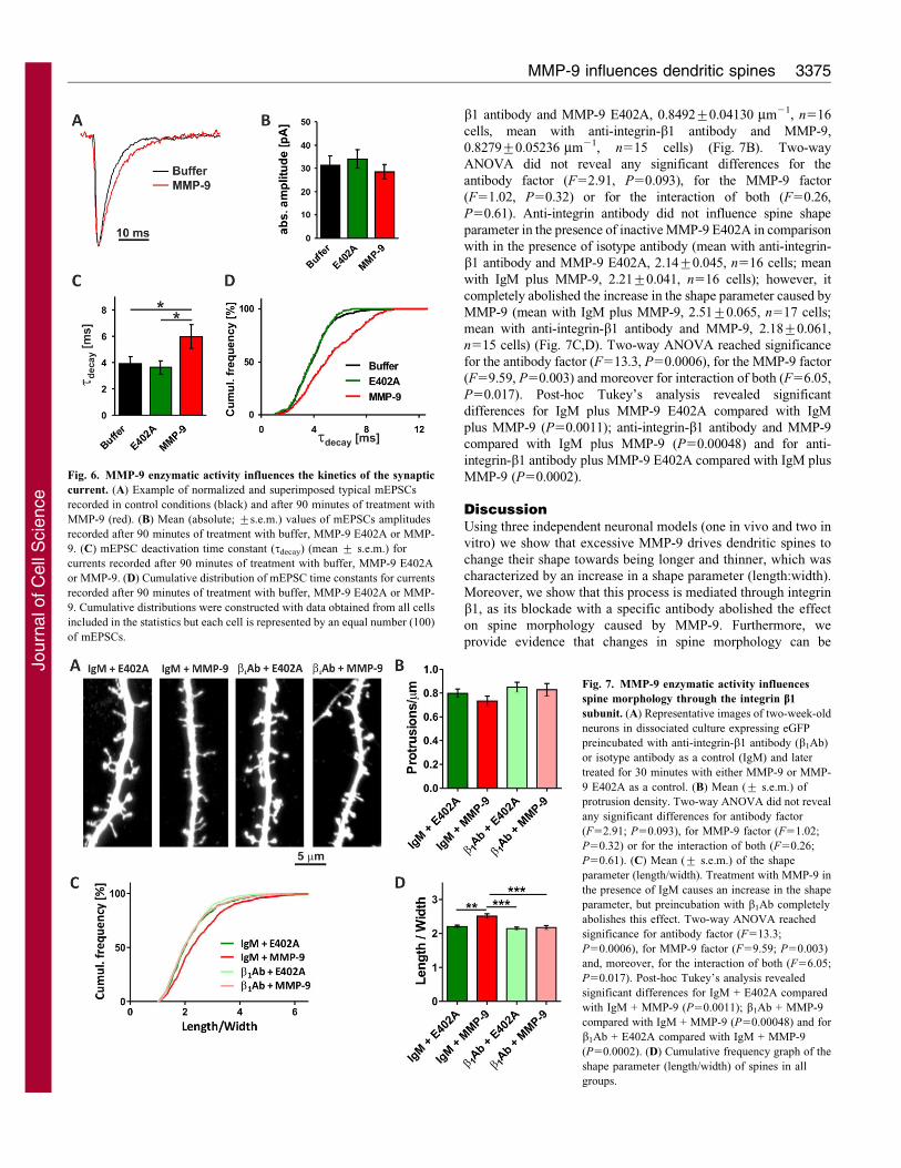
The enzymatic activity of MMP-9 affects spine physiology
Because it is known that alterations of spine morphology associated
with plasticity phenomena, such as LTP and LTD, are accompanied
by profound functional changes at glutamatergic synapses, we
wanted to test whether MMP-9 activity, besides affecting spine
geometry, also influences the electrophysiological properties of
synapses in our model. To this end, we used patch-clamping and
recorded glutamatergic miniature excitatory synaptic currents
(mEPSCs) in the whole-cell mode (Vm5–70 mV) from
dissociated hippocampal neurons. Neurons were incubated in the
presence of autoactivating MMP-9, MMP-9 E402A or protein
buffer for up to 90 minutes and the impact of treatment was
checked after 30, 60 and 90 minutes. We found that such treatment
did not significantly affect the mean current amplitude of minis
(31.38¡4.02 pA, n56; 34.08¡4.05 pA, n57; 28.53¡3.07 pA,
n57 for MMP-9, E402A and buffer, respectively; P.0.05,
unpaired Student’s t-test, Fig. 6A,B). However, the functional
impact of synaptic currents depends not only on amplitude but also
on the timecourse. In particular, synaptic current duration (typically
assessed as the weighted decay time constant) can strongly affect
synaptic integration and this parameter is known to undergo a
developmental increase that is strictly correlated with alterations in
synapse geometry (Cathala et al., 2005; Wall et al., 2002).
Treatment of neurons with MMP-9, MMP-9 E402A or buffer had
no significant effect on the mEPSC decay time course in neurons
treated for 30 or 60 minutes (data not shown). However,
90 minutes of treatment with MMP-9, resulted in a significant
increase in the decay time constant with respect to buffer- or MMP-
9-E402A-treated neurons (5.96¡0.91 ms, n57; 3.89¡0.53 ms,
n56; 3.61¡0.51 ms, n56 for MMP-9, MMP-9 E402A and buffer
Fig. 4. MMP-9 enzymatic activity makes dendritic spines in dissociated hippocampal culture longer and thinner but does not change the dendritic arbor.
(A) Live imaging of 15 DIV hippocampal neurons, expressing eGFP, incubated with MMP-9 does not reveal changes in the structure and number of neurites.
(B) Maximal projections of confocal scans of a dissociated hippocampal culture transfected with a plasmid carrying eGFP under the control of the b-actin
promoter. After reaching an age of at least 15 DIV, cells were incubated for 30 and 90 minutes with 400 ng/ml of active MMP-9, or for 90 minutes with inactive
MMP-9 E402A or the buffer used for elution of recombinant protein from the affinity column. (C). Mean (¡ s.e.m.) protrusion density. One-way ANOVA did not
reveal significant differences between groups of mean protrusions density calculated per cell (F50.936; P50.4353). (D) Mean (¡ s.e.m.) shape parameter
(length/width). One-way ANOVA revealed significant differences between groups of mean shape parameter calculated per cell (F515.61; P,0.0001). Post-hoc
Tukey’s tests reached significance for buffer compared with MMP-9 at 30 minutes (*P,0.05) and 90 minutes (***P,0.001), for MMP-9 E402A compared with
MMP-9 at 30 minutes (**P,0.01) and for MMP-9 E402A compared with MMP-9 at 90 minutes (***P,0.001). (E) The cumulative frequency of the shape
parameter (length/width).
MMP-9 influences dendritic spines 3373
Jour
nal o
f Cel
l Sci
ence

treatment, respectively; P,0.05, unpaired Student’s t-test,
Fig. 6A,C). The MMP-9-induced slowdown of the mEPSC decay
phase with respect to buffer or MMP-9-E402A-treated groups is
particularly clear in the cumulative distribution of the decay time
constants (Fig. 6D). Treatment with MMP-9 (or with MMP-9
E402A) for up to 90 minutes had no effect on mEPSC onset
kinetics (data not shown).
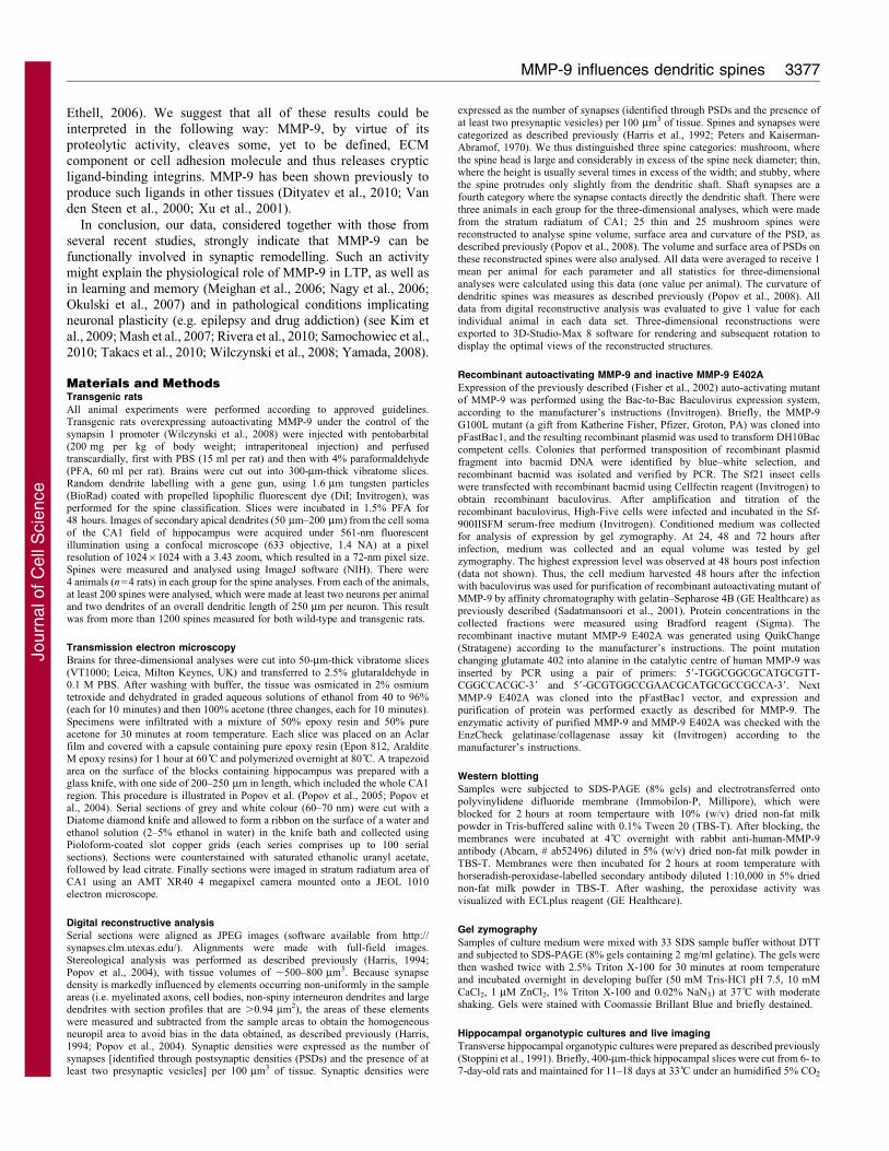
The MMP-9 enzymatic activity influences spine
morphology through engagement of integrin b1 subunit
It has been shown previously that MMP-9 can exert its function in
neurons through engaging the integrin b1 subunit (Michaluk et al.,
2009; Nagy et al., 2006; Wang et al., 2008) and that integrins are
involved in spine regulation (Bourgin et al., 2007; Shi and Ethell,
2006). Therefore, we decided to block the function of integrin b1
with a specific antibody and determine whether this would affect
the action of MMP-9 on spine morphology. Dissociated
hippocampal cultures were pretreated with either anti-integrin-b1
antibody or an isotype antibody (IgM), as a control, and were later
incubated with either MMP-9 or MMP-9 E402A for 30 minutes
(Fig. 7A). We did not observe any significant differences in spine
density between the analysed groups (mean with IgM plus MMP-9
E402A, 0.7981¡0.0362 mm21, n516 cells; mean with IgM plus
MMP-9, 0.733¡0.041 mm21, n517 cells; mean with anti-integrin-
Fig. 5. MMP-9 enzymatic activity does not change the localization and abundance of synaptic markers. A dissociated hippocampal culture was transfected
with plasmid caring eGFP under the b-actin promoter and, after reaching 15 DIV, cells were incubated for 90 minutes with 400 ng/ml of MMP-9 or inactive
MMP-9 E402A, or buffer. Cells were fixed and stained for bassoon. (A) Examples of dendrites in GFP-expressing neurons, which were stained for bassoon after
treatment with buffer, MMP-9 E402A or MMP-9. The furthest right-hand panel shows a magnification of the merge panels. (B) Mean (¡ s.e.m.) percentage of
bassoon-positive protrusions. (C) Density of bassoon clusters in the examined GFP-positive dendrites. (D) Examples of dendrites from GFP-expressing neurons
that were stained for homer-1 after treatment with buffer, MMP-9 E402A or MMP-9. The furthest right-hand panel shows a magnification of the merged panel.
(E) Mean (¡ s.e.m.) percentage of homer-1-positive protrusions. (F) Density of homer-1 clusters in the examined GFP-positive dendrites.
Journal of Cell Science 124 (19)3374
Jour
nal o
f Cel
l Sci
ence
b1 antibody and MMP-9 E402A, 0.8492¡0.04130 mm21, n516
cells, mean with anti-integrin-b1 antibody and MMP-9,
0.8279¡0.05236 mm21, n515 cells) (Fig. 7B). Two-way
ANOVA did not reveal any significant differences for the
antibody factor (F52.91, P50.093), for the MMP-9 factor
(F51.02, P50.32) or for the interaction of both (F50.26,
P50.61). Anti-integrin antibody did not influence spine shape
parameter in the presence of inactive MMP-9 E402A in comparison
with in the presence of isotype antibody (mean with anti-integrin-
b1 antibody and MMP-9 E402A, 2.14¡0.045, n516 cells; mean
with IgM plus MMP-9, 2.21¡0.041, n516 cells); however, it
completely abolished the increase in the shape parameter caused by
MMP-9 (mean with IgM plus MMP-9, 2.51¡0.065, n517 cells;
mean with anti-integrin-b1 antibody and MMP-9, 2.18¡0.061,
n515 cells) (Fig. 7C,D). Two-way ANOVA reached significance
for the antibody factor (F513.3, P50.0006), for the MMP-9 factor
(F59.59, P50.003) and moreover for interaction of both (F56.05,
P50.017). Post-hoc Tukey’s analysis revealed significant
differences for IgM plus MMP-9 E402A compared with IgM
plus MMP-9 (P50.0011); anti-integrin-b1 antibody and MMP-9
compared with IgM plus MMP-9 (P50.00048) and for anti-
integrin-b1 antibody plus MMP-9 E402A compared with IgM plus
MMP-9 (P50.0002).
DiscussionUsing three independent neuronal models (one in vivo and two in
vitro) we show that excessive MMP-9 drives dendritic spines to
change their shape towards being longer and thinner, which was
characterized by an increase in a shape parameter (length:width).
Moreover, we show that this process is mediated through integrin
b1, as its blockade with a specific antibody abolished the effect
on spine morphology caused by MMP-9. Furthermore, we
provide evidence that changes in spine morphology can be
Fig. 6. MMP-9 enzymatic activity influences the kinetics of the synaptic
current. (A) Example of normalized and superimposed typical mEPSCs
recorded in control conditions (black) and after 90 minutes of treatment with
MMP-9 (red). (B) Mean (absolute; ¡s.e.m.) values of mEPSCs amplitudes
recorded after 90 minutes of treatment with buffer, MMP-9 E402A or MMP-
9. (C) mEPSC deactivation time constant (tdecay) (mean ¡ s.e.m.) for
currents recorded after 90 minutes of treatment with buffer, MMP-9 E402A
or MMP-9. (D) Cumulative distribution of mEPSC time constants for currents
recorded after 90 minutes of treatment with buffer, MMP-9 E402A or MMP-
9. Cumulative distributions were constructed with data obtained from all cells
included in the statistics but each cell is represented by an equal number (100)
of mEPSCs.
Fig. 7. MMP-9 enzymatic activity influences
spine morphology through the integrin b1
subunit. (A) Representative images of two-week-old
neurons in dissociated culture expressing eGFP
preincubated with anti-integrin-b1 antibody (b1Ab)
or isotype antibody as a control (IgM) and later
treated for 30 minutes with either MMP-9 or MMP-
9 E402A as a control. (B) Mean (¡ s.e.m.) of
protrusion density. Two-way ANOVA did not reveal
any significant differences for antibody factor
(F52.91; P50.093), for MMP-9 factor (F51.02;
P50.32) or for the interaction of both (F50.26;
P50.61). (C) Mean (¡ s.e.m.) of the shape
parameter (length/width). Treatment with MMP-9 in
the presence of IgM causes an increase in the shape
parameter, but preincubation with b1Ab completely
abolishes this effect. Two-way ANOVA reached
significance for antibody factor (F513.3;
P50.0006), for MMP-9 factor (F59.59; P50.003)
and, moreover, for the interaction of both (F56.05;
P50.017). Post-hoc Tukey’s analysis revealed
significant differences for IgM + E402A compared
with IgM + MMP-9 (P50.0011); b1Ab + MMP-9
compared with IgM + MMP-9 (P50.00048) and for
b1Ab + E402A compared with IgM + MMP-9
(P50.0002). (D) Cumulative frequency graph of the
shape parameter (length/width) of spines in all
groups.
MMP-9 influences dendritic spines 3375
Jour
nal o
f Cel
l Sci
ence

associated with modifications of the decay time of synaptic
currents.
To study the influence MMP-9 on spines in vivo we usedtransgenic rats overexpressing autoactivating MMP-9 under thecontrol of the synapsin I promoter. In comparison with control
(WT rats), intensified MMP-9 activity caused an increase in thespine length:width ratio. Furthermore, we confirmed our lightmicroscopy data with electron microscopy, demonstrating clearly
that there is an increase in the largest spine category, thin spines, atthe expense of mushroom spines, which decrease as a percentageof the total spine complement. Interestingly, Bilousova et al. have
observed a decreased spine-head area and increased length ofspines in CA1 and CA3 regions of the hippocampus in Fmr1-knockout mouse, which among various pathological featuresoverexpresses MMP-9 (Bilousova et al., 2009). This phenotype
was rescued by treatment with minocycline, a tetracyclinederivative that, among other effects, decreases the expressionand activity of MMP-9 (Yao et al., 2004).
To avoid subjective discrimination between spines and filopodiawe utilized a shape parameter defined as the ratio of the length tothe maximal width of the spine. We decided to do this becausethere is no coherent definition of a filopodium and many research
groups use different shape parameters to distinguish between thesestructures and, moreover, there is also no clear functional criterionof filopodia.
For our studies, we have produced a recombinant autoactivatingmutant of MMP-9, as well as its inactive form MMP-9 E402A, andcharacterized both proteins, which were then used in hippocampal
cultures. These new experimental tools allowed us to address, forthe first time, directly the issue of whether enzymatic activity andnot just protein–protein interactions are responsible for the effect ofMMP-9 on spines. This was important because MMP-9 has several
structural protein domains and has been shown to have biologicalactivity in the absence of enzymatic activity (Ezhilarasan et al.,2009; Redondo-Munoz et al., 2010). Furthermore, it has been
reported that MMP-9 is capable of binding many surface proteins,such as integrins (Bjorklund et al., 2004; Redondo-Munoz et al.,2008; Stefanidakis et al., 2003), LRP-1 (Van den Steen et al., 2006)
and CD44 (Bourguignon et al., 1998). We demonstrated clearlythat lack of enzymatic activity renders the MMP-9 inactive mutantMMP-9 E402A incapable of affecting either spine morphology orchanging the shape parameter in hippocampal cultures.
Initially, the effects of the recombinant MMP-9 proteins weredemonstrated in live organotypic hippocampal cultures. Wang et al.(Wang et al., 2008) also noted the effect of application of
recombinant MMP-9 on spine morphology, namely on increasingspine-head volume. However, no detailed analyses of the spinegeometry were provided in that study. To extend further ourobservations we also employed dissociated hippocampal cultures,
which in this case are more amenable to experimental manipulation.Incubation of neurons at 15 DIV with recombinant MMP-9 alsocaused a significant increase in length:width parameter, which was
notable after only 30 minutes of incubation. This result is inagreement with previous observations of Bilousova et al.(Bilousova et al., 2009), who also applied recombinant MMP-9
for 1 hour in dissociated hippocampal culture and demonstrated anincrease in the number of filopodia-like long spines with smallheads and a concomitant decrease in the number of mushroom-
shaped short spines with large heads. Furthermore, it was recentlyshown that 6 hours of treatment with 5 mM NMDA caused anincrease in spine number, and the appearance of new small spines
and maturation of existing spines, which was blocked by both theMMP-2 and MMP-9 inhibitor, and by the knockout of ICAM-5,
the presumed MMP-2 and MMP-9 substrate (Tian et al., 2007). Theproposed model assumes that MMP-2 and/or MMP-9 enzymaticactivity, upon neuronal stimulation, leads to maturation of spinesand elongation of filopodia (Tian et al., 2007). Interestingly, the
recent findings of Conant et al. (Conant et al., 2010) support thenotion that ICAM-5 cleavage by MMPs (also including MMP-3 andMMP-7) can occur as early as 15 minutes after neuronal stimulation
by either NMDA treatment or induction of LTP.
We have also shown that the changes in spine morphologyinduced by MMP-9 treatment are not accompanied by significant
changes in the abundance and localization of the presynapticmarker bassoon, which suggests that MMP-9 activity does notaffect synaptic contacts of dendritic protrusions. Moreover, weobserved that incubation of neurons with MMP-9 caused a slowing
of the mEPSC decay phase (Fig. 6A–D) without affecting theabsolute amplitude. The increase in decay time is intriguing, asneuronal development comprises both conversion of spine shape
(from filopodial into mushroom-like) and an increase in synapticcurrents (Cathala et al., 2005; Wall et al., 2002). Thus, theappearance of filopodia and slowing of mEPSCs might suggest an
MMP-9-induced ‘juvenalization’ of glutamatergic synapses in ourmodel. However, the precise mechanism of mEPSC slowingdescribed here is not clear. Several potential processes mightunderlie the change in the timecourse of mEPSCs, including a
subunit switch (Kumar et al., 2002), lateral receptor mobility(Heine et al., 2008), endocytosis or exocytosis of receptors (Lee etal., 2004), changes in synapse geometry (Barbour et al., 1994;
Cathala et al., 2005) and post-translational modulation of synapticreceptors (Lee, 2006). Notably, Cathala et al. (Cathala et al., 2005)have attributed the developmental speeding of glutamatergic
EPSCs, to changes in the synapse geometry, a mechanism thatmight be consistent with the changes in spine shape and mEPSCkinetics that we observed here. However, it should be stressed that
the induction of filopodial spine shape by MMP-9, as describedhere, need not be the only mechanism whereby these enzymescould affect the glutamatergic synapse. For instance, MMPs couldchange the synaptic cleft geometry, the relative localization of
releasing sites and of postsynaptic densities and alter theimmediate surrounding of the synapse (thus affecting thediffusion of the agonist to the receptors), giving rise to a change
in synaptic glutamate transients, thereby altering mEPSC kinetics.Moreover, the fact that 30 minutes of treatment with MMP-9 wassufficient to induce a significant alteration in spine shape, whereas
90 minutes were needed for MMP-9 to induce a change in themEPSC kinetics, suggests that additional factors, other than spinegeometry, might also be involved. Further studies are needed to
elucidate the extent to which MMP-9 treatment modulates theshape and function of glutamatergic synapses by either common orseparate mechanisms.
Finally, we have shown that the influence of MMP-9 on spines
depends on integrin b1 activation, which agrees with previousreports that MMP-9 acts through this integrin subunit (Michaluket al., 2009; Nagy et al., 2006; Wang et al., 2008) and that MMP-
9 can bind integrins (Redondo-Munoz et al., 2008; Rolli et al.,2003; Stefanidakis et al., 2003). The influence of integrins onspine morphology reported previously agrees with those results
(Shi and Ethell, 2006). In particular, RGD peptides that mimicintegrin ligands, and can stimulate integrin signalling, can causeelongation of spines and formation of new filopodia (Shi and
Journal of Cell Science 124 (19)3376
Jour
nal o
f Cel
l Sci
ence
Ethell, 2006). We suggest that all of these results could beinterpreted in the following way: MMP-9, by virtue of its
proteolytic activity, cleaves some, yet to be defined, ECMcomponent or cell adhesion molecule and thus releases crypticligand-binding integrins. MMP-9 has been shown previously to
produce such ligands in other tissues (Dityatev et al., 2010; Vanden Steen et al., 2000; Xu et al., 2001).
In conclusion, our data, considered together with those from
several recent studies, strongly indicate that MMP-9 can befunctionally involved in synaptic remodelling. Such an activitymight explain the physiological role of MMP-9 in LTP, as well as
in learning and memory (Meighan et al., 2006; Nagy et al., 2006;Okulski et al., 2007) and in pathological conditions implicatingneuronal plasticity (e.g. epilepsy and drug addiction) (see Kim etal., 2009; Mash et al., 2007; Rivera et al., 2010; Samochowiec et al.,
2010; Takacs et al., 2010; Wilczynski et al., 2008; Yamada, 2008).
Materials and MethodsTransgenic rats
All animal experiments were performed according to approved guidelines.Transgenic rats overexpressing autoactivating MMP-9 under the control of thesynapsin 1 promoter (Wilczynski et al., 2008) were injected with pentobarbital(200 mg per kg of body weight; intraperitoneal injection) and perfusedtranscardially, first with PBS (15 ml per rat) and then with 4% paraformaldehyde(PFA, 60 ml per rat). Brains were cut out into 300-mm-thick vibratome slices.Random dendrite labelling with a gene gun, using 1.6 mm tungsten particles(BioRad) coated with propelled lipophilic fluorescent dye (DiI; Invitrogen), wasperformed for the spine classification. Slices were incubated in 1.5% PFA for48 hours. Images of secondary apical dendrites (50 mm–200 mm) from the cell somaof the CA1 field of hippocampus were acquired under 561-nm fluorescentillumination using a confocal microscope (633 objective, 1.4 NA) at a pixelresolution of 102461024 with a 3.43 zoom, which resulted in a 72-nm pixel size.Spines were measured and analysed using ImageJ software (NIH). There were4 animals (n54 rats) in each group for the spine analyses. From each of the animals,at least 200 spines were analysed, which were made at least two neurons per animaland two dendrites of an overall dendritic length of 250 mm per neuron. This resultwas from more than 1200 spines measured for both wild-type and transgenic rats.
Transmission electron microscopy
Brains for three-dimensional analyses were cut into 50-mm-thick vibratome slices(VT1000; Leica, Milton Keynes, UK) and transferred to 2.5% glutaraldehyde in0.1 M PBS. After washing with buffer, the tissue was osmicated in 2% osmiumtetroxide and dehydrated in graded aqueous solutions of ethanol from 40 to 96%(each for 10 minutes) and then 100% acetone (three changes, each for 10 minutes).Specimens were infiltrated with a mixture of 50% epoxy resin and 50% pureacetone for 30 minutes at room temperature. Each slice was placed on an Aclarfilm and covered with a capsule containing pure epoxy resin (Epon 812, AralditeM epoxy resins) for 1 hour at 60 C̊ and polymerized overnight at 80 C̊. A trapezoidarea on the surface of the blocks containing hippocampus was prepared with aglass knife, with one side of 200–250 mm in length, which included the whole CA1region. This procedure is illustrated in Popov et al. (Popov et al., 2005; Popov etal., 2004). Serial sections of grey and white colour (60–70 nm) were cut with aDiatome diamond knife and allowed to form a ribbon on the surface of a water andethanol solution (2–5% ethanol in water) in the knife bath and collected usingPioloform-coated slot copper grids (each series comprises up to 100 serialsections). Sections were counterstained with saturated ethanolic uranyl acetate,followed by lead citrate. Finally sections were imaged in stratum radiatum area ofCA1 using an AMT XR40 4 megapixel camera mounted onto a JEOL 1010electron microscope.
Digital reconstructive analysis
Serial sections were aligned as JPEG images (software available from http://synapses.clm.utexas.edu/). Alignments were made with full-field images.Stereological analysis was performed as described previously (Harris, 1994;Popov et al., 2004), with tissue volumes of ,500–800 mm3. Because synapsedensity is markedly influenced by elements occurring non-uniformly in the sampleareas (i.e. myelinated axons, cell bodies, non-spiny interneuron dendrites and largedendrites with section profiles that are .0.94 mm2), the areas of these elementswere measured and subtracted from the sample areas to obtain the homogeneousneuropil area to avoid bias in the data obtained, as described previously (Harris,1994; Popov et al., 2004). Synaptic densities were expressed as the number ofsynapses [identified through postsynaptic densities (PSDs) and the presence of atleast two presynaptic vesicles] per 100 mm3 of tissue. Synaptic densities were
expressed as the number of synapses (identified through PSDs and the presence ofat least two presynaptic vesicles) per 100 mm3 of tissue. Spines and synapses werecategorized as described previously (Harris et al., 1992; Peters and Kaiserman-Abramof, 1970). We thus distinguished three spine categories: mushroom, wherethe spine head is large and considerably in excess of the spine neck diameter; thin,where the height is usually several times in excess of the width; and stubby, wherethe spine protrudes only slightly from the dendritic shaft. Shaft synapses are afourth category where the synapse contacts directly the dendritic shaft. There werethree animals in each group for the three-dimensional analyses, which were madefrom the stratum radiatum of CA1; 25 thin and 25 mushroom spines werereconstructed to analyse spine volume, surface area and curvature of the PSD, asdescribed previously (Popov et al., 2008). The volume and surface area of PSDs onthese reconstructed spines were also analysed. All data were averaged to receive 1mean per animal for each parameter and all statistics for three-dimensionalanalyses were calculated using this data (one value per animal). The curvature ofdendritic spines was measures as described previously (Popov et al., 2008). Alldata from digital reconstructive analysis was evaluated to give 1 value for eachindividual animal in each data set. Three-dimensional reconstructions wereexported to 3D-Studio-Max 8 software for rendering and subsequent rotation todisplay the optimal views of the reconstructed structures.
Recombinant autoactivating MMP-9 and inactive MMP-9 E402A
Expression of the previously described (Fisher et al., 2002) auto-activating mutantof MMP-9 was performed using the Bac-to-Bac Baculovirus expression system,according to the manufacturer’s instructions (Invitrogen). Briefly, the MMP-9G100L mutant (a gift from Katherine Fisher, Pfizer, Groton, PA) was cloned intopFastBac1, and the resulting recombinant plasmid was used to transform DH10Baccompetent cells. Colonies that performed transposition of recombinant plasmidfragment into bacmid DNA were identified by blue–white selection, andrecombinant bacmid was isolated and verified by PCR. The Sf21 insect cellswere transfected with recombinant bacmid using Cellfectin reagent (Invitrogen) toobtain recombinant baculovirus. After amplification and titration of therecombinant baculovirus, High-Five cells were infected and incubated in the Sf-900IISFM serum-free medium (Invitrogen). Conditioned medium was collectedfor analysis of expression by gel zymography. At 24, 48 and 72 hours afterinfection, medium was collected and an equal volume was tested by gelzymography. The highest expression level was observed at 48 hours post infection(data not shown). Thus, the cell medium harvested 48 hours after the infectionwith baculovirus was used for purification of recombinant autoactivating mutant ofMMP-9 by affinity chromatography with gelatin–Sepharose 4B (GE Healthcare) aspreviously described (Sadatmansoori et al., 2001). Protein concentrations in thecollected fractions were measured using Bradford reagent (Sigma). Therecombinant inactive mutant MMP-9 E402A was generated using QuikChange(Stratagene) according to the manufacturer’s instructions. The point mutationchanging glutamate 402 into alanine in the catalytic centre of human MMP-9 wasinserted by PCR using a pair of primers: 59-TGGCGGCGCATGCGTT-CGGCCACGC-39 and 59-GCGTGGCCGAACGCATGCGCCGCCA-39. NextMMP-9 E402A was cloned into the pFastBac1 vector, and expression andpurification of protein was performed exactly as described for MMP-9. Theenzymatic activity of purified MMP-9 and MMP-9 E402A was checked with theEnzCheck gelatinase/collagenase assay kit (Invitrogen) according to themanufacturer’s instructions.
Western blotting
Samples were subjected to SDS-PAGE (8% gels) and electrotransferred ontopolyvinylidene difluoride membrane (Immobilon-P, Millipore), which wereblocked for 2 hours at room tempertaure with 10% (w/v) dried non-fat milkpowder in Tris-buffered saline with 0.1% Tween 20 (TBS-T). After blocking, themembranes were incubated at 4 C̊ overnight with rabbit anti-human-MMP-9antibody (Abcam, # ab52496) diluted in 5% (w/v) dried non-fat milk powder inTBS-T. Membranes were then incubated for 2 hours at room temperature withhorseradish-peroxidase-labelled secondary antibody diluted 1:10,000 in 5% driednon-fat milk powder in TBS-T. After washing, the peroxidase activity wasvisualized with ECLplus reagent (GE Healthcare).
Gel zymography
Samples of culture medium were mixed with 33 SDS sample buffer without DTTand subjected to SDS-PAGE (8% gels containing 2 mg/ml gelatine). The gels werethen washed twice with 2.5% Triton X-100 for 30 minutes at room temperatureand incubated overnight in developing buffer (50 mM Tris-HCl pH 7.5, 10 mMCaCl2, 1 mM ZnCl2, 1% Triton X-100 and 0.02% NaN3) at 37 C̊ with moderateshaking. Gels were stained with Coomassie Brillant Blue and briefly destained.
Hippocampal organotypic cultures and live imaging
Transverse hippocampal organotypic cultures were prepared as described previously(Stoppini et al., 1991). Briefly, 400-mm-thick hippocampal slices were cut from 6- to7-day-old rats and maintained for 11–18 days at 33 C̊ under an humidified 5% CO2
MMP-9 influences dendritic spines 3377
Jour
nal o
f Cel
l Sci
ence

atmosphere on Millipore inserts in culture medium (MEM plus HEPES, 25% horseserum and 25% Hanks solution). After 8 DIV cultures were transfected with eitherpcDNA3.1-eGFP or a pCX-mRFP1 using a biolistic method (Helios Gene Gun, Bio-Rad) and were used for experiments 3 days after transfection (De Roo et al., 2008a).Brief imaging sessions were carried out with an Olympus Fluoview 300 systemcoupled to a single photon laser. We focused on secondary or tertiary apicaldendrites from CA1 field of hippocampus using a 403 objective (0.8 NA) and 103digital zoom (final resolution 25 pixel size; steps between scans of 0.4 mm). Z-stackswere analysed manually using ImageJ software (NIH).
Dissociated hippocampal cultures
Dissociated hippocampal cultures from P0 (postnatal day 0) Wistar rats wereprepared as described below. Brains were removed and hippocampi were isolatedon ice in dissociation medium DM; 81.8 mM Na2SO4, 30 mM K2SO4, 5.8 mMMgCl2, 0.25 mM CaCl2, 1 mM HEPES pH 7.4, 20 mM glucose; 1 mM kynureicacid; 0.001% Phenol Red), hippocampi were later incubated twice for 15 minutesat 37 C̊ with 100 units of papain (Worthington, NY) in DM and rinsed three timesin DM and subsequently three times in plating medium [MEM, 10% fetal bovineserum (FBS) and 1% penicilin-streptomycin]. Hippocampi were triturated inplating medium until no clumps were visible and cells were diluted 1:10 inOptiMEM (Invitrogen), centrifuged for 10 minutes at room temperature, at208.5 g. The resulting cell pellet was suspended in plating medium, cells werecounted and plated at density 120,000 cells per 18-mm-diameter coverslip(Assistent, Germany) coated with 1 mg/ml poly-L-lysine (Sigma) and 2.5 mg/mllaminin (Roche). At 3 hours after plating medium was exchanged for maintenancemedium (Neurobasal-A without Phenol Red, 2% B-27 supplement, 1% penicillin-streptomycin, 0.5 mM glutamine, 12.5 mM glutamate, 25 mM b-mercaptoethanol)and cells were kept at 37 C̊, under a humidified 5% CO2 atmosphere for 2 weeks.All experiments were performed at 14–19 days in vitro (DIV). Cells weretransfected using Effectene (Qiagen), according to the manufacturer’s protocol, at10 DIV with plasmid carrying eGFP under the control of the b-actin promoter.
Cell stimulation
Cells were incubated for 30–90 minutes with 400 ng/ml of recombinant MMP-9 orMMP-9 E402A in maintenance medium or the buffer (50 mM Tris-HCl pH 7.5,400 mM NaCl, 10 mM CaCl2 and 2% DMSO) used for elution of recombinantproteins from the affinity column, which was diluted at least 1:125 in themaintenance medium, so the final concentration of DMSO in the culture did notexceed 0.016%. As indicated, cells were also incubated overnight with anti-CD29(integrin b1 chain) antibody (BD Pharmingen, no. 555002) or isotype antibody(IgM; BD Pharmingen no. 553957) at a final concentration of 40 mg/ml.
Immunostaining and confocal microscopy
Cells were fixed for 10 minutes at room temperature with 4% PFA, washed withPBS, permeabilized for 7 minutes with 0.1% Triton X-100 in PBS and blocked for1 hour at room temperature with 10% normal goat serum in PBS. After blocking,cells were incubated overnight with mouse anti-GFP antibody (Millipore #MAB3580) and rabbit anti-bassoon antibody (Synaptic Systems no. 141003), andguinea-pig anti-homer-1 antibody (Synaptic systems no. 160004), and then werewashed and incubated with fluorescent (Alexa-Fluor-488-, -568 or -647-conjugated) secondary anti-rabbit-IgG antibody (Invitrogen) for 40 minutes atroom temperature, washed and mounted. Fluorescent specimens were examinedunder a confocal microscope (TCS SP2; Leica) equipped with a 633 1.32 NA oilimmersion objective using the 488 nm line of an argon laser (for excitation ofAlexa Fluor 488), the 633 nm line of a helium-neon laser (for excitation of AlexaFluor 647) and the 543 nm line of a helium-neon laser (for excitation of AlexaFluor 568) of an at a pixel resolution of 204862048 and 1.653 optical zoom. Theresulting pixel size was 70.45 nm. The Z-stacks of optical slices were acquired in0.2 mm steps. The sum of Z-stacks of secondary or higher order dendrites (mostlyof pyramidal neurons) were analysed with ImageJ software (NIH). We discardedfrom the analyses all objects (protrusions) with an area smaller than 0.2 mm2 owingto the planar resolution of confocal setup. Colocalization of the synaptic markerwith dendritic protrusions was determined manually using merged images. Thepercentage of clusters colocalized with protrusions relative to total was thencalculated (Verpelli et al., 2010). All measurements were expressed as mean ¡s.e.m.
Electrophysiology
Miniature excitatory postsynaptic currents (mEPSC) were recorded in the whole-cell configuration of the patch-clamp technique at a membrane voltage of –70 mV,using the Axopatch 200B amplifier (Molecular Devices Corporation, Sunnyvale,CA). Signals were low-pass filtered at 5 kHz and acquired at 50 kHz in the gap-free mode using the analogue-to-digital converter Digidata 1440A (MolecularDevices Corporation). The intrapipette solution contained: 137 mM CsCl, 1 mMCaCl2, 2 mM MgCl2, 11 mM BAPTA (tetra cesium salt), 2 mM ATP and 10 mMHEPES pH 7.2 with CsOH. The composition of the external solution was:137 mM NaCl, 5 mM KCl, 2 mM CaCl2, 1 mM MgCl2, 20 mM glucose and
10 mM HEPES pH 7.2 with NaOH. mEPSCs were recorded in the presence of1 mM tetrodotoxin (TTX) to suppress neuronal excitability and 5 mM SR-95531was added to the external solution to block the GABAergic synaptic transmission.The presence of magnesium (2 mM) in the external saline and the highly negativemembrane voltage (–70 mV) were expected to eliminate the NMDA-receptor-mediated component of synaptic currents. All recordings were performed at roomtemperature (21–24 C̊). Chemicals were from Sigma except for TTX (LaToxan,France). The mEPSCs decaying phase was fitted to a single exponential functiony(t)5A exp(–t/tdecay), where A is the amplitude and tdecay is the decay timeconstant. To assess the impact of MMP-9 on mEPSCs, neurons were incubated forup to 90 minutes in culture medium with recombinant MMP-9 at concentration of400 ng/ml. Control recordings were performed in parallel on two groups ofneurons from the same preparation: one treated for the same time duration withrespective volume of MMP-9 dilution buffer and the second group treated for thesame time and with the same amount of enzymatically inactive MMP-9 E402A.The effect of MMP-9 was assessed for neurons treated for 30, 60 and 90 minutes.
Statistics
Data were tested for normal distribution using the Kolmogorov–Smirnov normalitytest and because all data passed that test, they were expressed as means ¡ s.e.m.Groups were compared using unpaired Student’s t-tests and if the number ofgroups was larger than two, we used a one-way or two-way ANOVA and a post-hoc Tukey’s test. The number of animals and/or the number of cells (n) used forstatistical evaluation of the means ¡ s.e.m values is provided, after thecorresponding mean value for each analysed group, in the Results section.
AcknowledgementsWe are grateful to Katherine Fisher (Pfizer) for providing the cDNAof the autoactivating MMP-9 mutant.
FundingThis work was supported by The Polish Ministry of Science and HigherEducation research grant P-N/030/2006 (J.W.M. and L.K.), the 7th FPEU grant Memstick (M.G.S. and L.K.), the Polish–Norwegian ResearchFund grant (PNRF-96, G.M.W. and L.K.) and fellowships from theFoundation for Polish Science (P.M.) and the European MolecularBiology Organization (J.W.). M.S. and J.W.M. were partially supportedby the Polish Ministry for Science grant no. N401 541540.
ReferencesAsahi, M., Asahi, K., Jung, J. C., del Zoppo, G. J., Fini, M. E. and Lo, E. H. (2000).
Role for matrix metalloproteinase 9 after focal cerebral ischemia: effects of geneknockout and enzyme inhibition with BB-94. J. Cereb. Blood Flow Metab. 20, 1681-1689.
Barbour, B., Keller, B. U., Llano, I. and Marty, A. (1994). Prolonged presence ofglutamate during excitatory synaptic transmission to cerebellar Purkinje cells. Neuron
12, 1331-1343.
Bilousova, T. V., Rusakov, D. A., Ethell, D. W. and Ethell, I. M. (2006). Matrixmetalloproteinase-7 disrupts dendritic spines in hippocampal neurons through NMDAreceptor activation. J. Neurochem. 97, 44-56.
Bilousova, T. V., Dansie, L., Ngo, M., Aye, J., Charles, J. R., Ethell, D. W. and
Ethell, I. M. (2009). Minocycline promotes dendritic spine maturation and improvesbehavioural performance in the fragile X mouse model. J. Med. Genet. 46, 94-102.
Bjorklund, M., Heikkila, P. and Koivunen, E. (2004). Peptide inhibition of catalyticand noncatalytic activities of matrix metalloproteinase-9 blocks tumor cell migrationand invasion. J. Biol. Chem. 279, 29589-29597.
Bourgin, C., Murai, K. K., Richter, M. and Pasquale, E. B. (2007). The EphA4receptor regulates dendritic spine remodeling by affecting beta1-integrin signalingpathways. J. Cell Biol. 178, 1295-1307.
Bourguignon, L. Y., Gunja-Smith, Z., Iida, N., Zhu, H. B., Young, L. J., Muller,
W. J. and Cardiff, R. D. (1998). CD44v(3,8-10) is involved in cytoskeleton-mediated tumor cell migration and matrix metalloproteinase (MMP-9) association inmetastatic breast cancer cells. J. Cell. Physiol. 176, 206-215.
Brown, T. E., Wilson, A. R., Cocking, D. L. and Sorg, B. A. (2009). Inhibition ofmatrix metalloproteinase activity disrupts reconsolidation but not consolidation of afear memory. Neurobiol. Learn. Mem. 91, 66-72.
Cathala, L., Holderith, N. B., Nusser, Z., DiGregorio, D. A. and Cull-Candy, S. G.
(2005). Changes in synaptic structure underlie the developmental speeding of AMPAreceptor-mediated EPSCs. Nat. Neurosci. 8, 1310-1318.
Chklovskii, D. B., Mel, B. W. and Svoboda, K. (2004). Cortical rewiring andinformation storage. Nature 431, 782-788.
Conant, K., Wang, Y., Szklarczyk, A., Dudak, A., Mattson, M. P. and Lim, S. T.(2010). Matrix metalloproteinase-dependent shedding of intercellular adhesionmolecule-5 occurs with long-term potentiation. Neuroscience 166, 508-521.
De Roo, M., Klauser, P., Mendez, P., Poglia, L. and Muller, D. (2008a). Activity-dependent PSD formation and stabilization of newly formed spines in hippocampalslice cultures. Cereb. Cortex 18, 151-161.
Journal of Cell Science 124 (19)3378
Jour
nal o
f Cel
l Sci
ence

De Roo, M., Klauser, P. and Muller, D. (2008b). LTP promotes a selective long-termstabilization and clustering of dendritic spines. PLoS Biol. 6, e219.
Dityatev, A., Schachner, M. and Sonderegger, P. (2010). The dual role of theextracellular matrix in synaptic plasticity and homeostasis. Nat. Rev. Neurosci. 11,735-746.
Ezhilarasan, R., Jadhav, U., Mohanam, I., Rao, J. S., Gujrati, M. and Mohanam, S.(2009). The hemopexin domain of MMP-9 inhibits angiogenesis and retards thegrowth of intracranial glioblastoma xenograft in nude mice. Int. J. Cancer 124, 306-315.
Fisher, K. E., Fei, Q., Laird, E. R., Stock, J. L., Allen, M. R., Sahagan, B. G. andStrick, C. A. (2002). Engineering autoactivating forms of matrix metalloproteinase-9and expression of the active enzyme in cultured cells and transgenic mouse brain.Biochemistry 41, 8289-8297.
Grabrucker, A., Vaida, B., Bockmann, J. and Boeckers, T. M. (2009).Synaptogenesis of hippocampal neurons in primary cell culture. Cell Tissue Res.
338, 333-341.
Gu, Z., Kaul, M., Yan, B., Kridel, S. J., Cui, J., Strongin, A., Smith, J. W.,
Liddington, R. C. and Lipton, S. A. (2002). S-nitrosylation of matrix metallopro-teinases: signaling pathway to neuronal cell death. Science 297, 1186-1190.
Harris, K. M. (1994). Serial electron microscopy as an alternative or compliment toconfocal microscopy for the study of synapses and dendritic spines in the centralnervous system. In Three-Dimensional Confocal Microscopy, Volume Investigation
Of Biological Specimens (ed. J. K. Stevens, L. R. Mills and J. E. Trogadis), pp. 421-445. New York: Academic Press.
Harris, K. M., Jensen, F. E. and Tsao, B. (1992). Three-dimensional structure ofdendritic spines and synapses in rat hippocampus (CA1) at postnatal day 15 and adultages: implications for the maturation of synaptic physiology and long-termpotentiation. J. Neurosci. 12, 2685-2705.
Heine, M., Groc, L., Frischknecht, R., Beique, J. C., Lounis, B., Rumbaugh, G.,
Huganir, R. L., Cognet, L. and Choquet, D. (2008). Surface mobility ofpostsynaptic AMPARs tunes synaptic transmission. Science 320, 201-205.
Holtmaat, A. and Svoboda, K. (2009). Experience-dependent structural synapticplasticity in the mammalian brain. Nat. Rev. Neurosci. 10, 647-658.
Holtmaat, A., Wilbrecht, L., Knott, G. W., Welker, E. and Svoboda, K. (2006).Experience-dependent and cell-type-specific spine growth in the neocortex. Nature
441, 979-983.
Kim, G. W., Kim, H. J., Cho, K. J., Kim, H. W., Cho, Y. J. and Lee, B. I. (2009). Therole of MMP-9 in integrin-mediated hippocampal cell death after pilocarpine-inducedstatus epilepticus. Neurobiol. Dis. 36, 169-180.
Konopka, W., Kiryk, A., Novak, M., Herwerth, M., Parkitna, J. R., Wawrzyniak,M., Kowarsch, A., Michaluk, P., Dzwonek, J., Arnsperger, T. et al. (2010).MicroRNA loss enhances learning and memory in mice. J. Neurosci. 30, 14835-14842.
Kumar, S. S., Bacci, A., Kharazia, V. and Huguenard, J. R. (2002). A developmentalswitch of AMPA receptor subunits in neocortical pyramidal neurons. J. Neurosci. 22,3005-3015.
Lee, H. K. (2006). Synaptic plasticity and phosphorylation. Pharmacol. Ther. 112, 810-832.
Lee, S. H., Simonetta, A. and Sheng, M. (2004). Subunit rules governing the sorting ofinternalized AMPA receptors in hippocampal neurons. Neuron 43, 221-236.
Mash, D. C., ffrench-Mullen, J., Adi, N., Qin, Y., Buck, A. and Pablo, J. (2007).Gene expression in human hippocampus from cocaine abusers identifies genes whichregulate extracellular matrix remodeling. PLoS ONE 2, e1187.
Matsuzaki, M., Honkura, N., Ellis-Davies, G. C. and Kasai, H. (2004). Structuralbasis of long-term potentiation in single dendritic spines. Nature 429, 761-766.
Meighan, S. E., Meighan, P. C., Choudhury, P., Davis, C. J., Olson, M. L., Zornes,
P. A., Wright, J. W. and Harding, J. W. (2006). Effects of extracellular matrix-degrading proteases matrix metalloproteinases 3 and 9 on spatial learning andsynaptic plasticity. J. Neurochem. 96, 1227-1241.
Michaluk, P., Mikasova, L., Groc, L., Frischknecht, R., Choquet, D. andKaczmarek, L. (2009). Matrix metalloproteinase-9 controls NMDA receptor surfacediffusion through integrin beta1 signaling. J. Neurosci. 29, 6007-6012.
Mizoguchi, H., Yamada, K., Niwa, M., Mouri, A., Mizuno, T., Noda, Y., Nitta, A.,Itohara, S., Banno, Y. and Nabeshima, T. (2007). Reduction of methamphetamine-induced sensitization and reward in matrix metalloproteinase-2 and -9-deficient mice.J. Neurochem. 100, 1579-1588.
Moeller, M. L., Shi, Y., Reichardt, L. F. and Ethell, I. M. (2006). EphB receptorsregulate dendritic spine morphogenesis through the recruitment/phosphorylation offocal adhesion kinase and RhoA activation. J. Biol. Chem. 281, 1587-1598.
Moser, M. B., Trommald, M. and Andersen, P. (1994). An increase in dendritic spinedensity on hippocampal CA1 pyramidal cells following spatial learning in adult ratssuggests the formation of new synapses. Proc. Natl. Acad. Sci. USA 91, 12673-12675.
Mott, J. D. and Werb, Z. (2004). Regulation of matrix biology by matrixmetalloproteinases. Curr. Opin. Cell Biol. 16, 558-564.
Mysore, S. P., Tai, C. Y. and Schuman, E. M. (2007). Effects of N-cadherin disruptionon spine morphological dynamics. Front. Cell. Neurosci. 1, 1.
Nagy, V., Bozdagi, O., Matynia, A., Balcerzyk, M., Okulski, P., Dzwonek, J., Costa,
R. M., Silva, A. J., Kaczmarek, L. and Huntley, G. W. (2006). Matrixmetalloproteinase-9 is required for hippocampal late-phase long-term potentiationand memory. J. Neurosci. 26, 1923-1934.
Niu, S., Yabut, O. and D’Arcangelo, G. (2008). The Reelin signaling pathwaypromotes dendritic spine development in hippocampal neurons. J. Neurosci. 28,10339-10348.
Okulski, P., Jay, T. M., Jaworski, J., Duniec, K., Dzwonek, J., Konopacki, F. A.,
Wilczynski, G. M., Sanchez-Capelo, A., Mallet, J. and Kaczmarek, L. (2007).
TIMP-1 abolishes MMP-9-dependent long-lasting long-term potentiation in theprefrontal cortex. Biol. Psychiatry 62, 359-362.
Peters, A. and Kaiserman-Abramof, I. R. (1970). The small pyramidal neuron of the
rat cerebral cortex. The perikaryon, dendrites and spines. Am. J. Anat. 127, 321-355.
Petrini, E. M., Lu, J., Cognet, L., Lounis, B., Ehlers, M. D. and Choquet, D. (2009).
Endocytic trafficking and recycling maintain a pool of mobile surface AMPA
receptors required for synaptic potentiation. Neuron 63, 92-105.
Pizzorusso, T., Medini, P., Berardi, N., Chierzi, S., Fawcett, J. W. and Maffei, L.
(2002). Reactivation of ocular dominance plasticity in the adult visual cortex. Science
298, 1248-1251.
Popov, V. I., Davies, H. A., Rogachevsky, V. V., Patrushev, I. V., Errington, M. L.,
Gabbott, P. L., Bliss, T. V. and Stewart, M. G. (2004). Remodelling of synaptic
morphology but unchanged synaptic density during late phase long-term potentiation
(LTP): a serial section electron micrograph study in the dentate gyrus in the
anaesthetised rat. Neuroscience 128, 251-262.
Popov, V., Medvedev, N. I., Davies, H. A. and Stewart, M. G. (2005). Mitochondria
form a filamentous reticular network in hippocampal dendrites but are present as
discrete bodies in axons: a three-dimensional ultrastructural study. J. Comp. Neurol.
492, 50-65.
Popov, V. I., Medvedev, N. I., Kraev, I. V., Gabbott, P. L., Davies, H. A., Lynch, M.,
Cowley, T. R., Berezin, V., Bock, E. and Stewart, M. G. (2008). A cell adhesion
molecule mimetic, FGL peptide, induces alterations in synapse and dendritic spinestructure in the dentate gyrus of aged rats: a three-dimensional ultrastructural study.
Eur. J. Neurosci. 27, 301-314.
Redondo-Munoz, J., Ugarte-Berzal, E., Garcia-Marco, J. A., del Cerro, M. H., Van
den Steen, P. E., Opdenakker, G., Terol, M. J. and Garcia-Pardo, A. (2008).Alpha4beta1 integrin and 190-kDa CD44v constitute a cell surface docking complex
for gelatinase B/MMP-9 in chronic leukemic but not in normal B cells. Blood 112,
169-178.
Redondo-Munoz, J., Ugarte-Berzal, E., Terol, M. J., Van den Steen, P. E.,
Hernandez del Cerro, M., Roderfeld, M., Roeb, E., Opdenakker, G., Garcia-
Marco, J. A. and Garcia-Pardo, A. (2010). Matrix metalloproteinase-9 promotes
chronic lymphocytic leukemia b cell survival through its hemopexin domain. Cancer
Cell 17, 160-172.
Rivera, S., Khrestchatisky, M., Kaczmarek, L., Rosenberg, G. A. and Jaworski,
D. M. (2010). Metzincin proteases and their inhibitors: foes or friends in nervoussystem physiology? J. Neurosci. 30, 15337-15357.
Rolli, M., Fransvea, E., Pilch, J., Saven, A. and Felding-Habermann, B. (2003).
Activated integrin alphavbeta3 cooperates with metalloproteinase MMP-9 in
regulating migration of metastatic breast cancer cells. Proc. Natl. Acad. Sci. USA
100, 9482-9487.
Sadatmansoori, S., MacDougall, J., Khademi, S., Cooke, L. S., Guarino, L., Meyer,
E. F. and Forough, R. (2001). Construction, expression, and characterization of abaculovirally expressed catalytic domain of human matrix metalloproteinase-9.
Protein Expr. Purif. 23, 447-452.
Samochowiec, A., Grzywacz, A., Kaczmarek, L., Bienkowski, P., Samochowiec, J.,
Mierzejewski, P., Preuss, U. W., Grochans, E. and Ciechanowicz, A. (2010).Functional polymorphism of matrix metalloproteinase-9 (MMP-9) gene in alcohol
dependence: family and case control study. Brain Res. 1327, 103-106.
Shi, Y. and Ethell, I. M. (2006). Integrins control dendritic spine plasticity inhippocampal neurons through NMDA receptor and Ca2+/calmodulin-dependent
protein kinase II-mediated actin reorganization. J. Neurosci. 26, 1813-1822.
Stefanidakis, M., Bjorklund, M., Ihanus, E., Gahmberg, C. G. and Koivunen, E.
(2003). Identification of a negatively charged peptide motif within the catalyticdomain of progelatinases that mediates binding to leukocyte beta 2 integrins. J. Biol.
Chem. 278, 34674-34684.
Sternlicht, M. D. and Werb, Z. (2001). How matrix metalloproteinases regulate cellbehavior. Annu. Rev. Cell Dev. Biol. 17, 463-516.
Stoppini, L., Buchs, P. A. and Muller, D. (1991). A simple method for organotypic
cultures of nervous tissue. J. Neurosci. Methods 37, 173-182.
Szklarczyk, A., Lapinska, J., Rylski, M., McKay, R. D. and Kaczmarek, L. (2002).
Matrix metalloproteinase-9 undergoes expression and activation during dendritic
remodeling in adult hippocampus. J. Neurosci. 22, 920-930.
Takacs, E., Nyilas, R., Szepesi, Z., Baracskay, P., Karlsen, B., Rosvold, T., Bjorkum,
A. A., Czurko, A., Kovacs, Z., Kekesi, A. K. et al. (2010). Matrix metalloproteinase-
9 activity increased by two different types of epileptic seizures that do not induce
neuronal death: a possible role in homeostatic synaptic plasticity. Neurochem. Int. 56,
799-809.
Tian, L., Stefanidakis, M., Ning, L., Van Lint, P., Nyman-Huttunen, H., Libert, C.,
Itohara, S., Mishina, M., Rauvala, H. and Gahmberg, C. G. (2007). Activation of
NMDA receptors promotes dendritic spine development through MMP-mediatedICAM-5 cleavage. J. Cell Biol. 178, 687-700.
Van den Steen, P. E., Proost, P., Wuyts, A., Van Damme, J. and Opdenakker, G.
(2000). Neutrophil gelatinase B potentiates interleukin-8 tenfold by aminoterminal
processing, whereas it degrades CTAP-III, PF-4, and GRO-alpha and leaves RANTESand MCP-2 intact. Blood 96, 2673-2681.
Van den Steen, P. E., Van Aelst, I., Hvidberg, V., Piccard, H., Fiten, P., Jacobsen,
C., Moestrup, S. K., Fry, S., Royle, L., Wormald, M. R. et al. (2006). Thehemopexin and O-glycosylated domains tune gelatinase B/MMP-9 bioavailability via
inhibition and binding to cargo receptors. J. Biol. Chem. 281, 18626-18637.
MMP-9 influences dendritic spines 3379
Jour
nal o
f Cel
l Sci
ence

Verpelli, C., Piccoli, G., Zanchi, A., Gardoni, F., Huang, K., Brambilla, D., Di Luca, M.,Battaglioli, E. and Sala, C. (2010). Synaptic activity controls dendritic spine morphologyby modulating eEF2-dependent BDNF synthesis. J. Neurosci. 30, 5830-5842.
Wall, M. J., Robert, A., Howe, J. R. and Usowicz, M. M. (2002). The speeding ofEPSC kinetics during maturation of a central synapse. Eur. J. Neurosci. 15, 785-797.
Wang, X. B., Bozdagi, O., Nikitczuk, J. S., Zhai, Z. W., Zhou, Q. and Huntley,G. W. (2008). Extracellular proteolysis by matrix metalloproteinase-9 drives dendriticspine enlargement and long-term potentiation coordinately. Proc. Natl. Acad. Sci.
USA 105, 19520-19525.Wilczynski, G. M., Konopacki, F. A., Wilczek, E., Lasiecka, Z., Gorlewicz, A.,
Michaluk, P., Wawrzyniak, M., Malinowska, M., Okulski, P., Kolodziej, L. R.et al. (2008). Important role of matrix metalloproteinase 9 in epileptogenesis. J. Cell
Biol. 180, 1021-1035.Xu, J., Rodriguez, D., Petitclerc, E., Kim, J. J., Hangai, M., Moon, Y. S., Davis,
G. E. and Brooks, P. C. (2001). Proteolytic exposure of a cryptic site within collagen typeIV is required for angiogenesis and tumor growth in vivo. J. Cell Biol. 154, 1069-1079.
Xu, T., Yu, X., Perlik, A. J., Tobin, W. F., Zweig, J. A., Tennant, K., Jones, T. andZuo, Y. (2009). Rapid formation and selective stabilization of synapses for enduringmotor memories. Nature 462, 915-919.
Yamada, K. (2008). Endogenous modulators for drug dependence. Biol. Pharm. Bull.
31, 1635-1638.Yang, Y., Wang, X. B., Frerking, M. and Zhou, Q. (2008). Spine expansion and
stabilization associated with long-term potentiation. J. Neurosci. 28, 5740-5751.Yao, J. S., Chen, Y., Zhai, W., Xu, K., Young, W. L. and Yang, G. Y. (2004).
Minocycline exerts multiple inhibitory effects on vascular endothelial growth factor-induced smooth muscle cell migration: the role of ERK1/2, PI3K, and matrixmetalloproteinases. Circ. Res. 95, 364-371.
Yong, V. W. (2005). Metalloproteinases: mediators of pathology and regeneration in theCNS. Nat. Rev. Neurosci. 6, 931-944.
Zhou, Q., Homma, K. J. and Poo, M. M. (2004). Shrinkage of dendritic spinesassociated with long-term depression of hippocampal synapses. Neuron 44, 749-757.
Journal of Cell Science 124 (19)3380
Jour
nal o
f Cel
l Sci
ence


![Gene therapy of gastric cancer using LIGHT-secreting human ...TRAIL gene therapy. Bhoopathi et al. [11] evaluated the role of matrix metalloproteinase (MMP)-2 in the tropism of UCB-MSCs](https://img.pdfslide.us/doc/110x75/5f6d428dd8e52917836a6dca/gene-therapy-of-gastric-cancer-using-light-secreting-human-trail-gene-therapy.jpg)