Embed Size (px)
Citation preview
The EMBO Journal Peer Review Process File - EMBO-2013-84477
© European Molecular Biology Organization 1
Manuscript EMBO-2013-84477 EPS8 controls dendritic spine density and synaptic plasticity through its Actin capping activity Elisabetta Menna, Stefania Zambetti, Raffaella Morini, Andrea Donzelli, Andrea Disanza, Daniela Calvigioni, Daniela Braida, Chiara Nicolini, Marta Orlando, Giuliana Fossati, Maria Cristina Regondi, Linda Pattini, Carolina Frassoni, Maura Francolini, Giorgio Scita, Mariaelvina Sala, Margaret Fahnestock, Michela Matteoli Corresponding author: Elisabetta Menna and Michela Matteoli, University of Milan Review timeline: Submission date: 14 January 2013 Editorial Decision: 11 February 2013 Revision received: 28 March 2013 Accepted: 15 April 2013 Editor: Karin Dumstrei Transaction Report: (Note: With the exception of the correction of typographical or spelling errors that could be a source of ambiguity, letters and reports are not edited. The original formatting of letters and referee reports may not be reflected in this compilation.)
1st Editorial Decision 11 February 2013
Thank you for submitting your manuscript to the EMBO Journal. Your study has now been seen by three referees and their comments are provided below. As you can see, the referees find the analysis interesting and important. However, they also raise some issues that should be resolved. The behavioral analysis needs to be more carefully considered and it is not clear if the defects observed are due to the capping function of Eps8. Given the positive comments on your paper, I would like to invite you to submit a suitably revised manuscript for our consideration. I should add that it is EMBO Journal policy to allow only a single round of revision and it is therefore important to address the raised concerns at this stage. When preparing your letter of response to the referees' comments, please bear in mind that this will form part of the Review Process File, and will therefore be available online to the community. For more details on our Transparent Editorial Process, please visit our website: http://www.nature.com/emboj/about/process.html Thank you for the opportunity to consider your work for publication. I look forward to your revision. _____ REFEREE REPORTS: Referee #1
The EMBO Journal Peer Review Process File - EMBO-2013-84477
© European Molecular Biology Organization 2
Here Menna et al. explore a cellular and physiological function for EPS8, an actin-capping protein, in synaptic plasticity and mice behavior. This is a logical extension of the previous study from the same group reporting of a role for EPS8 in axonal filopodia dynamics that is important for synapse formation. Shifting the focus to mature dendrites, the authors find that EPS8 is recruited to the spine head during LTP, and inhibiting EPS8 capping activity blocks spine enlargement and plasticity. Furthermore, mice lacking EPS8 show immature spines, abnormal EEG activity, impaired cognitive function and sociability. This is a tour de force study combining multiple experimental approaches from biochemisty, molecularbiology, immunocytochemistry, fluorescence live imaging, electrophysiology, to assays of mice behavior. The work is important in highlighting the roles for EPS8-dependent modulation of spine plasticity and long-term synaptic strength changes that are likely relevant for learning and social behaviour. Nevertheless, as it stands, the manuscript suffers from technical issues that weaken the main conclusions. The following points require careful consideration. Specific comments/questions: Social behavior in EPS8KO mice is assessed using a single test only, and therefore, this point should be toned down in the abstract and the introduction. In general, for the spine analysis shown (Figures 2-5), please state how many spines, how many neurons, and from how many cultures/preparation were involved for each condition. Figure 1A, B. What is the basis for the difference in spatial learning exhibited within the EPS8KO mice group (that is apparent in the cumulative distributions of % animals reaching the criterion), in which half of the mutant mice perform not appreciably worse than the wild type mice? Interestingly, this is consistent for the two different paradigms tested. Figure 1C-E. Do EPS8KO mice show a bimodal deficit in the behaviors tested here as have been found for spatial learning (see point above) if compared across individual animals? Do EPS8KO mice show higher seizure activity? Figure 2D,E and Figure 3A,B. EPS8 could play a role not only in governing spine length but also the spine head size by affecting actin filament branching. Moreover, AMPA receptor number is correlated to spine head size. Therefore, it would be informative to analyse the spine structure data by showing the relative distributions of different spine types rather than limiting to spine length and density. Figure 3B. Spine length information is missing here. The same parameters should be shown for comparing the results of overexpression of EPS8 and the loss of expression of EPS8. LTP examined here is not the conventional LTP elicited by electrical stimulation studied in hippocampal slices but is a chemically induced form of LTP in dissociated cultures. This needs to be clarified in the abstract and throughout the text. Figure 4A. Fluorescence images shown here are of questionable quality. For example, the v-Glut1 puncta appear saturated. It would also be helpful to show DIC images to demonstrate the healthy appearance of neurons. Figure 5D. The peptide experiments should also be performed in EPS8KO cultures in parallel to demonstrate the specificity of the blocking peptide effect to EPS8. Note that in the right panel, the x-axis label is missing. Figure 6. The relevance of the specific reduction of EPS8 in autism patients in the context of present work highlighting EPS8KO with spine plasticity deficits needs to be further substantiated by a simultaneous comparison of additional postsynaptic markers and actin modulators between control and autism brain tissue. Supplementary Figure 2. It is curious that despite the robust increase in spine density and the pre
The EMBO Journal Peer Review Process File - EMBO-2013-84477
© European Molecular Biology Organization 3
and the postsynaptic areas, the mEPSC frequency is not altered in EPS8KO neurons under basal conditions. Is this due to the decrease in mEPSC amplitude where many of the events fall below detection or due to a reduction in presynaptic release probability? Supplementary Figure 4. This is an important result, and the summary shown in panel C should be included as part of Figure 5. The images could be left as the supplementary data. Referee #2 The manuscript by Menna et al. 'Eps8 controls dendritic density and synaptic plasticity through its actin capping activity' deals with the question of actin remodeling by Eps8 in the synapse and the accompanied physiological changes and alterations of animal behaviour. The authors present a battery of behaviour experiments on Eps8 knockout mice that show deficits in learning, novel object recognition and social interaction. These deficits could be correlated with impaired spine morphogenesis (increase in immature spines) and alterations in cell culture electrophysiology. In cell culture transfection of the wild type Eps8, but not a mutant deficient in capping is able to rescue, leading to the conclusion that Eps8 capping is the crucial activity for synaptic plasticity. In general the data are interesting and shed some new light on the role of Eps8 in synaptic physiology. The authors present a significant body of good quality data. Unfortunately, the presentation of data appears superficial and largely confusing, partly due to the attempt to oversimplify the complex role of Eps8 in neurons. In particular a reader with little knowledge in behaviour or electrophysiology will have a hard time to understand the rationale behind the experiments. Without a clear explanation of the experimental rationale and a deeper discussion of the results, the work appears largely descriptive. I was hoping that this would be picked up again in the discussion. However, the discussion is weak - simply summarizing the current literature on actin in the synapse. I could not find any discussion or explanation of the presented data. Concerning the interpretation of data and conclusions there are two major critical points. First in my view, the connection to autism is largely overstated and is missing solid grounds. The behavioural phenotype, as well as the synaptic density and maturation defects are well known from the Fragile X Syndrome patients and mouse model. The data presented here seem to essentially describe intellectual disability (ID) more than ASD (autism spectrum disorder). In the behavioural screen of the Eps8 ko mice conducted by the authors, I don't see any specific reference to ASD hallmarks, except for the social interaction deficit, which is also common to ID. Eps8 ko mice show no signs of stereotypic or repetitive behaviour or communication defects, no coordination problems, no nesting deficits, etc. One should not forget that ASD has a high prevalence in ID (15-70%), and the study on spine density in ASD patients (Hustler and Zhang 2010, cited by the authors), performed in the cortex and not in hippocampus, finds that "high spine densities were associated with decreased brain weights and were most commonly found in ASD subjects with lower levels of cognitive functioning". Therefore it is not surprising that reduced Eps8 levels were found in ASD patients. However, the causality based on protein levels in patients remains unclear. Is there any evidence for genetic mutations in Eps8 in ASD? Second, contrary to what the authors state it is not clear that the observed defects in Eps8 ko mice are exclusively due to the capping function of Eps8. However, no data are presented on actin dynamics in spines. There are no data provided on the Rac1 pathway, which has been shown previously to be activated by the Eps8/Abi1/SOS1 complex (Innocenti et al 2002, Disanza et al 2004). There is no analysis in Eps8 ko brain (or hippocampus) extracts or synaptosomes of the activation levels of Rac1 (GTP-bound Rac1) compared to wild type. The authors mention several times the balance between capping and Arp2/3 activity during spine stabilization. Now, Arp2/3 is activated by WAVE, which is in turn activated by Rac1 binding to Sra1 in the WAVE complex (Steffen et al 2004). How can it be excluded that also, or even mainly, Arp2/3 function is affected in Eps8 ko mice? In addition there are a number of issues which need to be addressed:

The EMBO Journal Peer Review Process File - EMBO-2013-84477
© European Molecular Biology Organization 4
no electrophysiology on slices is provided. This is standard in order to judge pre- and postsynaptic parameters and synaptic plasticity, LTP and LTD. the Eps8 knockout is systemic (conventional mutant), but the information on the knockout and the expression of Eps8 in brain is not there. Where in brain is Eps8 expressed? What neuronal populations express EPS8? Is it expressed in glia? One also wants to see a basic histological investigation of the different brain regions in order to exclude developmental defects (e.g. cortical displacement of neurons, lamination defects). Otherwise any behavioural phenotype is difficult to reconcile with defects in synaptic plasticity. is Eps8 in the synaptic compartment and if yes, is it found pre- or postsynaptic (EM, synaptosomes)? what about the other Eps8 family members? Are any of them overexpressed in the knockout? ESP8 is also a bundling protein. Why is this left aside in the interpretation? According to previous publications by the authors they should have the tools to distinguish capping versus bundling as a requirement for spine morphogenesis. The authors inhibit Abi1 binding by injecting a competing peptide. Binding to Abi is relevant not only for the capping activity, but also for the Rac1 pathway activation (the ternary complex is functional as GEF to activate Rac1, Innocenti et al 2002). How can this experiment then serve to distinguish which function of Eps8 (capping or Rac1 activation) is involved in spine stabilization ? The authors state: "As a further support of the crucial role of capping activity in dendritic spines during LTP, high Eps8 capping activity impaired actin cytoskeleton remodeling and spine formation, possibly due to the saturation of actin barbed ends. Indeed, exogenous expression of the Eps8 actin capping mutant, Eps8H1, did not prevent spine remodeling (Supplemental figure 4)". This seems to contradict experiments (Fig. 3) where overexpression of wild type Eps8 increased synapse density and synaptic maturation and growth, while the Eps8H1 mutant was unable to show this effect. Are we looking at different mechanisms ? Referee #3 The manuscript "EPPS8 CONTROLS DENDRITIC SPINE DENSITY AND SYNAPTIC PLASTICITY THROUGH ITS ACTIN CAPPING ACTIVITY" by Menna et al. report that Eps8 is required for synaptic plasticity. Specifically, the authors showed first that Eps8 KO mice are impaired in learning. Second, Eps8 KO neurons had aberrant synaptic growth and long thin spines. Third, neuronal cultures from Eps8 KO hippocampi lacked synaptic strengthening when long term potentiation (LTP) inducing protocols were applied. Consistently, Eps8 was recruited to the spines upon LTP induction. The authors then depicted the molecular mechanism of Eps8 action and found that the capping activity of Eps8 is crucial for dendritic spine enlargement and plasticity. Finally, the authors found that Eps8 is reduced in postmortem fusiform gyrus tissue from autistic patients. The presented work is very impressive. The authors succeeded to analyze the behavioural phenotype of their mice, i.e. the learning and memory defect, and then linked this phenotype to abnormal spine morphology and the preclusion of synaptic potentiation. It was then an outstanding effort that the authors managed to decipher the molecular mechanism of Eps8 function. This is truly a great paper and should be published as it is. I am actually a bit surprised about my own words and would like to apologize: It has never happened to me that I simply accepted a paper as it is. Normally, there are some smaller and bigger obstacles that prevent the direct sending-off of a manuscript to the printer. In this exceptional case, I would be simply happy to see the paper in print as it is.
The EMBO Journal Peer Review Process File - EMBO-2013-84477
© European Molecular Biology Organization 5
1st Revision - authors' response 28 March 2013
We are now resubmitting a completely revised version taking into account all the points raised by the Reviewers. We have performed more experiments, added new figure panels and extensively revised the text. Specific answers to all Reviewers’ concerns are provided below. Referee #1 We thank the Reviewer for stating that “the work is important in highlighting the roles for EPS8-dependent modulation of spine plasticity and long-term synaptic strength changes that are likely relevant for learning and social behavior”. Specific comments: Social behavior in EPS8KO mice is assessed using a single test only, and therefore, this point should be toned down in the abstract and the introduction R- We have followed the reviewer’s suggestions and toned down in both abstract and introduction the sentence referring to deficits in social behavior occurring in Eps8 KO mice. In general, for the spine analysis shown (Figures 2-5), please state how many spines, how many neurons, and from how many cultures/preparation were involved for each condition. R- We have specified in the legends of figures 2-5 the number of examined neurons and the number of independent experiments performed. Figure 1A, B. What is the basis for the difference in spatial learning exhibited within the EPS8KO mice group (that is apparent in the cumulative distributions of % animals reaching the criterion), in which half of the mutant mice perform not appreciably worse than the wild type mice? Interestingly, this is consistent for the two different paradigms tested. Figure 1C-E. Do EPS8KO mice show a bimodal deficit in the behaviors tested here as have been found for spatial learning (see point above) if compared across individual animals? R- Figure 1A, B: A possible explanation could reside in a form of compensation memory. During acquisition hippocampal and striatal systems work in parallel, competing with each other (Chang et al., 2003 a and b).With extensive training (as something becomes a “habit”), behaviour becomes dependent upon the dorsal lateral striatum as previously shown (Graybeil et al., 2008). Thus, it is possible that some KO mice develop a good strategy to perform the mazes through the dorsal lateral striatum involvement even when the behavior is spatial and continues to require hippocampal processing. Figure 1C-E: In the object recognition (C) or sociability (E) tasks, all the animals showed a clear impairment. In the step through latency (D), only one mouse showed a greater latency compared to the remaining KO group. Do EPS8KO mice show higher seizure activity? R- Despite altered EEG profile, Eps8 KO mice do not display spontaneous seizures, as already stated in the original text (page 10). We have now added that spontaneous seizures were not detected in Eps8 KO mice even after mice handling (page 10). Figure 2D,E and Figure 3A,B. EPS8 could play a role not only in governing spine length but also the spine head size by affecting actin filament branching. Moreover, AMPA receptor number is correlated to spine head size. Therefore, it would be informative to analyse the spine structure data by showing the relative distributions of different spine types rather than limiting to spine length and density. R- As required by the Reviewer, we have now provided quantitation regarding distribution of different spine types (fig. 3; Results page 14; Methods page 6). Figure 3B. Spine length information is missing here. The same parameters should be shown for comparing the results of overexpression of EPS8 and the loss of expression of EPS8.
The EMBO Journal Peer Review Process File - EMBO-2013-84477
© European Molecular Biology Organization 6
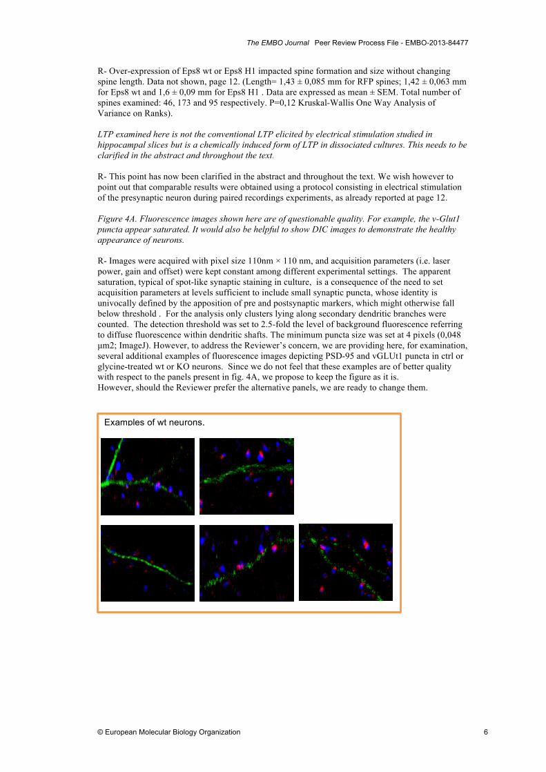
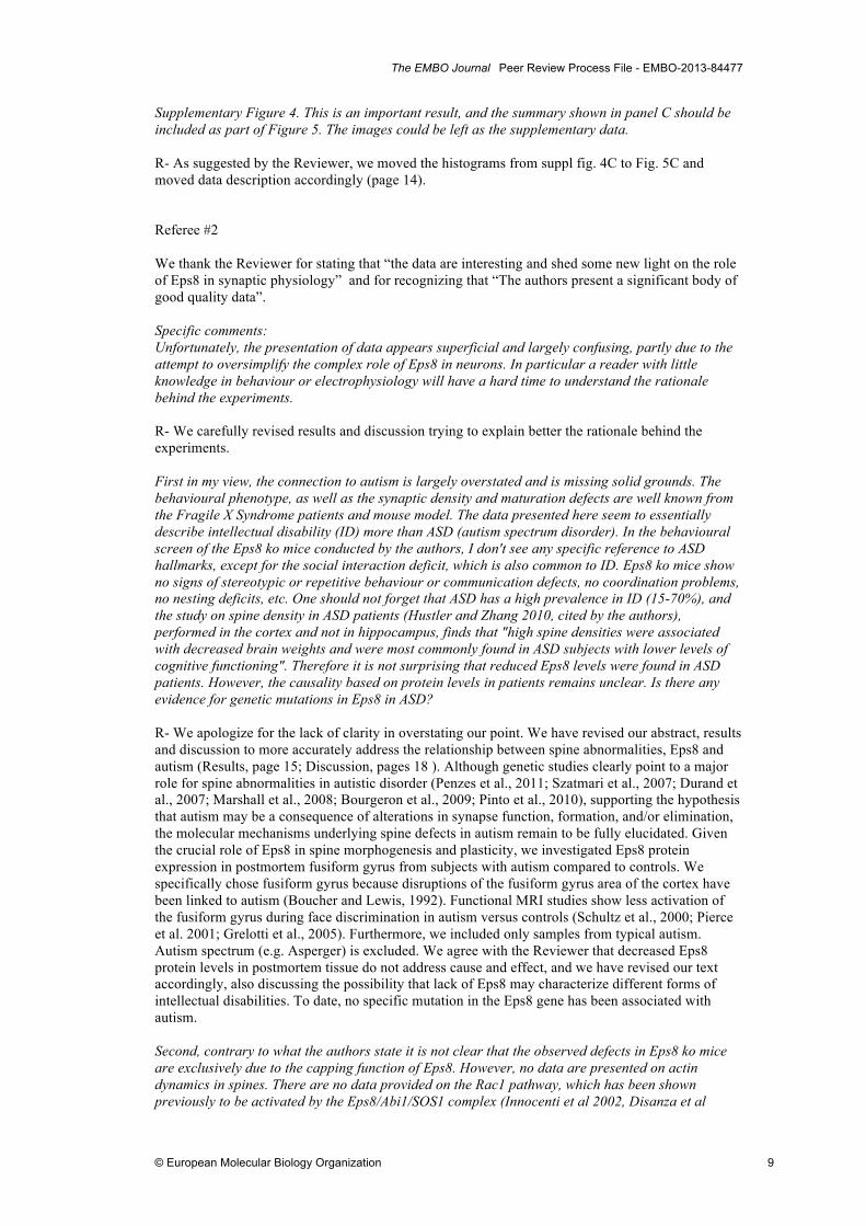
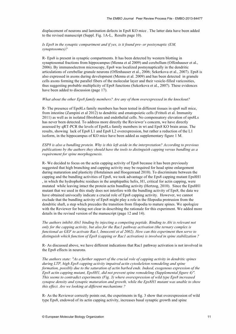
R- Over-expression of Eps8 wt or Eps8 H1 impacted spine formation and size without changing spine length. Data not shown, page 12. (Length= 1,43 ± 0,085 mm for RFP spines; 1,42 ± 0,063 mm for Eps8 wt and 1,6 ± 0,09 mm for Eps8 H1 . Data are expressed as mean ± SEM. Total number of spines examined: 46, 173 and 95 respectively. P=0,12 Kruskal-Wallis One Way Analysis of Variance on Ranks). LTP examined here is not the conventional LTP elicited by electrical stimulation studied in hippocampal slices but is a chemically induced form of LTP in dissociated cultures. This needs to be clarified in the abstract and throughout the text. R- This point has now been clarified in the abstract and throughout the text. We wish however to point out that comparable results were obtained using a protocol consisting in electrical stimulation of the presynaptic neuron during paired recordings experiments, as already reported at page 12. Figure 4A. Fluorescence images shown here are of questionable quality. For example, the v-Glut1 puncta appear saturated. It would also be helpful to show DIC images to demonstrate the healthy appearance of neurons. R- Images were acquired with pixel size 110nm × 110 nm, and acquisition parameters (i.e. laser power, gain and offset) were kept constant among different experimental settings. The apparent saturation, typical of spot-like synaptic staining in culture, is a consequence of the need to set acquisition parameters at levels sufficient to include small synaptic puncta, whose identity is univocally defined by the apposition of pre and postsynaptic markers, which might otherwise fall below threshold . For the analysis only clusters lying along secondary dendritic branches were counted. The detection threshold was set to 2.5-fold the level of background fluorescence referring to diffuse fluorescence within dendritic shafts. The minimum puncta size was set at 4 pixels (0,048 µm2; ImageJ). However, to address the Reviewer’s concern, we are providing here, for examination, several additional examples of fluorescence images depicting PSD-95 and vGLUt1 puncta in ctrl or glycine-treated wt or KO neurons. Since we do not feel that these examples are of better quality with respect to the panels present in fig. 4A, we propose to keep the figure as it is. However, should the Reviewer prefer the alternative panels, we are ready to change them.
Examples of wt neurons, control conditions

The EMBO Journal Peer Review Process File - EMBO-2013-84477
© European Molecular Biology Organization 7
Examples of wt neurons, glycine-treated
Examples of Eps8 KO neurons, control conditions

The EMBO Journal Peer Review Process File - EMBO-2013-84477
© European Molecular Biology Organization 8
To demonstrate that neurons are in fact healthy, we have provided quantitative data of input resistance before and after LTP protocol application (Results page 15). Neurons after chemical LTP are viable and healthy as demonstrated by normal values of input resistance. These data have been added to the text (page 13). To demonstrate that neurons are in fact healthy, we have provided quantitative data of input resistance before and after LTP protocol application (Results page 13). Figure 5D. The peptide experiments should also be performed in EPS8KO cultures in parallel to demonstrate the specificity of the blocking peptide effect to EPS8. Note that in the right panel, the x-axis label is missing. R- Following Reviewer’s suggestion we have carried out the peptide experiments also in Eps8 KO cultures. Injection of either blocking or scrambled peptide in Eps8 KO neurons does not change LTP (no differences in mEPSC frequency or amplitude, fig. 5 G and H, results page 14). Also, we now show that the blocking peptide does not change per se basal synaptic properties in either wt or KO neurons (Fig. 5 I and L, results page 14). Figure 6. The relevance of the specific reduction of EPS8 in autism patients in the context of present work highlighting EPS8KO with spine plasticity deficits needs to be further substantiated by a simultaneous comparison of additional postsynaptic markers and actin modulators between control and autism brain tissue. R- We examined and quantified the synaptosomal protein SNAP-25 as an additional synaptic marker to control for specificity of Eps8. We show lack of SNAP-25 changes in autism versus control samples. We have added new panels in figure 6 (panels D-F) presenting these additional data (Results, page 15). Supplementary Figure 2. It is curious that despite the robust increase in spine density and the pre and the postsynaptic areas, the mEPSC frequency is not altered in EPS8KO neurons under basal conditions. Is this due to the decrease in mEPSC amplitude where many of the events fall below detection or due to a reduction in presynaptic release probability? R- We favor the possibility that lack of alteration in mEPSC frequency derives from decrease in mEPSC amplitude, thus resulting in many of the events falling below detection. We cannot however exclude a role of Eps8 at the presynaptic level. We thank the Reviewer for raising this point which has now been mentioned in the revised text (page 12).
Examples of Eps8 KO neurons, glycine-
The EMBO Journal Peer Review Process File - EMBO-2013-84477
© European Molecular Biology Organization 9
Supplementary Figure 4. This is an important result, and the summary shown in panel C should be included as part of Figure 5. The images could be left as the supplementary data. R- As suggested by the Reviewer, we moved the histograms from suppl fig. 4C to Fig. 5C and moved data description accordingly (page 14). Referee #2 We thank the Reviewer for stating that “the data are interesting and shed some new light on the role of Eps8 in synaptic physiology” and for recognizing that “The authors present a significant body of good quality data”. Specific comments: Unfortunately, the presentation of data appears superficial and largely confusing, partly due to the attempt to oversimplify the complex role of Eps8 in neurons. In particular a reader with little knowledge in behaviour or electrophysiology will have a hard time to understand the rationale behind the experiments. R- We carefully revised results and discussion trying to explain better the rationale behind the experiments. First in my view, the connection to autism is largely overstated and is missing solid grounds. The behavioural phenotype, as well as the synaptic density and maturation defects are well known from the Fragile X Syndrome patients and mouse model. The data presented here seem to essentially describe intellectual disability (ID) more than ASD (autism spectrum disorder). In the behavioural screen of the Eps8 ko mice conducted by the authors, I don't see any specific reference to ASD hallmarks, except for the social interaction deficit, which is also common to ID. Eps8 ko mice show no signs of stereotypic or repetitive behaviour or communication defects, no coordination problems, no nesting deficits, etc. One should not forget that ASD has a high prevalence in ID (15-70%), and the study on spine density in ASD patients (Hustler and Zhang 2010, cited by the authors), performed in the cortex and not in hippocampus, finds that "high spine densities were associated with decreased brain weights and were most commonly found in ASD subjects with lower levels of cognitive functioning". Therefore it is not surprising that reduced Eps8 levels were found in ASD patients. However, the causality based on protein levels in patients remains unclear. Is there any evidence for genetic mutations in Eps8 in ASD? R- We apologize for the lack of clarity in overstating our point. We have revised our abstract, results and discussion to more accurately address the relationship between spine abnormalities, Eps8 and autism (Results, page 15; Discussion, pages 18 ). Although genetic studies clearly point to a major role for spine abnormalities in autistic disorder (Penzes et al., 2011; Szatmari et al., 2007; Durand et al., 2007; Marshall et al., 2008; Bourgeron et al., 2009; Pinto et al., 2010), supporting the hypothesis that autism may be a consequence of alterations in synapse function, formation, and/or elimination, the molecular mechanisms underlying spine defects in autism remain to be fully elucidated. Given the crucial role of Eps8 in spine morphogenesis and plasticity, we investigated Eps8 protein expression in postmortem fusiform gyrus from subjects with autism compared to controls. We specifically chose fusiform gyrus because disruptions of the fusiform gyrus area of the cortex have been linked to autism (Boucher and Lewis, 1992). Functional MRI studies show less activation of the fusiform gyrus during face discrimination in autism versus controls (Schultz et al., 2000; Pierce et al. 2001; Grelotti et al., 2005). Furthermore, we included only samples from typical autism. Autism spectrum (e.g. Asperger) is excluded. We agree with the Reviewer that decreased Eps8 protein levels in postmortem tissue do not address cause and effect, and we have revised our text accordingly, also discussing the possibility that lack of Eps8 may characterize different forms of intellectual disabilities. To date, no specific mutation in the Eps8 gene has been associated with autism. Second, contrary to what the authors state it is not clear that the observed defects in Eps8 ko mice are exclusively due to the capping function of Eps8. However, no data are presented on actin dynamics in spines. There are no data provided on the Rac1 pathway, which has been shown previously to be activated by the Eps8/Abi1/SOS1 complex (Innocenti et al 2002, Disanza et al

The EMBO Journal Peer Review Process File - EMBO-2013-84477
© European Molecular Biology Organization 10
2004). There is no analysis in Eps8 ko brain (or hippocampus) extracts or synaptosomes of the activation levels of Rac1 (GTP-bound Rac1) compared to wild type. The authors mention several times the balance between capping and Arp2/3 activity during spine stabilization. Now, Arp2/3 is activated by WAVE, which is in turn activated by Rac1 binding to Sra1 in the WAVE complex (Steffen et al 2004). How can it be excluded that also, or even mainly, Arp2/3 function is affected in Eps8 ko mice? R- Part of the experiments suggested by the Reviewer have already been performed and published. In particular, measurement of RacGTP levels by CRIB assay revealed equal levels of RacGTP in lysates of brain cortex and hippocampus derived from WT and Eps8 null mice (Menna et al., 2009), indicating that in brain Eps8 is not required for Rac activation. Indeed, we have also demostrated that in neuronal cells the capping function of Eps8 is predominant (Vaggi et al 2011). As additional proof, exposing hippocampal neurons to the specific Rac1 inhibitor, NSC23766, resulted in a reduction in the number of filopodia, while Eps8 removal resulted in increase in filopodia density (Menna et al., 2009). These data indicate that the impact of loss of Eps8 on Rac activity is negligible in CNS. Similar results have also been reported by some of the authors of this manuscript but using cerebellar neurons (Offenhauser et al. Cell 2006). These considerations have now been added to the revised text (page 14). Anyway, following the Reviewer’s suggestion, we measured the levels of Rac-GTP in mature cultures from wt or Eps8KO mice by using G-LISA™ Rac Activation Assay Biochem Kit™. These data have been added to the revised version of the manuscript (Suppl. Fig.4D; page 14). As expected, Rac activity appeared to be not affected by Eps8 removal. In keeping with this latter notion and based on the fact that Rac through the WAVE/SCAR controls Arp2/3 function, we do not expect any alteration in the activity of the latter complex. These considerations have been added to the text (page 14). No electrophysiology on slices is provided. This is standard in order to judge pre- and postsynaptic parameters and synaptic plasticity, LTP and LTD. R- We agree that these experiments will provide important information and we plan indeed to carry them out. As the Reviewer is certainly aware, performing electrophysiology on slices is technically challenging and would require a significant amount of additional time that is probably incompatible with a timely publication of the results we obtained so far. We also would like to point out that the addition of electrophysiology data, while interesting, would add an additional levels of complexity to a study that, as stated by Reviewer 1, appears already as “ a tour de force study combining multiple experimental approaches from biochemistry, molecular biology, immunocytochemistry, fluorescence live imaging, electrophysiology, to assays of mice behavior”. The Eps8 knockout is systemic (conventional mutant), but the information on the knockout and the expression of Eps8 in brain is not there. Where in brain is Eps8 expressed? What neuronal populations express EPS8? Is it expressed in glia? One also wants to see a basic histological investigation of the different brain regions in order to exclude developmental defects (e.g. cortical displacement of neurons, lamination defects). Otherwise any behavioural phenotype is difficult to reconcile with defects in synaptic plasticity. R- All this analysis has been already carried out and published (Sekerkova et al., 2007; Offenhauser et al.2006). Eps8 has been detected in many regions of the gray matter, including olfactory bulb, anterior olfactory nuclei, basal forebrain, cerebral cortex, hippocampus, septal nuclei, amygdala, thalamus, hypothalamus, colliculi, pontine nuclei, cerebellum, cochlear nuclear complex and inferior olive, while the white matter was generally unstained (Sekerkova et al., 2007). In the adult, Eps8 is expressed at higher levels in scattered neurons in layers II and III of the cerebral cortex and in the hippocampus, two areas classically implicated in higher cognitive functions, and in the cerebellum (Offenhauser et al., 2006). In the rat cerebellum, Eps8 protein was restricted to the excitatory interneurons innervated by mossy fibers, granule cells and unipolar brush cells (Sekerkova et al., 2007). Eps8 has not been detected in cerebellar astrocytes (Sekerkova et al., 2007), although it is present in cultured astrocytes from hippocampus (our unpublished observations). Eps8 has also been detected in the tips of the stereocilia in mammalian IHCs and OHCs (Zampini et al., 2011). For the sake of completeness, we added most of this information to the text (page 17). Following the Reviewer’s request, we have now performed histological investigation of different brain regions of wt and Eps8KO adult mice, which allowed to exclude developmental defects such as cortical
The EMBO Journal Peer Review Process File - EMBO-2013-84477
© European Molecular Biology Organization 11
displacement of neurons and lamination defects in Eps8 KO mice . The latter data have been added to the revised manuscript (Suppl. Fig. 1A-L, Results page 10). Is Eps8 in the synaptic compartment and if yes, is it found pre- or postsynaptic (EM, synaptosomes)? R- Eps8 is present in synaptic compartments. It has been detected by western blotting in synaptosomal fractions from hippocampus (Menna et al 2009) and cerebellum (Offenhauser et al., 2006). By immunoelectron microscopy, Eps8 was localized postsynaptically in the dendritic articulations of cerebellar granule neurons (Offenhauser et al., 2006; Sekerkova et al., 2007). Eps8 is also expressed in axons during development (Menna et al. 2009) and has been detected in granule cells axons forming the parallel fibers of the molecular layer and their vesicle-filled varicosities, thus suggesting probable multiplicity of Eps8 functions (Sekerkova et al., 2007). These evidences have been added to discussion (page 17). What about the other Eps8 family members? Are any of them overexpressed in the knockout? R- The presence of Eps8Ls family members has been tested in different tissues in eps8 null mice, from intestine (Zampini et al 2012) to dendritic and ematopoietic cells (Frittoli et al. Immunity 2011) as well as in isolated fibroblasts and endothelial cells. No compensatory elevation of eps8Ls has never been detected. To address more directly the Reviewer’s concern, we have directly assessed by qRT-PCR the levels of Eps8Ls family members in wt and Eps8 KO brain areas. The results, showing lack of Eps8 L1 and Eps8 L2 overexpression, but rather a reduction of the L1 isoform, in the hippocampus of KO mice have been added as supplementary figure 1 M. ESP8 is also a bundling protein. Why is this left aside in the interpretation? According to previous publications by the authors they should have the tools to distinguish capping versus bundling as a requirement for spine morphogenesis. R- We decided to focus on the actin capping activity of Eps8 because it has been previously suggested that high branching and capping activity may be required for head spine enlargement during maturation and plasticity (Hotulainen and Hoogenraad 2010). To discriminate between the capping and the bundling activities of Eps8, we took advantage of the Eps8 capping mutant Eps8H1 , in which the hydrophobic residues in the amphipathic helix, H1, critical for actin capping, were mutated while leaving intact the protein actin bundling activity (Hertzog, 2010). Since the Eps8H1 mutant that we used in this study does not interfere with the bundling activity of Eps8, the data we have obtained univocally indicate a crucial role of Eps8 capping activity. However, we cannot exclude that the bundling activity of Eps8 might play a role in the filopodia protrusion from the dendritic shaft, a step which precedes the transition from filopodia to mature spines. We apologize with the Reviewer for being not clear in describing the rationale for this experiment. We added more details in the revised version of the manuscript (page 12 and 14). The authors inhibit Abi1 binding by injecting a competing peptide. Binding to Abi is relevant not only for the capping activity, but also for the Rac1 pathway activation (the ternary complex is functional as GEF to activate Rac1, Innocenti et al 2002). How can this experiment then serve to distinguish which function of Eps8 (capping or Rac1 activation) is involved in spine stabilization ? R- As discussed above, we have different indications that Rac1 pathway activation is not involved in the Eps8 effects in neurons. The authors state: "As a further support of the crucial role of capping activity in dendritic spines during LTP, high Eps8 capping activity impaired actin cytoskeleton remodeling and spine formation, possibly due to the saturation of actin barbed ends. Indeed, exogenous expression of the Eps8 actin capping mutant, Eps8H1, did not prevent spine remodeling (Supplemental figure 4)". This seems to contradict experiments (Fig. 3) where overexpression of wild type Eps8 increased synapse density and synaptic maturation and growth, while the Eps8H1 mutant was unable to show this effect. Are we looking at different mechanisms ? R- As the Reviewer correctly points out, the experiments in fig. 3 show that overexpression of wild type Eps8, endowed of its actin capping activity, increases basal synaptic growth and spine

The EMBO Journal Peer Review Process File - EMBO-2013-84477
© European Molecular Biology Organization 12
formation. In these conditions, LTP application does not produce any further increase in spine formation (see revised supplementary fig. 4 A-C and fig.5C ). This is not contradictory. Our interpretation of this result is that overexpressed Eps8 saturates barbed ends of actin filament by capping them, thus altering actin filament growth and dynamics, eventually precluding additional spine formation upon LTP. Conversely, overexpression of the Eps8H1 mutant, which is unable to bind barbed ends, has no effects on actin capping and dynamics. Thus, upon LTP application no impairment in spine formation is detected as expected. Based on the request of reviewer 1 who, given the interest of these results, asked to move them from Supplementary fig. 4 to Fig. 5, we took the opportunity to rephrase the description of the experiment in order to make the concept clearer (page 14). Referee #3 We thank the reviewer for stating “The presented work is very impressive. The authors succeeded to analyze the behavioural phenotype of their mice, i.e. the learning and memory defect, and then linked this phenotype to abnormal spine morphology and the preclusion of synaptic potentiation. It was then an outstanding effort that the authors managed to decipher the molecular mechanism of Eps8 function. This is truly a great paper and should be published as it is”. We are sincerely proud of this comment. Acceptance letter 15 April 2013
Thank you for submitting your revised manuscript to The EMBO Journal. I asked referee #1 to take a look at the revised version and I have now heard back from the referee. As you can below, the referee appreciates the introduced changes and support publication here. I am therefore very pleased to accept the paper for publication here. Thank you for contributing to the EMBO Journal! Referee #1 The authors have fully addressed my prior concerns in the revision.