Embed Size (px)
Citation preview
AN INVESTIGATION ON THE EFFECT OF SOME
HEAVY METALS ON SOIL MYCOFLORA OF URBAN
ENVIRONMENT
THESIS SUBMITTED TO UNIVERSITY OF KALYANI FOR THE
DEGREE OF DOCTOR OF PHILOSOPHY (PH.D) IN SCIENCE
Kaoushik K Mukherjee, M.Sc
Department of Environmental Science
University of Kalyani
Kalyani-741235
West Bengal
INDIA
2014
University of Kalyani FACULTY OF SCIENCE
No……………………. Date: January 16, 2014
TO WHOM IT MAY CONCERN
I have the pleasure to certify that Shri Kaoushik K Mukherjee, has completed
his Ph.D. work entitled "AN INVESTIGATION ON THE EFFECT OF SOME
HEAVY METALS ON SOIL MYCOFLORA OF URBAN ENVIRONMENT", under
my supervision. Shri Mukherjee has completed his work through repeated
laboratory analysis in the Department of Environmental Science, University of
Kalyani, Nadia, West Bengal. This work is original and has not been
submitted earlier for any degree or award.
(Prof. S. C. Santra)
Supervisor
Phone: +91-33-2582750 Ext: 291, 292
+91-33-2580 8749 (Direct)
Fax: +91-33-2580 8749 (Direct)
+91-33-2582 8282
E-mail: [email protected]
Prof. S. C. Santra Professor
Department of Environmental Science
University of Kalyani
Kalyani - 741235, Nadia, West Bengal,
INDIA
Acknowledgement
It is a matter of great privilege to extend my sincere thanks to Professor (Dr.) S. C.
Santra, Department of Environmental Science, University of Kalyani, at the very outset
under whose able leadership, guidance and supervision, this work has been carried out. A
constant source of inspiration and support, I am deeply indebted to his encouragement
and sustained navigation throughout the entire span of the present work. I express my
deep gratitude to Dr.(Mrs.) Rina Bhattacharya, Associate Professor and Head,
Department of Environmental Science, University of Kalyani. I also express my heartfelt
thanks to Professor (Dr.) Dilip Kumar Khan, Professor (Dr.) Debasish Das, Dr. (Mrs.)
Soma Mukherjee, Associate Professor, Department of Environmental Science, University
of Kalyani for their valuable suggestions and cooperation throughout the entire period of
my research work. I must also mention the moral support extended to me by Dr. Tapas
Bandyopadhyay, Associate Professor, Department of Molecular Biology &
Biotechnology, University of Kalyani and Dr. Arindam Bhattacharyya, Associate
Professor, Department of Zoology, University of Calcutta at different times. I would like
to extend my sincere thanks to Professor Rattan Lal Hangloo, Vice Chancellor, University
of Kalyani and his predecessors who had allowed me to take up the research. I am also
grateful to the Registrar and all my officer colleagues who have always wished me best in
my academic pursuit.
My special thanks to Dr. Alok Chandra Samal for his tremendous support throughout the
entire period of my research work in the field work, laboratory experiments and thesis
writing. I also extend my special thanks to Ms. Satabdi Banerjee who has frequently
accompanied me in carrying out my experiments even in other institutions.
I would also like thank Sandip, Sukalyan, Jayjit, Piyal, Debargha, Amrita, Dipanwita,
Debojyoti, Soumen and Sourav for extending their supportive hands at different points of
time and at difficult situations. I will sure carry the nice moments of working with them.
My thanks to other research scholars of the Department of Environmental Science too.
My sincere thanks to all the non teaching staff of the Department of Environmental
Science and the Ph. D Section of Registrar‘s Department, University of Kalyani for their
cooperation and support at various stages of my research.
Last but not the least, I would like to offer a tribute of love, admiration and respect to my
parents Shri K. D. Mukherjee and Smt. Sova Mukherjee, my unmarried aunt Smt. Minati
Mukherjee and my uncle Shri M. L. Mukherjee who have always been a tremendous
source of inspiration throughout my life and also during this prolonged span of my effort
when I had to tide over a series of difficult situation. In fact they are the ones who have
always wanted to see me stuck to academics and taught me to remember that Vidhyan
Sarvatra Pujwathe since my very childhood. And finally my wife Rita and daughter little
love Sohini who being so close to my heart have never allowed me to feel fatigued at any
point of time and without their huge support, even not being directly related to this effort,
it was simply not possible for me to accomplish this modest academic task.
Let me sincerely apologise to all my other well wishers and friends whose names I could
not mention owing to the limitation a frame of page always bears with it.
(Kaoushik K Mukherjee)
CONTENTS
Chapter Page No.
1. Introduction 1-5
1.1 Essentiality of metals 2
2. Review of literature 6-49
2.1 Metals toxicity of fungi 6
2.2 Resistance and tolerance 7
2.3 Environmental influence on heavy metal toxicity towards
fungi
9
2.4 Fungi in polluted habitats 12
2.5 Interactions between toxic metals and fungi 15
2.5.1 Extracellular precipitation and complexation 16
2.5.2. Metal binding to cell walls 16
2.5.3 Transport of toxic metal cations 20
2.5.4 Intracellular fate of toxic metals 24
2.6. Metal transformations 33
2.7 Accumulation of metals by macrofungi 35
2.8 Biotechnological aspects of fungal metal accumulation 36
2.9 Metal removal by inducing, excreted or derived biomolecules 41
2.10 Metal recovery 41
2.11 Biosorption of meals and radionuclides by fungal biomass 42
3. Aims and Objectives 50
4. Materials and Methods 51-58
4.1 Sampling of soil 51
4.2 Isolation of metal resistant microorganisms 51
4.3 Media compositions 52
4.4 Standard plate counts 52
4.5 Isolation of pure cultures 52
4.6 Maintenance of cultures 53
4.7 Selection of tolerant strains 54
4. 8 Heavy metal analysis of contaminated soil 54
4.9 Identification of fungi 54
4.10 Growth optimization of media, pH and Temperature 54

4.11 Antibiotic sensitivity test 55
4.12 Heavy metal tolerance assessment 55
4.13 Cross metal resistance assay 55
4.14 Metal toxicity of the fungal isolates 56
4.15 Metal removal by selected strains 56
4.16 Metal biosorption by fungal biomass 56
4.17 Metal adsorption by Chitosan 57
4.18 Assay of metallothionein protein 57
4.19 Scanning Electron Microscopic(SEM) study 58
5. Results 59 - 94
5.1 Soil analysis 59
5.2 Isolation of Fungal strains from contaminated Soil 61
5.3 Identification of selected strain 64
5.4 Optimization of pH and Temperature on growth of selected
fungal strain
66
5.5 Antibiotic sensitivity test 67
5.6 Metal Tolerance Level 68
5.7 Cross Metal Tolerance 69
5.8 Growth response of fungal strain in different metal
concentration
71
5.9 Test for metal removal in vitro 74
5.10 Metal accumulation by live fungal mycelium 82
5.11 Metal uptake by dead fungal mycelium 85
5.12 Metal adsorption by chitosan 88
5.13. Analysis of metallothionein 89
5.14. Scanning Electron Microscopy (SEM) study of the fungal
isolates
91
6. Discussion 95 -102
7. conclusion 103 -106
8. Reference 107 -135
Publication
LIST OF TABLES
Table No. Table Name Page No.
Table 1.1 Classification of metal ions 3
Table 2.1 Macromolecular constituents of fungal cell walls
(adapted from Peberdy, 1990)
16
Table 2.2 Some examples of metal and actinide accumulation by
fungi
37
Table 2.3 Biosorption by fungal biomass (mg g-1
) 43
Table 2.4 The biosorptive capacity of various fungal organisms 46
Table 4.1 Morphological and biochemical characteristics of the
isolated fungal species.
53
Table 5.1 Soil characteristics of the sampling sites 60
Table 5.2 Morphological and biochemical characteristics of the
isolated fungal species
61
Table 5.3 Morphological characters of selected strain 64
Table- 5.4 Effect of pH for growth of isolated fungal strains (in
terms of mycelia weight after 6days of incubation)
66
Table-5. 5 Effect of temperature for growth of isolated fungal
strains (in terms of mycelial weight after 6days of
incubation)
67
Table: 5.6 Antibiotic sensitivity test with the fungal strains 68
Table 5.7 Metal Tolerance Level (MTL) study 69
Table 5. 8 Cross metal resistance 70
Table 5.9 Arsenic removal by AsC1 strain (Aspergillus flavus ) 75
Table 5.10 Arsenic removal by AsB3 strain (Aspergillus niger ) 76
Table 5.11 Chromium removal (%) of CrTW1 strain (Aspergillus
niger)
78
Table 5.12 Chromium removal by CrTW3 strain (Penicillium sp.) 79
Table 5.13 Cadmium removal by CdEP strain (Aspergillus flavus) 81
Table 5.14 Adsorption of Arsenic in Chitosan 88
Table 5.15 Adsorption of Chromium in Chitosan 88
Table 5.16 Adsorption of Cadmium by Chitosan 89
Table 5.17 Estimation of Metallothionein in metal treated and
control strains
90
LIST OF FIGURES
Figure No. Figure Name Page No.
Fig. 1.1 Diagrammatic representation of metal-fungal
interactions
3
Fig. 2.1 Diagrammatic representation of metal interactions with
fungi. The metal species are represented in cationic
form. All the interactions shown may be involved in
fungal survival and/or metal detoxification.
9
Fig. 2.2 Mechanisms of heavy metal uptake on microbial
surface
15
Fig. 2.3 Metalloregulation of the CUP1 locus in
Saccharomyces cerevisiae. The Cu-ACE1
metalloprotein complex interacts with upstream
sequences from the CUP1 coding sequences and
facilitates transcription of the MT gene with the CUP1
locus. Translation of the MT mRNA yields MT protein
which acts to regulate the cytosolic concentration of
copper ions (modified after Mehra & Winge, 1991).
27
Fig. 2.4 Structures of glutathione and the metal-binding
polypeptide, (-glutamylcysteinyl)3-glycine. Peptide
bonds between glutamate and cysteine utilize the side
chain, or -carboxylate of glutamate (outlined) rather
than the az-carboxylate characteristic of proteins
whose synthesis is ribosome dependent (modified after
Steffens, 1990).
28
Fig. 5.1 Microphotograph of AsC1 65
Fig. 5.2 Microphotograph of AsB3 65
Fig. 5.3 Microphotograph of CrTW1 65
Fig. 5.4 Microphotograph of CrTW3 65
Fig. 5.5 Microphotograph of CdEP 65
Fig. 5.6 Cross metal tolerance by fungal strains 70
Fig. 5.7 Inhibition zone by metal 71
Fig. 5.8 Growth response of AsC1 in different doses of arsenic 71
Fig. 5.9 Growth response of AsB3 in different doses of arsenic 72
Fig. 5.10 Growth response of CrTW1 in different doses of
chromium
73
Fig. 5.11 Growth response of CrTW3 in different doses of
chromium
73
Fig. 5.12 Growth response of CdEP in different doses of
cadmium
74
Fig. 5.13 Arsenic removal by AsC1 strain 76
Fig. 5.14 Arsenic removal by AsB3 strain 77
Fig. 5.15 Chromium removal by CrTW1 strain 79
Fig. 5.16 Chromium removal by CrTW3 strain 80
Fig. 5.17 Cadmium removal by CdEP strain 81
Fig. 5.18 Accumulation of Arsenic by live fungal mycelium of
AsC1
82
Fig. 5.19 Accumulation of Arsenic by live fungal mycelium of
AsB3
83
Fig. 5.20 Accumulation of Chromium by live fungal mycelium
of CrTW1
84
Fig. 5.21 Accumulation of Chromium by live fungal mycelium
of CrTW3
84
Fig. 5.22 Accumulation of Cadmium by live fungal mycelium of
CdEP
85
Fig. 5.23 Arsenic accumulation in dead mycelium of AsC1 and
AsB3 strain
86
Fig. 5.24 Accumulation of chromium by dead mycelium of
CrTW1 and CrTW3 strain
87
Fig. 5.25 Accumulation of Cadmium by dead mycelium of
CdEP strain
87
Fig. 5.26 Metallothionein activity of the studied fungal strains 90
Fig. 5.27 Morphological changes in Arsenic treated fungal
strain(AsC1) observed using Scanning Electron
Microscopy (SEM)
92
Fig. 5.28 Morphological changes in Arsenic treated fungal
strain(AsB3) observed using Scanning Electron
Microscopy (SEM)
92
Fig. 5.29 Morphological changes in Chromium treated fungal
strain(CrTW1) observed using Scanning Electron
Microscopy (SEM)
93
Fig. 5.30 Morphological changes in Chromium treated fungal
strain(CrTW3) observed using Scanning Electron
Microscopy (SEM)
93
Fig. 5.31 Morphological changes in Cadmium treated fungal
strain(CdEP) observed using Scanning Electron
Microscopy (SEM)
94
Chapter - 1
1
INTRODUCTION
Fungi are ubiquitous in natural environments and important in industrial processes. A
range of morphologies are found from unicellular yeasts to polymorphic and
filamentous fungi, many of which have complex macroscopic fruiting bodies. Their
most important roles are as decomposers of organic materials, with concomitant
nutrient cycling, as pathogens and symbionts of animals and plants and as spoilage
organisms of natural and synthetic materials like wood, paint, leather, food and
fabrics.
The fungi are utilized as producers of economically important substances, e.g.
ethanol, citric acid, antibiotics, polysaccharides, enzymes and vitamins. The study of
the interaction between toxic metals and fungi has long been of scientific interest.
This is because metal toxicity has been, and remains the basis of many fungicidal
preparations used in the control of plant pathogens and to preserve natural and
synthetic materials (Horsfall, 1956; Ross, 1975). Early work often had a
phytopathological perspective relating to the assessment of toxicity (Somers, 1961).
Subsequent observations on the ability of fungi to resist and adapt to toxic metals led
to further work on the physiological, bio- chemical and genetical explanations for
these phenomena (Ashida, 1965; Ross, 1975; Gadd 1986a). Research has been
stimulated particularly by the continuous refinement in biochemical and molecular
techniques and by the general appreciation of filamentous fungi and yeasts as model
eukaryotic cells systems. In an environmental context, accelerating pollution of the
natural environment by toxic metals, metalloids, radionuclides and organo-
metal(loid)s have also led to increased interest because of the ubiquitous and
sometimes dominant presence of fungi in metal-polluted habitats, their uptake and
translocation of toxic metals and radionuclides to fruit bodies of edible fungi and the
significance of mycorrhizal fungi in polluted habitats (Gadd, 1986a; Brown & Hall,
1990). While amelioration of metal phytotoxicity by mycorrhizal fungi is relevant to
land reclamation (Colpaert & van Assche, 1987), another area of applied interest is
the use of fungal (and other microbial) biomass for the detoxification of
metal/radionuclide-containing industrial effluents (Gadd, 1986b, 1990a, 1992a,
2010).
Chapter - 1
2
Physico-chemical, biochemical and genetical aspects of cellular interactions are
included mainly with reference to 'microfungi' and yeasts, though mention is also
made for mycorrhizas and macrofungi in polluted habitats. Biotechnological
discussion relates only to the use of fungal biomass and products for environmental
protection. Several important topics such as the roles of essential metal ions in fungal
metabolism and differentiation (Jones & Gadd, 1990), yeast flocculation
(Kuriyama, et al., 1991), iron acquisition and function (Winkelmann, et al., 1987;
Winkelmann, 1992), interactions of metals with lichens (Nieboer, et al., 1978;
Nash, 1990) and (organo) metal-containing fungicides and their effects (Cooney &
Wuertz, 1989) are not covered in detail.
1.2 Essentiality of metals
Metals are directly and/or indirectly involved in all aspects of fungal growth,
metabolism and differentiation. While many metals are essential, e.g. K, Na, Mg, Ca,
Mn, Fe, Cu, Zn, Co and Ni, many others have no apparent essential function, e.g. Rb,
Cs, Al, Cd, Ag, Au, Hg and Pb. However, all these elements can interact with fungal
cells and can be accumulated by physico-chemical mechanisms and transport systems
of varying specificity (Gadd, 1988). Most essential and inessential metals exhibit
toxicity above a certain concentration, which vary depending on the organism, the
physico- chemical properties of the metal and environmental factors. This may
necessitate expression of a detoxification mechanism if the organism is to survive
(Fig. 1). Although the connotations of toxicity and bioaccumulation are most often
linked with inessential metals, the potential toxicity of many essential metals should
not be overlooked. A good example is provided by calcium, an important intracellular
second messenger in fungal growth and differentiation, which can also be highly toxic
within cells owing to precipitation of phosphates. Fungi, like other eukaryotic cells,
maintain cytosolic free Ca2+
at around 0 1 µM by means of a variety of transport
systems located on the plasma and vacuolar membranes which effect influx/efflux and
vacuolar compartmentation; cytosolic free Ca2+
may also be modulated by interaction
with calcium-binding proteins (Miller, et al., 1990; Gadd & Brunton, 1992). Many
attempts have been made to define metals in relation to biological effects using their
physical, chemical and biological properties. Metals and metalloids can be considered
to include all the elements except the noble gases and H, B, C, N, 0.
Chapter - 1
3
Fig.1.1 Diagrammatic representation of metal-fungal interactions.
The metals are broadly classified as type A, type B and transition metal. The details
are given in Table - 1.
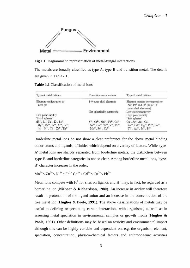
Table 1.1 Classification of metal ions
Borderline metal ions do not show a clear preference for the above metal binding
donor atoms and ligands, affinities which depend on a variety of factors. While 'type-
A' metal ions are sharply separated from borderline metals, the distinction between
'type-B' and borderline categories is not so clear. Among borderline metal ions, ‗type-
B‘ character increases in the order:
Mn2+
< Zn2+
< Ni2+
< Fe2+
Co2+
< Cd2+
< Cu2+
< Pb2+
Metal ions compete with H+ for sites on ligands and H
+ may, in fact, be regarded as a
borderline ion (Nieboer & Richardson, 1980). An increase in acidity will therefore
result in protonation of the ligand anion and an increase in the concentration of the
free metal ion (Hughes & Poole, 1991). The above classifications of metals may be
useful in defining or predicting certain interactions with organisms, as well as in
assessing metal speciation in environmental samples or growth media (Hughes &
Poole, 1991). Other definitions may be based on toxicity and environmental impact
although this can be highly variable and dependent on, e.g. the organism, element,
speciation, concentration, physico-chemical factors and anthropogenic activities
Chapter - 1
4
(Babich & Stotzky, 1980; Gadd, 1990b). In fungi, metal effects can vary between
organisms, strains, the stage of growth and even different vegetative and reproductive
forms of the same organism (Gadd & Mowll, 1985; Sabie & Gadd, 1990). A given
organism may exhibit several mechanisms by which potentially toxic metal species
are detoxified, both dependent and independent of metabolism and all affected by
environmental factors. Furthermore, other elements not considered to be true metals
according to chemical definitions may have some metallic properties and exhibit
varying degrees of toxicity as well as accumulation by fungi. These include
metalloids, and the actinides and lanthanides, e.g. uranium and thorium. The alkali
metals are not usually included in discussions on metal toxicity but nevertheless
exhibit a dramatic range of biological properties as exemplified by K+, Na+ and Cs
+
as respectively representing an essential non-toxic, an essential but potentially-toxic,
and an inessential potentially-toxic metal ion. Caesium, and many actinides, are of
current concern because of environmental contamination, accumulation by the biota,
including fungi, and transfer to other organisms including humans (Gadd, 1992a).
Current definitions of the term 'heavy metal' are particularly variable. These are often
defined as a group of approximately 65 metallic elements, of density greater than 5,
with the general ability to exert toxic effects on microbial and other life forms (Gadd
& Griffiths, 1978; Nieboer & Richardson, 1980). A further complication is that
'heavy elements' may be defined as Pb, As, Cd, Hg, Sb, Se, TI, In, Bi, and Te
(Fergusson, 1990). This is again a diverse group and, in fact, As, Sb, Bi, Se and Te
have elemental structures more typical of non metals (Fergusson, 1990; Morgan &
Stumm, 1991). Organometallic compounds, simply defined as a compound
containing at least one metal-carbon bond (Thayer, 1988), frequently exhibit
enhanced toxicity, hence they are used as fungicides. When such compounds contain
metalloid elements, the term organo-metalloid may be used. Such substances should
also be included in discussions of metal interactions with fungi because of their
significance as environmental pollutants and as fungicides and also because some
may be synthesized by fungi as a result of exposure to and transformation of the
'parental' metal species.
The present study was planned to isolate and characterize metal tolerant fungal strains
from metal contaminated natural sites and also to assess their possible utility in
soil/waste water/industrial effluent bioremediation.
Chapter - 1
5
The brief outline of the thesis is given below:
Chapter – 1: Introduction
Chapter – 2: Review of Literature
Chapter – 3: Aims and Objective
Chapter – 4: Materials and Methods
Chapter – 5: Results
Chapter – 6: Discussion
Chapter – 7: Conclusion
Chapter – 8: References
Chapter – 9: Publications
Chapter - 2
6
REVIEW OF LITERATURE
2.1 Metals toxicity of fungi
Toxic metals can exert harmful effects principally as a result of their strong
coordinating abilities (Ochiai, 1987). Toxic effects include the blocking of functional
groups of biologically important molecules (e.g. enzymes and transport systems for
essential nutrients and ions), the displacement and/or substitution of essential metal
ions from biomolecules and functional cellular units, conformational modification,
denaturation and inactivation of enzymes and disruption of cellular and organelles
membrane integrity (Ochiai, 1987). Because of the wide spectrum of potentially toxic
interactions between metals and fungi, almost every aspect of their metabolism,
growth and differentiation may be affected, depending on the organism, metal species
and concentration and physico-chemical factors (Gadd, 1986a, c; Gadd & White,
1989a). Thus, toxic symptoms may vary widely between different fungi and for
different metal species. A prerequisite for direct toxic interactions is contact between
the active metal species and cellular components (Gadd & White, 1989a). The cell
membrane is an obvious initial site of action for a toxic metal species and membrane
damage can result in loss of mobile cellular solutes, e.g. K+, and increased
permeability of the cell to external materials (Norris & Kelly, 1977; Kuypers &
Roomans, 1979; Mowll & Gadd, 1983; White & Gadd, 1987 a, b; Laurence, et
al., 1989). Indirect mechanisms of metal toxicity may involve free radicals, which are
deleterious to cells as they can take part in chain reactions which involve the
breakdown of biological macromolecules. Consequently, aerobic organisms possess
protective enzymes such as superoxide dismutases, which are metalloenzymes
containing either Mn, Fe or Cu/Zn (Greco et al., 1990), which eliminate the radicals
produced by normal metabolism. Major targets in cells are membranes, where lipid
peroxidation, whereby the alkyl chains of lipids are converted to peroxyalkyl radicals
and fatty acid hydroperoxides, is initiated (Mehlhorn, 1986). Lipid-soluble
complexes of transition elements such as Fe (II) may undergo the Fenton reaction
[(1), (2)] with the hydroperoxides and accelerate this process (McCord & Day,
1978).
Fe2 + H2O2-* Fe
3+ +OH
-+OH (1)
Chapter - 2
7
O2-+ Fe3+ O> 02+ Fe
2+ (2)
Complexes and free ions of these cations may also undergo this reaction in aqueous
solution. In animal systems, metal ions such as Hg2+
, Cd2+
, Pb2+
and Ag2+
induce free
radical toxicity as a result of their reactions with thiols or enzymes which normally
protect against these reactive species (Mehlhorn, 1986). A little work on this aspect
has been carried out with fungi except for Cu2+
and Saccharomyces cerevisiae (Greco
et al., 1990).
Organometallic compounds are of increasing environmental significance because of
their use in the chemical and petroleum industries and as biocides. Organometals are
generally more toxic towards fungi than corresponding free metal ions and the
toxicity of their compounds varies with the number and with the identity of the
organic groups (Blunden, et al., 1984; Cooney & Wuertz, 1989). Major effects of
organotins and organoleads are disruption of mitochondrial membranes and action as
C1-/OH- ionophores. In this way, they depolarize electrochemical gradients and
consequently interfere with energy conservation (Blunden et al., 1984; Cooney &
Wuertz, 1989). In fact, organotin-resistant mutants of S. cerevisiae can exhibit
increased respiration rates (Dupont et al., 1990) and modification of the organotin-
binding site on the inner mitochondrial membrane (Lancashire & Griffiths, 1975).
Organometals may also damage membranes by the production of free radicals since
the carbon-metal bond readily reacts with available radicals to produce peroxyalkyl
radicals which can result in lipid peroxidation (Mehlhorn, 1986). The organometallic
compounds may also exert a disruptive effect on cell membranes and cause a loss of
K+ (Cooney et al., 1989).
2.2 Resistance and tolerance
Fungal survival in the presence of toxic metals mainly depends on intrinsic
biochemical and structural properties, physiological and/or genetic adaptation
including morphological changes and environmental modification of metal speciation,
availability and toxicity The relative importance of each often been difficult to
determine (Gadd & Griffiths, 1978; Gadd, 1990 b, 1992 b). Arbitrary terms such as
' resistance ' and ' tolerance ' which are used rather loosely and often interchangeably
in the literature are generally based on the ability to grow on a certain metal
concentration in laboratory media (Trevors, et al., 1986; Gadd, 1992b, c). It is
Chapter - 2
8
probably more appropriate to define 'resistance' as the ability of an organism to
survive metal toxicity by means of a mechanism produced in direct response to the
metal species concerned, e.g. synthesis of metallothioneins or y-glutamyl peptides
(Mehra & Winge, 1991). 'Metal tolerance' may be defined as the ability of an
organism to survive metal toxicity by means of intrinsic properties and/or
environmental modification of toxicity (Gadd, 1992b, c). Intrinsic properties that can
determine survival include possession of impermeable pigmented cell walls,
extracellular polysaccharide and metabolite excretion, especially where this leads to
detoxification of the metal species by binding or precipitation (Gadd, 1990b).
However, in many cases distinctions are difficult because of the involvement of
several direct and indirect physico-chemical and biological mechanisms in survival in
field and laboratory. Biological mechanisms implicated in fungal survival (as distinct
from environmental modification of toxicity) include extracellular precipitation,
complexation and crystallization, transformation of metal species by, e.g. oxidation,
reduction, methylation and dealkylation, biosorption to cell walls, pigments and
extracellular polysaccharide, decreased transport or impermeability, efflux,
intracellular compartmentation and precipitation and/or sequestration (Fig. 2.1) (Ross,
1975; Gadd & Griffiths, 1978; Gadd, 1988, 1990a, b, 1992a,b,c; Brown & Hall,
1990; Mehra & Winge, 1991). A particular organism may directly and/or indirectly
rely on several survival strategies. For example, metallothionein synthesis is a
mechanism of Cu2+
resistance in Saccharomyces cerevisiae yet Cu2+
binding or
precipitation around the cell wall and intracellular transport are also components of
the total cellular response (Gadd & White, 1989a). Fungus species Cunninghamella
elegans has great ability to accumulate phosphorus in the form of poly phosphate
(Lima et al., 2003) to metabolize xenobiotic recalcitrant substances such as PAH
(Cerniglia, 1997); azodyes used in textile industry (Ambrosio and Takaki, 2004);
Oxidation of dibenzothiophene (Schlenk, et al., 1994); biotransformation of drugs
(Zhang et al,. 1996); as well as able to tolerate and remove cadmium (Lima et al,.
2013). Detailed accounts of metal resistance and tolerance of fungi can be found
elsewhere (Ross, 1975; Gadd, 1986a, 1990b, 1992b,2010; Mehra & Winge, 1991).
Chapter - 2
9
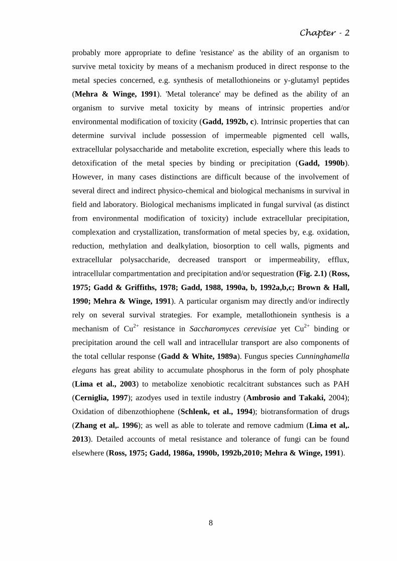
Fig. 2.1 Diagrammatic representation of metal interactions with fungi. The metal
species are represented in cationic form. All the interactions shown may be involved
in fungal survival and/or metal detoxification.
2.3 Environmental influence on heavy metal toxicity towards fungi
The physico-chemical properties of a given environment or growth medium determine
metal speciation and therefore biological availability and toxicity as well as other
essential and inessential metal-organism interactions. Where such factors as pH, Eh,
the presence of other anions and cations, particulate and soluble organic matter, and
clay minerals decrease biological availability and toxicity may be reduced (Gadd &
Griffiths, 1978). It is therefore clear that the concentration of free metal ion, defined
as pM = -log [M] (Hughes & Poole, 1991), may be an important consideration in
many cases, although toxicity may still be exerted by many inorganic and organic
metal complexes in the absence of free metal cations, M+.
Chapter - 2
10
In simple terms, if excess metal ion (M) is in the presence of a strongly binding ligand
(L), [Mfree] = [Mtotal] - [Ltotal]. If the ligand is in excess, and unprotonated, Lt = ML +
L, [ML] ~[Mt] and the pM may be calculated:
K1 = [ML]/[M] [L] = [Mt]/[M] [Lt-Mt],
pM = -log [M] = log K1 + log ([L1-M1]/ [Mt]).
If the ligand is protonated, the calculation becomes Lt = ML +L+HL+H2L ... HnL
(Hughes & Poole, 1991). In relation to the complex situation which occurs in the
environment and in growth media, the multiplicity of metal-ligand binding
interactions that can occur may mean that calculation of pM is not possible.
The pH can affect metal-fungal responses by effects on metal speciation and mobility
and indirectly by influencing other aspects of cell physiology and metabolism.
Increasing pH can result in the formation and precipitation of metal hydroxides or
oxides. In aqueous solution, divalent metal cations form multiple hydroxylated
species, some forming precipitates (Collins & Stotzky, 1989; Hughes & Poole,
1991).
At higher pH values, some precipitates re dissolve giving anionic hydroxometallate
complexes, e.g. Zn(OH)42 (Hughes & Poole, 1991). The pH values at which
hydroxylated species form varies between different metals. The different
hydroxylated forms possess different toxicities (Collins & Stotzky, 1989). The
formation of hydrolysis products may also favour biosorption.
Though decreasing pH may increase the concentration of free metal ions in solution
as mentioned previously, H+ may compete with metal ions for cellular binding sites
and reduce potential interactions with cells. External pH may also affect the
speciation of metal-binding ligands and therefore metal complexation. Many toxic
interactions of heavy metals with fungal cells can be interpreted in terms of effect of
pH on metal speciation and availability (Newby & Gadd, 1986; Gadd, 1986a).
Fungal toxicity of Cd may increase with increasing pH. A reduction in metal toxicity
at low pH may result from H+ competition and also pH effects on intracellular uptake.
This has been described for those fungi, e.g. Penicillium ochrochloron, that are
capable of growing in saturated CuS04 and high concentrations of other heavy metal
salts (Gadd et al., 1984). With increasing pH intracellular Cu accumulation
Chapter - 2
11
dramatically increased in P. ochrochloron and at pH 6 and above this fungus was
sensitive to quite low concentrations of Cu (Gadd & White, 1989).
Oxidation-reduction potential (Eh) may affect heavy metal availability and toxicity. In
a reducing environment (negative Eh), insoluble metal sulphides may form which
exhibit little or no toxicity. Sulphide precipitation is a detoxification mechanism
found in several fungi (see later). The Eh may also effect speciation, e.g. chromium
exists as Cr6+
or Cr3+
depending on the Eh. Cr6+
exhibits the greater toxicity towards
hyphal growth and sporulation in several fungi (Collins & Stotzky, 1989).
Inorganic anions may affect toxicity by forming inorganic complexes, as mentioned
previously and by precipitation of (Gadd & Griffiths, 1978). Increasing Cl-, for
example, decreased Cd toxicity towards several filamentous fungi, possibly as a result
of the formation of negative Cd-Cl coordination complexes (Babich & Stotzky,
1982; Collins & Stotzky, 1989). Other cations may affect fungitoxicity of heavy
metals by competing for binding sites on cell surfaces including membranes and for
transport mechanisms. Heavy metal toxicity towards fungi was also reduced in hard
water an effect attributed to CO32-
(Babich & Stotzky, 1981). Many heavy metal salts
are of low solubility, e.g. sulphides and phosphates, and therefore readily precipitate
out of solution. Silver chloride is highly insoluble and the concentration of free Ag+
in solution may be negligible in many cases, particularly in growth media (Hughes &
Poole, 1991).
Clay minerals can adsorb heavy metal cations and reduce their potential toxicity
(Gadd & Griffiths, 1978; Babich & Stotzky, 1980). In general, minerals with a high
Cation Exchange Capacity (CEC) are more effective in reducing toxicity than
minerals with a lower CEC (Babich & Stotzky, 1977a, b; Bewley & Stotzky, 1983).
Montmorillonite was more effective than kaolinite, with a lower CEC, in protecting
fungi from Cd, Ni and Pb toxicity (Babich & Stotzky, 1978, 1979, 1982). Such
effects are also being influenced by soil pH (Collins & Stotzky, 1989).
Dissolved and particulate organic matter in the environment, and in growth media,
can influence metal toxicity by complexation and binding, generally reducing toxicity.
Examples of such organic materials include natural and synthetic chelating agents,
including plant, fungal and other microbial exudates and metabolites, humic acids and
soil colloids, and soluble organic chemicals and mixtures. It is commonly observed
Chapter - 2
12
that metal toxicity towards fungi is reduced in complex media compared to simple
defined media and such effects are also being influenced by pH for the reasons
outlined previously (Gadd & Griffiths, 1978; Collins & Stotzky, 1989).
2.4 Fungi in polluted habitats
A range of fungi from all major taxonomic groups may be found in metal-polluted
habitats and the ability to survive and grow in the presence of potentially toxic
concentrations is frequently encountered (Ross, 1975; Gadd, 1986a; Baldi, et al.,
1990; Turnau, 1991). In general terms, toxic metals are believed to affect fungal
populations by reducing abundance and species diversity and selecting for a
resistant/tolerant population (Babich & Stotzky, 1985; Duxbury, 1985). However,
the effect of toxic metals on microbial abundance in natural habitats varies with the
metal species and organisms present and with environmental factors (Gadd &
Griffiths, 1978; Duxbury, 1985).
General reductions in fungal numbers have often been noted in soils polluted with Cu,
Cd, Pb, As and Zn (Babich & Stotzky, 1985). Along a steep gradient of Cu and Zn in
soil towards a brass mill, fungal biomass decreased by 75 % (Nordgren, et al., 1983)
while fungal numbers were reduced in glucose-supplemented soil by Cd or Zn, the
former metal being of greater toxicity (Bewley & Stotzky, 1983). However,
numerical estimates alone which have built-in inadequacies may provide little
meaningful information unless possible changes in fungal groups and species are
considered. There is evidence that such changes can occur in response to metal
exposure.
In Cu and Zn polluted soil, Geomyces and Paecilomyces sp. and some sterile forms
increased with increasing pollution, whereas Penicillium and Oidiodendron sp.
declined at polluted sites (Nordgren et al., 1983). Trichocladium asperum,
Trichoderma hamatum, Zygorrhynchus moelleri and Chrysosporium pannorum were
isolated more frequently from an organomercurial treated golf green than from
untreated locations, whereas Chaetomium, Fusarium, Penicillium and Paecilomyces
sp. were greatly reduced (Williams & Pugh, 1975). Several other studies have
indicated sensitivity of Penicillium sp. towards heavy metals (Duxbury, 1985).
However, some of the best examples of microbial metal tolerance are also found in
the genus Penicillium, which underlines the fact that metal responses may be strain
Chapter - 2
13
specific. As noted above, P. ochrochloron can grow in saturated CuSO4 and it is
frequently isolated from industrial effluents (Stokes & Lindsay, 1979) whereas P.
lilacinum comprised 23% of all fungi isolated from soil polluted by mine drainage
(Tatsuyama et al., 1975). In soil polluted with cadmium dust, Strobilurus tenacellus,
Mycena ammoniaca, Auriscalpium vulgare and Armillaria lutea were the most
common Basidiomycetes (Turnau, 1991).
Heavy metal pollution of plant surfaces is wide spread and while there may be
significant decrease in total microbial numbers (including bacteria) on phylloplanes,
numbers of filamentous fungi and non-pigmented yeasts are generally little affected
(Bewley, 1979, 1980; Bewley & Campbell, 1980; Mowll & Gadd, 1985) and if
metal-supplemented isolation media are used only fungi may be isolated (Bewley,
1980). On polluted oak leaves Aureobasidium pullulans and Cladosporium sp. were
the most numerous organisms and a greater proportion were metal-tolerant when
compared with control isolates (Bewley & Campbell, 1978; Bewley, 1980). In fact,
numbers of A. pullulans showed a good positive correlation with lead concentrations,
whether derived from industrial or vehicular sources and it often became the dominant
organism, in some cases comprising up to 97% of the leaf surface population on a
numerical basis (Bewley & Campbell, 1980; Mowll & Gadd, 1985). However,
adaptation was unnecessary for growth of A. pullulans on polluted leaf surfaces and
the ability to tolerate high concentrations of lead occurred in isolates from the
unpolluted site (Gadd, 1984; Mowll & Gadd, 1985).
In contrast, the numbers of the ballistospore producing yeast, Sporobolomyces roseus
and heterotrophic bacteria were very low or absent from polluted samples while
numbers of S. roseus showed a significant negative correlation with increasing lead
concentrations (Mowll & Gadd, 1985). Smith (1977) also studied the effect of heavy
metals on phylloplane fungi and found that Aureobasidium pullulans, Epicoccum sp.
and Phialophora verrucosa were tolerant, Gnomia platani, Cladosporium and
Pleurophomella sp. were of intermediate tolerance while Pestalotiopsis and
Chaetomium sp. were sensitive. Mercury tolerant fungi have been isolated from the
surfaces of seeds treated with mercury compounds. These include Pyrenophora
avenae, Penicillium crustosum, Cladosporium cladosporoides, Syncephalastrum
racemosum and Ulocladium atrum (Ross & Old, 1973; Greenaway, et al., 1974).
Chapter - 2
14
It seems therefore that elevated concentrations of heavy metals can affect the
qualitative and quantitative composition of fungal populations though it must be
stressed that it may be difficult to separate metal effects from those of other
environmental components. Phylloplane mycoflora may be subject to the influence of
other potential toxicants, e.g. SO2 (Babich & Stotzky, 1980), while polluted soils
may be nutrient-poor, of variable pH and also contain additional toxicants (Gadd &
Griffiths, 1978). These factors may affect fungal populations. Furthermore, there are
well known difficulties in obtaining meaningful assessments of fungal numbers and
diversity which cannot be ignored. Despite this, it is less apparent that certain fungi
can exhibit considerable tolerance towards toxic metals and can become dominant
microorganisms in some polluted habitats. However, while species diversity may be
reduced in certain cases, resistance/tolerance can be exhibited by fungi from both
polluted and non-polluted habitats. Fungal numbers were reduced and there were
alterations in species composition in polluted soil near a zinc smelter (Jordan &
Lechevalier, 1975). However, there was little difference in the zinc tolerance of fungi
isolated from either site and most could exhibit 50% growth at 700ppm Zn2+
. At
control sites, Zn-tolerant genera included Bdellospora, Verticillium and Paecilomyces
with Penicillium, Torula and Aureobasidium at polluted sites. In another study, fungal
populations in soil contaminated with Ni, Cn, Fe and Co were not significantly
different from those at control sites (Freedman & Hutchinson, 1980). Cu and
Nitolerant fungi (defined as being capable of growth at approximately 16 mM Cu2+
and/or Ni2+
) were isolated from both control and contaminated sites, the predominant
genera being Penicillium (60%) followed by Trichoderma, Rhodotorula,
Oidiodendron, Mortierella and Mucor. Examination of microfungi isolated from
unpolluted and copper-polluted forest soil confirmed that species from the polluted
site were usually copper-tolerant although there was little evidence for adaptation in
isolates from sites with short or long histories of pollution (Arnebrant, Baath &
Nordgren, 1987). Such studies indicate that in many cases survival must be
dependent on intrinsic properties of the organisms rather than adpative changes
(which are generally studied under laboratory conditions) and physico-chemical
properties of that environment. These will include changes associated with the metal
pollution, which may modulate toxicity and affect species composition (Gadd, 1986a,
1990b).
Chapter - 2
15
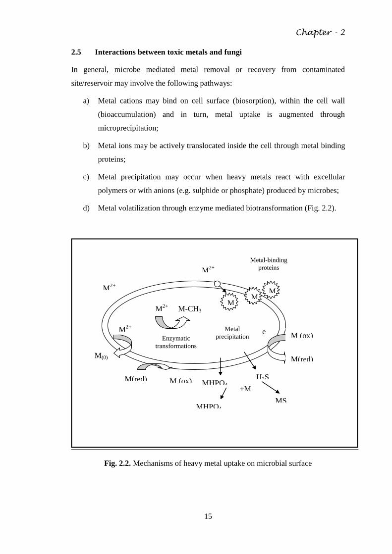
2.5 Interactions between toxic metals and fungi
In general, microbe mediated metal removal or recovery from contaminated
site/reservoir may involve the following pathways:
a) Metal cations may bind on cell surface (biosorption), within the cell wall
(bioaccumulation) and in turn, metal uptake is augmented through
microprecipitation;
b) Metal ions may be actively translocated inside the cell through metal binding
proteins;
c) Metal precipitation may occur when heavy metals react with excellular
polymers or with anions (e.g. sulphide or phosphate) produced by microbes;
d) Metal volatilization through enzyme mediated biotransformation (Fig. 2.2).
Fig. 2.2. Mechanisms of heavy metal uptake on microbial surface
M M
M M-CH3
M2+
M2+
Metal-binding
proteins
Enzymatic
transformations
Metal
precipitation
M2+
M(0)
M(red)
M (ox)
M2+
M (ox)
M(red)
e
MHPO4
H2S MHPO4
2
MS
+M2+
Chapter - 2
16
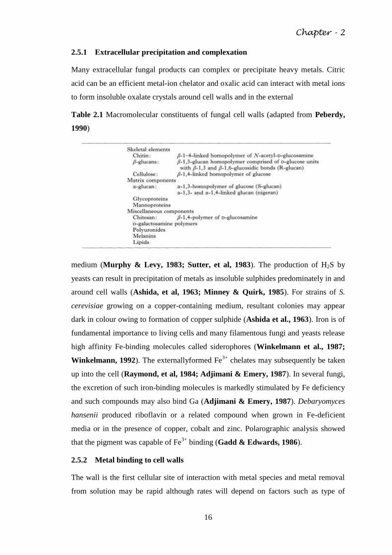
2.5.1 Extracellular precipitation and complexation
Many extracellular fungal products can complex or precipitate heavy metals. Citric
acid can be an efficient metal-ion chelator and oxalic acid can interact with metal ions
to form insoluble oxalate crystals around cell walls and in the external
Table 2.1 Macromolecular constituents of fungal cell walls (adapted from Peberdy,
1990)
medium (Murphy & Levy, 1983; Sutter, et al, 1983). The production of H2S by
yeasts can result in precipitation of metals as insoluble sulphides predominately in and
around cell walls (Ashida, et al, 1963; Minney & Quirk, 1985). For strains of S.
cerevisiae growing on a copper-containing medium, resultant colonies may appear
dark in colour owing to formation of copper sulphide (Ashida et al., 1963). Iron is of
fundamental importance to living cells and many filamentous fungi and yeasts release
high affinity Fe-binding molecules called siderophores (Winkelmann et al., 1987;
Winkelmann, 1992). The externallyformed Fe3+
chelates may subsequently be taken
up into the cell (Raymond, et al, 1984; Adjimani & Emery, 1987). In several fungi,
the excretion of such iron-binding molecules is markedly stimulated by Fe deficiency
and such compounds may also bind Ga (Adjimani & Emery, 1987). Debaryomyces
hansenii produced riboflavin or a related compound when grown in Fe-deficient
media or in the presence of copper, cobalt and zinc. Polarographic analysis showed
that the pigment was capable of Fe3+
binding (Gadd & Edwards, 1986).
2.5.2 Metal binding to cell walls
The wall is the first cellular site of interaction with metal species and metal removal
from solution may be rapid although rates will depend on factors such as type of
Chapter - 2
17
metal ion and biomass, concentration of metal and environmental factors.
Metabolism- independent association of metal species to fungal walls may include ion
exchange, adsorption, complexation, precipitation and crystallization (Mullen et al.,
1992). 'Biosorption' is a term that is frequently used to describe such non-directed
physico-chemical interactions between metals (including radionuclide) species and
microbial biomass particularly in a biotechnological context (Shumate &
Strandberg, 1985).
The fungal cell wall thus has important protective properties and so may act as a
barrier, controlling the uptake of solutes into the cell including potentially toxic metal
species (Gadd & Griffiths, 1980 a; Gadd, 1986a, b; Ono, et al, 1988) and also
indirectly affecting the intracellular ionic composition by restriction of cellular water
(Peberdy, 1990). The wall is mainly composed of polysaccharides, some of which
may have associated protein, with other components including lipids and melanins
(Table 2.2). All filamentous fungi, with the exception of the Oomycetes, contain
chitin with (1-3)-fl-glucan as skeletal components. The Oomycetes, together with
Hyphochytridiomycetes possess cellulosic walls (Peberby, 1990). In yeasts, skeletal
components consist of (1-3)-fl-D-glucans with (1-6)-,8-linkages at branch points, the
degree of branching determining crystallinity (Bartnicki-Garcia, 1973). Matrix
components in yeasts comprise a mannan-protein complex, while (1-3)-cc-glucan
normally occurring at the outer surface of the wall, is the major matrix polymer in
filamentous fungi (Peberdy, 1990). Heterogeneous glycoproteins are also found in
filamentous fungal walls (Peberdy, 1990).
It is clear, therefore, that metal biosorption in fungal cell walls may be complex,
involving different components and mechanisms, and variable depending on wall
structure and composition, the later itself being affected by the presence of toxic
metals (Gadd & Griffiths, 1980a; Newby & Gadd, 1987; Venkateswerlu &
Stotzky, 1989; Venkateswerlu, et al, 1989). A variety of potential sites may be
involved in metal sequestration including carboxyl, amine, hydroxyl, phosphate and
sulphydryl groups although their relative significance is usually difficult to resolve
(Strandberg, et al, 1981). However, primary interactions probably involve binding to
carboxyl and phosphate groups which may be enhanced by electrostatic attraction to
other negatively charged functional groups (Tobin, et al, 1990). Metabolism-
independent biosorption is frequently rapid and unaffected over moderate ranges of
Chapter - 2
18
temperature, e.g. 4-30 °C (Norris & Kelly, 1977; De Rome & Gadd, 1987a, b;
Junghans & Straube, 1991). In Rhizopus arrhizus, biosorption was related to ionic
radius for, e.g. La3+
, Mn2+
, Cu2+
, Zn2+
, Cd2+
, Ba2+
, Hg2+
, Pb2+
, UO22+
and Ag+, but not
Cr3+
or the alkali metal cations, Na+, K
+, Cs
+ and Rb
+, which were not taken up by the
cell walls (Tobin, et al, 1984). Low external pH often decreases biosorption of, e.g.,
Cu2+
, Cd2+
, Zn2+
, Mn2+
and Co2+
(Fuhrmann & Rothstein, 1968; Paton & Budd,
1972; Roomans et al., 1979; Gadd & Mowll, 1985; De Rome & Gadd, 1987 a, b;
Venkateswerlu & Stotzky, 1989; Gadd, 1990a) while other anions and cations may
have the same effect by, e.g. precipitation by phosphates, and removal of metal from
solution or by competition for binding sites, e.g., Mg2+
, Ca2+
, Zn2+
, Cu2+
and Mn2+
(Tobin, et al, 1987, 1988; Gadd, 1990a). The later effects may be dependent on the
relative concentrations and chemical behaviour of the different metal species present.
Biomass concentration may also effect fungal biosorption. At a given equilibrium
concentration, the uptake of metals when expressed on a unit dry weight or cell basis,
is generally greater at lower cell densities than at high ones (Itoh, et al, 1975; De
Rome & Gadd, 1987a; Junghans & Straube, 1991). Since growth conditions affect
the structure and composition of fungal cell walls, their manipulation offers potential
for biosorbency for specific purposes (Volesky, 1990). Chitin and chitosan have
received attention as significant metal-biosorbing substances in cell walls of fungi.
Chitin is a polymer of N-acetyl-D-glucosamine; chitosan is deacetylated chitin
(Volesky, 1990). A variety of metal ions are readily bound by chitin although alkali
metals, NH4+
, Ca2+
and Mg2+
are not sequestered (Muzzarelli, et al, 1986). In R.
arrhizus coordination of uranium to the amine nitrogen of chitin and simultaneous
adsorption in the cell wall chitin structure are followed by slower precipitation of
uranyl hydroxide (Tsezos & Volesky, 1982a). Accumulation of hydrolysis products
continues until final equilibrium (Tsezos, 1983). The mechanism of thorium
deposition involved localization at the outer region of the wall in R. arrhizus.
Coordination to amine nitrogen still occurred but the hydrolysis of Th4+
to Th(OH)4 at
pH 4 resulted in deposition at the chitin surface. The formation of hydrolysis products
favours biosorptionat pH 2. Where Th4+
is the main thorium species, lower
biosorption occurred with a general dispersal of Th4+
through the wall (Tsezos &
Volesky, 1982 b). The chitin-chitosan content of fungal walls varies between species,
e.g. Neurospora crassa (26 % of the wall dry weight) and R. nigricans (53 Go), and
this may change during growth (Volesky, 1990). Purified chitin and chitosan
Chapter - 2
19
derivatives may also have a biosorptive ability although this may be influenced by the
extraction method employed. Isolated chitin from R. arrhizus showed decreased
uranium biosorption in comparison with intact biomass (Tsezos, 1986). Chitin
phosphate and chitosan phosphate were able to remove metal and actinide species
from solution, both substances showing a markedly greater affinity for UO221
than
other metal species including Cu2+
, Co2+
, Cd2+
, Mn2+
Zn2+
Mg2+
, Ni2+
and Ca2+
(Sakaguchi & Nakajima, 1982). Non-phosphorylated chitin and chitosan were not
efficient biosorbents (Sakaguchi & Nakajima, 1982). Insoluble chitosan-glucan
complexes and glucans possessing amino- or sugar acid groups from Aspergillus
niger exhibit biosorptive properties and may efficiently remove transition metal ions
from solution (Muzzarelli et al., 1986). Synthesized chitosan derivatives, e.g. N-[2-
(1,2- dihydroxyethyl)tetra-hydrofuryl] chitosan, can remove uranium from brines at
an order of magnitude greater than the intact biomass of R. arrhizus (Muzzarelli et
al., 1986; Macaskie & Dean, 1990a). Hydroxide treatment of fungi to expose
chitin/chitosan, may also improve biosorptive abilities (Wales & Sagar, 1990).
Melanins are important fungal pigments which enhance the survival of many species
in response to environmental stress (Bell & Wheeler, 1986). Fungal melanins are
located in and/or exterior to cell walls where they may appear as electron-dense
deposits and granules on electron micrographs. These granules may be released into
the external medium and be termed 'extracellular melanin', although this is generally
of identical composition to wall-associated melanin. 'Extracellular melanins' are more
correctly defined as melanins synthesized exterior to cells by secretion of phenol
oxidases which oxidize phenolics or secretion of phenols that are subsequently
oxidized (Bell & Wheeler, 1986). Fungal phenolic polymers and melanins contain
phenolic units, peptides, carbohydrates, aliphatic hydrocarbons and fatty acids and
therefore possess many potential metal-binding sites (Saiz-Jimenez & Shafizadeh,
1984; Senesi, et al, 1987; Sakaguchi & Nakajima, 1987). Oxygen-containing
groups in these substances, including carboxyl, phenolic and alcoholic hydroxyl,
carbonyl and methoxyl groups may be particularly important in metal binding (Gadd,
1988). The order of binding ability of fungal phenolic polymers and melanins
followed the sequence Cu > Ca > Mg > Zn (Zunino & Martin, 1977).
A variety of melanin types occur in fungi. Although it is apparent that many fungi can
form extracellular DOPA (3,4-dihydroxyphenylalanine)melanin via tyrosinase-
Chapter - 2
20
mediated oxidation of tyrosine, there is still no firm evidence for the existence of
DOPA- melanin in fungal cell walls (Bell & Wheeler, 1986). Melanins in cell walls
of Basidiomycetes are derived from y-glutaminyl-3,4-dihydroxybenzene (GDHB) or
catechol. In Ascomycetes and Deuteromycetes, wall melanins are generally
synthesized by the pentaketide pathway via 1 ,8-dihydroxynaphthalene (DHN) as the
immediate precursor. Other extracellular dark pigments may be referred to as
'heterogeneous' melanins (Bell & Wheeler, 1986). A variety of heavy metals can
induce or accelerate melanin production in fungi and melanized cell forms, e.g.
chlamydospores, can have high capacities to adsorb metals with virtually all the metal
being located in the cell wall (Mowll and Gadd, 1984). Melanin from Aureobasidium
pullulans can bind significant amounts of Cu2+
and Fe3+
(Gadd, 1984; Senesi et al.,
1987; Gadd & De Rome, 1988) as well as organometallic compounds, e.g. tributyltin
chloride (Gadd, 1990).
2.5.3 Transport of toxic metal cations
The plasma and vacuolar membranes are the main transport membranes of fungi and
the basic energetics as well as detailed kinetic analysis have been established for a
variety of solutes, particularly carbon and/or nitrogen sources (Sanders, 1988, 1990).
Most work on metal ion transport in fungi has concerned K+ and Ca
2+ largely because
of their great importance in fungal growth, metabolism and differentiation. The
transport of toxic metal species is still poorly understood. Transport systems in fungal
(and plant) cell membranes are usually classed as either carrier or channel systems. In
the former the conformational changes in the transport protein are believed to result in
alternate exposure of the transport binding site(s) on each side of the membrane.
Carriers include all metabolically-coupled and H+-
gradient driven transport systems.
Fluxes through such systems saturate with respect to ligand concentration and if a
current is carried with respect to the membrane potential (∆Ψ) (Sanders, 1990). Ion
channels are a class of proteins that function as gated pores in the plasma membrane
allowing the flow of ions down electrical and/or chemical gradients (Gustin et al.,
1986). Channels have higher turnover rates than carriers, 107-8
s-1
compared to 102-4
S-
1 respectively (Sanders, 1990). To date, the main primary transport systems which
have been characterized in fungi derive energy from ATP hydrolysis and pump H+
electrogenically from the cytosol, creating a transmembrane electrochemical H+
gradient (∆~µH+), negative and alkaline inside. Such an electrochemical gradient
Chapter - 2
21
(∆~µH+) has an electrical component, the membrane potential (∆Ψ) ,a chemical
component and the pH gradient (∆pH) which are interconvertible and which can drive
the transport of ionizable substances across membranes. Such secondary gradient-
coupled transport systems exist for a variety of inorganic and organic solutes which
are energized by coupling with passive reflux of H+; proton and solute fluxes may be
in the same (symport) or opposing (antiport) directions (Sanders, 1990). In fungi and
yeasts, three main classes of H+- pumping ATPases have been identified in
mitochondrial, vacuolar and plasma membranes (Serrano, 1984, 1985) though it is
now suggested that transport ATPases include a Ca2+
-ATPase located on the
endoplasmic reticulum (Goffeau et al., 1990). The main function of the mitochondrial
ATPase is ATP synthesis via the mitochondrial respiratory chain. It is the plasma
membrane and vacuolar ATPases that are associated with ion transport and
intracellular compartmentation and regulation of intracellular pH (Jones & Gadd,
1990). The transport of monovalent cations is linked to the action of the plasma
membrane H+-ATPase that expels protons creating the transmembrane
electrochemical proton gradient (∆~µH+) previously described. K
+ uniport in fungi
appears to be electrically coupled with H+ efflux. Under usual cellular and
environmental conditions, H+ is extruded in an apparent 1: 1 ratio to K
+ uptake with
H+ able to be extruded against 50: 1 concentration gradients and K+ to be taken up
against 1000:1 such gradients (Borst-Pauwels, 1981). However, it is now known that
under K+-deficiency, K
+ influx may be coupled with H
+ influx in a H
+-K
+ symport
system. It has been suggested that observations of stoichiometric K+/H
+ exchange
arise because the H+ pump maintains electroneutrality during K
+ uptake by efflux of
one equivalent of H+ per equivalent of charge flow mediated by symport (Sanders,
1988); the net inflow of positive charge is approximately twice the net K+ uptake
(Rodriguez-Navarro et al 1986). One kind of K+ uniport is via channels in the fungal
plasma membrane (Gustin et al., 1986). Since such channels are not active at the
hyperopolarizing (negative) potentials found in intact cells, their physiological role is
unclear (Sanders, 1988). It now appears that the plasma membrane of Saccharomyces
cerevisiae may have at least 2 classes of K+-selective channels, the first of which is
voltage gated (Gustin et al., 1986) and the second possibly operated via second
messengers and sensitive to blocking by divalent cations (Van de Mortel et al, 1990).
Chapter - 2
22
Potentially toxic metal ions, e.g. Cu2+
, Co2+
, Ni2+
, Cd2+
, Mn2+
, mercurials and other
organometallic compounds, can inhibit the yeast plasma membrane ATPase by means
of various binding interactions, both specific and non-specific (Ochiai, 1987; White
& Gadd, 1987 a, b). Such deleterious effects lead to a reduced ability in maintenance
of the electrochemical gradients described. For H+ efflux, heavy metal inhibition
generally increased with increasing metal concentration with a toxicity sequence of
Cd2+
> Cu2+
> Ni2+
> Zn2+
> Co2+
, Mn2+
. These metals also inhibited K+ uptake with a
toxicity sequence of Cd2+
> Cu2+
> Ni2+
> Co2+
, Mn2+
> Zn2+
. K+ uptake was
considerably more affected by these metals than was H+ efflux which suggested that
the inhibition of K+ uptake was not solely a result of inhibition of the H
+-ATPase and
other factors such as increased membrane permeability to K+ may have contributed
(White & Gadd, 1987 a, b).
Many divalent cations, such as Mg2+
, Ca2+
, Cu2+
, Zn2+
and Mn2+
, which are essential
for growth and metabolism need to be accumulated from the external environment.
However, above certain concentrations, many are toxic and may cause impairment of
cell metabolism including ionic nutrition and ultimately cell death. Inessential metals
may also be accumulated, e.g. Cd2+
and Hg2+
. Some divalent cations, such as Ca2+
and
Mg2+
, may enter S. cerevisiae as low-affinity substrates of the monovalent cation
transport system but this is probably of little significance in relation to the systems
that will now be described.
In filamentous fungi and yeasts, energy-dependent transport of many divalent cations
has been demonstrated, the apparent affinity series being Mg2+
, Co2+
, Zn2+
> Mn2+
>
Ni2+
> Ca2+
> Sr2+
(Fuhrmann & Rothstein, 1968). However, differences in
accumulation may not be due to differences in affinities for the transport mechanism
as some reductions in the net rate of Ca2+
and Sr2+
uptake, for example, may be due to
increased leakage or efflux of these cations. In addition, depending on the metal and
its concentration, there may be effects on the structural integrity of the membrane
(Gadd & Mowll, 1983; White & Gadd, 1987a). As with monovalent cations, uptake
of divalent cations is inhibited or halted by metabolic inhibitors, low temperatures and
the absence of energy-yielding substrates (Gadd & White, 1989 a; Sabie & Gadd,
1990; Starling & Ross, 1990). It follows that uptake thus depends on the metabolic
state of the cell and may vary with different growth media and conditions. For
maximal uptake rates, cells require adequate K+ and phosphate (Fuhrmann &
Chapter - 2
23
Rothstein, 1968; Okorokov et al., 1975; Roomans et al., 1979) and the phosphate
requirement may be indirect and related to the energetic state of the cell as shown for
Mn2+
(Borst-Pauwels, 1981).
Divalent cation transport is dependent on plasma membrane H+-ATPase activity and a
similar affinity sequence of divalent cations for stimulation of the ATPase as for
transport has been demonstrated (Fuhrmann & Rothstein, 1968). Diethylstilboestrol
(DES) is an effective in vitro and in vivo inhibitor of the plasma membrane H+-
ATPase (Bowman et al., 1978; Serrano, 1985) and also inhibits the uptake of divalent
cations (Borst-Pauwels, 1981; White & Gadd, 1987a). However, a direct role of the
H+- ATPase has been discounted for several reasons. Initial transport rates of Mn
2+
and Sr2+
in S. cerevisiae were similar, yet Mn2+
strongly stimulated the plasma
membrane H+-ATPase whereas Sr
2+ had no effect (Roomans et al., 1979;
Nieuwenhuis, et al, 1981). Even though influx rates of these two ions were similar,
much more Mn2+
was subsequently accumulated than Sr2+
which suggested efflux
systems and vacuolar Mn2+
compartmentalization (Nieuwenhuis et al., 1981).
Calcium ions enter cells via ligand or voltage-gated channels. These are membrane
proteins which, when in the open position, allow passive flux of Ca2+
down the
electrochemical gradient, ∆~µ2ca2+
(Miller et al., 1990). Influx of Ca2+
may be
difficult to distinguish because of its energy-dependent accumulation in fungal
vacuoles (Eilam et al., 1985 a).
It appears that the major role of the plasma membrane H+-ATPase in the uptake of
divalent cations is to energize the cell membrane by creating the electrochemical
gradient previously mentioned. Transport depends on the membrane potential
(Boutry, et al, 1977; Borst- Pauwels, 1981; Eilam & Chernichovsky, 1987; White
& Gadd, 1987a; Budd, 1988, 1989) and uptake is inhibited by substances, such as
protonophoric uncouplers and high concentrations of external K+, which depolarize
the cell membrane (Roomans et al., 1979; Borbolla & Pena, 1980; Gadd & White,
1989 a). Conversely, divalent cation uptake may be enhanced under conditions where
the membrane potential is increased, e.g. by enhanced K+ efflux (Eilam, 1984; Eilam,
et al, 1985 b; Gadd & Mowll, 1985; Eilam & Cherni- chovsky, 1987). For S.
cerevisiae treated with high concentrations of ATPase inhibitors, e.g. DCCD, the
resulting hyperpolarization of the membrane caused increased Ca2+
uptake. The
increase in A3f balanced the decrease in pH so that the overall value of (∆~µH+)
Chapter - 2
24
remained almost constant (Eilam, et al 1984). However, changes in (∆Ψ) only partly
account for glucose-stimulated Ca2+
influx in this yeast with intracellular acidification
also being involved (Eilam, et al, 1990). Furthermore, it should be stressed that
inhibitor studies should be treated with caution since the enhancement of M2+
uptake
in yeast induced by several plasma membrane ATPase inhibitors was due to increased
cation permeability of the membrane rather than hyperpolarization (Borst-Pauwels,
1988). Similar conditions may apply to interactions of yeast with those metal cations
or xenobiotics which are potentially toxic and induce K+ efflux (Gadd & Mowll,
1983; Gadd, 1986 a, c; Theuvenet et al., 1987; Borst-Pauwels, 1988; Belde et al.,
1988).
2.5.4 Intracellular fate of toxic metals
Metal-binding proteins and peptides of filamentous fungi and yeasts
Metal-binding proteins are important in the modulation of intracellular concentrations
of both potentially-toxic and essential metal ions. The super family of proteins called
metallothioneins, first discovered in mammalian systems, may achieve these roles by
binding metal ions to cysteine thiolate groups (Hamer, 1986). It is now clear that a
variety of metal binding polypeptides are found in animals, plants and fungi and there
has been some progress in devising a coherent nomenclature (Rauser, 1990).
Polypeptides are designated 'metallothioneins' if they have properties which include
low molecular mass, high metal content, high cysteine (Cys) content, lack aromatic
amino acids and histidine, possess abundant Cys-X- Cys sequences (where X is an
amino acid other than Cys), spectroscopic properties characteristic of metal thiolates,
and metal-thiolate clusters, all are the characteristics of equine renal metallothionein.
Sub division into three classes has been recommended (Rauser, 1990):
Class I: Polypeptides with locations of cysteine closely related to those in equine renal
metallothionein.
Class II: Polypeptides with locations of cysteine only distantly related to those in
equine renal metallothionein.
Class III: Atypical, non-translationally synthesized metal thiolate polypeptides.
Class I includes most vertebrate forms as well as some fungal metallothioneins, e.g.
those of Neurospora crassa and Agaricus bisporus (Lerch & Beltramini, 1983;
Chapter - 2
25
Munger & Lerch, 1985). Class II metallothioneins are found in Saccharomyces
cerevisiae (Steffens, 1990). Algae, plants and certain fungi, e.g. Schizosaccharomyces
pombe and Candida glabrata produce class III metallothionein (Rauser, 1990). The
last occur as metal-binding complexes which behave like entities of relative molecular
mass 10000-13800 in gel filtration (Rauser, 1990). However, these complexes are
aggregates of a heterogeneous group of polypeptides for which no single name is
suitable. In this article, the term metal y-glutamyl peptide will be used (Winge et al.,
1989) although, in the context of plants, this may be inappropriate because of the
diversity of y-glutamyl di- and tripeptides which may occur (Rauser, 1990).
Metallothioneins are small, cysteine-rich polypeptides that can bind essential metals,
e.g. Cu and Zn, as well as inessential metals such as Cd.
Copper resistance in S. cerevisiae can be mediated by the induction of a 6573 Da
cysteine-rich protein, copper-metallothionein (Cu-MT) (Hamer, 1986; Butt &
Ecker, 1987; Fogel, et al, 1988). This protein normally functions to maintain low
concentrations of intracellular copper ions and thus prevent futile transcription of the
CUP1 structural gene (Wright, et al, 1988). Although yeast metallothionein can also
bind Cd and Zn in vitro, it is not transcriptionally induced by these ions and
apparently does not protect against them (Butt et al., 1984; Winge et al., 1985). S.
cerevisiae contains a single Cu-MT gene which is present in the CUP1 locus located
on chromosome VIII (Fogel & Welch, 1982; Karin et al., 1984). Copper-resistant S.
cerevisiae strains (CUP1r) contain 2-14 or more copies of the CUP1 locus, which are
randomly repeated, and may grow on media containing ≥ 2 mm copper (Fogel, Welch
& Karin, 1983; Karin et al., 1984). Copper-sensitive strains (cup1s) do not grow at
concentrations ≥ 0.15 mM (Butt & Ecker, 1987). Continuous subculture of S.
cerevisiae in the presence of increasing copper concentrations can select for hyper-
resistant strains (White & Gadd, 1986) which appear disomic for chromosome VIII.
The disomic chromosomes exhibit differential patterns of CUP1 gene amplification
which indicates that the copper resistance mechanism involves not only amplification
of the CUP1 locus on the chromosomes but also disomy or aneuploidy of
chromosome VIII (Fogel et al., 1983; Butt & Ecker, 1987). The DNA fragments of
the CUP1 locus that confer copper resistance have been cloned (Fogel & Welch,
1982;Henderson, et al, 1985) and has shown evidence that the CUP1 locus was
present as multiple copies in CUP1 strains which was obtained after restriction
Chapter - 2
26
enzyme analysis of the cloned DNA (Butt & Ecker, 1987; Fogel et al., 1988). The
basic repeating unit 'CUP1 locus' is composed of 2-0 kb DNA fragments containing,
with respect to restriction enzymes, a unique Xba I site and two sites for Kpn I and
Sau3A. The full nucleotide sequence of the CUP1 locus has been determined (Karin
et al., 1984). Copper resistance in S. Service depends on amplification of the CUP1
locus (Butt & Ecker, 1987; Fogel et al., 1988). In simple terms, the gene
amplification model suggests that because of the proposed homology of DNA
sequences at the junction of CUP1 repeats, one or more units are looped out. If the
loop is replicated, the copy number increases (amplification) but if the loop is
degraded, the copy number decreases (deamplification) (Fogel et al., 1983; Butt &
Ecker, 1987; Fogel et al., 1988). For more precise accounts of this model, readers
should consult the detailed reviews available (Hamer, et al, 1985; Thiele & Hamer,
1986; Hamer, 1986; Butt & Ecker, 1987; Fogel et al., 1988). It should be noted that
Cu-MT is not directly involved in Cu2+
uptake in S. cerevisiae (Lin & Kosman,
1990).
The mechanism by which copper ions induce transcription of the Cu-MT gene has
received detailed attention (Thiele & Hamer, 198&6; Butt & Ecker, 1987; Thiele,
1988; Furst et al., 1988; Furst & Hamer, 1989; Culotta et al., 1989; Welch et al.,
1989; Hu, et al, 1990). It is now known that a trans-acting regulatory protein,
encoded by the ACE1 locus (Thiele, 1988; Furst et al., 1988) and also known as the
CUP2 locus (Welch et al., 1989; Buchman et al., 1989) activates transcription of the
Cu-MT gene in response to excess copper (or silver) ions (Hu et al., 1990). Although
the ACE1 gene is constitutively expressed in the absence or presence of copper, the
apoprotein cannot bind DNA (Furst et al., 1988). However, in the presence of Cu (I)
[or Ag (I)], the amino-terminal portion of the ACE1 protein (amino acids 1-122)
undergoes a conformational change which allows the protein specifically to bind to
the CUP1 upstream activator sequence (UAS) (Furst et al., 1988; Huibregtse, et al,
1989). This metal-dependent DNA-binding domain is rich in cysteines and basic
amino acids and bears structural similarities to yeast Cu-MT itself (Furst et al., 1988;
Furst & Hamer, 1989; Hu et al., 1990; Dameron et al., 1991). It is proposed that
the cysteine residues of ACE1 form a poly-nuclear Cu(i)-S core in which cysteine
thiolates coordinate multiple Cu(i) ions and Cu(i) ions which are bound by multiple
sulphur bonds (Hu et al., 1990). Wrapping the protein around this Cu(i)-S scaffold
Chapter - 2
27
would lead to the formation of positively charged loop structures that are postulated
specifically to bind DNA and activate transcription (Furst et al., 1988). It therefore
appears that the role of Cu in yeast Cu-MT regulation is to allow ACE1 to bind to the
control sequences of the metallothionein gene (Fig. 2.3) (Hu et al., 1990); the Cu
cluster organizes and stabilizes the conformation of the N-terminal domain of ACEl
for specific DNA binding (Dameron et al., 1991).
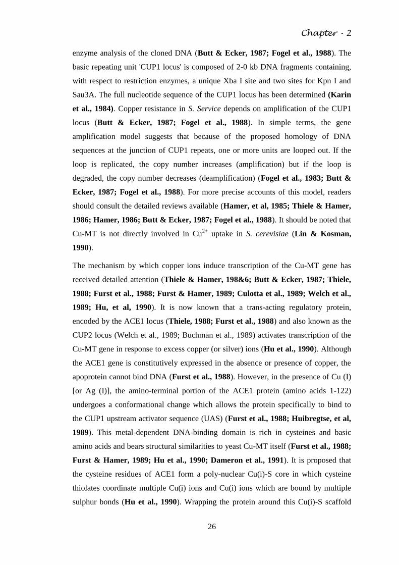
Fig. 2.3 Metalloregulation of the CUP1 locus in Saccharomyces cerevisiae.
The Cu-ACE1 metalloprotein complex interacts with upstream sequences from the
CUP1 coding sequences and facilitates transcription of the MT gene with the CUP1
locus. Translation of the MT mRNA yields MT protein which acts to regulate the
cytosolic concentration of copper ions (modified after Mehra & Winge, 1991).
Chapter - 2
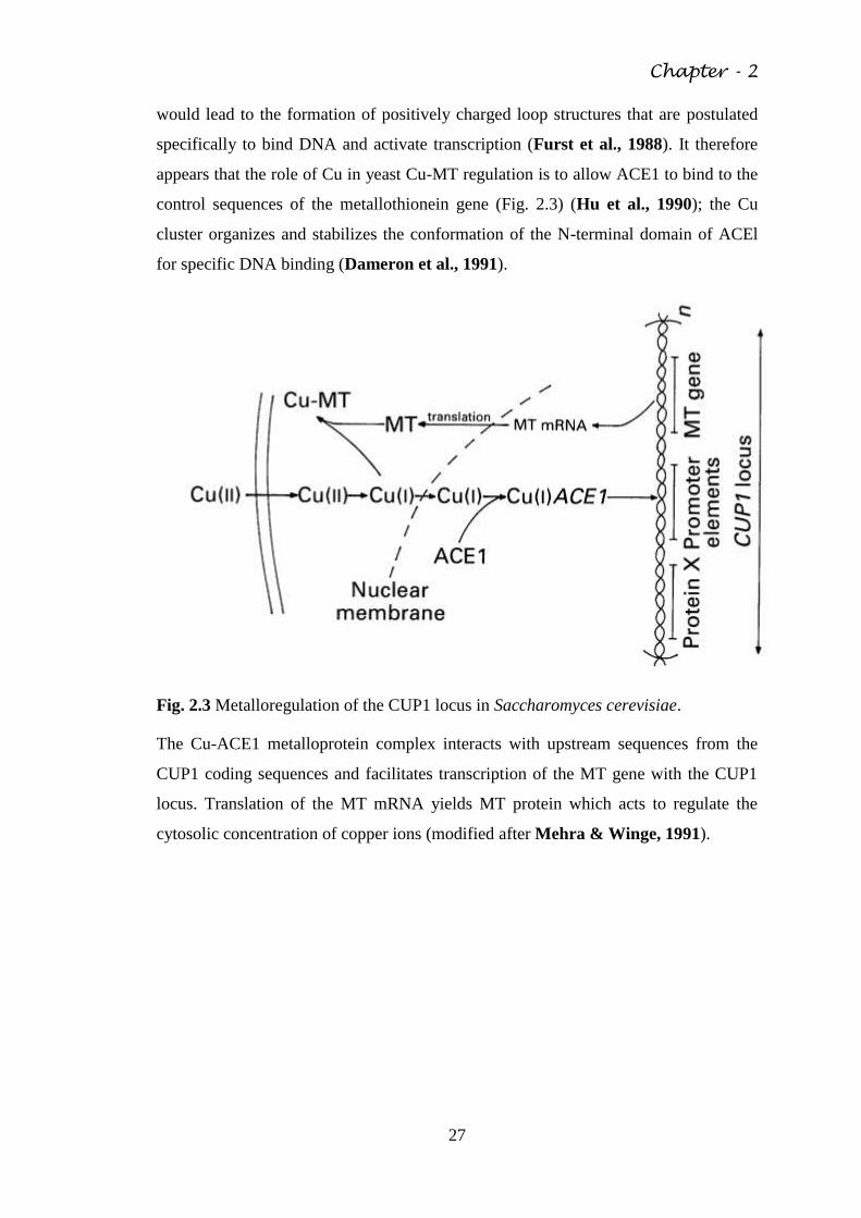
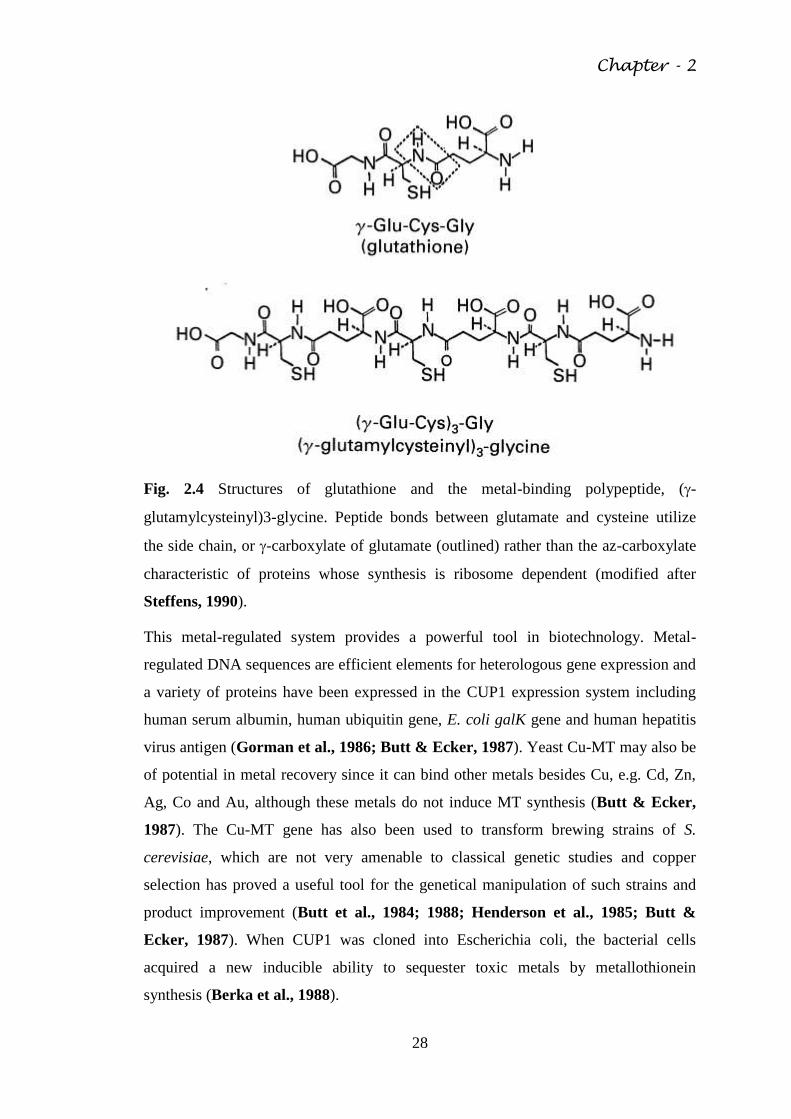
28
Fig. 2.4 Structures of glutathione and the metal-binding polypeptide, (-
glutamylcysteinyl)3-glycine. Peptide bonds between glutamate and cysteine utilize
the side chain, or -carboxylate of glutamate (outlined) rather than the az-carboxylate
characteristic of proteins whose synthesis is ribosome dependent (modified after
Steffens, 1990).
This metal-regulated system provides a powerful tool in biotechnology. Metal-
regulated DNA sequences are efficient elements for heterologous gene expression and
a variety of proteins have been expressed in the CUP1 expression system including
human serum albumin, human ubiquitin gene, E. coli galK gene and human hepatitis
virus antigen (Gorman et al., 1986; Butt & Ecker, 1987). Yeast Cu-MT may also be
of potential in metal recovery since it can bind other metals besides Cu, e.g. Cd, Zn,
Ag, Co and Au, although these metals do not induce MT synthesis (Butt & Ecker,
1987). The Cu-MT gene has also been used to transform brewing strains of S.
cerevisiae, which are not very amenable to classical genetic studies and copper
selection has proved a useful tool for the genetical manipulation of such strains and
product improvement (Butt et al., 1984; 1988; Henderson et al., 1985; Butt &
Ecker, 1987). When CUP1 was cloned into Escherichia coli, the bacterial cells
acquired a new inducible ability to sequester toxic metals by metallothionein
synthesis (Berka et al., 1988).
Chapter - 2
29
It should be mentioned that a Cd-resistant strain of S. cerevisiae synthesized a
cytoplasmic Cd-binding protein when grown in Cd-containing medium (Joho, et al,
1985 a; Joho, et al, 1985 b). This protein (9kDa) exhibits the characteristics of a
metallothionein, being rich in cysteine (18 mol%) and having a high Cd content (63
,tg Cd (mg protein)-1
). There was a high similarity in amino acid composition between
Cd and Cu-MT of S. cerevisiae (Inouhe et al., 1989). The Cd-MT of Cd- resistant S.
cerevisiae is also encoded by the CUP1 gene and transcription of the CUP1 gene
occurs in the absence of added metal ions unlike the inducible transcription in the Cu-
resistant strain. Cadmium and copper further increase the rate of transcription of the
CUP1 gene in the Cd-resistant strain (Tohoyama et al., 1992).
The similarity in structure between glutathione and y-glutamyl peptides indicates that
biosynthesis of these peptides shares common features with that of glutathione
(Steffens, 1990) and in fact they may be viewed as linear polymers of the y-glutamyl-
cysteine (y-Glu-Cys) portion of glutathione (Grill et al., 1989). Because of the
repetitive y-glutamic acid bonds, they cannot be regarded as primary gene products
and must be formed by a ribosome- independent enzyme reaction (Steffens, 1990)
(Fig. 2.4). Linkage of glutathione metabolism with that of y- glutamyl peptides has
been kinetically demonstrated in vivo (Grill, et al, 1987; Steffens, 1990) and also by
using mutants of S. pombe which lacked the glutathione biosynthetic enzymes, y-
glutamylcysteine synthetase and glutathione synthetase. These were unable to
synthesise glutathione or (y-Glu-Cys)n-Gly peptides and were hypersensitive to Cd
(Mutoh & Hayashi, 1988). This work provided the first genetical evidence that y-
glutamyl peptides were essential for Cd-detoxification and clearly demonstrated
biochemical linkage with glutathione metabolism. It is now known that y-glutamyl
peptides are synthesized by the enzyme y-glutamyl- cysteine dipeptidyl transpeptidase
('phytochelatin synthase'). This catalyses the transfer of the y- glutamylcysteine
dipeptide moiety of glutathione to an acceptor glutathione molecule or a growing
chain of (y-Glu-Cys)n-Gly oligomers, thus synthesizing metal-binding y-glutamyl
peptides (Grill et al., 1989). The primary reaction can be expressed:
Cd2+
is the best activator of the enzyme followed by Ag+, Bi
3+, Pb
2+, Zn
2+, Cu
2+, Hg
2+
and Au+ and it appears that the reaction product (y-glutamyl peptide) chelates the
Chapter - 2
30
activating metal ions thus terminating the reaction (Grill et al., 1989; Loeffler et al.,
1989). In cell-free systems of S. pombe, an additional pathway has also been
discovered which involves yEC polymerization from (yEC)n and glutathione to
(yEC)n+1 followed by addition of glycine with glutathione synthetase (Hayashi et al.,
1991).
The originally discovered two y-glutamyl peptides in S. pombe, cadystin A and B (n =
2 and 3, respectively), were synthesized in response to Cd2+
(Murasugi, et al 1981)
though other metals including Cu2+
, Pb2+
, Zn2+
and Ag+ were also effective (Grill et
al., 1987; Hayashi, et al., 1988), a situation analogous to that in vascular plants
(Hayashi et al., 1988; Rauser, 1990). However, the greatest amount of peptide
synthesis occurs with Cd and it seems larger peptides bind Cd more strongly than
smaller ones (Hayashi et al., 1986, 1988). When S. pombe was exposed to Cu(ii), a
Cu-y-glutamyl peptide was synthesized similar in structure to the Cd-y-glutamyl
peptide (Reese et al., 1988). However, only one Cu-y-glutamyl peptide complex
which did not contain labile sulphur was observed; the metal was bound as Cu(i)
(Reese et al., 1988).
Cadmium induced (but not copper-induced) y- glutamyl peptides contain labile
sulphur. Cadmium stimulates the production of S2-
in S. pombe and Candida glabrata
and a portion of this may be incorporated within the Cd-y-glutamyl peptide
complexes (Mehra et al., 1988; Reese & Winge, 1988; Winge et al., 1989). The
sulphide content is dependent on growth conditions and incubation time and
concentrations of S2-
, ranging from 0.1 to 1.5 mol (mol peptide)-1, have been
demonstrated (Winge et al., 1989). Incorporation of sulphide enhances the stability
and metal content of the peptide complex (the molar sulphide: cadmium ratios ranged
from < 0 06 in low sulphide forms to 0.8 in high sulphide forms) and this enhanced
stability may increase the efficacy of the complex in maintaining a low intracellular
concentration of Cd2+
(Winge et al., 1989). Complexes with a S: Cd ratio of 0.7
contain a 20 A diameter CdS crystallite coated with the y-glutamyl peptides. These
complexes are quantum particles exhibiting size-dependent electronic states
(Dameron et al., 1991), a feature of relevance to solid state physics since the
crystallites exhibit properties analogous to those of semiconductor clusters (Dameron
et al., 1989). The extracellular and cell- associated CdS colloid formed by interaction
of Cd2+
with S2-
may also contribute to Cd2+
detoxification.
Chapter - 2
31
Candida (Torulopsis) glabrata is unique in expressing both metallothionein and y-
glutamyl peptide synthesis for metal detoxification in a metal-specific manner
(Mehra et al., 1988, 1992a, b). C. glabrata synthesizes two distinct MT polypeptides
in response to copper which exhibit repeats of the Cys-X-Cys sequence, common in
mammalian MT, and coordinates copper in Cu(i)-thiolate clusters. The cysteine
content of the two molecules is 30 mol 0 (S. cerevisiae Cu-MT = 22 mol %; N. crassa
Cu-MT = 28 mol %) with the copper content being between 10 and 13 mol per mol
protein for each (Mehra et al., 1988). Two metallothionein genes (MT-I and MT-Il)
from C. glabrata have been cloned and sequenced. The former is present as a single
copy but multiple (3-9) and tandemly arranged copies of one MT-II gene are present
in different strains. C. glabrata strains hyper-resistant to Cu showed further stable
chromosomal amplification (>30 copies) of the MT-II gene (Mehra, Garey &
Winge, 1990). Both genes were inducible by Ag but not by Cd, the later reducing
amounts of both MT-I and MT-II mRNAs (Mehra et al., 1989). One MT-II gene,
designated MT-IIa, was amplified in several wild-type strains of C. glabrata while
another MT-IIb, occurs as a single copy (Mehra et al., 1990). The amplified MT-IIa
gene contains autonomously replicating sequences (ARS) (Mehra et al., 1992 b). The
Cd-y-glutamyl peptide of C. glabrata has n = 2 whereas peptides with n = 3 and 4 are
more common in S. pombe and plants (Grill et al., 1987; Reese et al., 1988). Thus, C.
glabrata can utilize two types of regulatory metal-binding molecules and the affinity
and/or coordination preference of the molecules may determine whether MT or y-
glutamyl peptides are synthesized (Mehra et al., 1988, 1992a, b).
The fungal vacuole is highly important organelle with functions including
macromolecular degradation, storage of metabolites and cytosolic ion and pH
homeostasis (Wiemken, Schellenberg & Urech, 1979; Davis, 1986; Wada et al.,
1987; Jones & Gadd, 1990). It is often considered analogous to the mammalian
lysosome because hydrolytic enzymes are contained in an acidic compartment
although it has greater similarity to vacuoles of plant cells (Matile, 1978; Achstetter
& Wolf, 1975; Klionsky, et al, 1990).
A vacuolar ATPase has been identified in several fungi, notably S. cerevisiae, S.
carlsbergensis and N. crassa (Okorokov et al., 1985; Bowman & Bowman, 1986;
Anraku et al., 1989) and is differentiated from mitochondrial and plasma membrane
ATPases by means of pH optima, subunit composition and structure, and inhibitor
Chapter - 2
32
sensitivity (Klionsky et al., 1990). The vacuolar ATPase utilizes the energy arising
from ATP hydrolysis to pump H+ into the vacuole resulting in the generation of an
electrochemical proton gradient of about 180 mV across the vacuolar membrane
(tonoplast) (Kakinuma, et al, 1981; Bowman & Bowman, 1986). In the presence of
an energy source, a pH gradient of 0-5-1V5 units can be generated across the
tonoplast (vacuolar pH generally ~pH 6) (Jones & Gadd, 1990) and a membrane
potential of- ~75 mV has been recorded for S. cerevisiae and 25-40 mV for N. crassa
(Klionsky et al., 1990). The electrochemical proton gradient energizes transport of
monovalent and divalent cations as well as other substances including basic amino
acids into the vacuole (Jones & Gadd, 1990; (Klionsky et al., 1990). The main
mechanism of transport appears to be proton antiport, although there is some evidence
of a pyrophosphatase activity in S. carlsbergensis which may be responsible for a PPi-
dependent pH gradient (Klionsky et al., 1990). The gradients of pH and electrical
potential difference across the tonoplast may be regulated through the interactions of a
membrane potential-dependent cation channel (Wada et al., 1987; Bertyl &
Slayman, 1990), chloride transport systems (Wada, et al, 1986) and the vacuolar
ATPase itself (Anraku et al., 1989).
The fungal vacuole has an important role in the regulation of the cytosolic
concentrations of metal ions, both for essential metabolic functions and the
detoxification of potentially-toxic metal ions. Many ions, including inorganic
phosphate (Pi) and monovalent and divalent metal cations, are preferentially located in
vacuoles (Ohsumi, et al, 1988). Ratios of [total ion]: [cytosolic ion] may be 4 for K+,
4-28 for Mg2+
, > 70 for Mn2+
and 10 for Ca2+
(Lichko, et al, 1982; Jones & Gadd,
1990). The mechanism of ion transport across the tonoplast is by H+ antiporters and in
S. carlsbergensis apparent Km values for the transport of Ca2+
, Mg2+
, Mn2+
, Zn2+
and
Pi are 004, 03, 08, 0-055-017 and 15 mm, respectively. It is assumed that transport
proceeds until cytosolic concentrations of ions approach these values (Okorokov et
al., 1985). These ionic substrates can stimulate vacuolar ATPase activity (Okorokov,
1985) although it is noteworthy that activity is inhibited by free Mg2+
at 04-05 mm
(Borst-Pauwels & Peters, 1981). Localization of metal ions in the vacuole enables
low cytosolic concentrations of, for example, Ca2+
to be maintained (Ohsumi &
Anraku, 1983; Eilam et al., 1985 a, b; Okorokov et al., 1985; Eilam &
Chernichovsky, 1987; Cornelius & Nakashima, 1987; Ohsumi et al., 1988).
Chapter - 2
33
Cytosolic concentrations of Mg2+
and Mn2+
may remain relatively constant, even
under considerable environmental perturbations (Lichko et al., 1982; Kihn, et al,
1988). There is further evidence for compartmentation of Zn2+
, Co2+
and iron in yeast
vacuoles, which may contribute to tolerant behaviour (White & Gadd, 1986, 1987a;
Raguzzi, et al, 1988). Mutants of S. cerevisiae defective in vacuolar function were
sensitive to Ca2+
and other toxic metals . Polyphosphates, the only inorganic
macromolecular anion found in the vacuole, have an important role in maintaining
ionic compartmentation and their biosynthesis accompanies vacuolar accumulation of
Mg2+
or Mn2+
(Okorokov et al., 1977, et al, 1980; Lichko et al., 1982; Okorokov et
al., 1983 a, b; Miller, 1984; Kihn et al., 1988). The significance of polyphosphate
granules as a store for divalent cations has long been appreciated (Doonan et al.,
1979; Roomans, 1980). In S. cerevisiae, vacuolar inclusions of Ca2+
-polyphosphate
complexes vary with the amount of available polyphosphate (Ohsumi et al., 1988)
although this was not the sole mechanism of Ca2+
'trapping' in the vacuole (Ohsumi
& Anraku, 1983). Vacuolar Zn2+
has also been associated with polyphosphate bodies
in yeast (Bilinski & Miller, 1983) while intravacuolar deposits have also been
observed in yeast grown in the presence of thorium (Gadd & White, 1989b). In the
absence of an energy source, precipitation of Mn2+
within yeast cells has been
demonstrated using Electron Spin Resonance (ESR) spectroscopy (Kihn et al., 1988).
2.6 Metal transformations
Fungi, as well as other microorganisms, can effect chemical transformations of metals
by oxidation, reduction, methylation and dealkylation (Gadd, 1992 a). Although in
contrast to bacteria detailed information is not available in several areas for fungi.
Some enzymatic metal transformations may be involved in survival since certain
transformed metal species are less toxic and/or more volatile than the original species.
Plasmid-mediated reduction of Hg2+
to Hg° is a well-characterized bacterial metal
resistance system and there is only a limited amount of evidence for an analogous
process in filamentous fungi and yeasts (Vonk & Sijpesteijn, 1973; Brunker &
Bott, 1974; Yannai, et al, 1991). Other reductions carried out by fungi include Ag+
to metallic Ag° which is deposited in and around cells (Kierans et al., 1990) and the
reduction of Cu (ii) to Cu (i) by cell wall associated materials in Debaryomyces
hansenii (Wakatsuki, et al, 1988; Wakatsuki et al., 1991 a, b). A copper-reducing
enzyme, copper reductase, catalyses Cu (II) reduction of Cu (I) with NADH or
Chapter - 2
34
NADPH as e- donor (Wakatsuki et al., 1991 a, b). The location of copper reductase
appears to be at the cell wall and this process may have a role in regulation of copper
uptake (Wakatsuki et al., 1991 b). Fungal reductions of metalloids include reduction
of tellurite to elemental tellurium, which can appear as a black deposit, located in the
endoplasmic reticulum (Corfield & Smith, 1970; Smith, 1974) and the reduction of
selenate or selenite to form amorphous selenium which imparts a red colour to fungal
colonies owing to deposition primarily in walls and membranes (Konetzka, 1977).
Methylation of Hg and other metals and metalloids can be catalyzed by several fungi
and may be viewed as a detoxification mechanism since methylated species are
usually more volatile and may be lost from the environment (Landner, 1971; Yannai
et al., 1991; Gadd, 1992a, b). There is a general agreement that methylation of Hg2+
involves methy- cobalamin (vitamin B12) (Craig, 1986; Thayer, 1988).
Biomethylation of As by transfer of carbonium ions (CH3+) from S-
adenosylmethionine (SAM) has also been demonstrated (Thayer, 1984, 1989;
Andreae, 1986). Original work by Challenger (1945) first demonstrated that
trimethylarsine could be produced by several fungi from arsenite. Biomethylation of
Se appears to be by an analogous mechanism to that of arsenic and several fungi can
produce organoselenium compounds, e.g. dimethylselenide from inorganic selenium
ones (Chau & Wong, 1986; Thayer, 1988; Thompson-Eagle, et al, 1989). Certain
fungi may be capable of producing volatile dimethyl telluride from tellurium salts
(Konetzka, 1977; Chau & Wong, 1986). Apart from these examples, fungal
involvement in the methylation of other metals and metalloids has not been examined
in detail. Organometallic compounds (which contain at least one metal-carbon bond)
arise in the environment naturally, by abiotic and biotic means, and because of
accidental and deliberate introduction. The later mainly results from the use of several
organometallic and organometalloid compounds as biocides, e.g. organoarsenicals,
organomercurials and organotins (Craig, 1986; Thayer, 1988; Cooney & Wuertz,
1989). Degradation involves the breaking of the metal-C bonds though detailed
information about such processes is rather sparse for fungi. It should be noted that
metal-C bonds can be disrupted by physico-chemical means, although fungal activity
may enhance the conditions necessary for abiotic attack by, e.g. alteration of pH or
metabolite excretion. Organotin degradation involves sequential removal of organic
groups from the tin atom, a process generally resulting in a decrease in toxicity
Chapter - 2
35
(Blunden & Chapman, 1986). Degradation of tributyltin oxide and tributyltin
naphthenate, used as wood preservatives, can be achieved by fungal action.
Breakdown products include mono- and dibutyltins (Barug, 1981; Orsler &
Holland, 1982). Organomercurials may be detoxified by organomercury lyase, the
resulting Hg2+
being then reduced to Hg0 by mercuric reductase (Tezuka &
Takasaki, 1988) a similar mechanism that found in bacteria. Trimethyl lead can be
degraded by Phaeolus schweinitzii, a process with implications for fungal- treatment
of wastes (Macaskie & Dean, 1990b).
2.7 Accumulation of metals by macrofungi
Elevated concentrations of heavy metals and radio nuclides often occur in the fruiting
bodies of basidiomycete fungi when growing in polluted environments (Brown &
Hall, 1990). This phenomenon is, as yet, poorly understood at the physiological level
but is of significance because of the widespread consumption of edible wild fungi in
many countries. In general, higher concentrations of Pb, Cd, Zn and Hg are found in
macrofungi from urban or industrial areas although there are differences in uptake
abilities between different species and different metals (Tyler, 1980; Bressa, et al,
1988; Lepsova & Mejstrik, 1989; Brown & Hall, 1990). Cadmium appears to be
accumulated to quite high values in macrofungi, averaging around 5 µg g-1
d.wt
though concentrations of up to 40 µg g-1
d. wt have been recorded (Byrne, et al,
1976). Average concentrations of As, Cu, Hg, Mn, Se and Zn in a range of up to 27
species were 1, 50, 2, 3, 30, 15 and 114 µg g-1
d. wt. respectively (Byrne et al., 1976).
However, a wide range of accumulation values can occur depending on the species
and location of growth. Caps of Laccaria amethystina exhibited total As
concentrations of 100-200 µg g-1
d. wt. (Stijve, et al, 1990; Byrne et al., 1990).
Dimethylarsinic acid was a major arsenic compound in cap extracts, providing
tentative evidence for biomethylation of this element (Byrne et al., 1990).
Accumulation of 110Ag and 203Hg was studied in Agaricus bisporus and
concentration factors (metal concentration in mushroom:metal concentration in
substrate) were found to be up to 40 and 37 respectively, with the highest Ag and Hg
contents recorded being 167 and 75 g g-1
d.wt respectively. Silver-protein complexes
were isolated which possibly corresponded with metallothioneins or analogous metal-
binding proteins (Byrne et al, 1990).
2.8 Biotechnological aspects of fungal metal accumulation
Chapter - 2
36
Fungi, in common with other microbial groups, can accumulate metals and
radionuclides from their external environment by means of the physico- chemical and
biological mechanisms already discussed. Living and dead cells are capable of metal/
radionuclide removal from solution as well as excreted or derived products, e.g. cell
wall constituents, pigments, polysaccharides, proteins and siderophores (Gadd, 1988;
Pighi, et al, 1989; Siegel, et al, 1990). The removal of radionuclides, metal or
organometal(loid) species, compounds or particulates from solution by microbial
biomass is now often referred to as 'biosorption', particularly where predominant
interactions are physico-chemical. Biosorption is an area of increasing
biotechnological interest since the removal of potentially toxic and/or valuable metals
and radionuclides from contaminated effluents can be used to detoxify them prior to
environmental discharge. Furthermore, appropriate treatment of loaded biomass can
enable recovery of valuable metals for recycling or further containment of highly
toxic and/or radioactive species. Detailed reviews of this research area are available
(Table 2.2) (Gadd, 1988, 1990a, 1992 a, b).
Although the basic features of metal accumulation are common to most microbial
groups, fungi possess a number of unique attributes which reflect their morphological
and physiological diversity. The majority of fungi exhibit a filamentous or hyphal
growth form which enables efficient colonization of substrate.
Table 2.2 Some examples of metal and actinide accumulation by fungi
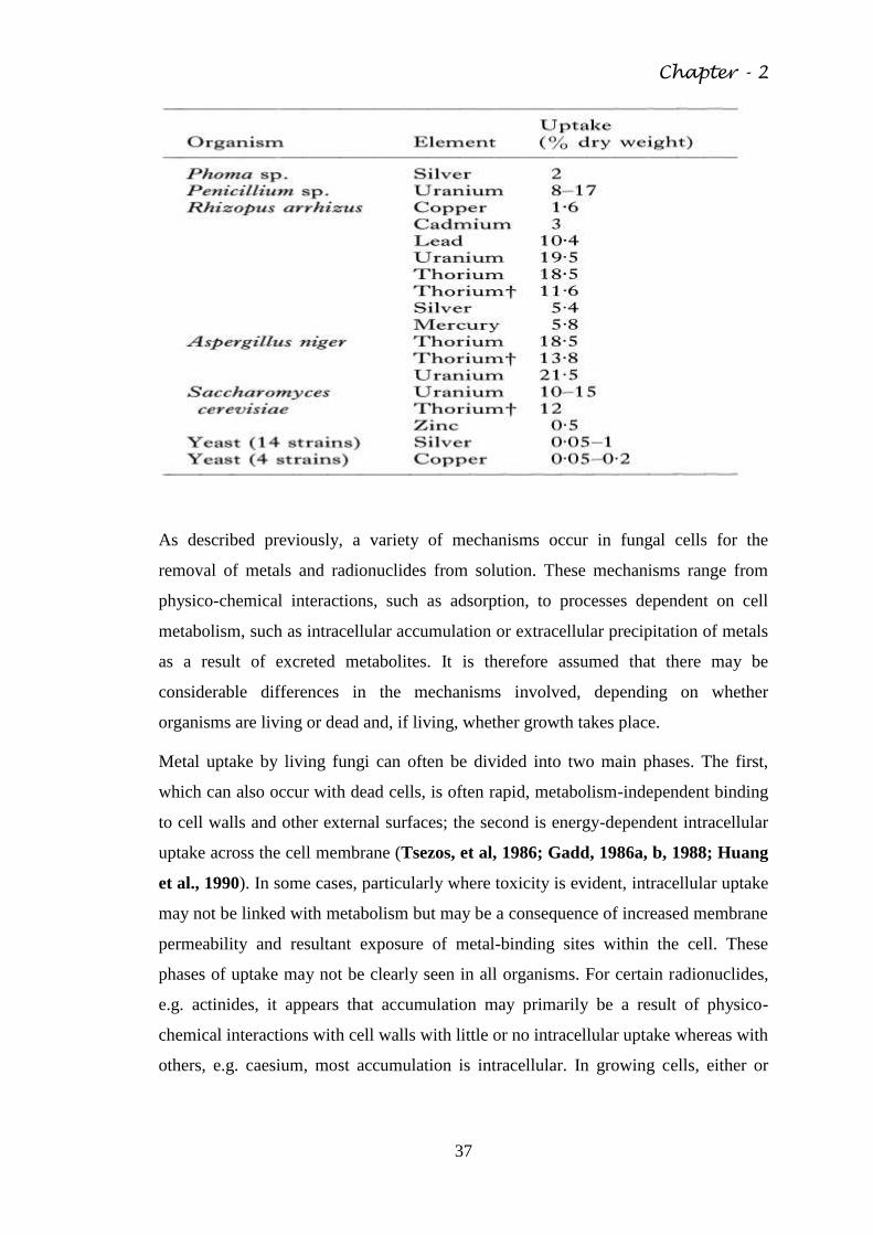
Chapter - 2
37
As described previously, a variety of mechanisms occur in fungal cells for the
removal of metals and radionuclides from solution. These mechanisms range from
physico-chemical interactions, such as adsorption, to processes dependent on cell
metabolism, such as intracellular accumulation or extracellular precipitation of metals
as a result of excreted metabolites. It is therefore assumed that there may be
considerable differences in the mechanisms involved, depending on whether
organisms are living or dead and, if living, whether growth takes place.
Metal uptake by living fungi can often be divided into two main phases. The first,
which can also occur with dead cells, is often rapid, metabolism-independent binding
to cell walls and other external surfaces; the second is energy-dependent intracellular
uptake across the cell membrane (Tsezos, et al, 1986; Gadd, 1986a, b, 1988; Huang
et al., 1990). In some cases, particularly where toxicity is evident, intracellular uptake
may not be linked with metabolism but may be a consequence of increased membrane
permeability and resultant exposure of metal-binding sites within the cell. These
phases of uptake may not be clearly seen in all organisms. For certain radionuclides,
e.g. actinides, it appears that accumulation may primarily be a result of physico-
chemical interactions with cell walls with little or no intracellular uptake whereas with
others, e.g. caesium, most accumulation is intracellular. In growing cells, either or
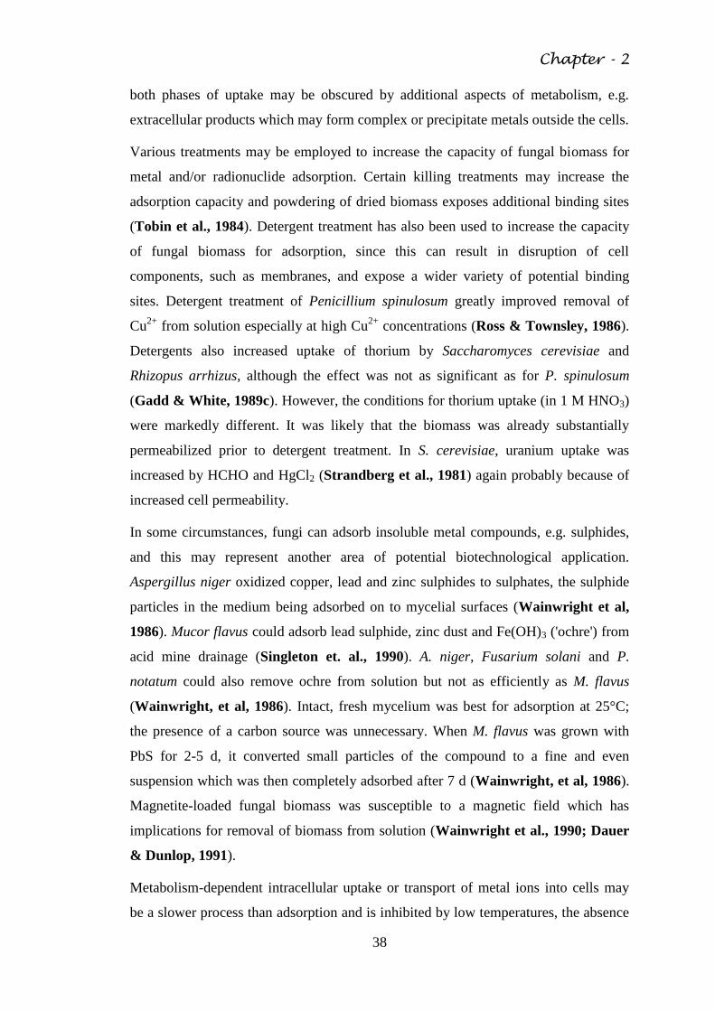
Chapter - 2
38
both phases of uptake may be obscured by additional aspects of metabolism, e.g.
extracellular products which may form complex or precipitate metals outside the cells.
Various treatments may be employed to increase the capacity of fungal biomass for
metal and/or radionuclide adsorption. Certain killing treatments may increase the
adsorption capacity and powdering of dried biomass exposes additional binding sites
(Tobin et al., 1984). Detergent treatment has also been used to increase the capacity
of fungal biomass for adsorption, since this can result in disruption of cell
components, such as membranes, and expose a wider variety of potential binding
sites. Detergent treatment of Penicillium spinulosum greatly improved removal of
Cu2+
from solution especially at high Cu2+
concentrations (Ross & Townsley, 1986).
Detergents also increased uptake of thorium by Saccharomyces cerevisiae and
Rhizopus arrhizus, although the effect was not as significant as for P. spinulosum
(Gadd & White, 1989c). However, the conditions for thorium uptake (in 1 M HNO3)
were markedly different. It was likely that the biomass was already substantially
permeabilized prior to detergent treatment. In S. cerevisiae, uranium uptake was
increased by HCHO and HgCl2 (Strandberg et al., 1981) again probably because of
increased cell permeability.
In some circumstances, fungi can adsorb insoluble metal compounds, e.g. sulphides,
and this may represent another area of potential biotechnological application.
Aspergillus niger oxidized copper, lead and zinc sulphides to sulphates, the sulphide
particles in the medium being adsorbed on to mycelial surfaces (Wainwright et al,
1986). Mucor flavus could adsorb lead sulphide, zinc dust and Fe(OH)3 ('ochre') from
acid mine drainage (Singleton et. al., 1990). A. niger, Fusarium solani and P.
notatum could also remove ochre from solution but not as efficiently as M. flavus
(Wainwright, et al, 1986). Intact, fresh mycelium was best for adsorption at 25°C;
the presence of a carbon source was unnecessary. When M. flavus was grown with
PbS for 2-5 d, it converted small particles of the compound to a fine and even
suspension which was then completely adsorbed after 7 d (Wainwright, et al, 1986).
Magnetite-loaded fungal biomass was susceptible to a magnetic field which has
implications for removal of biomass from solution (Wainwright et al., 1990; Dauer
& Dunlop, 1991).
Metabolism-dependent intracellular uptake or transport of metal ions into cells may
be a slower process than adsorption and is inhibited by low temperatures, the absence
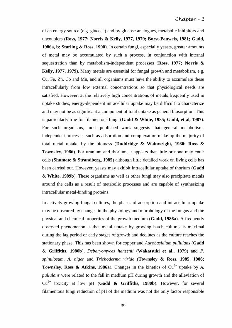
Chapter - 2
39
of an energy source (e.g. glucose) and by glucose analogues, metabolic inhibitors and
uncouplers (Ross, 1977; Norris & Kelly, 1977, 1979; Borst-Pauwels, 1981; Gadd,
1986a, b; Starling & Ross, 1990). In certain fungi, especially yeasts, greater amounts
of metal may be accumulated by such a process, in conjunction with internal
sequestration than by metabolism-independent processes (Ross, 1977; Norris &
Kelly, 1977, 1979). Many metals are essential for fungal growth and metabolism, e.g.
Cu, Fe, Zn, Co and Mn, and all organisms must have the ability to accumulate these
intracellularly from low external concentrations so that physiological needs are
satisfied. However, at the relatively high concentrations of metals frequently used in
uptake studies, energy-dependent intracellular uptake may be difficult to characterize
and may not be as significant a component of total uptake as general biosorption. This
is particularly true for filamentous fungi (Gadd & White, 1985; Gadd, et al, 1987).
For such organisms, most published work suggests that general metabolism-
independent processes such as adsorption and complexation make up the majority of
total metal uptake by the biomass (Duddridge & Wainwright, 1980; Ross &
Townsley, 1986). For uranium and thorium, it appears that little or none may enter
cells (Shumate & Strandberg, 1985) although little detailed work on living cells has
been carried out. However, yeasts may exhibit intracellular uptake of thorium (Gadd
& White, 1989b). These organisms as well as other fungi may also precipitate metals
around the cells as a result of metabolic processes and are capable of synthesizing
intracellular metal-binding proteins.
In actively growing fungal cultures, the phases of adsorption and intracellular uptake
may be obscured by changes in the physiology and morphology of the fungus and the
physical and chemical properties of the growth medium (Gadd, 1986a). A frequently
observed phenomenon is that metal uptake by growing batch cultures is maximal
during the lag period or early stages of growth and declines as the culture reaches the
stationary phase. This has been shown for copper and Aurobasidium pullulans (Gadd
& Griffiths, 1980b), Debaryomyces hansenii (Wakatsuki et al., 1979) and P.
spinulosum, A. niger and Trichoderma viride (Townsley & Ross, 1985, 1986;
Townsley, Ross & Atkins, 1986a). Changes in the kinetics of Cu2+
uptake by A.
pullulans were related to the fall in medium pH during growth and the alleviation of
Cu2+
toxicity at low pH (Gadd & Griffiths, 1980b). However, for several
filamentous fungi reduction of pH of the medium was not the only factor responsible
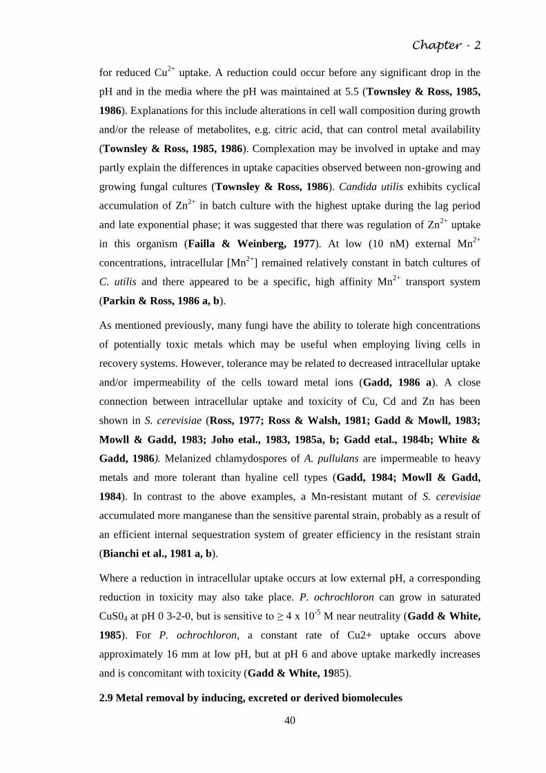
Chapter - 2
40
for reduced Cu2+
uptake. A reduction could occur before any significant drop in the
pH and in the media where the pH was maintained at 5.5 (Townsley & Ross, 1985,
1986). Explanations for this include alterations in cell wall composition during growth
and/or the release of metabolites, e.g. citric acid, that can control metal availability
(Townsley & Ross, 1985, 1986). Complexation may be involved in uptake and may
partly explain the differences in uptake capacities observed between non-growing and
growing fungal cultures (Townsley & Ross, 1986). Candida utilis exhibits cyclical
accumulation of Zn2+
in batch culture with the highest uptake during the lag period
and late exponential phase; it was suggested that there was regulation of Zn2+
uptake
in this organism (Failla & Weinberg, 1977). At low (10 nM) external Mn2+
concentrations, intracellular [Mn2+
] remained relatively constant in batch cultures of
C. utilis and there appeared to be a specific, high affinity Mn2+
transport system
(Parkin & Ross, 1986 a, b).
As mentioned previously, many fungi have the ability to tolerate high concentrations
of potentially toxic metals which may be useful when employing living cells in
recovery systems. However, tolerance may be related to decreased intracellular uptake
and/or impermeability of the cells toward metal ions (Gadd, 1986 a). A close
connection between intracellular uptake and toxicity of Cu, Cd and Zn has been
shown in S. cerevisiae (Ross, 1977; Ross & Walsh, 1981; Gadd & Mowll, 1983;
Mowll & Gadd, 1983; Joho etal., 1983, 1985a, b; Gadd etal., 1984b; White &
Gadd, 1986). Melanized chlamydospores of A. pullulans are impermeable to heavy
metals and more tolerant than hyaline cell types (Gadd, 1984; Mowll & Gadd,
1984). In contrast to the above examples, a Mn-resistant mutant of S. cerevisiae
accumulated more manganese than the sensitive parental strain, probably as a result of
an efficient internal sequestration system of greater efficiency in the resistant strain
(Bianchi et al., 1981 a, b).
Where a reduction in intracellular uptake occurs at low external pH, a corresponding
reduction in toxicity may also take place. P. ochrochloron can grow in saturated
CuS04 at pH 0 3-2-0, but is sensitive to ≥ 4 x 10⁻⁵
M near neutrality (Gadd & White,
1985). For P. ochrochloron, a constant rate of Cu2+ uptake occurs above
approximately 16 mm at low pH, but at pH 6 and above uptake markedly increases
and is concomitant with toxicity (Gadd & White, 1985).
2.9 Metal removal by inducing, excreted or derived biomolecules
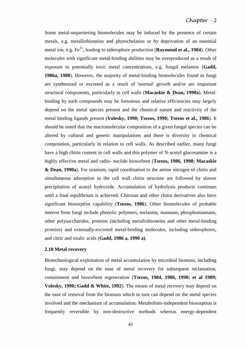
Chapter - 2
41
Some metal-sequestering biomolecules may be induced by the presence of certain
metals, e.g. metallothioneins and phytochelatins or by deprivation of an essential
metal ion, e.g. Fe3+
, leading to siderophore production (Raymond et al., 1984). Other
molecules with significant metal-binding abilities may be overproduced as a result of
exposure to potentially toxic metal concentrations, e.g. fungal melanins (Gadd,
1986a, 1988). However, the majority of metal-binding biomolecules found in fungi
are synthesized or excreted as a result of 'normal' growth and/or are important
structural components, particularly in cell walls (Macaskie & Dean, 1990a). Metal-
binding by such compounds may be fortuitous and relative efficiencies may largely
depend on the metal species present and the chemical nature and reactivity of the
metal binding ligands present (Volesky, 1990; Tsezos, 1990; Tsezos et al., 1986). It
should be noted that the macromolecular composition of a given fungal species can be
altered by cultural and genetic manipulations and there is diversity in chemical
composition, particularly in relation to cell walls. As described earlier, many fungi
have a high chitin content in cell walls and this polymer of N-acetyl glucosamine is a
highly effective metal and radio- nuclide biosorbent (Tsezos, 1986, 1990; Macaskie
& Dean, 1990a). For uranium, rapid coordination to the amine nitrogen of chitin and
simultaneous adsorption in the cell wall chitin structure are followed by slower
precipitation of uranyl hydroxide. Accumulation of hydrolysis products continues
until a final equilibrium is achieved. Chitosan and other chitin derivatives also have
significant biosorptive capability (Tsezos, 1986). Other biomolecules of probable
interest from fungi include phenolic polymers, melanins, mannans, phosphomannans,
other polysaccharides, proteins (including metallothioneins and other metal-binding
proteins) and externally-excreted metal-binding molecules, including siderophores,
and citric and oxalic acids (Gadd, 1986 a, 1990 a).
2.10 Metal recovery
Biotechnological exploitation of metal accumulation by microbial biomass, including
fungi, may depend on the ease of metal recovery for subsequent reclamation,
containment and biosorbent regeneration (Tsezos, 1984, 1986, 1990; et al 1989;
Volesky, 1990; Gadd & White, 1992). The means of metal recovery may depend on
the ease of removal from the biomass which in turn can depend on the metal species
involved and the mechanism of accumulation. Metabolism-independent biosorption is
frequently reversible by non-destructive methods whereas energy-dependent

Chapter - 2
42
intracellular accumulation and compartmentation/binding to induced proteins etc. are
often irreversible and require destructive recovery (Gadd, 1988). The later may be
achieved by incineration or dissolution in strong acids or alkalis (Brierley et al.,
1985). Most work has concentrated on non-destructive desorption which should
ideally be highly efficient, economical and result in minimal damage to the
biosorbent. Dilute mineral acids (~ 0 1 M), e.g. HNO3, HCI or H2SO4, can be
effective for the removal of heavy metals and radionuclides from fungal biomass
(Tsezos et al., 1987) although at higher concentrations (> 1 M) or with prolonged
exposure there may be irreversible damage which reduces the potential of the biomass
to be reused (Tsezos, 1984). Organic complexing agents may be effective, although
relatively expensive, desorbers without affecting integrity of the biomass. EDTA
treatment may also be useful in distinguishing surface complex formation from
intracellular, unexchangeable metal (Galun et al., 1983; Tsezos et al., 1987).
Carbonates and/or bicarbonates are efficient desorption agents with potential for
cheap, nondestructive metal recovery. Of several elution systems examined for
uranium desorption from R. arrhizus, NaHCO3 exhibited> 90 % efficiency of removal
and high uranium concentration factors (Tsezos, 1984). The solid: liquid ratios can
exceed 120:1 (mg:ml) for a 1 M NaHCO3 solution with eluate uranium concentrations
of at least 1-98 x 104 mg 1-1 (Tsezos, 1984). The solid:liquid (S/L) ratio, i.e. the ratio
of weight of loaded biomass to the eluant volume required for complete recovery,
needs to be maximized in any recovery process in order to yield an eluate of minimal
volume yet containing the highest possible metal concentration (Tsezos, 1990).
2.11 Biosorption of meals and radionuclides by fungal biomass
In a recent study by Mishra and Malik (2012) the effectiveness of fungal isolates,
Aspergillus lentulus (FJ 172995) for concurrent removal of heavy metals like
chromium, copper and lead from industrial effluent was examined. They inferred that
Cr, Cu, Pb, and Ni tolerant Aspergillus lentulus accumulated a significant amount of
each metal. The removal metals from synthetic solutions showed the trend like Pb2+
(100%) > Cr 3+
(*79%) > Cu2+
(78%) > Ni2+
(42%) after 5 days. When the same
fungal strain was used to treat the multiple metal contamining electroplating effluent,
the metal concentrations declined by 71%, 56%, and 100% for Cr, Cu and Pb
respectively within 11 days.Congeevaram et al 2007 concluded in another study that
mainly pH was attributed to organism specific physiology of biosorption. Many
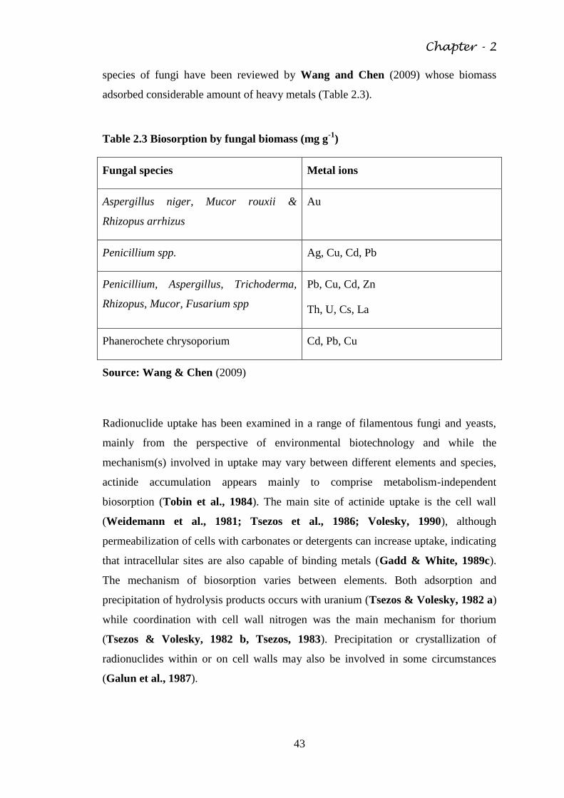
Chapter - 2
43
species of fungi have been reviewed by Wang and Chen (2009) whose biomass
adsorbed considerable amount of heavy metals (Table 2.3).
Table 2.3 Biosorption by fungal biomass (mg g-1
)
Fungal species Metal ions
Aspergillus niger, Mucor rouxii &
Rhizopus arrhizus
Au
Penicillium spp. Ag, Cu, Cd, Pb
Penicillium, Aspergillus, Trichoderma,
Rhizopus, Mucor, Fusarium spp
Pb, Cu, Cd, Zn
Th, U, Cs, La
Phanerochete chrysoporium Cd, Pb, Cu
Source: Wang & Chen (2009)
Radionuclide uptake has been examined in a range of filamentous fungi and yeasts,
mainly from the perspective of environmental biotechnology and while the
mechanism(s) involved in uptake may vary between different elements and species,
actinide accumulation appears mainly to comprise metabolism-independent
biosorption (Tobin et al., 1984). The main site of actinide uptake is the cell wall
(Weidemann et al., 1981; Tsezos et al., 1986; Volesky, 1990), although
permeabilization of cells with carbonates or detergents can increase uptake, indicating
that intracellular sites are also capable of binding metals (Gadd & White, 1989c).
The mechanism of biosorption varies between elements. Both adsorption and
precipitation of hydrolysis products occurs with uranium (Tsezos & Volesky, 1982 a)
while coordination with cell wall nitrogen was the main mechanism for thorium
(Tsezos & Volesky, 1982 b, Tsezos, 1983). Precipitation or crystallization of
radionuclides within or on cell walls may also be involved in some circumstances
(Galun et al., 1987).
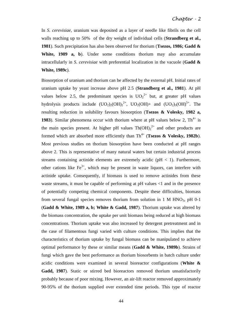
Chapter - 2
44
In S. cerevisiae, uranium was deposited as a layer of needle like fibrils on the cell
walls reaching up to 50% of the dry weight of individual cells (Strandberg et al.,
1981). Such precipitation has also been observed for thorium (Tsezos, 1986; Gadd &
White, 1989 a, b). Under some conditions thorium may also accumulate
intracellularly in S. cerevisiae with preferential localization in the vacuole (Gadd &
White, 1989c).
Biosorption of uranium and thorium can be affected by the external pH. Initial rates of
uranium uptake by yeast increase above pH 2.5 (Strandberg et al., 1981). At pH
values below 2.5, the predominant species is UO22+
but, at greater pH values
hydrolysis products include (UO2)2(OH)22+
, UO2(OH)+ and (UO2)3(OH)5+
. The
resulting reduction in solubility favours biosorption (Tsezos & Volesky, 1982 a,
1983). Similar phenomena occur with thorium where at pH values below 2, Th4+
is
the main species present. At higher pH values Th(OH)22+
and other products are
formed which are absorbed more efficiently than Th4+
(Tsezos & Volesky, 1982b).
Most previous studies on thorium biosorption have been conducted at pH ranges
above 2. This is representative of many natural waters but certain industrial process
streams containing actinide elements are extremely acidic (pH < 1). Furthermore,
other cations like Fe3+
, which may be present in waste liquors, can interfere with
actinide uptake. Consequently, if biomass is used to remove actinides from these
waste streams, it must be capable of performing at pH values <1 and in the presence
of potentially competing chemical components. Despite these difficulties, biomass
from several fungal species removes thorium from solution in 1 M HNO3, pH 0-1
(Gadd & White, 1989 a, b; White & Gadd, 1987). Thorium uptake was altered by
the biomass concentration, the uptake per unit biomass being reduced at high biomass
concentrations. Thorium uptake was also increased by detergent pretreatment and in
the case of filamentous fungi varied with culture conditions. This implies that the
characteristics of thorium uptake by fungal biomass can be manipulated to achieve
optimal performance by these or similar means (Gadd & White, 1989b). Strains of
fungi which gave the best performance as thorium biosorbents in batch culture under
acidic conditions were examined in several bioreactor configurations (White &
Gadd, 1987). Static or stirred bed bioreactors removed thorium unsatisfactorily
probably because of poor mixing. However, an air-lift reactor removed approximately
90-95% of the thorium supplied over extended time periods. This type of reactor
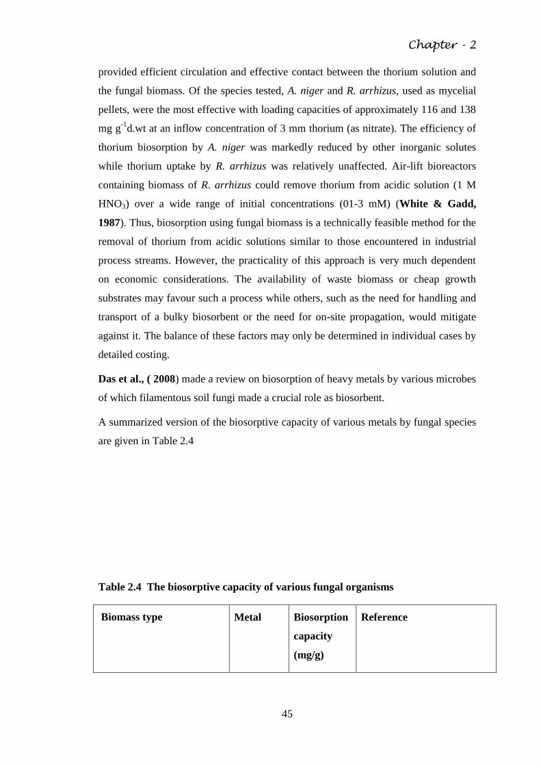
Chapter - 2
45
provided efficient circulation and effective contact between the thorium solution and
the fungal biomass. Of the species tested, A. niger and R. arrhizus, used as mycelial
pellets, were the most effective with loading capacities of approximately 116 and 138
mg g-1
d.wt at an inflow concentration of 3 mm thorium (as nitrate). The efficiency of
thorium biosorption by A. niger was markedly reduced by other inorganic solutes
while thorium uptake by R. arrhizus was relatively unaffected. Air-lift bioreactors
containing biomass of R. arrhizus could remove thorium from acidic solution (1 M
HNO3) over a wide range of initial concentrations (01-3 mM) (White & Gadd,
1987). Thus, biosorption using fungal biomass is a technically feasible method for the
removal of thorium from acidic solutions similar to those encountered in industrial
process streams. However, the practicality of this approach is very much dependent
on economic considerations. The availability of waste biomass or cheap growth
substrates may favour such a process while others, such as the need for handling and
transport of a bulky biosorbent or the need for on-site propagation, would mitigate
against it. The balance of these factors may only be determined in individual cases by
detailed costing.
Das et al., ( 2008) made a review on biosorption of heavy metals by various microbes
of which filamentous soil fungi made a crucial role as biosorbent.
A summarized version of the biosorptive capacity of various metals by fungal species
are given in Table 2.4
Table 2.4 The biosorptive capacity of various fungal organisms
Biomass type Metal Biosorption
capacity
(mg/g)
Reference
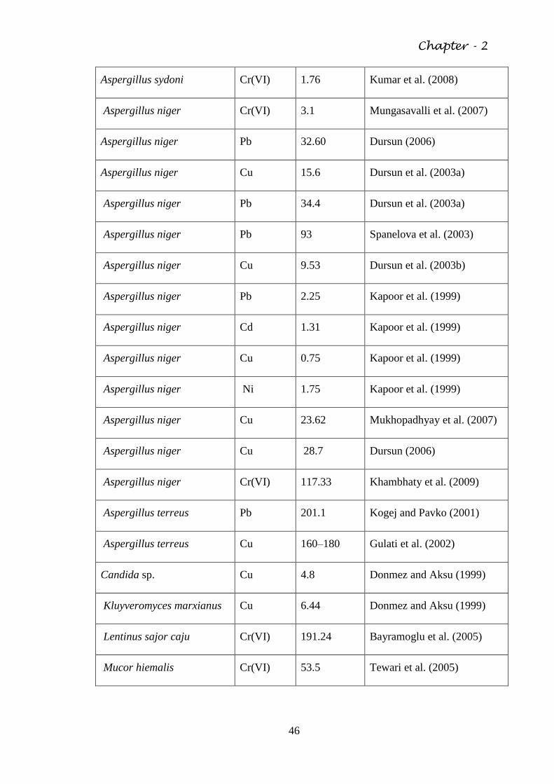
Chapter - 2
46
Aspergillus sydoni Cr(VI) 1.76 Kumar et al. (2008)
Aspergillus niger Cr(VI) 3.1 Mungasavalli et al. (2007)
Aspergillus niger Pb 32.60 Dursun (2006)
Aspergillus niger Cu 15.6 Dursun et al. (2003a)
Aspergillus niger Pb 34.4 Dursun et al. (2003a)
Aspergillus niger Pb 93 Spanelova et al. (2003)
Aspergillus niger Cu 9.53 Dursun et al. (2003b)
Aspergillus niger Pb 2.25 Kapoor et al. (1999)
Aspergillus niger Cd 1.31 Kapoor et al. (1999)
Aspergillus niger Cu 0.75 Kapoor et al. (1999)
Aspergillus niger Ni 1.75 Kapoor et al. (1999)
Aspergillus niger Cu 23.62 Mukhopadhyay et al. (2007)
Aspergillus niger Cu 28.7 Dursun (2006)
Aspergillus niger Cr(VI) 117.33 Khambhaty et al. (2009)
Aspergillus terreus Pb 201.1 Kogej and Pavko (2001)
Aspergillus terreus Cu 160–180 Gulati et al. (2002)
Candida sp. Cu 4.8 Donmez and Aksu (1999)
Kluyveromyces marxianus Cu 6.44 Donmez and Aksu (1999)
Lentinus sajor caju Cr(VI) 191.24 Bayramoglu et al. (2005)
Mucor hiemalis Cr(VI) 53.5 Tewari et al. (2005)
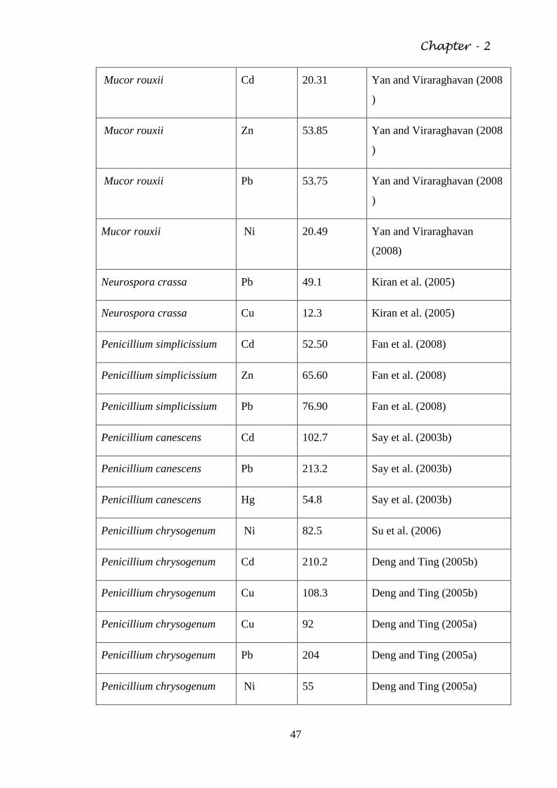
Chapter - 2
47
Mucor rouxii Cd 20.31 Yan and Viraraghavan (2008
)
Mucor rouxii Zn 53.85 Yan and Viraraghavan (2008
)
Mucor rouxii Pb 53.75 Yan and Viraraghavan (2008
)
Mucor rouxii Ni 20.49 Yan and Viraraghavan
(2008)
Neurospora crassa Pb 49.1 Kiran et al. (2005)
Neurospora crassa Cu 12.3 Kiran et al. (2005)
Penicillium simplicissium Cd 52.50 Fan et al. (2008)
Penicillium simplicissium Zn 65.60 Fan et al. (2008)
Penicillium simplicissium Pb 76.90 Fan et al. (2008)
Penicillium canescens Cd 102.7 Say et al. (2003b)
Penicillium canescens Pb 213.2 Say et al. (2003b)
Penicillium canescens Hg 54.8 Say et al. (2003b)
Penicillium chrysogenum Ni 82.5 Su et al. (2006)
Penicillium chrysogenum Cd 210.2 Deng and Ting (2005b)
Penicillium chrysogenum Cu 108.3 Deng and Ting (2005b)
Penicillium chrysogenum Cu 92 Deng and Ting (2005a)
Penicillium chrysogenum Pb 204 Deng and Ting (2005a)
Penicillium chrysogenum Ni 55 Deng and Ting (2005a)
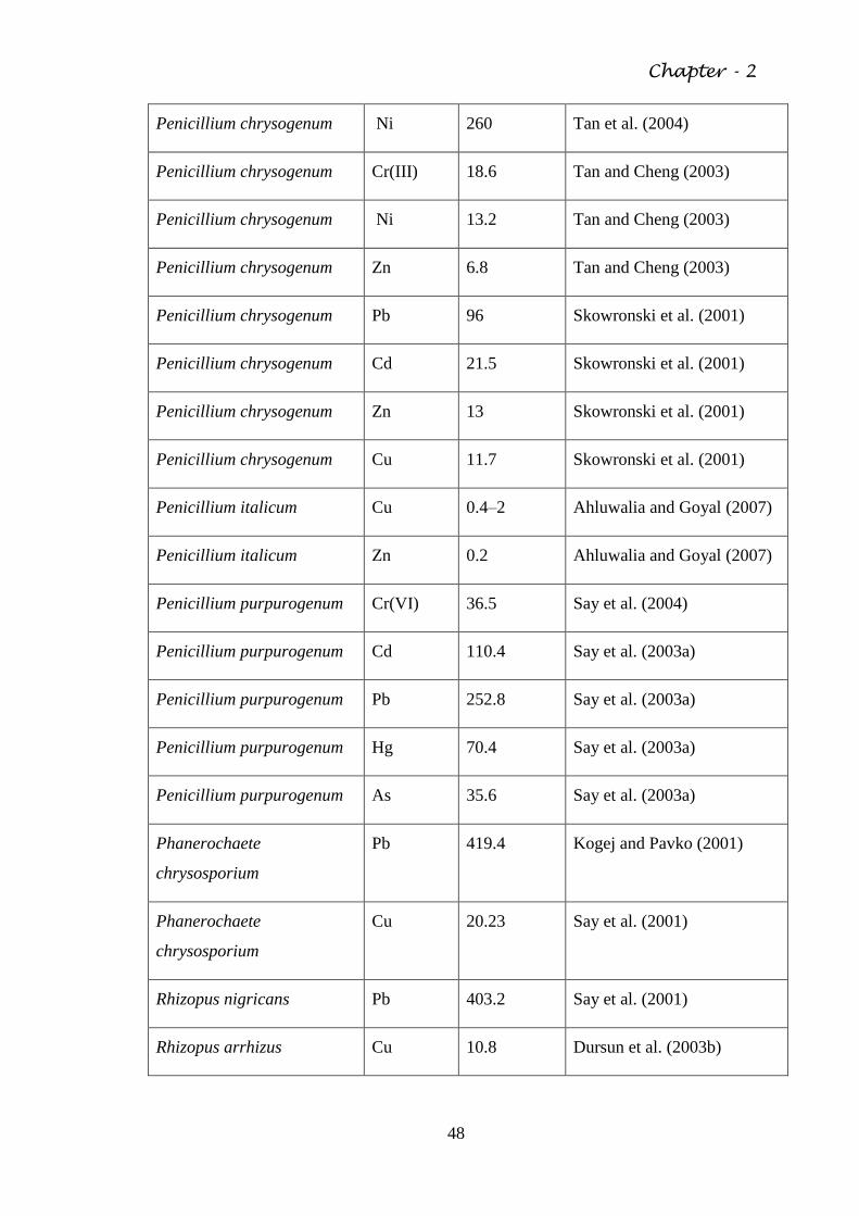
Chapter - 2
48
Penicillium chrysogenum Ni 260 Tan et al. (2004)
Penicillium chrysogenum Cr(III) 18.6 Tan and Cheng (2003)
Penicillium chrysogenum Ni 13.2 Tan and Cheng (2003)
Penicillium chrysogenum Zn 6.8 Tan and Cheng (2003)
Penicillium chrysogenum Pb 96 Skowronski et al. (2001)
Penicillium chrysogenum Cd 21.5 Skowronski et al. (2001)
Penicillium chrysogenum Zn 13 Skowronski et al. (2001)
Penicillium chrysogenum Cu 11.7 Skowronski et al. (2001)
Penicillium italicum Cu 0.4–2 Ahluwalia and Goyal (2007)
Penicillium italicum Zn 0.2 Ahluwalia and Goyal (2007)
Penicillium purpurogenum Cr(VI) 36.5 Say et al. (2004)
Penicillium purpurogenum Cd 110.4 Say et al. (2003a)
Penicillium purpurogenum Pb 252.8 Say et al. (2003a)
Penicillium purpurogenum Hg 70.4 Say et al. (2003a)
Penicillium purpurogenum As 35.6 Say et al. (2003a)
Phanerochaete
chrysosporium
Pb 419.4 Kogej and Pavko (2001)
Phanerochaete
chrysosporium
Cu 20.23 Say et al. (2001)
Rhizopus nigricans Pb 403.2 Say et al. (2001)
Rhizopus arrhizus Cu 10.8 Dursun et al. (2003b)
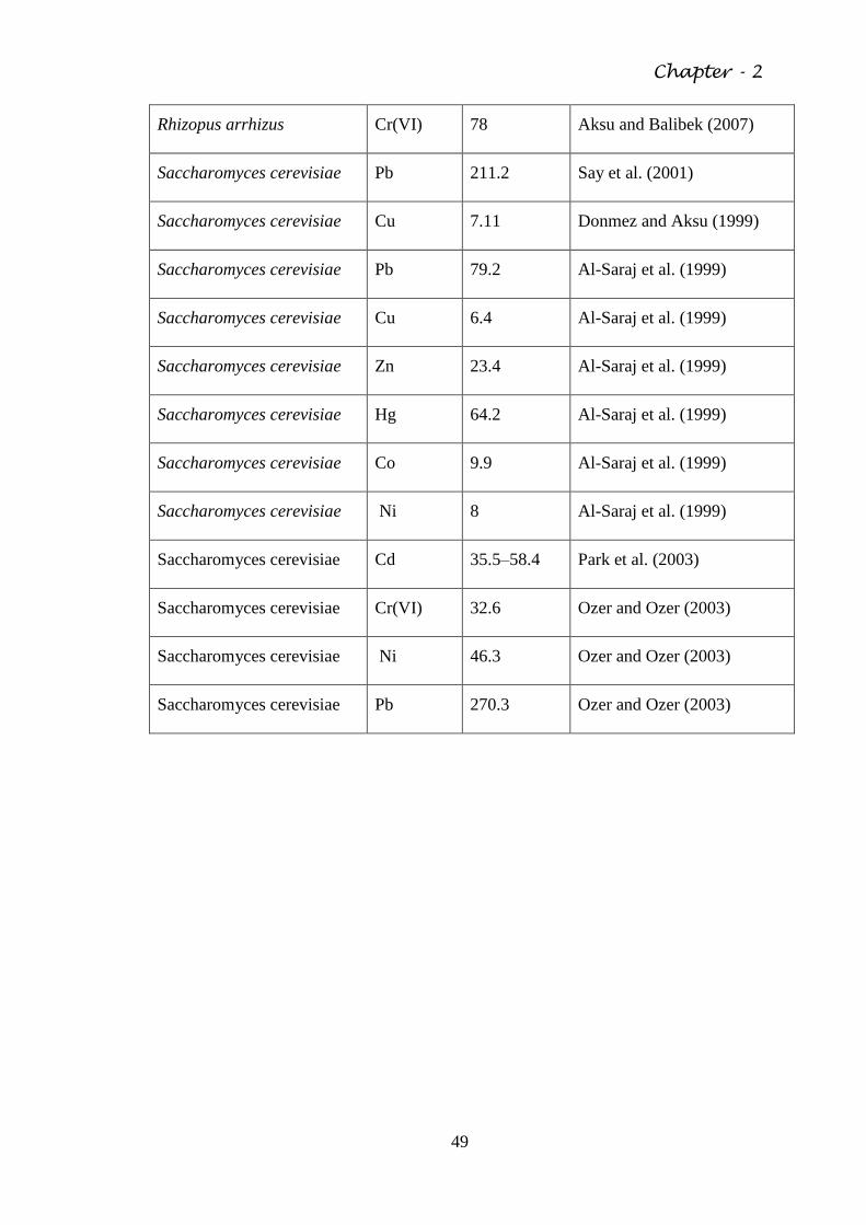
Chapter - 2
49
Rhizopus arrhizus Cr(VI) 78 Aksu and Balibek (2007)
Saccharomyces cerevisiae Pb 211.2 Say et al. (2001)
Saccharomyces cerevisiae Cu 7.11 Donmez and Aksu (1999)
Saccharomyces cerevisiae Pb 79.2 Al-Saraj et al. (1999)
Saccharomyces cerevisiae Cu 6.4 Al-Saraj et al. (1999)
Saccharomyces cerevisiae Zn 23.4 Al-Saraj et al. (1999)
Saccharomyces cerevisiae Hg 64.2 Al-Saraj et al. (1999)
Saccharomyces cerevisiae Co 9.9 Al-Saraj et al. (1999)
Saccharomyces cerevisiae Ni 8 Al-Saraj et al. (1999)
Saccharomyces cerevisiae Cd 35.5–58.4 Park et al. (2003)
Saccharomyces cerevisiae Cr(VI) 32.6 Ozer and Ozer (2003)
Saccharomyces cerevisiae Ni 46.3 Ozer and Ozer (2003)
Saccharomyces cerevisiae Pb 270.3 Ozer and Ozer (2003)
Chapter - 3
50
AIMS AND OBJECTIVES
During the last decades, extensive attention has been paid on the management of
environmental pollution caused by hazardous materials such as heavy metal and
complex organic molecules. Decontamination of such toxicants from the soil, water
around the industrial area, waste dumps and similar contaminated sites has been a
challenge for a long time. A number of methods have been developed for the removal
of heavy metals from liquid wastes/contaminated site lechates such as precipitation,
evaporation, electroplating, ion exchange, membrane processes etc. All these methods
have several disadvantages such as unpredictable metal ion removal, high reagent
requirement, generation of toxic sludge etc. Microbial treatment specially the use of
fungi and bacteria provide an innovative biotechnological approach for much better
cleanup process of contaminated sites/materials/environment.
The present study shall therefore be targeted at identifying the mechanism by which
the mycoflora develop tolerance against the toxic heavy metals, so that the same
could be applied in removing the metal contaminants from the soil by the way of
bioremediation. In general fungi shall naturally be viewed with greater potentialities
because of their aggressive growth, greater biomass production and extensive hypal
richness in soil.
These prime objectives are as followings:
To understand the nature of toxicity induced by various toxic metals on soil
fungi;
To study the role of soil component on bioavailability in fungal system;
To evaluate the metal–metal interaction and its biotic effect with respect to
fungal system;
To understand the mechanism of metal tolerance and removal by selected
fungi.
Chapter -4
51
MATERIALS AND METHODS
4.1 Sampling of soil
Soil samples were collected from four pre-selected contaminated sites along with a
control (comparatively non-contaminated) site in and around Kolkata city during
2005-2007. The sites are as follows:
(i) Waste dumping site of Dhapa areas of Kolkata;
(ii) Tannery waste dump sites along Eastern Metropolitan Bypass areas of
Kolkata;
(iii) Contaminated site within the area of Central Bus Terminus, Esplanade
Area, Kolkata ;
(iv) Waste dump sites within the premises of Calcutta Medical College
Hospital, Kolkata;
(v) Victoria Memorial Monument premises, Kolkata as non-contaminated
control site.
Each site has known metal contaminants as per previous report. 100gms of surface
soil was collected aseptically in poly bag and were stored at -20 C before further
analysis.
4.2 Isolation of metal resistant microorganisms
Fungal strains were isolated from soil using conventional dilution plate culture
techniques. One gm of pre-weighted soil was suspended in 10ml of sterilized distilled
water and then serially diluted to 10-3
dilution and 1ml of such dilutant was plated in
sterile petridish containing pre-coated media. The soil dilutant was spread over the
plate and incubated at 28± 2oC for six days. Three fungal media viz. Potato Dextrose
Agar, Malt agar and Czapek dox Agar were used for this purpose.
Chapter -4
52
4.3 Media compositions
Potato Dextrose (PD) Agar was prepared using 250gms of potato boiled in 100ml of
distilled water for 30 minutes and the filtrate was mixed with 2gms of dextrose and 20
gms of agar powder per liter.
Malt Agar medium was prepared with mixture of malt extract and agar powder 15gms
per liter. Identically Czapekdox (CD) medium was prepared (glucose -2gm, NaNO3 –
2.5gm, KCl -0.5gm, MgSO4,7H2O -0.5gm, FeSO4 – 0.5gm, ZnSO4, 7H2O – 0.5gm,
KH2PO4 – 1gm and agar powder 15gms per liter) (APHA, 1992).
4.4 Standard plate counts
Heterotrophic plate counts were done by Pour Plate Method after incubation for 48
hours in plate count agar (APHA, 1992). Samples were serial-diluted with sterilized
distilled water for getting appropriate counts. All microbiological operations were
done aseptically. Finally to isolate the preliminary metal resistant fungal strains
isolated from the sampling sites ,medium in respective plates were added with 50 ppm
of metals like arsenic, cadmium and chromium.
4.5 Isolation of pure cultures
In nature, microbial populations do not segregate themselves by genomic or specific
identities but exist in coherent mixture of all existing types. In the laboratory this
mixed population needs to be separated into pure cultures. As the pure cultures
contain only one specific type of organism, these are very much suitable for study of
their properties.
By the streak-plate dilution technique i.e. by spreading a loopful of culture over the
surface of a agar plate and incubating, pure cultures were obtained by identifying and
culturing distinct single colonies with sufficient distance from other colonies
(Trivedy and Goel, 1984).
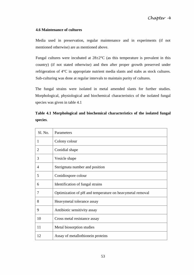
Chapter -4
53
4.6 Maintenance of cultures
Media used in preservation, regular maintenance and in experiments (if not
mentioned otherwise) are as mentioned above.
Fungal cultures were incubated at 28±2°C (as this temperature is prevalent in this
country) (if not stated otherwise) and then after proper growth preserved under
refrigeration of 4°C in appropriate nutrient media slants and stabs as stock cultures.
Sub-culturing was done at regular intervals to maintain purity of cultures.
The fungal strains were isolated in metal amended slants for further studies.
Morphological, physiological and biochemical characteristics of the isolated fungal
species was given in table 4.1
Table 4.1 Morphological and biochemical characteristics of the isolated fungal
species.
Sl. No. Parameters
1 Colony colour
2 Conidial shape
3 Vesicle shape
4 Sterigmata number and position
5 Conidiospore colour
6 Identification of fungal strains
7 Optimization of pH and temperature on heavymetal removal
8 Heavymetal tolerance assay
9 Antibiotic sensitivity assay
10 Cross metal resistance assay
11 Metal biosorption studies
12 Assay of metallothionein proteins
Chapter -4
54
4.7 Selection of tolerant strains
For determining metal tolerance limit of these fungal strains, the strains were allowed
to grow in Czapek dox agar media incorporating different metal concentration. After
incubating for appropriate periods, tolerant strains were detected and identified by
visible prominence of growth i. e colony formation and size.
4. 8 Heavy metal analysis of contaminated soil
Each soil sample (1gm) was taken in the digestion tube (100ml) with addition of 10ml
of HNO3, HClO4 (1:2) solution and heated in sand bath until clear solution was
developed. The solutions were filtered through Whatman No1 filter paper and volume
was made up to 50ml by adding distilled water. Each soil sample was digested in
triplicate for analysis of contaminated heavy metal content separately. Blank (control)
samples contained 10ml of HNO3, HClO4 (1:2) solution were also digested and those
were diluted in the same way for comparison. Finally all metal analysis (Arsenic,
Chromium and Cadmium) were performed by using Atomic Absorption
Spectrophotometer with flow injection hydride generation (Perkin Elmer Analyst 400)
(APHA, 1998).
4.9 Identification of fungi
Isolated fungal culture strains were identified on basis of macroscopic (colony
character, morphology, colour, texture, shape, diameter and appearance of colony)
and microscopic (septation in mycelium, presence of specific reproductive structures,
shape and structure of conidia and conidiospore etc) characteristic using fungal
identification manuals (Thorn and Raper, 1945; Domsch et al., 1980 Ellis and
Ellis, 1992; Zafar et al., 2006).
4.10 Growth optimization of media, pH and temperature
Isolated strains were grown in sterilized Czapekdox broth medium for establishment
of the optimum temperature, pH that supports the exuberant growth of those species.
The pH of the medium was adjusted using dilute HCl or NaOH. The temperature
ranges were varied from 25°C- 35°C, and pH was from 3 to 9.
Chapter -4
55
The fungal strains were inoculated into a series of 250ml conical flasks containing
varied concentration of each pre-selected metal (arsenic, chromium and cadmium) in
CDB medium. The pH was varied from 3 to 9 (3, 5, 7 and 9). The pH of the medium
was adjusted using dilute (N/10) HCl or NaOH. For optimization of temperature
against each pH the representative culture was incubated at different temperatures (25
to 35oC). Fungal biomass of each flask was harvested and weight was measured
separately. The residual heavy metal of CD broth medium was also examined. Based
upon the heavy metal removal and biomass data, the optimal pH and temperature
were determined (Pujol et al., 1997).
4.11 Antibiotic sensitivity test
Antibiotic (antifungal) sensitivity of each metal tolerant fungal strains was also
measured using the same cup assay method described earlier (Pujol et al., 1997). The
antibiotics used for this assay were Nystatin, Griseofulvin and Kanamycin.
4.12 Heavy metal tolerance assessment
To explore the tolerance of the isolates to the heavy metals, the fungal strains were
exposed to varying concentrations of heavy metal in Czapek dox broth. To each
freshly prepared growth medium, selected metal was amended with concentration
which varied between 50ppm to 5000ppm. After 6 days of incubation, the extents of
colony growth (in diameter) was compared with the control (i.e. growth of fungi in
medium without metal). All the experiments were carried out in triplicate
(Congeevaram et al., 2007).
4.13 Cross metal resistance assay
Cross heavy metal resistance of the fungal isolates was determined by conventional
cup assay method (Dhar et al., 2004). Selected metal tolerant fungal strains were
grown separately in Czapekdox agar medium supplemented with diverse
concentration (sub-lethal to lethal) of each heavy metal separately. Then cups were
prepared where various other metallic solutions (50ppm to 500ppm) were poured. For
each test triplicates were made and incubated for 6 days. Zone of inhibition of fungal
Chapter -4
56
growth around the cup was examined and measured in terms of MIC in each case
separately.
4.14 Metal toxicity of the fungal isolates
The effect of heavy metals on the growth of the fungal isolates was determined
separately by inoculating the isolates in Czapek dox broth medium supplemented with
preselected metal concentration (sub-lethal concentration) of each heavy metal against
each fungal isolates were done separately. These inoculated flasks were incubated in
shaker-incubator at 28± 2oC. Control were maintained and consisted of inoculated
medium without the supplementation of heavy metal. The weight of fungal mycelium
were taken at regular intervals (3, 6, 9 and 12 days) for study the growth responses
against the metal concentrations (Dugal et al., 2012).
4.15 Metal removal by selected strains
To study metal removal capacity of selected fungi, the strains were grown in 50
ml. Czapek Dox broth (pH 7) containing 20 ppm, 100ppm and 1000ppm of respective
metal solution (arsenic, chromium and cadmium). The cultures were incubated in
28±2°C in a shaker incubator. A control set was maintained side by side. The removal
percentage was determined in 3, 6 and 9 days interval. The inocula were 2 ml. of fresh
spore suspension of inoculum strength: 4-5x 10³ c.f.u./ml. For determination of
residual metal in the filtrate, the filtrate (passed through 0.22µ Milipore filter) was
taken out and residual metal estimation was done by AAS(Perkin Elmer model
Analyst 400) (Ahmed et al., 2006).
4.16 Metal biosorption by fungal biomass
2gms of each wet weight from the fungal biomass thus harvested in previous
experiment were washed with clean distilled water and then homogenized for 15 min
in a blender. The biomass was then digested with HNO3, HCl mixture (1:2) solution
in digestion flask as mentioned earlier and further cell wall bound heavy metals
(arsenic, chromium and cadmium) were measured separately using Atomic
Absorption Spectrometer(AAS). Control set was maintained using non metal tolerant
species. The metal absorption capacity of chitosan was also measured for comparison
in the same way (Ahmed et al 2006).
Chapter -4
57
The metal adsorption by the dead fungal biomass was also studied using 6-7 days
grown 2gms of oven dried fungal mat in 50ml of different concentration of metal
solution with continuous shaking. After 3days the fungal biomass was harvested
through centrifugation. The bioadsorption capacity dead fungal biomass was
measured by AAS after digestion (Ahmed et al 2006).
4.17 Metal adsorption by Chitosan
To study the bioadsorption of metals by chitosan, 20ppm and 100ppm of metal
concentration were prepared for chromium, cadmium and arsenic respectively. The
metal solutions were prepared form standard solutions of Chromium (1000 ppm),
Arsenic (1000 ppm) and Cadmium (1000 ppm) (Merck Germany). 50 ml of solution
of each metal concentration were taken and 500 mg of chitosan was added (Sigma-
Aldrich). Then the mixture was continuously stirred using magnetic stirrer for 6 hours
at room temperature (30 °C). After that the solution was filtered and the filtrate was
measured by Atomic Absorption Spectroscopy assisted with FIAS (Perkin Elmer
Model Analyst 400). The test was performed in varying pH to analyse the relation of
metal accumulation by chitosan against different pH (Sewandi et al., 2011).
4.18 Assay of metallothionein protein
Spore suspension of wild non metal tolerant and metal tolerant fungal strains (test
organisms) were inoculated in Czapekdox broth medium supplemented with sub-
lethal concentration of respective heavy metal and allowed to incubate in a shaker at
28+2°C for 96 hours. The mycelium was then harvested and washed thrice with
distilled water under aseptic conditions. The excess water was blotted off and
preserved at -20°C for preparation of cell free extract of each strain separately. For
each experiment triplicate was run in aseptic way. Metallothionein was measured
according to the method of Virarengo et al. (1997). In the preparation of sample,
mycelium of both wild (non metal tolerant) and metal tolerant strain were
homogenized separately in a mixture of 0.5 mol l-1
sucrose, 0.02 mol l-1
tris-HCl
buffer (pH 8.6) with added 6x10⁻⁶
mol l-1
leupeptine, 5x10⁻³
mol l-1
phenyl methyl
sulphonyl fluoride (PMSF) as antiproteolytic agent and 0.01% β-mercaptoethanol as
reducing agent. The homogenate was then centrifuged at 30000xg for 20mins to
obtain the supernatant containing metallothionein. The collected supernatant was then
Chapter -4
58
treated with ethanol- chloroform as described by Kimura et al., (1979). 1.05 ml of
cold (-200C) absolute ethanol and 80μl of chloroform were added to aliquot of 1 ml of
supernatant. The samples were then centrifuged at 6000x g for 10 mins at 0-40
C. The
collected supernatant was combined with 1 mg RNA and 40μl 37% HCl and
subsequently with three volume of cold ethanol. The samples were maintained at –200
C for 1 h and centrifuged in a swinging rotor at 6000x g for 10 min. The pellets
containing metallothionein was then washed with 87 % ethanol and 1 % chloroforms
in homogenizing buffer, centrifuged at 6000x g for 10 min and dried under nitrogen
stream. The pellets were re-suspended in 150 μl NaCl (0.25 mol l-1
) and subsequently
150l HCl (1N) containing EDTA (0.004 mol l-1
) and 4.2 ml NaCl (2 mol l-1
)
containing 5,5-dithiobis-2-nitrobenzoic acid (DTMB) (4.3x 10⁻⁴
mol l-1
) buffered with
0.2 mol l-1
Na-Phosphate (pH 8) were added . The mixture was finally centrifuged at
3000g for 5 mins. The absorbance of supernatant was measured
spectrophotometrically at 412 nm. The metallothionein concentration was estimated
considering reduced glutathione (GSH) as reference standard (Pal et al., 2005).
4.19 Scanning Electron Microscopic(SEM) study
The mycelium of metal tolerant fungal strains and their respective wild strains were
washed with buffer and dehydrated in a series of ethanol water solution (30,50, 70 and
90% ethanol, 5 minutes each) and dried at a critical point under a CO2 atmosphere for
30 minutes. Then mounting was made on aluminium stubs, and mycelium was coated
with 90Ao thick gold palladium coating in a polaron Sc 7640 Sputter coate for 30 min.
Coated mycelium were viewed at 15KV with Scanning Electron Microscopy (IB2 Ion
Coater HITACHI S-530) (Mishra et al., 2013).
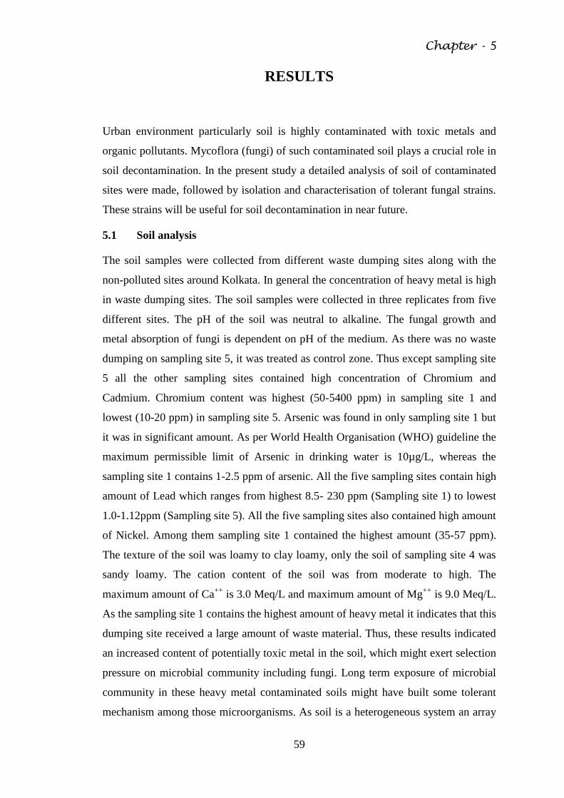
Chapter - 5
59
RESULTS
Urban environment particularly soil is highly contaminated with toxic metals and
organic pollutants. Mycoflora (fungi) of such contaminated soil plays a crucial role in
soil decontamination. In the present study a detailed analysis of soil of contaminated
sites were made, followed by isolation and characterisation of tolerant fungal strains.
These strains will be useful for soil decontamination in near future.
5.1 Soil analysis
The soil samples were collected from different waste dumping sites along with the
non-polluted sites around Kolkata. In general the concentration of heavy metal is high
in waste dumping sites. The soil samples were collected in three replicates from five
different sites. The pH of the soil was neutral to alkaline. The fungal growth and
metal absorption of fungi is dependent on pH of the medium. As there was no waste
dumping on sampling site 5, it was treated as control zone. Thus except sampling site
5 all the other sampling sites contained high concentration of Chromium and
Cadmium. Chromium content was highest (50-5400 ppm) in sampling site 1 and
lowest (10-20 ppm) in sampling site 5. Arsenic was found in only sampling site 1 but
it was in significant amount. As per World Health Organisation (WHO) guideline the
maximum permissible limit of Arsenic in drinking water is 10µg/L, whereas the
sampling site 1 contains 1-2.5 ppm of arsenic. All the five sampling sites contain high
amount of Lead which ranges from highest 8.5- 230 ppm (Sampling site 1) to lowest
1.0-1.12ppm (Sampling site 5). All the five sampling sites also contained high amount
of Nickel. Among them sampling site 1 contained the highest amount (35-57 ppm).
The texture of the soil was loamy to clay loamy, only the soil of sampling site 4 was
sandy loamy. The cation content of the soil was from moderate to high. The
maximum amount of Ca++
is 3.0 Meq/L and maximum amount of Mg++
is 9.0 Meq/L.
As the sampling site 1 contains the highest amount of heavy metal it indicates that this
dumping site received a large amount of waste material. Thus, these results indicated
an increased content of potentially toxic metal in the soil, which might exert selection
pressure on microbial community including fungi. Long term exposure of microbial
community in these heavy metal contaminated soils might have built some tolerant
mechanism among those microorganisms. As soil is a heterogeneous system an array
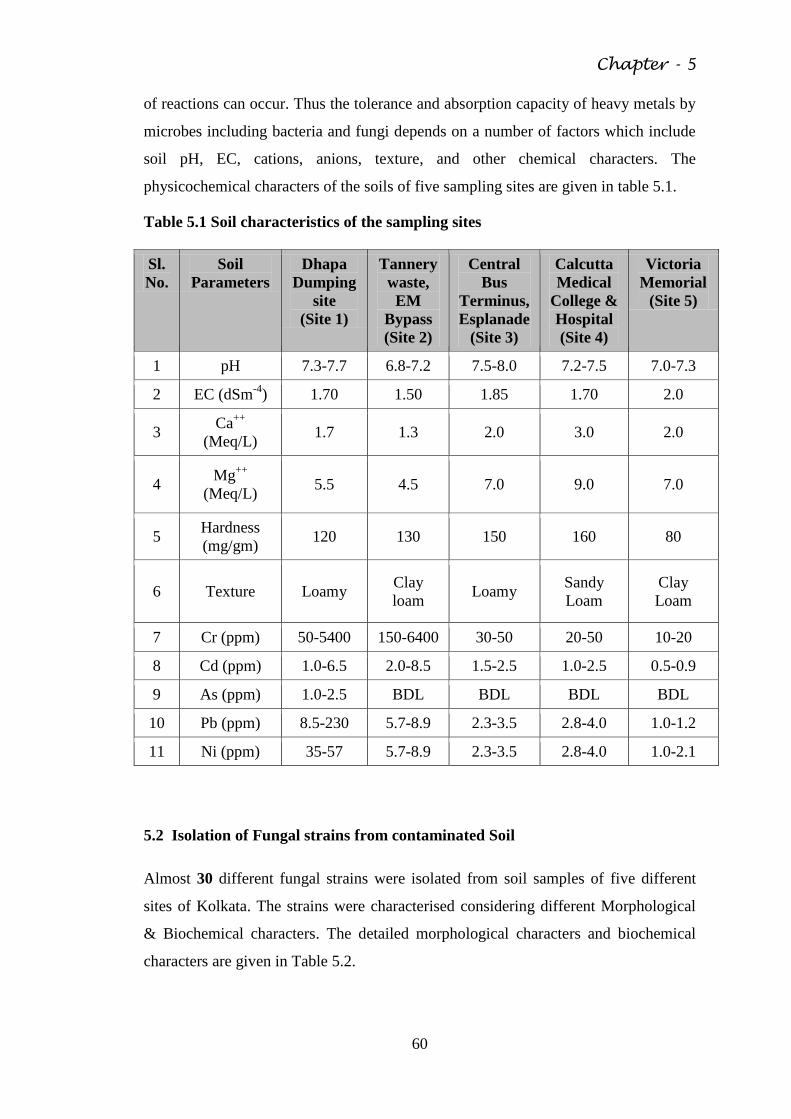
Chapter - 5
60
of reactions can occur. Thus the tolerance and absorption capacity of heavy metals by
microbes including bacteria and fungi depends on a number of factors which include
soil pH, EC, cations, anions, texture, and other chemical characters. The
physicochemical characters of the soils of five sampling sites are given in table 5.1.
Table 5.1 Soil characteristics of the sampling sites
Sl.
No.
Soil
Parameters
Dhapa
Dumping
site
(Site 1)
Tannery
waste,
EM
Bypass
(Site 2)
Central
Bus
Terminus,
Esplanade
(Site 3)
Calcutta
Medical
College &
Hospital
(Site 4)
Victoria
Memorial
(Site 5)
1 pH 7.3-7.7 6.8-7.2 7.5-8.0 7.2-7.5 7.0-7.3
2 EC (dSm-4
) 1.70 1.50 1.85 1.70 2.0
3 Ca
++
(Meq/L) 1.7 1.3 2.0 3.0 2.0
4 Mg
++
(Meq/L) 5.5 4.5 7.0 9.0 7.0
5 Hardness
(mg/gm) 120 130 150 160 80
6 Texture Loamy Clay
loam Loamy
Sandy
Loam
Clay
Loam
7 Cr (ppm) 50-5400 150-6400 30-50 20-50 10-20
8 Cd (ppm) 1.0-6.5 2.0-8.5 1.5-2.5 1.0-2.5 0.5-0.9
9 As (ppm) 1.0-2.5 BDL BDL BDL BDL
10 Pb (ppm) 8.5-230 5.7-8.9 2.3-3.5 2.8-4.0 1.0-1.2
11 Ni (ppm) 35-57 5.7-8.9 2.3-3.5 2.8-4.0 1.0-2.1
5.2 Isolation of Fungal strains from contaminated Soil
Almost 30 different fungal strains were isolated from soil samples of five different
sites of Kolkata. The strains were characterised considering different Morphological
& Biochemical characters. The detailed morphological characters and biochemical
characters are given in Table 5.2.
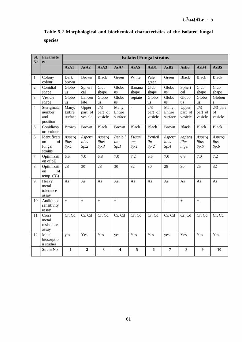
Chapter - 5
61
Table 5.2 Morphological and biochemical characteristics of the isolated fungal
species
Sl.
No
Paramete
rs Isolated Fungal strains
AsA1 AsA2 AsA3 AsA4 AsA5 AsB1 AsB2 AsB3 AsB4 AsB5
1 Colony
colour
Dark
brown
Brown Black Green White Pale
green
Green Black Black Black
2 Conidial
shape
Globo
us
Spheri
cal
Club
shape
Globo
us
Banana
shape
Club
shape
Globo
us
Spheri
cal
Club
shape
Club
shape
3 Vesicle
shape
Globo
us
Lanceo
late
Globo
us
Globo
us
septate Globo
us
Globo
us
Globo
us
Globo
us
Globou
s
4 Sterigmata
number
and
position
Many,
Entire
surface
Upper
part of
vesicle
2/3
part of
vesicle
Many,
Entire
surface
- 2/3
part of
vesicle
Many,
Entire
surface
Upper
part of
vesicle
2/3
part of
vesicle
2/3 part
of
vesicle
5 Conidiosp
ore colour
Brown Brown Black Brown Black Black Brown Black Black Black
6 Identificati
on of
fungal
strains
Asperg
illus
Sp.1
Asperg
illus
Sp.2
Asperg
illus
Sp.3
Penicil
lin
Sp.1
Fusari
um
Sp.1
Penicil
lin
Sp.2
Asperg
illus
Sp.4
Asperg
illus
niger
Asperg
illus
Sp.5
Aspergi
llus
Sp.6
7 Optimizati
on of pH
6.5 7.0 6.8 7.0 7.2 6.5 7.0 6.8 7.0 7.2
8 Optimizati
on of
temp. (oC)
28 30 28 30 32 30 28 30 25 32
9 Heavy
metal
tolerance
assay
As As As As As As As As As As
10 Antibiotic
sensitivity
assay
+ + + + - - - + + -
11 Cross
metal
resistance
assay
Cr, Cd Cr, Cd Cr, Cd Cr, Cd Cr, Cd Cr, Cd Cr, Cd Cr, Cd Cr, Cd Cr, Cd
12 Metal
biosorptio
n studies
yes Yes Yes yes Yes Yes yes Yes Yes Yes
Strain No 1 2 3 4 5 6 7 8 9 10
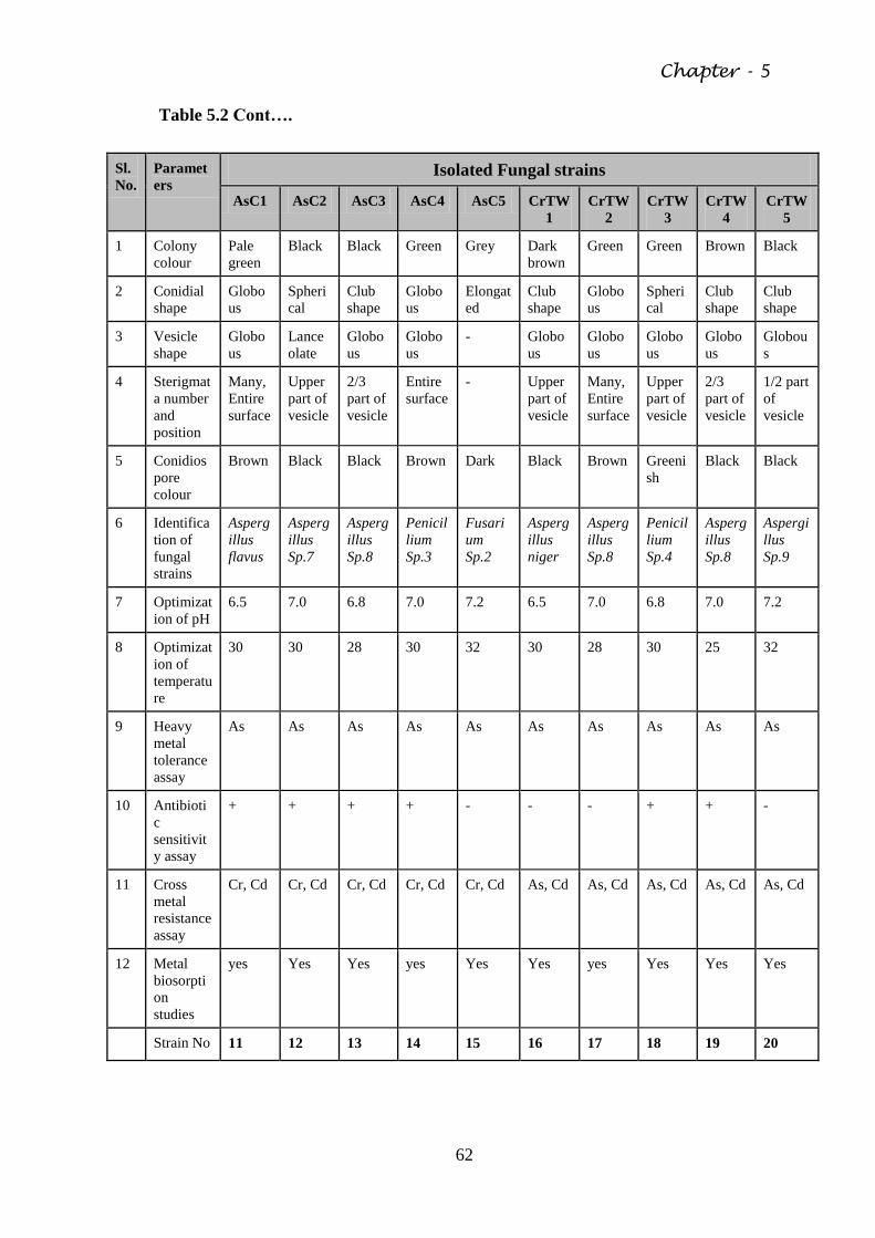
Chapter - 5
62
Table 5.2 Cont….
Sl.
No.
Paramet
ers Isolated Fungal strains
AsC1 AsC2 AsC3 AsC4 AsC5 CrTW
1
CrTW
2
CrTW
3
CrTW
4
CrTW
5
1 Colony
colour
Pale
green
Black Black Green Grey Dark
brown
Green Green Brown Black
2 Conidial
shape
Globo
us
Spheri
cal
Club
shape
Globo
us
Elongat
ed
Club
shape
Globo
us
Spheri
cal
Club
shape
Club
shape
3 Vesicle
shape
Globo
us
Lance
olate
Globo
us
Globo
us
- Globo
us
Globo
us
Globo
us
Globo
us
Globou
s
4 Sterigmat
a number
and
position
Many,
Entire
surface
Upper
part of
vesicle
2/3
part of
vesicle
Entire
surface
- Upper
part of
vesicle
Many,
Entire
surface
Upper
part of
vesicle
2/3
part of
vesicle
1/2 part
of
vesicle
5 Conidios
pore
colour
Brown Black Black Brown Dark Black Brown Greeni
sh
Black Black
6 Identifica
tion of
fungal
strains
Asperg
illus
flavus
Asperg
illus
Sp.7
Asperg
illus
Sp.8
Penicil
lium
Sp.3
Fusari
um
Sp.2
Asperg
illus
niger
Asperg
illus
Sp.8
Penicil
lium
Sp.4
Asperg
illus
Sp.8
Aspergi
llus
Sp.9
7 Optimizat
ion of pH
6.5 7.0 6.8 7.0 7.2 6.5 7.0 6.8 7.0 7.2
8 Optimizat
ion of
temperatu
re
30 30 28 30 32 30 28 30 25 32
9 Heavy
metal
tolerance
assay
As As As As As As As As As As
10 Antibioti
c
sensitivit
y assay
+ + + + - - - + + -
11 Cross
metal
resistance
assay
Cr, Cd Cr, Cd Cr, Cd Cr, Cd Cr, Cd As, Cd As, Cd As, Cd As, Cd As, Cd
12 Metal
biosorpti
on
studies
yes Yes Yes yes Yes Yes yes Yes Yes Yes
Strain No 11 12 13 14 15 16 17 18 19 20
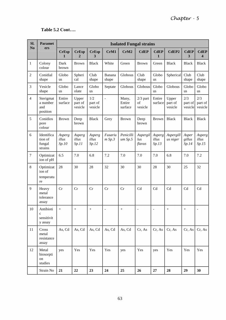
Chapter - 5
63
Table 5.2 Cont….
Sl.
No
Paramet
ers Isolated Fungal strains
CrEsp
1
CrEsp
2
CrEsp
3
CrM1 CrM2 CdEP CdEP
1
CdEP2 CdEP
3
CdEP
4
1 Colony
colour
Dark
brown
Brown Black White Green Brown Green Black Black Black
2 Conidial
shape
Globo
us
Spheri
cal
Club
shape
Banana
shape
Globous Club
shape
Globo
us
Spherical Club
shape
Club
shape
3 Vesicle
shape
Globo
us
Lance
olate
Globo
us
Septate Globous Globous Globo
us
Globous Globo
us
Globo
us
4 Sterigmat
a number
and
position
Entire
surface
Upper
part of
vesicle
1/2
part of
vesicle
- Many,
Entire
surface
2/3 part
of
vesicle
Entire
surface
Upper
part of
vesicle
2/3
part of
vesicle
2/3
part of
vesicle
5 Conidios
pore
colour
Brown Deep
brown
Black Grey Brown Deep
brown
Brown Black Black Black
6 Identifica
tion of
fungal
strains
Asperg
illus
Sp.10
Asperg
illus
Sp.11
Asperg
illus
Sp.12
Fusariu
m Sp.3
Penicilli
um Sp.5
Aspergil
lus
flavus
Asperg
illus
Sp.13
Aspergill
us niger
Asper
gillus
Sp.14
Asperg
illus
Sp.15
7 Optimizat
ion of pH
6.5 7.0 6.8 7.2 7.0 7.0 7.0 6.8 7.0 7.2
8 Optimizat
ion of
temperatu
re
28 30 28 32 30 30 28 30 25 32
9 Heavy
metal
tolerance
assay
Cr Cr Cr Cr Cr Cd Cd Cd Cd Cd
10 Antibioti
c
sensitivit
y assay
+ + + - + - - + + -
11 Cross
metal
resistance
assay
As, Cd As, Cd As, Cd As, Cd As, Cd Cr, As Cr, As Cr, As Cr, As Cr, As
12 Metal
biosorpti
on
studies
yes Yes Yes Yes yes Yes yes Yes Yes Yes
Strain No 21 22 23 24 25 26 27 28 29 30
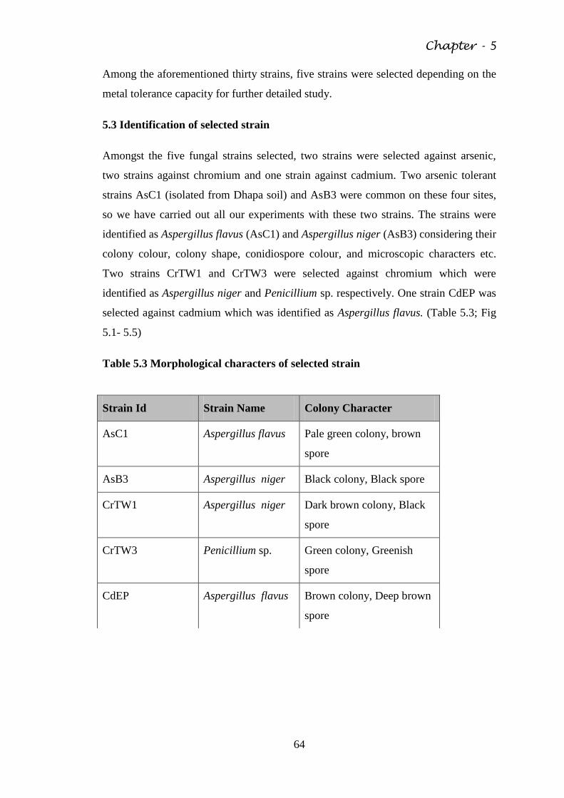
Chapter - 5
64
Among the aforementioned thirty strains, five strains were selected depending on the
metal tolerance capacity for further detailed study.
5.3 Identification of selected strain
Amongst the five fungal strains selected, two strains were selected against arsenic,
two strains against chromium and one strain against cadmium. Two arsenic tolerant
strains AsC1 (isolated from Dhapa soil) and AsB3 were common on these four sites,
so we have carried out all our experiments with these two strains. The strains were
identified as Aspergillus flavus (AsC1) and Aspergillus niger (AsB3) considering their
colony colour, colony shape, conidiospore colour, and microscopic characters etc.
Two strains CrTW1 and CrTW3 were selected against chromium which were
identified as Aspergillus niger and Penicillium sp. respectively. One strain CdEP was
selected against cadmium which was identified as Aspergillus flavus. (Table 5.3; Fig
5.1- 5.5)
Table 5.3 Morphological characters of selected strain
Strain Id Strain Name Colony Character
AsC1 Aspergillus flavus Pale green colony, brown
spore
AsB3 Aspergillus niger Black colony, Black spore
CrTW1 Aspergillus niger Dark brown colony, Black
spore
CrTW3 Penicillium sp. Green colony, Greenish
spore
CdEP Aspergillus flavus Brown colony, Deep brown
spore
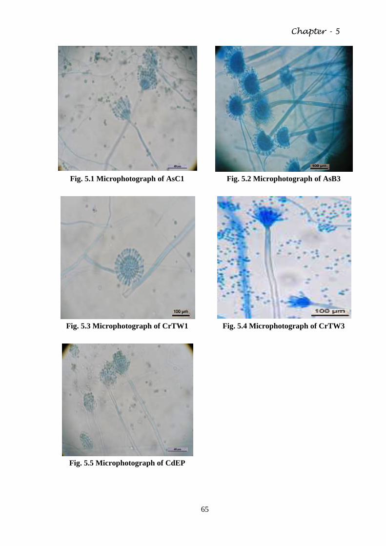
Chapter - 5
65
Fig. 5.1 Microphotograph of AsC1 Fig. 5.2 Microphotograph of AsB3
Fig. 5.3 Microphotograph of CrTW1
Fig. 5.4 Microphotograph of CrTW3
Fig. 5.5 Microphotograph of CdEP
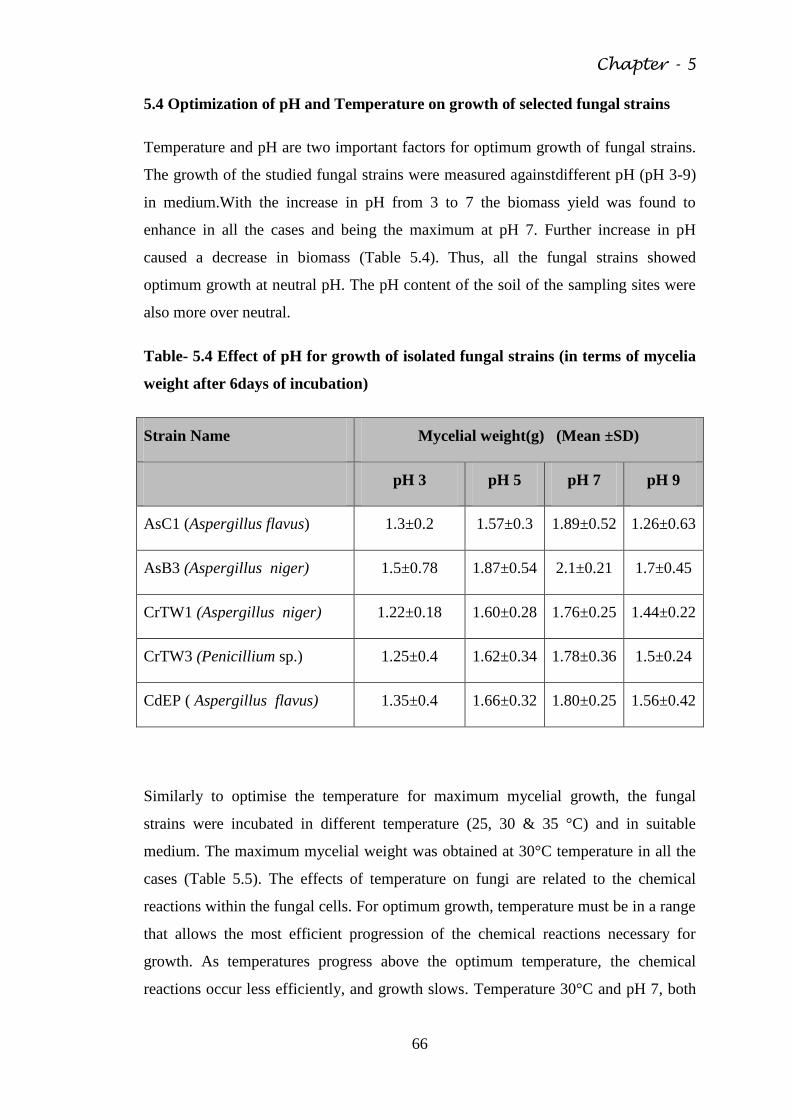
Chapter - 5
66
5.4 Optimization of pH and Temperature on growth of selected fungal strains
Temperature and pH are two important factors for optimum growth of fungal strains.
The growth of the studied fungal strains were measured againstdifferent pH (pH 3-9)
in medium.With the increase in pH from 3 to 7 the biomass yield was found to
enhance in all the cases and being the maximum at pH 7. Further increase in pH
caused a decrease in biomass (Table 5.4). Thus, all the fungal strains showed
optimum growth at neutral pH. The pH content of the soil of the sampling sites were
also more over neutral.
Table- 5.4 Effect of pH for growth of isolated fungal strains (in terms of mycelia
weight after 6days of incubation)
Strain Name Mycelial weight(g) (Mean ±SD)
pH 3 pH 5 pH 7 pH 9
AsC1 (Aspergillus flavus) 1.3±0.2 1.57±0.3 1.89±0.52 1.26±0.63
AsB3 (Aspergillus niger) 1.5±0.78 1.87±0.54 2.1±0.21 1.7±0.45
CrTW1 (Aspergillus niger) 1.22±0.18 1.60±0.28 1.76±0.25 1.44±0.22
CrTW3 (Penicillium sp.) 1.25±0.4 1.62±0.34 1.78±0.36 1.5±0.24
CdEP ( Aspergillus flavus) 1.35±0.4 1.66±0.32 1.80±0.25 1.56±0.42
Similarly to optimise the temperature for maximum mycelial growth, the fungal
strains were incubated in different temperature (25, 30 & 35 °C) and in suitable
medium. The maximum mycelial weight was obtained at 30°C temperature in all the
cases (Table 5.5). The effects of temperature on fungi are related to the chemical
reactions within the fungal cells. For optimum growth, temperature must be in a range
that allows the most efficient progression of the chemical reactions necessary for
growth. As temperatures progress above the optimum temperature, the chemical
reactions occur less efficiently, and growth slows. Temperature 30°C and pH 7, both
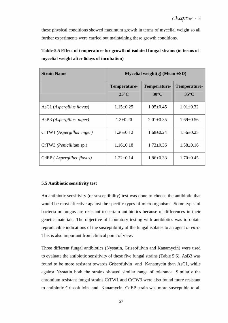
Chapter - 5
67
these physical conditions showed maximum growth in terms of mycelial weight so all
further experiments were carried out maintaining these growth conditions.
Table-5.5 Effect of temperature for growth of isolated fungal strains (in terms of
mycelial weight after 6days of incubation)
Strain Name Mycelial weight(g) (Mean ±SD)
Temperature-
25°C
Temperature-
30°C
Temperature-
35°C
AsC1 (Aspergillus flavus) 1.15±0.25 1.95±0.45 1.01±0.32
AsB3 (Aspergillus niger) 1.3±0.20 2.01±0.35 1.69±0.56
CrTW1 (Aspergillus niger) 1.26±0.12 1.68±0.24 1.56±0.25
CrTW3 (Penicillium sp.) 1.16±0.18 1.72±0.36 1.58±0.16
CdEP ( Aspergillus flavus) 1.22±0.14 1.86±0.33 1.70±0.45
5.5 Antibiotic sensitivity test
An antibiotic sensitivity (or susceptibility) test was done to choose the antibiotic that
would be most effective against the specific types of microorganism. Some types of
bacteria or fungus are resistant to certain antibiotics because of differences in their
genetic materials. The objective of laboratory testing with antibiotics was to obtain
reproducible indications of the susceptibility of the fungal isolates to an agent in vitro.
This is also important from clinical point of view.
Three different fungal antibiotics (Nystatin, Griseofulvin and Kanamycin) were used
to evaluate the antibiotic sensitivity of these five fungal strains (Table 5.6). AsB3 was
found to be more resistant towards Griseofulvin and Kanamycin than AsC1, while
against Nystatin both the strains showed similar range of tolerance. Similarly the
chromium resistant fungal strains CrTW1 and CrTW3 were also found more resistant
to antibiotic Griseofulvin and Kanamycin. CdEP strain was more susceptible to all
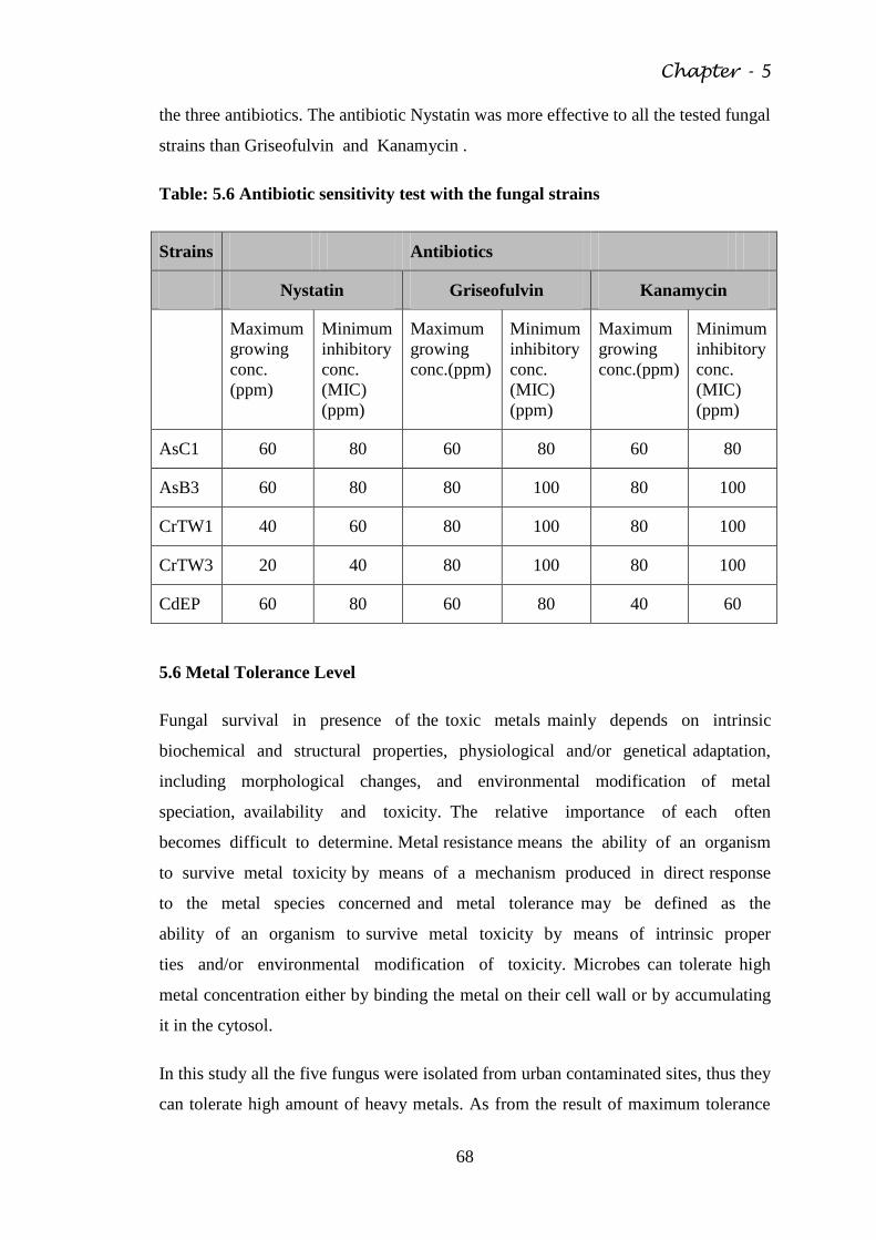
Chapter - 5
68
the three antibiotics. The antibiotic Nystatin was more effective to all the tested fungal
strains than Griseofulvin and Kanamycin .
Table: 5.6 Antibiotic sensitivity test with the fungal strains
Strains Antibiotics
Nystatin Griseofulvin Kanamycin
Maximum
growing
conc.
(ppm)
Minimum
inhibitory
conc.
(MIC)
(ppm)
Maximum
growing
conc.(ppm)
Minimum
inhibitory
conc.
(MIC)
(ppm)
Maximum
growing
conc.(ppm)
Minimum
inhibitory
conc.
(MIC)
(ppm)
AsC1 60 80 60 80 60 80
AsB3 60 80 80 100 80 100
CrTW1 40 60 80 100 80 100
CrTW3 20 40 80 100 80 100
CdEP 60 80 60 80 40 60
5.6 Metal Tolerance Level
Fungal survival in presence of the toxic metals mainly depends on intrinsic
biochemical and structural properties, physiological and/or genetical adaptation,
including morphological changes, and environmental modification of metal
speciation, availability and toxicity. The relative importance of each often
becomes difficult to determine. Metal resistance means the ability of an organism
to survive metal toxicity by means of a mechanism produced in direct response
to the metal species concerned and metal tolerance may be defined as the
ability of an organism to survive metal toxicity by means of intrinsic proper
ties and/or environmental modification of toxicity. Microbes can tolerate high
metal concentration either by binding the metal on their cell wall or by accumulating
it in the cytosol.
In this study all the five fungus were isolated from urban contaminated sites, thus they
can tolerate high amount of heavy metals. As from the result of maximum tolerance
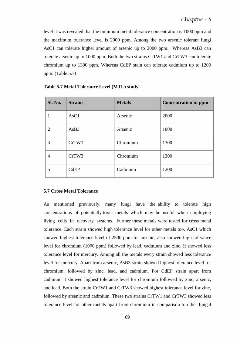
Chapter - 5
69
level it was revealed that the minimum metal tolerance concentration is 1000 ppm and
the maximum tolerance level is 2000 ppm. Among the two arsenic tolerant fungi
AsC1 can tolerate higher amount of arsenic up to 2000 ppm. Whereas AsB3 can
tolerate arsenic up to 1000 ppm. Both the two strains CrTW1 and CrTW3 can tolerate
chromium up to 1300 ppm. Whereas CdEP stain can tolerate cadmium up to 1200
ppm. (Table 5.7)
Table 5.7 Metal Tolerance Level (MTL) study
Sl. No. Strains Metals Concentration in ppm
1 AsC1 Arsenic 2000
2 AsB3 Arsenic 1000
3 CrTW1 Chromium 1300
4 CrTW3 Chromium 1300
5 CdEP Cadmium 1200
5.7 Cross Metal Tolerance
As mentioned previously, many fungi have the ability to tolerate high
concentrations of potentially toxic metals which may be useful when employing
living cells in recovery systems. Further these metals were tested for cross metal
tolerance. Each strain showed high tolerance level for other metals too. AsC1 which
showed highest tolerance level of 2500 ppm for arsenic, also showed high tolerance
level for chromium (1000 ppm) followed by lead, cadmium and zinc. It showed less
tolerance level for mercury. Among all the metals every strain showed less tolerance
level for mercury. Apart from arsenic, AsB3 strain showed highest tolerance level for
chromium, followed by zinc, lead, and cadmium. For CdEP strain apart from
cadmium it showed highest tolerance level for chromium followed by zinc, arsenic,
and lead. Both the strain CrTW1 and CrTW3 showed highest tolerance level for zinc,
followed by arsenic and cadmium. These two strains CrTW1 and CrTW3 showed less
tolerance level for other metals apart from chromium in comparison to other fungal
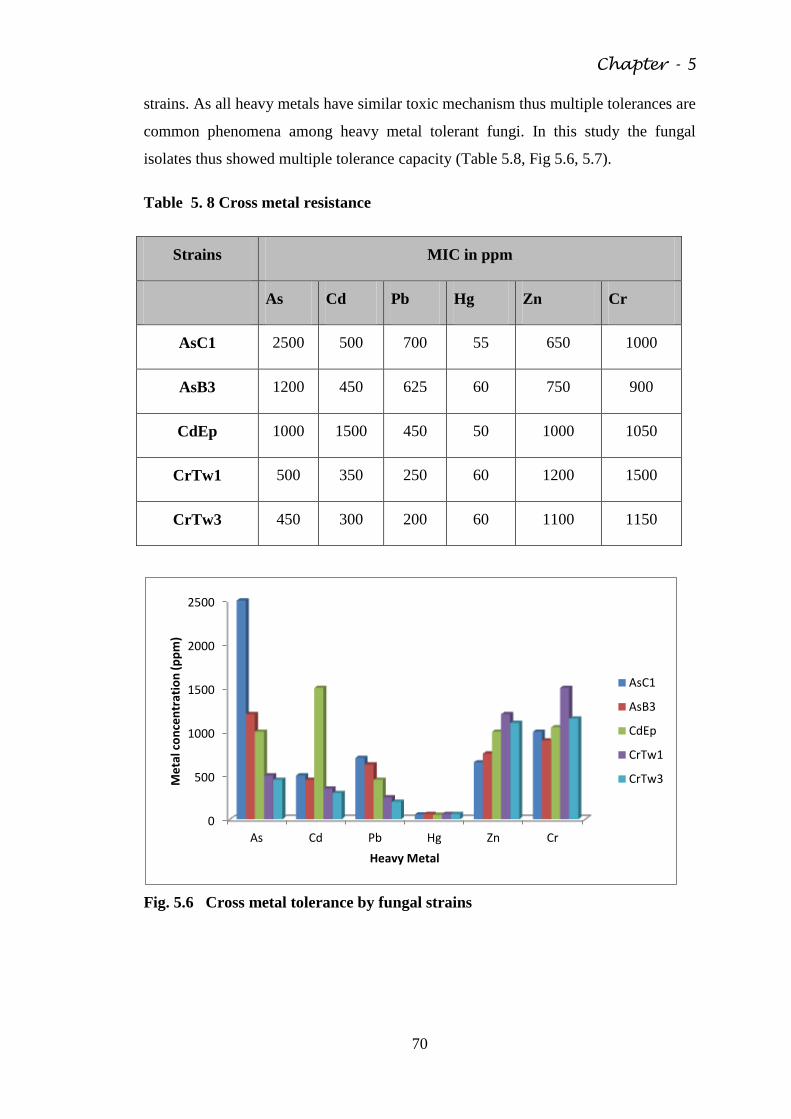
Chapter - 5
70
strains. As all heavy metals have similar toxic mechanism thus multiple tolerances are
common phenomena among heavy metal tolerant fungi. In this study the fungal
isolates thus showed multiple tolerance capacity (Table 5.8, Fig 5.6, 5.7).
Table 5. 8 Cross metal resistance
Strains MIC in ppm
As Cd Pb Hg Zn Cr
AsC1 2500 500 700 55 650 1000
AsB3 1200 450 625 60 750 900
CdEp 1000 1500 450 50 1000 1050
CrTw1 500 350 250 60 1200 1500
CrTw3 450 300 200 60 1100 1150
Fig. 5.6 Cross metal tolerance by fungal strains
0
500
1000
1500
2000
2500
As Cd Pb Hg Zn Cr
Me
tal c
on
cen
trat
ion
(p
pm
)
Heavy Metal
AsC1
AsB3
CdEp
CrTw1
CrTw3
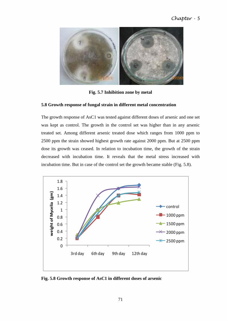
Chapter - 5
71
Fig. 5.7 Inhibition zone by metal
5.8 Growth response of fungal strain in different metal concentration
The growth response of AsC1 was tested against different doses of arsenic and one set
was kept as control. The growth in the control set was higher than in any arsenic
treated set. Among different arsenic treated dose which ranges from 1000 ppm to
2500 ppm the strain showed highest growth rate against 2000 ppm. But at 2500 ppm
dose its growth was ceased. In relation to incubation time, the growth of the strain
decreased with incubation time. It reveals that the metal stress increased with
incubation time. But in case of the control set the growth became stable (Fig. 5.8).
0
0.2
0.4
0.6
0.8
1
1.2
1.4
1.6
1.8
3rd day 6th day 9th day 12th day
we
igh
t o
f M
yce
lia (
gm)
control
1000 ppm
1500 ppm
2000 ppm
2500 ppm
Fig. 5.8 Growth response of AsC1 in different doses of arsenic
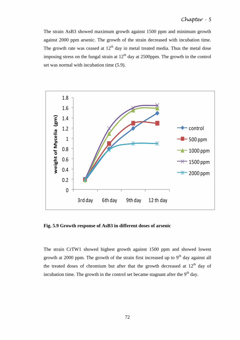
Chapter - 5
72
The strain AsB3 showed maximum growth against 1500 ppm and minimum growth
against 2000 ppm arsenic. The growth of the strain decreased with incubation time.
The growth rate was ceased at 12th
day in metal treated media. Thus the metal dose
imposing stress on the fungal strain at 12th
day at 2500ppm. The growth in the control
set was normal with incubation time (5.9).
0
0.2
0.4
0.6
0.8
1
1.2
1.4
1.6
1.8
3rd day 6th day 9th day 12 th day
we
igh
t o
f M
yce
lia
(g
m)
control
500 ppm
1000 ppm
1500 ppm
2000 ppm
Fig. 5.9 Growth response of AsB3 in different doses of arsenic
The strain CrTW1 showed highest growth against 1500 ppm and showed lowest
growth at 2000 ppm. The growth of the strain first increased up to 9th
day against all
the treated doses of chromium but after that the growth decreased at 12th
day of
incubation time. The growth in the control set became stagnant after the 9th
day.
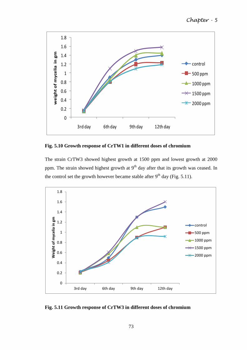
Chapter - 5
73
0
0.2
0.4
0.6
0.8
1
1.2
1.4
1.6
1.8
3rd day 6th day 9th day 12th day
we
igh
t o
f m
yce
lia
in
gm
control
500 ppm
1000 ppm
1500 ppm
2000 ppm
Fig. 5.10 Growth response of CrTW1 in different doses of chromium
The strain CrTW3 showed highest growth at 1500 ppm and lowest growth at 2000
ppm. The strain showed highest growth at 9th
day after that its growth was ceased. In
the control set the growth however became stable after 9th
day (Fig. 5.11).
Fig. 5.11 Growth response of CrTW3 in different doses of chromium
0
0.2
0.4
0.6
0.8
1
1.2
1.4
1.6
1.8
3rd day 6th day 9th day 12th day
We
igh
t o
f m
yce
lia in
gm
control
500 ppm
1000 ppm
1500 ppm
2000 ppm
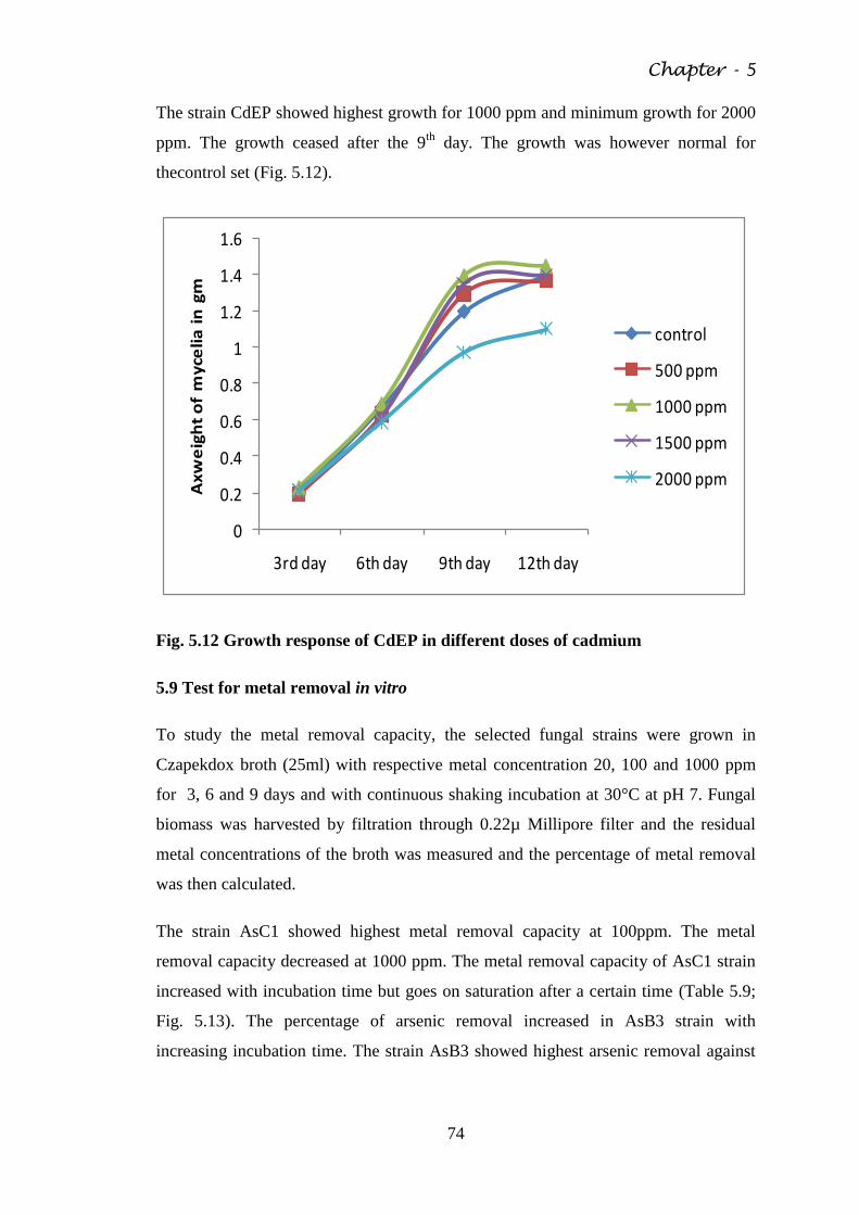
Chapter - 5
74
The strain CdEP showed highest growth for 1000 ppm and minimum growth for 2000
ppm. The growth ceased after the 9th
day. The growth was however normal for
thecontrol set (Fig. 5.12).
0
0.2
0.4
0.6
0.8
1
1.2
1.4
1.6
3rd day 6th day 9th day 12th day
Ax
we
igh
t o
f m
yce
lia
in
gm
control
500 ppm
1000 ppm
1500 ppm
2000 ppm
Fig. 5.12 Growth response of CdEP in different doses of cadmium
5.9 Test for metal removal in vitro
To study the metal removal capacity, the selected fungal strains were grown in
Czapekdox broth (25ml) with respective metal concentration 20, 100 and 1000 ppm
for 3, 6 and 9 days and with continuous shaking incubation at 30°C at pH 7. Fungal
biomass was harvested by filtration through 0.22µ Millipore filter and the residual
metal concentrations of the broth was measured and the percentage of metal removal
was then calculated.
The strain AsC1 showed highest metal removal capacity at 100ppm. The metal
removal capacity decreased at 1000 ppm. The metal removal capacity of AsC1 strain
increased with incubation time but goes on saturation after a certain time (Table 5.9;
Fig. 5.13). The percentage of arsenic removal increased in AsB3 strain with
increasing incubation time. The strain AsB3 showed highest arsenic removal against
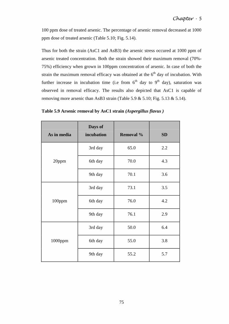
Chapter - 5
75
100 ppm dose of treated arsenic. The percentage of arsenic removal decreased at 1000
ppm dose of treated arsenic (Table 5.10; Fig. 5.14).
Thus for both the strain (AsC1 and AsB3) the arsenic stress occured at 1000 ppm of
arsenic treated concentration. Both the strain showed their maximum removal (70%-
75%) efficiency when grown in 100ppm concentration of arsenic. In case of both the
strain the maximum removal efficacy was obtained at the 6th
day of incubation. With
further increase in incubation time (i.e from 6th
day to 9th
day), saturation was
observed in removal efficacy. The results also depicted that AsC1 is capable of
removing more arsenic than AsB3 strain (Table 5.9 & 5.10; Fig. 5.13 & 5.14).
Table 5.9 Arsenic removal by AsC1 strain (Aspergillus flavus )
As in media
Days of
incubation Removal % SD
20ppm
3rd day 65.0 2.2
6th day 70.0 4.3
9th day 70.1 3.6
100ppm
3rd day 73.1 3.5
6th day 76.0 4.2
9th day 76.1 2.9
1000ppm
3rd day 50.0 6.4
6th day 55.0 3.8
9th day 55.2 5.7
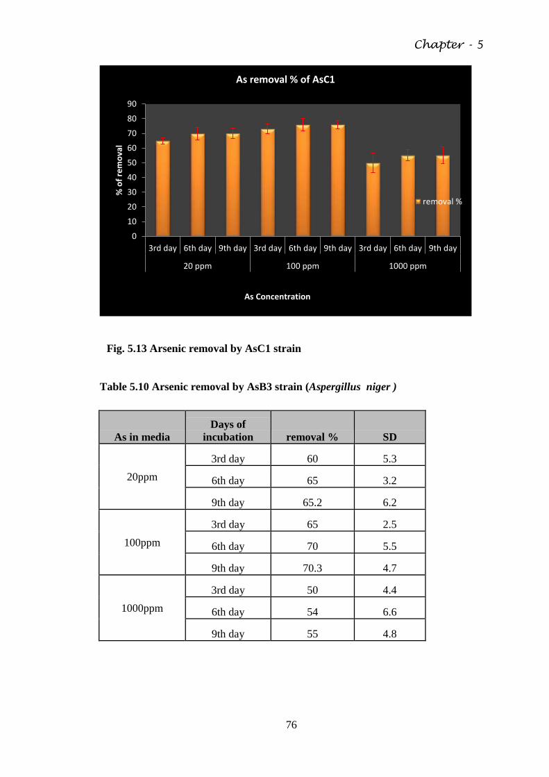
Chapter - 5
76
Fig. 5.13 Arsenic removal by AsC1 strain
Table 5.10 Arsenic removal by AsB3 strain (Aspergillus niger )
As in media
Days of
incubation removal % SD
20ppm
3rd day 60 5.3
6th day 65 3.2
9th day 65.2 6.2
100ppm
3rd day 65 2.5
6th day 70 5.5
9th day 70.3 4.7
1000ppm
3rd day 50 4.4
6th day 54 6.6
9th day 55 4.8
0
10
20
30
40
50
60
70
80
90
3rd day 6th day 9th day 3rd day 6th day 9th day 3rd day 6th day 9th day
20 ppm 100 ppm 1000 ppm
% o
f re
mo
val
As Concentration
As removal % of AsC1
removal %
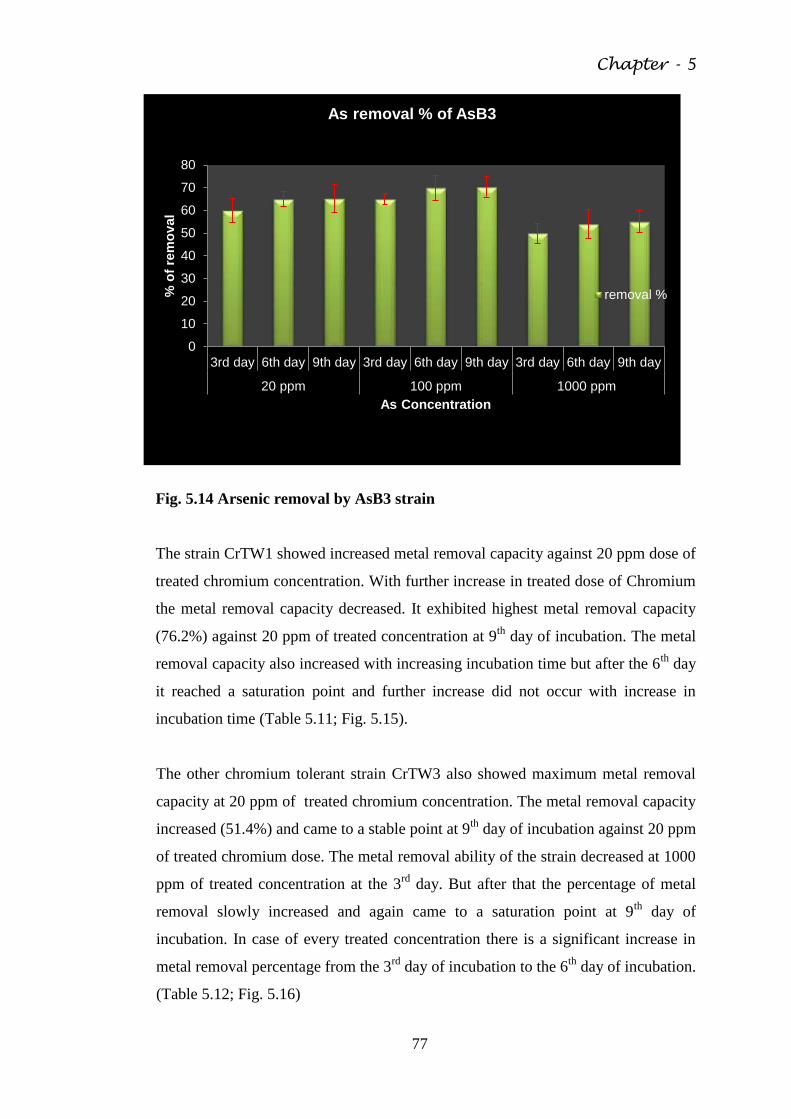
Chapter - 5
77
Fig. 5.14 Arsenic removal by AsB3 strain
The strain CrTW1 showed increased metal removal capacity against 20 ppm dose of
treated chromium concentration. With further increase in treated dose of Chromium
the metal removal capacity decreased. It exhibited highest metal removal capacity
(76.2%) against 20 ppm of treated concentration at 9th
day of incubation. The metal
removal capacity also increased with increasing incubation time but after the 6th
day
it reached a saturation point and further increase did not occur with increase in
incubation time (Table 5.11; Fig. 5.15).
The other chromium tolerant strain CrTW3 also showed maximum metal removal
capacity at 20 ppm of treated chromium concentration. The metal removal capacity
increased (51.4%) and came to a stable point at 9th
day of incubation against 20 ppm
of treated chromium dose. The metal removal ability of the strain decreased at 1000
ppm of treated concentration at the 3rd
day. But after that the percentage of metal
removal slowly increased and again came to a saturation point at 9th
day of
incubation. In case of every treated concentration there is a significant increase in
metal removal percentage from the 3rd
day of incubation to the 6th
day of incubation.
(Table 5.12; Fig. 5.16)
0
10
20
30
40
50
60
70
80
3rd day 6th day 9th day 3rd day 6th day 9th day 3rd day 6th day 9th day
20 ppm 100 ppm 1000 ppm
% o
f re
mo
val
As Concentration
As removal % of AsB3
removal %
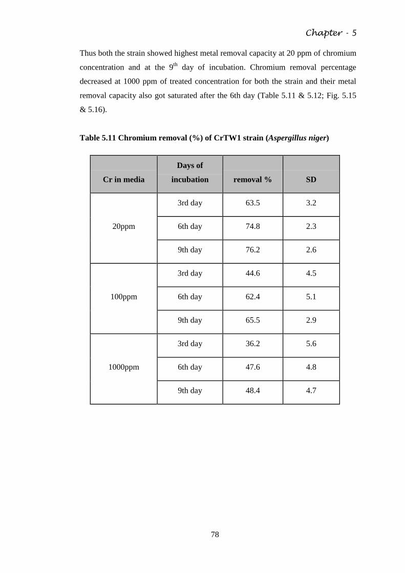
Chapter - 5
78
Thus both the strain showed highest metal removal capacity at 20 ppm of chromium
concentration and at the 9th
day of incubation. Chromium removal percentage
decreased at 1000 ppm of treated concentration for both the strain and their metal
removal capacity also got saturated after the 6th day (Table 5.11 & 5.12; Fig. 5.15
& 5.16).
Table 5.11 Chromium removal (%) of CrTW1 strain (Aspergillus niger)
Cr in media
Days of
incubation removal % SD
20ppm
3rd day 63.5 3.2
6th day 74.8 2.3
9th day 76.2 2.6
100ppm
3rd day 44.6 4.5
6th day 62.4 5.1
9th day 65.5 2.9
1000ppm
3rd day 36.2 5.6
6th day 47.6 4.8
9th day 48.4 4.7
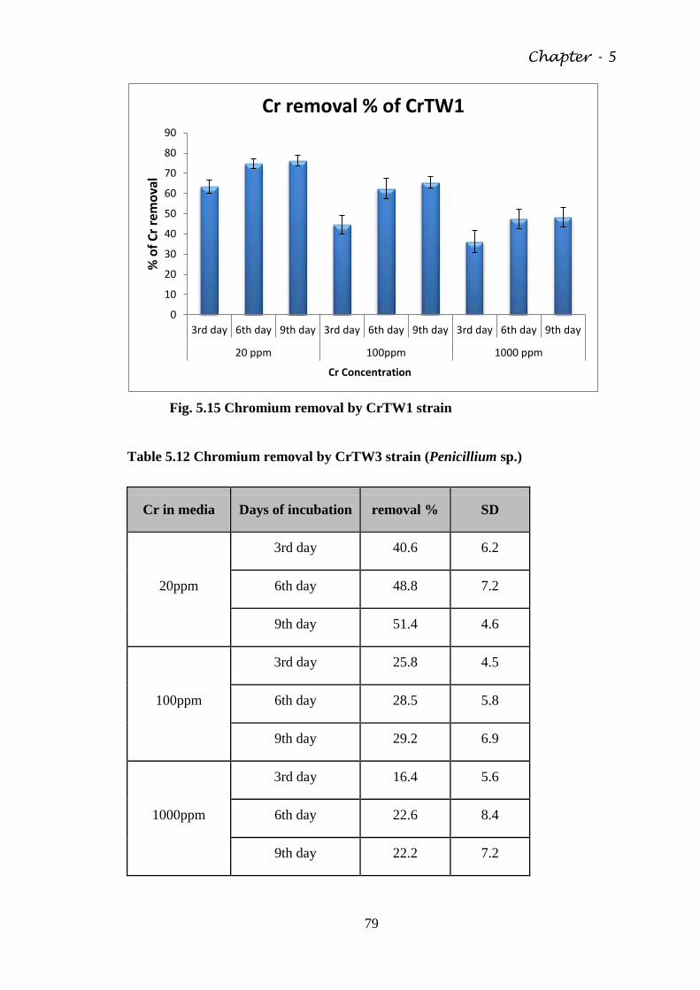
Chapter - 5
79
Fig. 5.15 Chromium removal by CrTW1 strain
Table 5.12 Chromium removal by CrTW3 strain (Penicillium sp.)
Cr in media Days of incubation removal % SD
20ppm
3rd day 40.6 6.2
6th day 48.8 7.2
9th day 51.4 4.6
100ppm
3rd day 25.8 4.5
6th day 28.5 5.8
9th day 29.2 6.9
1000ppm
3rd day 16.4 5.6
6th day 22.6 8.4
9th day 22.2 7.2
0
10
20
30
40
50
60
70
80
90
3rd day 6th day 9th day 3rd day 6th day 9th day 3rd day 6th day 9th day
20 ppm 100ppm 1000 ppm
% o
f C
r re
mo
val
Cr Concentration
Cr removal % of CrTW1
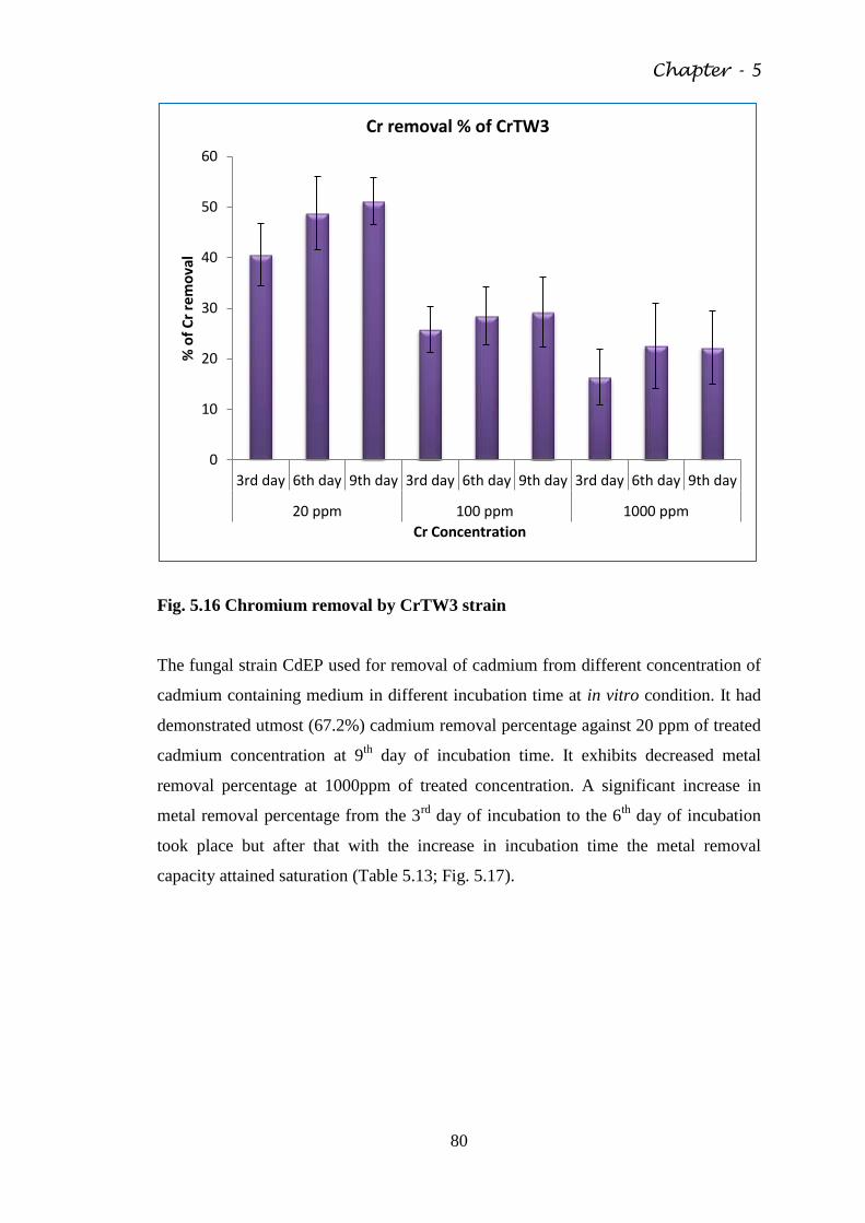
Chapter - 5
80
Fig. 5.16 Chromium removal by CrTW3 strain
The fungal strain CdEP used for removal of cadmium from different concentration of
cadmium containing medium in different incubation time at in vitro condition. It had
demonstrated utmost (67.2%) cadmium removal percentage against 20 ppm of treated
cadmium concentration at 9th
day of incubation time. It exhibits decreased metal
removal percentage at 1000ppm of treated concentration. A significant increase in
metal removal percentage from the 3rd
day of incubation to the 6th
day of incubation
took place but after that with the increase in incubation time the metal removal
capacity attained saturation (Table 5.13; Fig. 5.17).
0
10
20
30
40
50
60
3rd day 6th day 9th day 3rd day 6th day 9th day 3rd day 6th day 9th day
20 ppm 100 ppm 1000 ppm
% o
f C
r re
mo
val
Cr Concentration
Cr removal % of CrTW3
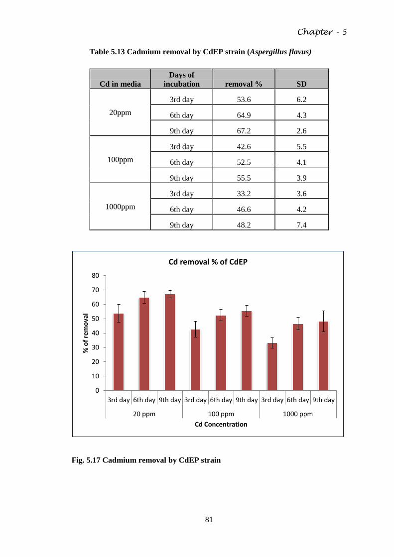
Chapter - 5
81
Table 5.13 Cadmium removal by CdEP strain (Aspergillus flavus)
Cd in media
Days of
incubation removal % SD
20ppm
3rd day 53.6 6.2
6th day 64.9 4.3
9th day 67.2 2.6
100ppm
3rd day 42.6 5.5
6th day 52.5 4.1
9th day 55.5 3.9
1000ppm
3rd day 33.2 3.6
6th day 46.6 4.2
9th day 48.2 7.4
Fig. 5.17 Cadmium removal by CdEP strain
0
10
20
30
40
50
60
70
80
3rd day 6th day 9th day 3rd day 6th day 9th day 3rd day 6th day 9th day
20 ppm 100 ppm 1000 ppm
% o
f re
mo
val
Cd Concentration
Cd removal % of CdEP
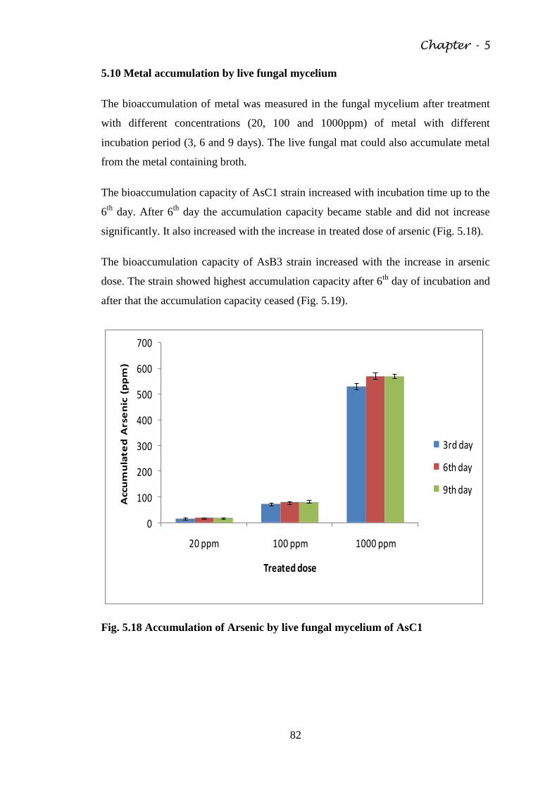
Chapter - 5
82
5.10 Metal accumulation by live fungal mycelium
The bioaccumulation of metal was measured in the fungal mycelium after treatment
with different concentrations (20, 100 and 1000ppm) of metal with different
incubation period (3, 6 and 9 days). The live fungal mat could also accumulate metal
from the metal containing broth.
The bioaccumulation capacity of AsC1 strain increased with incubation time up to the
6th
day. After 6th
day the accumulation capacity became stable and did not increase
significantly. It also increased with the increase in treated dose of arsenic (Fig. 5.18).
The bioaccumulation capacity of AsB3 strain increased with the increase in arsenic
dose. The strain showed highest accumulation capacity after 6th
day of incubation and
after that the accumulation capacity ceased (Fig. 5.19).
0
100
200
300
400
500
600
700
20 ppm 100 ppm 1000 ppm
Acc
um
ula
ted
Ars
en
ic (
pp
m)
Treated dose
3rd day
6th day
9th day
Fig. 5.18 Accumulation of Arsenic by live fungal mycelium of AsC1
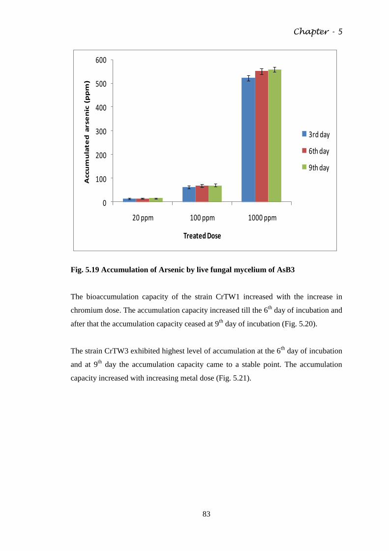
Chapter - 5
83
0
100
200
300
400
500
600
20 ppm 100 ppm 1000 ppm
Accu
mu
late
d a
rse
nic
(p
pm
)
Treated Dose
3rd day
6th day
9th day
Fig. 5.19 Accumulation of Arsenic by live fungal mycelium of AsB3
The bioaccumulation capacity of the strain CrTW1 increased with the increase in
chromium dose. The accumulation capacity increased till the 6th
day of incubation and
after that the accumulation capacity ceased at 9th
day of incubation (Fig. 5.20).
The strain CrTW3 exhibited highest level of accumulation at the 6th
day of incubation
and at 9th
day the accumulation capacity came to a stable point. The accumulation
capacity increased with increasing metal dose (Fig. 5.21).
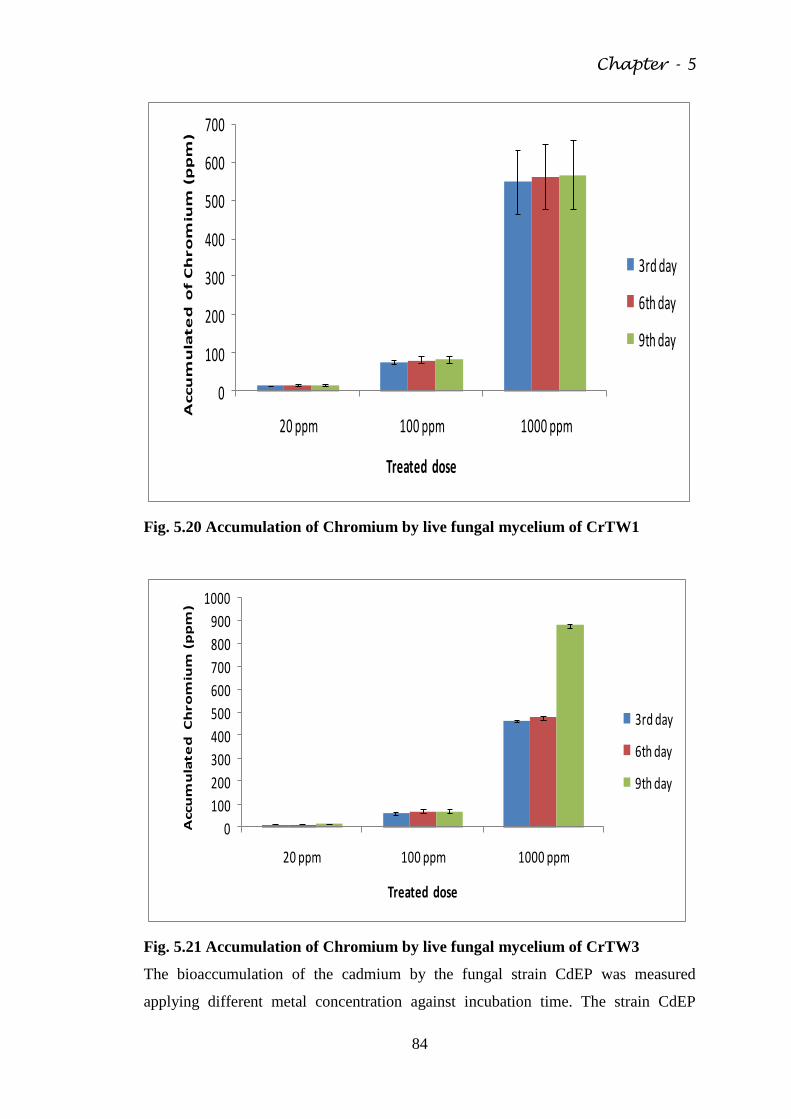
Chapter - 5
84
0
100
200
300
400
500
600
700
20 ppm 100 ppm 1000 ppm
Accu
mu
late
d o
f C
hro
miu
m (
pp
m)
Treated dose
3rd day
6th day
9th day
Fig. 5.20 Accumulation of Chromium by live fungal mycelium of CrTW1
0
100
200
300
400
500
600
700
800
900
1000
20 ppm 100 ppm 1000 ppm
Accu
mu
late
d C
hro
miu
m (
pp
m)
Treated dose
3rd day
6th day
9th day
Fig. 5.21 Accumulation of Chromium by live fungal mycelium of CrTW3
The bioaccumulation of the cadmium by the fungal strain CdEP was measured
applying different metal concentration against incubation time. The strain CdEP
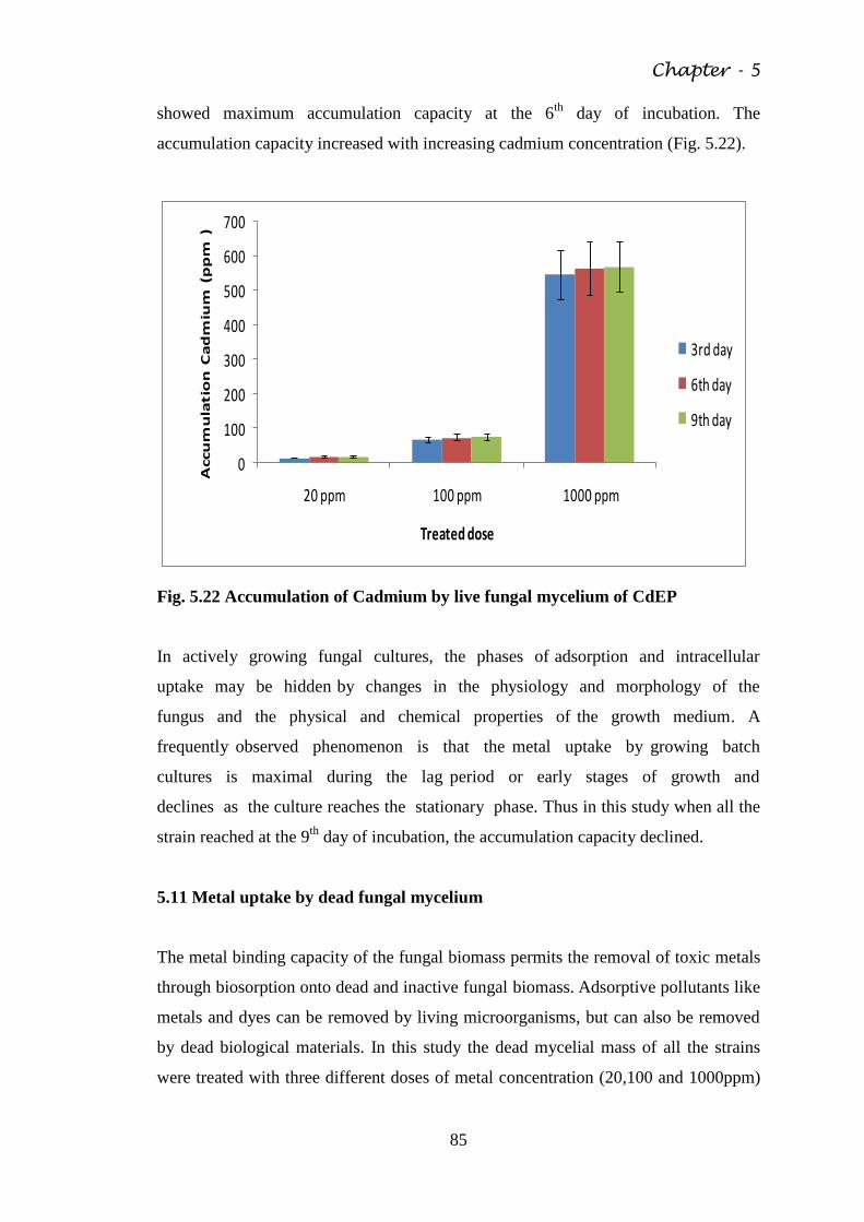
Chapter - 5
85
showed maximum accumulation capacity at the 6th
day of incubation. The
accumulation capacity increased with increasing cadmium concentration (Fig. 5.22).
0
100
200
300
400
500
600
700
20 ppm 100 ppm 1000 ppm
Accu
mu
latio
n C
ad
miu
m (
pp
m )
Treated dose
3rd day
6th day
9th day
Fig. 5.22 Accumulation of Cadmium by live fungal mycelium of CdEP
In actively growing fungal cultures, the phases of adsorption and intracellular
uptake may be hidden by changes in the physiology and morphology of the
fungus and the physical and chemical properties of the growth medium. A
frequently observed phenomenon is that the metal uptake by growing batch
cultures is maximal during the lag period or early stages of growth and
declines as the culture reaches the stationary phase. Thus in this study when all the
strain reached at the 9th
day of incubation, the accumulation capacity declined.
5.11 Metal uptake by dead fungal mycelium
The metal binding capacity of the fungal biomass permits the removal of toxic metals
through biosorption onto dead and inactive fungal biomass. Adsorptive pollutants like
metals and dyes can be removed by living microorganisms, but can also be removed
by dead biological materials. In this study the dead mycelial mass of all the strains
were treated with three different doses of metal concentration (20,100 and 1000ppm)
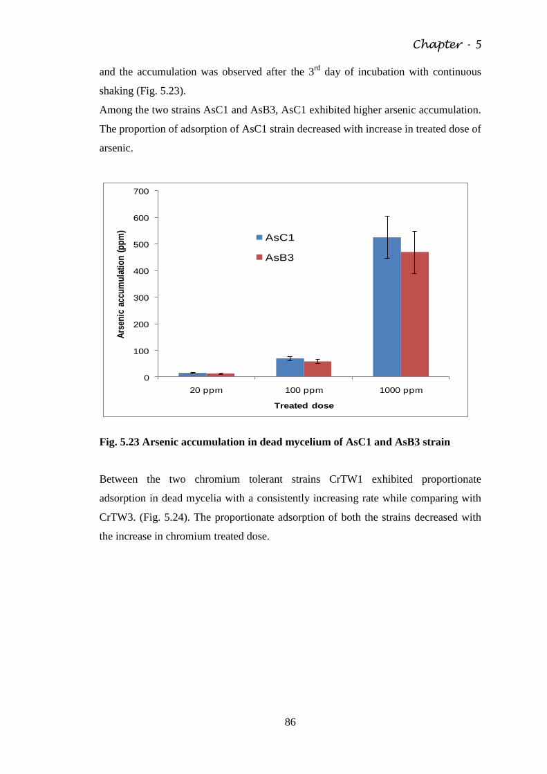
Chapter - 5
86
and the accumulation was observed after the 3rd
day of incubation with continuous
shaking (Fig. 5.23).
Among the two strains AsC1 and AsB3, AsC1 exhibited higher arsenic accumulation.
The proportion of adsorption of AsC1 strain decreased with increase in treated dose of
arsenic.
0
100
200
300
400
500
600
700
20 ppm 100 ppm 1000 ppm
Ars
enic
acc
um
ula
tio
n (
pp
m)
Treated dose
AsC1
AsB3
Fig. 5.23 Arsenic accumulation in dead mycelium of AsC1 and AsB3 strain
Between the two chromium tolerant strains CrTW1 exhibited proportionate
adsorption in dead mycelia with a consistently increasing rate while comparing with
CrTW3. (Fig. 5.24). The proportionate adsorption of both the strains decreased with
the increase in chromium treated dose.
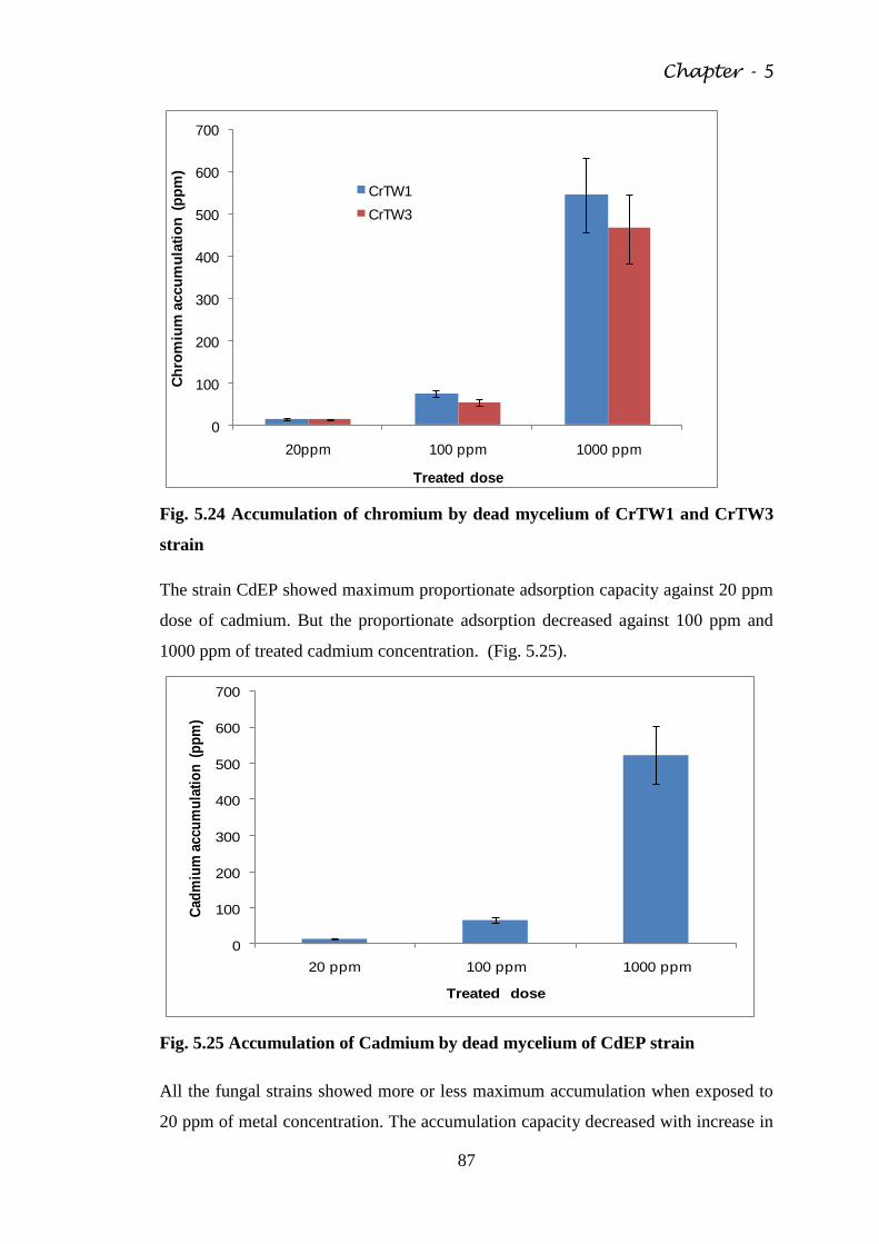
Chapter - 5
87
0
100
200
300
400
500
600
700
20ppm 100 ppm 1000 ppm
Ch
rom
ium
accu
mu
lati
on
(p
pm
)
Treated dose
CrTW1
CrTW3
Fig. 5.24 Accumulation of chromium by dead mycelium of CrTW1 and CrTW3
strain
The strain CdEP showed maximum proportionate adsorption capacity against 20 ppm
dose of cadmium. But the proportionate adsorption decreased against 100 ppm and
1000 ppm of treated cadmium concentration. (Fig. 5.25).
0
100
200
300
400
500
600
700
20 ppm 100 ppm 1000 ppm
Cad
miu
m a
ccu
mu
lati
on
(p
pm
)
Treated dose
Fig. 5.25 Accumulation of Cadmium by dead mycelium of CdEP strain
All the fungal strains showed more or less maximum accumulation when exposed to
20 ppm of metal concentration. The accumulation capacity decreased with increase in
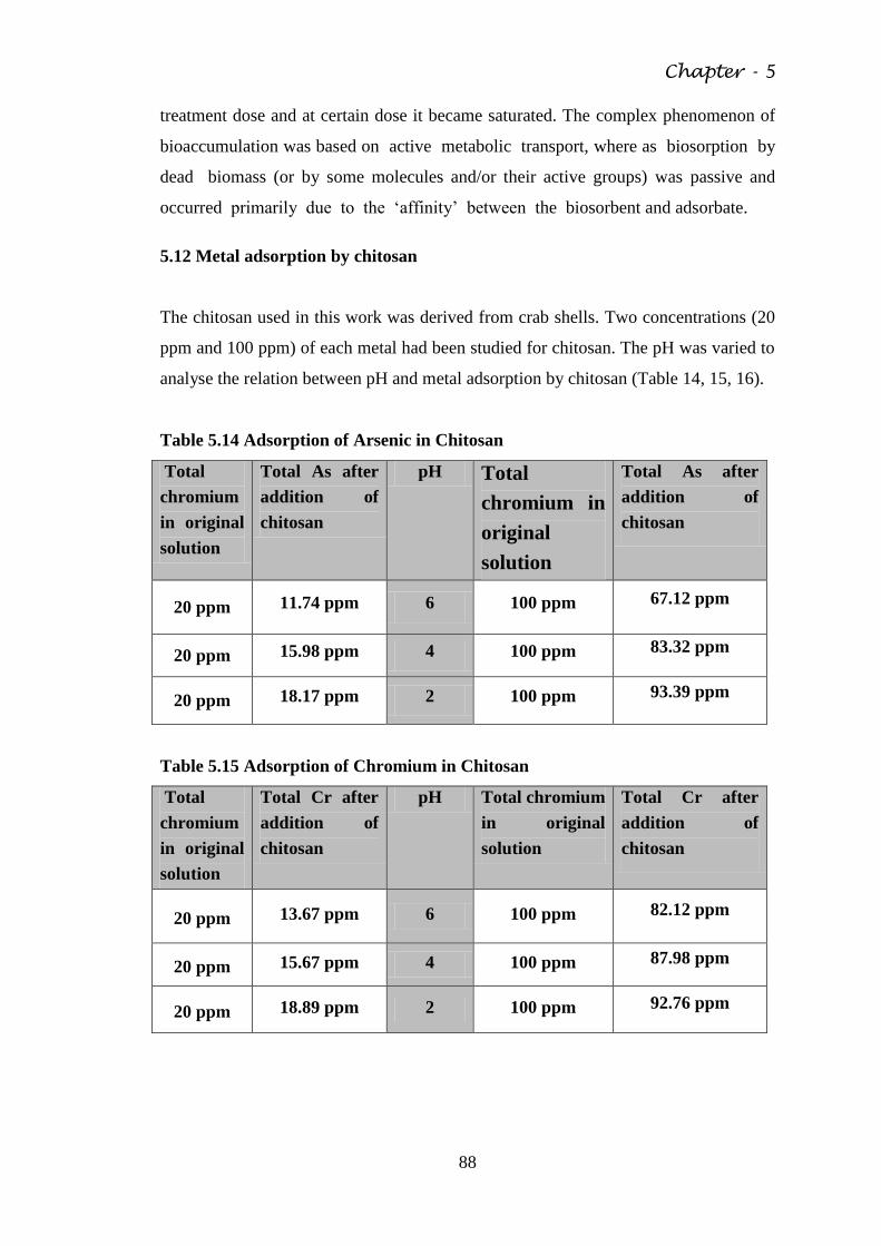
Chapter - 5
88
treatment dose and at certain dose it became saturated. The complex phenomenon of
bioaccumulation was based on active metabolic transport, where as biosorption by
dead biomass (or by some molecules and/or their active groups) was passive and
occurred primarily due to the ‗affinity‘ between the biosorbent and adsorbate.
5.12 Metal adsorption by chitosan
The chitosan used in this work was derived from crab shells. Two concentrations (20
ppm and 100 ppm) of each metal had been studied for chitosan. The pH was varied to
analyse the relation between pH and metal adsorption by chitosan (Table 14, 15, 16).
Table 5.14 Adsorption of Arsenic in Chitosan
Total
chromium
in original
solution
Total As after
addition of
chitosan
pH Total
chromium in
original
solution
Total As after
addition of
chitosan
20 ppm 11.74 ppm 6 100 ppm 67.12 ppm
20 ppm 15.98 ppm 4 100 ppm 83.32 ppm
20 ppm 18.17 ppm 2 100 ppm 93.39 ppm
Table 5.15 Adsorption of Chromium in Chitosan
Total
chromium
in original
solution
Total Cr after
addition of
chitosan
pH Total chromium
in original
solution
Total Cr after
addition of
chitosan
20 ppm 13.67 ppm 6 100 ppm 82.12 ppm
20 ppm 15.67 ppm 4 100 ppm 87.98 ppm
20 ppm 18.89 ppm 2 100 ppm 92.76 ppm
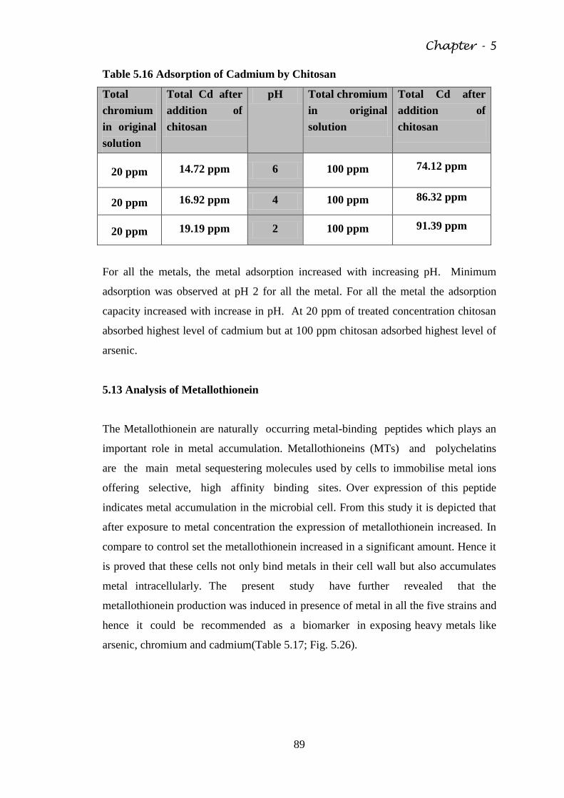
Chapter - 5
89
Table 5.16 Adsorption of Cadmium by Chitosan
Total
chromium
in original
solution
Total Cd after
addition of
chitosan
pH Total chromium
in original
solution
Total Cd after
addition of
chitosan
20 ppm 14.72 ppm 6 100 ppm 74.12 ppm
20 ppm 16.92 ppm 4 100 ppm 86.32 ppm
20 ppm 19.19 ppm 2 100 ppm 91.39 ppm
For all the metals, the metal adsorption increased with increasing pH. Minimum
adsorption was observed at pH 2 for all the metal. For all the metal the adsorption
capacity increased with increase in pH. At 20 ppm of treated concentration chitosan
absorbed highest level of cadmium but at 100 ppm chitosan adsorbed highest level of
arsenic.
5.13 Analysis of Metallothionein
The Metallothionein are naturally occurring metal-binding peptides which plays an
important role in metal accumulation. Metallothioneins (MTs) and polychelatins
are the main metal sequestering molecules used by cells to immobilise metal ions
offering selective, high affinity binding sites. Over expression of this peptide
indicates metal accumulation in the microbial cell. From this study it is depicted that
after exposure to metal concentration the expression of metallothionein increased. In
compare to control set the metallothionein increased in a significant amount. Hence it
is proved that these cells not only bind metals in their cell wall but also accumulates
metal intracellularly. The present study have further revealed that the
metallothionein production was induced in presence of metal in all the five strains and
hence it could be recommended as a biomarker in exposing heavy metals like
arsenic, chromium and cadmium(Table 5.17; Fig. 5.26).
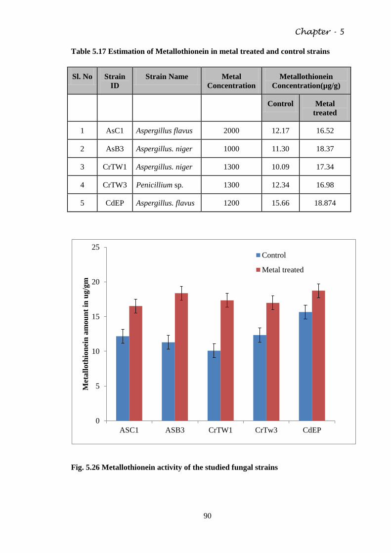
Chapter - 5
90
Table 5.17 Estimation of Metallothionein in metal treated and control strains
Sl. No Strain
ID Strain Name Metal
Concentration Metallothionein
Concentration(µg/g)
Control Metal
treated
1 AsC1 Aspergillus flavus 2000 12.17 16.52
2 AsB3 Aspergillus. niger 1000 11.30 18.37
3 CrTW1 Aspergillus. niger 1300 10.09 17.34
4 CrTW3 Penicillium sp. 1300 12.34 16.98
5 CdEP Aspergillus. flavus 1200 15.66 18.874
Fig. 5.26 Metallothionein activity of the studied fungal strains
0
5
10
15
20
25
ASC1 ASB3 CrTW1 CrTw3 CdEP
Met
all
oth
ion
ein
am
ou
nt
in u
g/g
m
Control
Metal treated
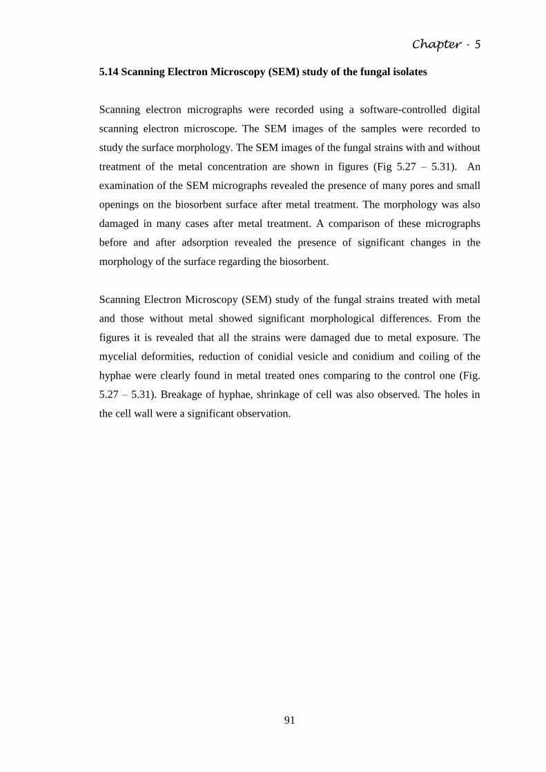
Chapter - 5
91
5.14 Scanning Electron Microscopy (SEM) study of the fungal isolates
Scanning electron micrographs were recorded using a software-controlled digital
scanning electron microscope. The SEM images of the samples were recorded to
study the surface morphology. The SEM images of the fungal strains with and without
treatment of the metal concentration are shown in figures (Fig 5.27 – 5.31). An
examination of the SEM micrographs revealed the presence of many pores and small
openings on the biosorbent surface after metal treatment. The morphology was also
damaged in many cases after metal treatment. A comparison of these micrographs
before and after adsorption revealed the presence of significant changes in the
morphology of the surface regarding the biosorbent.
Scanning Electron Microscopy (SEM) study of the fungal strains treated with metal
and those without metal showed significant morphological differences. From the
figures it is revealed that all the strains were damaged due to metal exposure. The
mycelial deformities, reduction of conidial vesicle and conidium and coiling of the
hyphae were clearly found in metal treated ones comparing to the control one (Fig.
5.27 – 5.31). Breakage of hyphae, shrinkage of cell was also observed. The holes in
the cell wall were a significant observation.
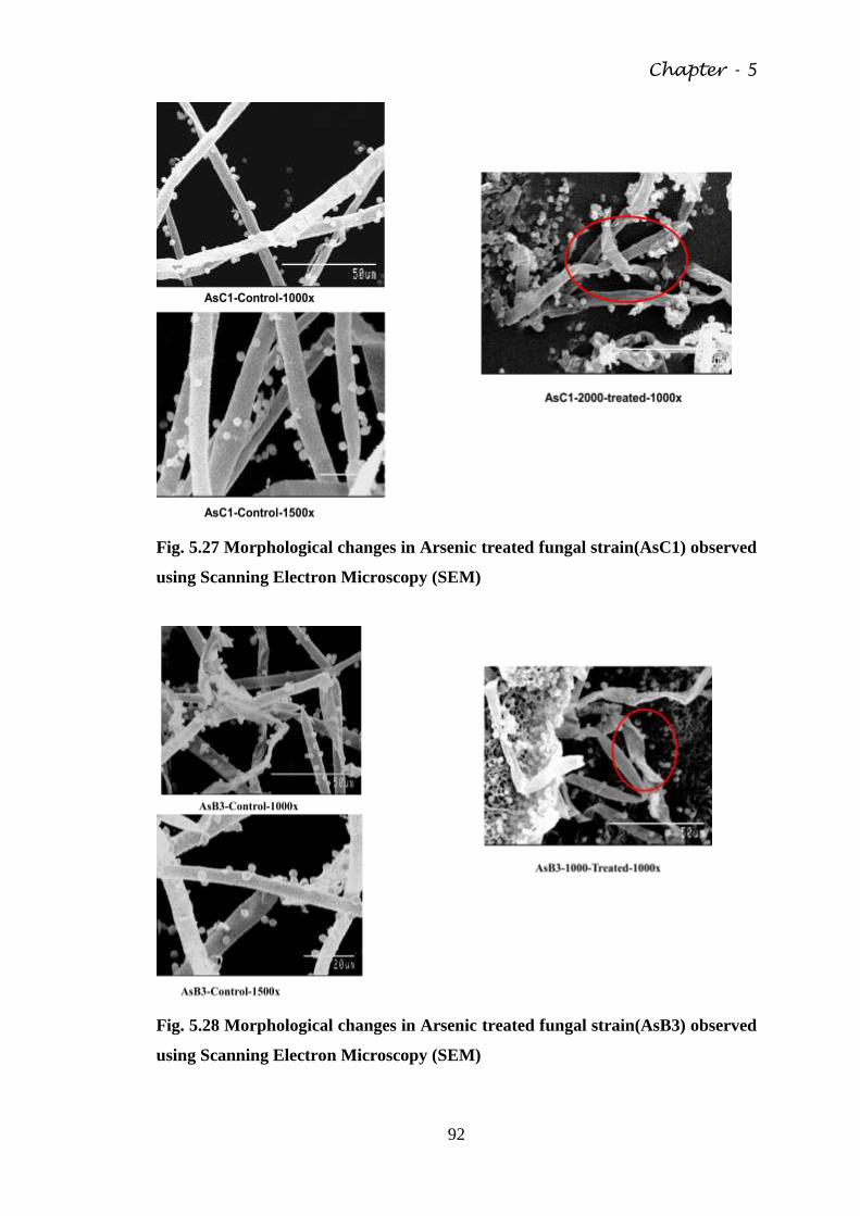
Chapter - 5
92
Fig. 5.27 Morphological changes in Arsenic treated fungal strain(AsC1) observed
using Scanning Electron Microscopy (SEM)
Fig. 5.28 Morphological changes in Arsenic treated fungal strain(AsB3) observed
using Scanning Electron Microscopy (SEM)
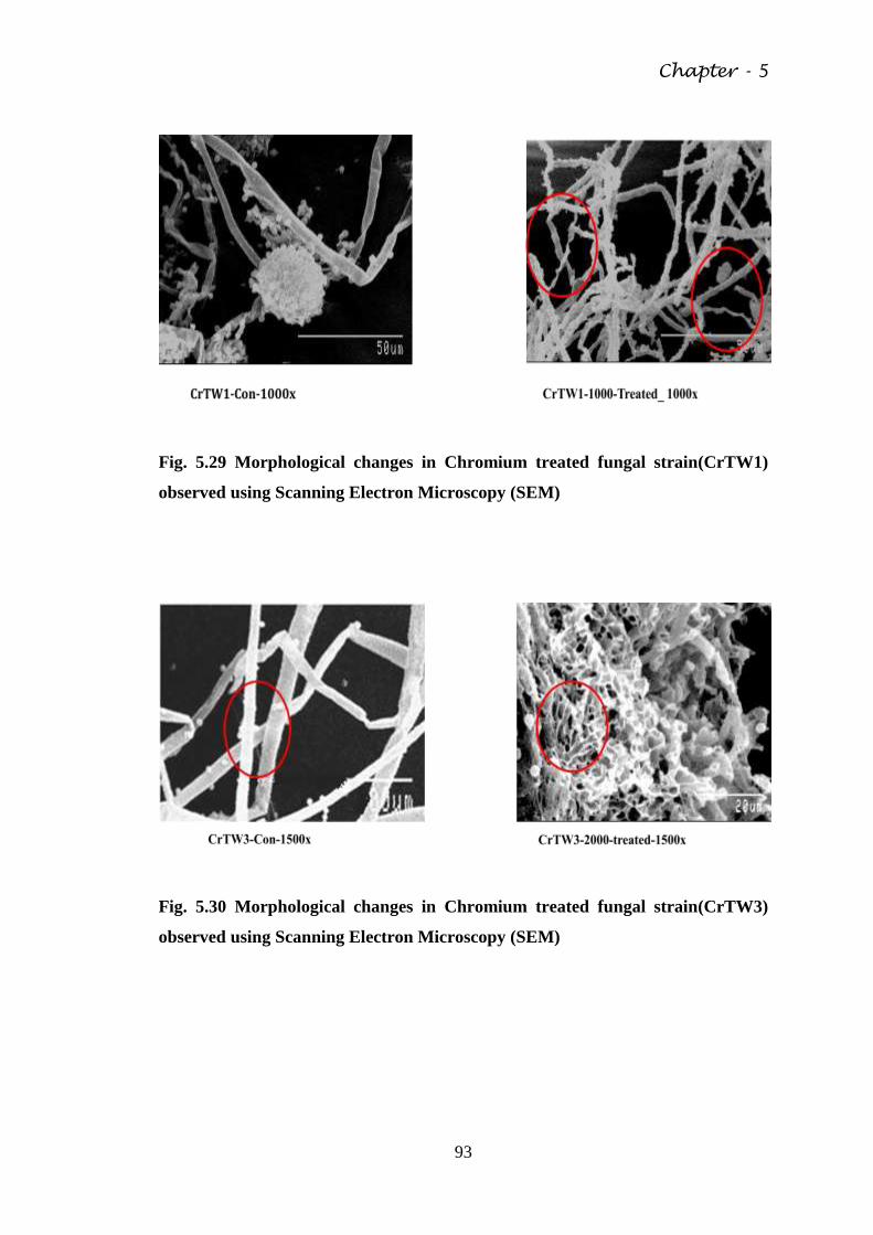
Chapter - 5
93
Fig. 5.29 Morphological changes in Chromium treated fungal strain(CrTW1)
observed using Scanning Electron Microscopy (SEM)
Fig. 5.30 Morphological changes in Chromium treated fungal strain(CrTW3)
observed using Scanning Electron Microscopy (SEM)
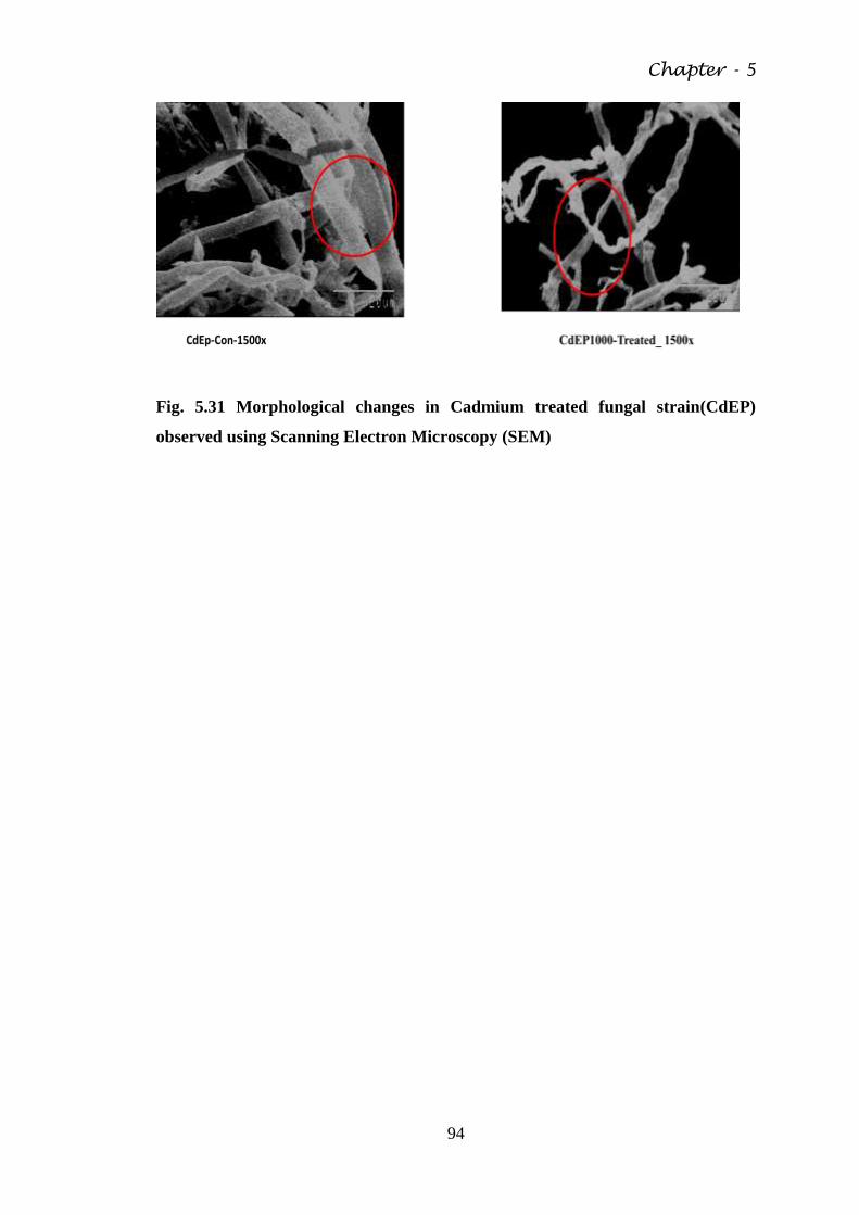
Chapter - 5
94
Fig. 5.31 Morphological changes in Cadmium treated fungal strain(CdEP)
observed using Scanning Electron Microscopy (SEM)
Chapter - 6
95
DISCUSSION
Heavy metal toxicity is an alarming problem in the recent era. The study of the
interaction between toxic metals and fungi has always remain a scientific interest. The
observations on the ability of fungi to resist and adapt to toxic metals stimulated
further work on the physiological, bio-chemical and genetical explanations for these
phenomena (Gadd, 1993). In fungi the metal toxicity may depend on the strain and
different stages of its growth. The detoxification mechanism of heavy metal also
varies from strain to strain. The survival of fungi in the presence of metal depends on
several factors like intrinsic biochemical and morphological properties, physiological
and genetical adaptation.
Microbial populations in metal polluted environments adapt to toxic concentrations of
heavy metals and become metal resistant (Prasenjit and Sumathi, 2005). Exposure
of microorganisms to heavy metals can lead to physiological adaptation or the
selection of mutants (Donmez and Aksu, 2001; Italiano et al., 2009) and such
changes may be associated with increased metal accumulation and sorption capacity
(Villegas et al., 2008). In this study five fungal strains were selected from a number
of fungal isolates depending on their metal tolerance capacity from the urban
contaminated soils around Kolkata city. The pH of the soil was neutral to alkaline.
The fungal growth and metal absorption of fungi is dependent on pH of the medium.
These soils contained significant amount heavy metals (Cd, As, Cr, Pb, Ni). Thus it
was assumed that the fungal strains would be tolerant to high concentration of heavy
metals. It has been reported that a number of fungi from all taxonomic groups may be
found in metal polluted soils and they possessed the ability to survive in that
environment by detoxification of the metal (Ross, 1975; Gadd, 1986a; Baldi,
Vaughan & Olson, 1990; Turnau, 1991). Usually metal toxicity reduces the
abundance of fungal population and thus only the metal tolerant species survive
(Babich & Stotzky, 1985; Duxbury, 1985). In this study all the selected five fungal
strains could resist and tolerate different heavy metals up to a certain range. The strain
AsC1 could tolerate arsenic concentration up to 2000ppm and AsB3 strain could
tolerate arsenic up to 1000 ppm. The strain CrTW1 and CrTW3 could tolerate
chromium up to 1300ppm. The strain CdEP could tolerate cadmium up to 1200ppm.
Chapter - 6
96
These studies revealed that all the strains could tolerate high level of respective heavy
metals. Thus they may possess some mechanism for metal tolerance.
The fungal strains were identified as Aspergillus flavus (AsC1), Aspergillus niger
(AsB3), Aspergillus niger (CrTw1), Penicillium sp. (CrTw3) and Aspergillus flavus
(CdEP) considering their morphological and microscopic characters. The suitable
media, pH, incubation temperature and time were selected for the studied fungal
strains according to their growth features. Temperature and pH are the two main
environmental factors for fungal growth and sporulation (Mehra and Jaitly, 1995).
The temperature can reach a point where growth stops and cell components begin to
be actually damaged by the heat. Enzymes are proteins that change structurally when
heated to their limit of tolerance. Likewise, membranes, which contain lipids, change
in structure and their function of protecting and regulating the internal environment of
the cell becomes compromised (Kamil et. al, 2011).
The antibiotic (Nystatin, Griseofulvin and Kanamycin) sensitivity test of the studied
fungi shown resistance to certain variable concentrations. From MIC test the
antibiotic Nystatin showed to be more effective to all the tested fungal strains than
Griseofulvin and Kanamycin which have importance from clinical point of view.
Rambali et al, (2001) studied the susceptibility testing of pathogenic fungi with
intraconazole.
The fungal strains which are resistant to arsenic can also tolerate other heavy metals
like Cd, Cr, Pb, Zn. This indicates that all the strains are positive for cross metal
tolerance test and they posses multi metal tolerance activity. Thus the detoxification
mechanism of the fungal strains are not metal specific but for only one metal it
showed maximum amount of tolerance limit. In this study the specific metal was
chosen for specific strains. Like arsenic was chosen for AsC1, AsB3; Chromium for
CrTW1 and CrTW3; and Cadmium for CdEP. Metals are directly and/or indirectly
involved in all aspects of fungal growth, metabolism and differentiation. While many
metals are essential (e.g. K, Na, Mg, Ca, Mn, Fe, Cu, Zn, Co, Ni etc.) , many others
have no apparent essential function (e.g. Rb, Cs, Al, Cd, Ag, Au, Hg, Pb etc.).
However, all these elements can interact with fungal cells and can be accumulated by
physico-chemical mechanisms and transport systems of varying specificity (Gadd,
1988).
Chapter - 6
97
The fungal strains could grow in respective metal containing media. All the strains
were also grown without metal as control set. In the presence of metal the strains
could grow up to a certain concentration but beyond that concentration their growth
decreased. All the strains were grown in metal treated concentration up to 9th
day of
incubation and after that their growth ceased, where as in the control set the growth
achieved a stable state. Both the strain AsC1 and AsB3 showed highest growth at
1500ppm. The two strains which were treated against chromium also showed highest
growth against 1500ppm. The strain CdEP showed highest growth against 1000ppm
dose of cadmium concentration. Thus all the selected strains exhibited high growth
rate against these metals up to a certain range, which proves that they can tolerate the
toxic effect of the metal. The metal can bind the cell wall or may accumulate within
the cytosol. The growth rate and biomass production of microorganisms is highly
affected by metal ion concentration. Often, growth rate and biomass production is
inversely proportional to the toxicity of the heavy metals. It decreases with increasing
toxic metal concentration in the growth medium. Acikel & Ersan (2010) also
reported that with the increase in Ni(II) ion concentrations in the range 0–50ppm, the
specific growth rates of R. delemar decreased significantly. In tolerant strains,
complex regulatory networks exist that ensures that the metabolic costs of the heavy
metal response are kept at a minimum without compromising the detoxification
process (Bellion et al., 2006). The change in growth parameters are also dependent on
the nature of metal present. Dursun et al. (2003b) observed the growth of Aspergillus
niger in presence of Cu(II), Pb(II), and Cr(VI) ions. Maximum biomass production by
A. niger had been reported in absence of metal ions while the growth was inhibited by
all concentrations of Cr(VI) tested. In this study the strains were tested against that
particular metal which it can tolerate at its highest amount.
These fungal strains can bio-accumulate heavy metal from the respective medium.
Bioaccumulation includes both bio-adsorption and bio-absorption. Bio-adsorption
involves the attachment of metal on the cell wall. The wall is the first cellular site of
interaction with metal species and metal removal from solution may be rapid although
rates will depend on factors such as type of metal ion and biomass, concentration of
metal and environmental factors.
The metal removal capacities of all the strain were high. The metal removal
percentage ranged from 30% to 76%. The metal removal percentage of strain AsB3
Chapter - 6
98
ranged from 50% to 70%. The highest metal removal percentage showed by AsC1
was 76% and the lowest was 50%. The metal removing percentage for the strain
CrTW1 ranged from 36% to 76.2% and for CrTW3 ranged from 22% to 51%. The
metal removing capacity of CdEP ranged from 33% to 67%. Among all the five
strains the strain CrTW3 showed lowest metal removing capacity. These results
indicated that all the five fungal strain could accumulate metal from the metal treated
broth thus resulting removal of the metal from the media. These kind of fungal strains
thus can be effective for the remediation of metal contaminated water and soil. Metal
tolerance property was reported from Penicilium genus (Gadd et al, 1984a, Gadd
and White, 1985). The metal removal capacity may depend on a number of factors
such as metal ion, environmental factor, and concentration of metal. Ahmad et al.
(2006) studied the biosorption by metal tolerant Aspergillus niger and Penicillium sp.
and concluded that fungi of metal contaminated soil have high level of metal
tolerance and biosorption properties.
The bioaccumulation capacity was also tested with live fungal mycelium. These
processes include both bio-adsorption and bio-absoprtion. All the strains can
accumulate metal up to 6th
days of incubation after that the accumulation capacity
become stagnant. Thus the strains can accumulate metal up to a certain time and
showed tolerance but after that the strain succumbs to the toxicity.
After uptake of the metal the cell wall is the first space where the metal interacts. The
metal biosorption in the fungal cell wall may be complex, involving different
compositions and mechanisms. The uptake of metal by cell wall is variable depending
on the wall structure. Chitin and chitosin have received attention as metal sorbing
components of the cell wall. While studying the metal bio-adsorption capacity of the
dead fungal mycelium in this case, it is found, that in all the fungal strains the metal
accumulation by fungal mycellium decreased with increase in treated metal dose.
Among the two strains AsC1 and AsB3, AsB3 strain showed highest arsenic
accumulation. Among the chromium tolerant strains CrTW3 strain showed highest
accumulation. The strain CdEP can accumulate significant amount of cadmium in the
dead mycelium. Thus among the five fungal strains, AsB3 strain can accumulate
highest metal in the cell wall.
Chitosan is a good source for adsorption of heavy metals. Chitosan is a biopolymer,
which is extracted from crustacean shells or from fungal biomass. The high porosity
Chapter - 6
99
of this natural polymer results in novel binding properties for metal ion such as
cadmium, copper, lead, mercury and chromium etc. Chitosan occur naturally in the
environment in large quantities and run second in abundance to cellulose. It has an
amine functional group which is strongly reactive with metal ions. Considerable
research has been done on the uptake of metal cations by chitosan. The amine groups
on chitosan bind metal cations at pH close to neutral. At low pH, chitosan is more
protonated and therefore it is able to bind anions by electrostatic attraction (Guibal,
2004). In this study chitosan could adsorb significant amount of metal from metal
treated solution. The adsorption capacity decreased with decrease in pH. At low pH
i.e, pH-2 chitosan could adsorb lowest amount of all three metals. At low pH the
amine group get protonated. That means chitosan get positively charged. Chromium,
cadmium and arsenic ions are also positively charged. As a result repulsive forces
occur between the metal ions and chitosan instead of attraction. Therefore at low pH
arsenic, chromium and cadmium uptake will be reduced.
The metallothionein protein was estimated to analyse the bioaccumulation of metal.
Metallothionein is a peptide which plays an important role in metal accumulation.
MTs have been found throughout the animal kingdom, in higher plants, in eukaryotic
microorganisms, and in many prokaryotes. MTs have been divided into three classes
based on their structural similarities. Class I, II and III with multiple isoforms within
each class. MT-I and MT-II are ubiquitously expressed and are stress inducible
(Thirumoorthy et al., 2007). The amino acid sequences of MTs from many
mammalian sources reveal that all contain approximately 61 amino acids of
remarkably similar composition. More importantly, all contain 20 cysteine residues
that remain invariant along the amino acid sequence. All cysteines are known to
participate in the coordination of 7 mol of Cd or zinc (Zn) per mol of MT (Kägi &
Vallee, 1960; Klaassen, et al., 1999). Coordination of these cysteine residues results
in a high binding affinity for Zn and Cd (Curtis et al., 1999). The increase in
metallothionein content reveals the accumulation of the metal. In this study all the
five strains showed increased amount of metallothionein content in comparison to the
control set. The strain CdEP showed highest amount of metallothionein followed by
AsB3 strains. According to previous results we have seen that AsB3 and CdEP strain
could accumulate high amount of metal among the five strains, thus the
metallothionein level of these two strains were also high among the five strains.
Chapter - 6
100
The microbes which can resist metal go through a range of morphological and ultra
structural changes. After intake of the metal, the toxic metal ion form complexes with
cellular membrane. This causes the loss of its integrity and impairs its function.
(Yilmazer and Saracoglu, 2009). It was reported that the morphology and
physiology of the cell changes with increased concentration of metal (de Siloniz et
al., 2002). In case of fungi, mycelia become short, dense, and broken in the metal
treated strains in comparison to the control strains. Improved aggregation of the
fungal hyphae can be one of the morphological strategies in response to toxic metals
(Gadd, 2007). Due to aggregation of hyphae the exposed surface area reduces and
facilitates high local concentrations of extracellular products (organic acids and
siderophores), metal precipitating agents, polysaccharides and pigments with metal
binding abilities (Baldriah, 2003; Dutton and Enans, 1996). Twisting and looping
of individual hyphae and formation of intertwined hyphal strands in response to
cadmium stress (Lilly et al., 1992) and decreased overall mycelial length in response
to Cd and Cu stress has been reported (Gadd, 2001). Saccharomyces cerevisiae and
Aureobasidium pullulans employ different mechanisms to overcome Pb toxicity as the
former accumulates Pb inside the cells while the later displays extracellular
sequestration. Scanning Electron Microscopy reveals that concomitant with the Pb(II)
accumulation, the cell surface of S. cerevisiae became rough and the amounts of
potassium, phosphorus and sulphur on the cell surface decreased (Suh et al., 1998). In
Taiwan one study was carried out with effect of Cu (II) and Zn (II) on growth and cell
morphology of Thraustochytrids isolated from fallen mangrove leaves (Lin et
al.2010). Schizochytrium cells exposed to >32 ppm concentration showed formation
of holes in the cell wall, shrinkage and general deformation of the cell structure as
depicted by light and scanning electron micrograph while at very high concentrations
(>250 ppm) cell lysis resulting in leakage of cellular content was observed. Baratelli
et al. (2007) investigated morphological and ultra structural changes in Paracoccus
sp. DE2007 strain isolated from Pb polluted site. The SEM image revealed that cells
were more deformed and had a higher tendency to aggregate. The Paracoccus sp.
DE2007 strain is able to capture lead extracellularly in its EPS envelopes, but does
not bioaccumulate intracellularly. Still increase in number of intracellular vesicles
was noticeable in presence of lead. This could be related to an increase in the
synthesis of lipid compounds that play an important role as a carbon reserve in
microorganisms under stress conditions such as the exposure to heavy metals. In this
Chapter - 6
101
study when the strains were exposed to high concentration of metal for a certain
period of incubation time the Scanning Electron Microscopy study revealed
morphological changes. The cells were deformed, the breakage of hyphae was
observed. Formations of holes in the cell wall, shrinked and wrinkled surface were
also depicted from the SEM analysis. Major holes in the cell wall were observed in
Penicillum sp. (CrTW3) strain.
The five strains were identified mostly form Aspergillius genus and one from
Penicilllium genus. AsC1 was identified as Aspergillus flavus, AsB3 was identified as
Aspergillus niger, CrTW1 as Aspergillus niger, CrTw3 as Penicillium sp. and CdEP
was identified as Aspergillus flavus. Previously several study reported various strains
from Aspergillus and Penicillium genus as heavy metal tolerant fungi. Liu et al.
(2009) reported Aspergillus flavus as copper and Zinc resistant strains which can
tolerate copper up to 400 ppm and zinc up to 800 ppm. Ezzouhri et al. (2009)
reported both Aspergillus and Penicillium strains as lead, copper and chromium
resistant strains. Parameswari et al. (2010) reported Aspergillus niger as Cr and Ni
resistant fungal strain. Congeevaram et al. (2007) isolated some strains, in which Cr-
resistant Aspergillus sp. and Micrococcus sp. survived to a maximum level of 10000
ppm. Valix et al. (2001) achieved nickel tolerance of up to 2000 ppm by Aspergillus
foetidus and Penicillium simplicissimum on solid media through step wise
acclimatization. Thus it is evident from the previous study that Aspergillus and
Penicillium genus had adapted to toxic effects of heavy metals and developed heavy
metal resistant mechanism.
The use of microorganism in heavy metal remediation has attracted a special attention
in recent decades. In the recent years extensive research and development has
occurred on biological methods, which have been considered as an eco-friendly
alternative for remediation of heavy metal contamination. Nevertheless, a large pool
of these studies have investigated microbial systems such as fungi, bacteria, and algae
as adsorbing agents for removal of heavy metals (Munoz et al., 2006; Pena-Castro,
2004). Briefly, biosorption studies involving easily available dead or live biomass are
a suitable low-cost approach to treat heavy metal pollution. In this study all the strains
can tolerate high amount of heavy metal. The strains can grow in the metal containing
media up to a certain period of incubation time. Both the dead and the live mycelia of
all the strains can accumulate metal from metal containing media.
Chapter - 6
102
Biosroption is a new and less expensive method for removal of heavy metals. Among
biosorbents, attempts have been made to use fungal biomass for the removal of heavy
metals. These strains could be helpful to build a metal removal strategy but more
intensive study is needed on the mechanism of metal absorption by the cell wall and
cytosol.
Chapter - 7
103
CONCLUSION
Heavy metal pollution has been a serious environmental threat of the recent era. Use
of wastewater in agriculture has increased in the recent years due to inherent
treatment capacity of soil and high contents of major micronutrients in it. However,
wastewater, particularly from industries, contains high concentrations of heavy metals
which enter into human beings and animals through food chain. Therefore, it is
desirable to remove these heavy metals from wastewater through low cost technology
before it is used in agriculture. Physico-chemical methods such as reverse osmosis,
solvent extraction, lime coagulation ion exchange and chemical precipitation for
removal of heavy metals from wastewater are very expensive and these do not remove
heavy metals from wastewater up to the desired level. Microorganisms including
fungi have been reported to exclude heavy metals from wastewater through
bioaccumulation and biosorption at low cost and in an eco-friendly way. Microbial
population in metal adapted environment build metal defence mechanism and adapt to
toxic concentration of heavy metal and ultimately became metal resistant. Different
species of Aspergillus have been reported as efficient chromium and nickel reducers
(Gopalan & Veeramani, 1994).
Biological mechanisms implicated in fungal survival (as distinct from
environmental modification of toxicity) include extracellular precipitation,
complexation and crystallization, transformation of metal species by oxidation,
reduction, methylation and dealkylation, biosorption to cell walls, pigments and
extracellular polysaccharide, decreased transport or impermeability, efflux,
intracellular compartmentation and precipitation and/or sequestration (Ross, 1975;
Gadd & Griffiths, 1978; Gadd, 1988, 1990a, b, 1992a-c; Brown & Hall,
1990; Mehra & Winge, 1991). A particular organism may directly and/or
indirectly rely on several survival strategies. For example, metallothionein
synthesis is a mechanism of Cu2+
resistance in Saccharomyces cerevisiae yet
Cu2+
binding or precipitation around the cell wall and intracellular transport are
also components of the total cellular response (Gadd & White, 1989a). In this
study detailed analysis have been done with the tolerance and bioaccumulation
capacity of five fungal strains.
Chapter - 7
104
The major findings of these studies are:
Five fungal strains were isolated namely AsC1, AsB3, CrTW1, CrTW3 and
CdEP out of total screening 30 fungal isolates. All these strains could
accumulate high amount of heavy metals. AsC1 and AsB3 were tested against
arsenic, CdEP strain for cadmium tolerance, CrTW1 and CrTW3 for
chromium.
AsC1 and CdEP were identified as Aspergillus flavus, AsB3 and CrTW1 were
identified as Aspergillus niger and CrTW3 was identified as Penicillium sp.
The optimum pH and temperature has been analysed for the five strains. At pH
7 and at a temperature of 30ºC all the further experiments were carried out.
From the antibiotic test it was found that the antibiotic Nystatin was more
effective to all the tested fungal strains than Griseofulvin and Kanamycin.
All the fungal strains could tolerate high level of metal concentration. AsC1
could tolerate arsenic up to 2000 ppm, AsB3 could tolerate arsenic upto 1000
ppm. CrTW1 and CrTW3 could tolerate chromium up to 1300 ppm. CdEP
could tolerate cadmium upto 1200 ppm.
All the fungal strains could accumulate other metals like lead, mercury, Zinc.
The strains tested for arsenic could also tolerate cadmium and chromium with
the other metals.
The strain AsC1 showed highest growth against 2000 ppm dose of treated
arsenic up to the 9th
day of incubation. The strain AsB3 showed highest
growth against 1500 ppm dose of treated arsenic up to 9th
day. The strain
CrTW1 and CrTW3 showed highest growth at 1500 ppm dose of treated
chromium up to 9th
day. The strain CdEP showed highest growth against 1000
ppm dose of treated cadmium. All the strains showed growth up to the 9th
day
and after that their growth ceased.
The metal removal capacity of all the strains was high. The metal removal
percentage ranged from 30% to 80%. The metal removal percentage of strain
AsB3 ranged from 50% to 70%. The strain AsC1 showed highest metal
removal percentage with 76% and lowest with 50%. The metal removing
percentage for CrTW1 ranged from 36% to 76% and for CrTW3 ranged from
22% to 51%. The metal removing capacity of CdEP ranged from 33% to 67%.
Chapter - 7
105
The metal bioaccumulation was also tested in the live mat of the fungal
strains. All the strains could accumulate considerable concentration of metal.
All the strains accumulated metal up to the 6th
day of incubation and after that
the accumulation capacity became stagnant.
For all the fungal strains the metal accumulation by the cell wall decreased
with increase in treated metal dose. Among the two strains AsC1 and AsB3,
AsB3 strain showed maximum metal accumulation by the cell wall. Among
the chromium tolerant strains CrTW3 strain showed maximum metal
accumulation by the cell wall. The strain CdEP can also accumulate
cosiderably high level of metal in the cell wall. The metal absorption capacity
by chitosan was also tested. Chitosan could adsorb metal to a significant level,
but the metal accumulation capacity decreased with decrease in pH. At low
pH2 chitosan adsorbed least amount of metal.
The metallothionin expression also increased upon metal exposure. The
content of metallothionin increased to a significant level in comparison to the
control set for every metal.
The morphological changes have been observed in the five fungal strains by
Scanning Electron Microscopy. Shrinkage of cell, breakage of hyphae,
mycelial deformities, reduction of conidial vessels were clearly shown in
metal treated fungal strains.
Filamentous fungi are employed in fermentation industries to generate diverse
metabolites for example antibiotics, enzymes, etc. The fungi showed a great
attraction for metal ions as compared to other microbes. These can gather metals
by means of biological and physiochemical mechanisms from their external
environment (Cabuk et al., 2004; Preetha & Viruthagiri, 2005). All the
filamentous fungi contain small quantity of protein, large amounts of polymer of
N-acetyl, chitin and chitosan, and deacetylated glucosamine on their cell wall.
Therefore, large number of potential binding sites are showed by free hydroxyl,
amine and carboxyl groups. The amine group containing nitrogen atom and the
hydroxyl group containing oxygen atom have the ability to bind a proton or a
metal ion.
Chapter - 7
106
The knowledge of present investigation will provide information about heavy
metal biosorption by filamentous fungus. This high absorption capacity of fungi
made them well suited for removal of heavy metals present in very low or diluted
concentration from polluted water, bioleaching, bioremediation of polluted sites
and effluent treatments. The information of present study will be helpful for
further assessment and management of natural biosorbent (fungus) which could
serve as an economically viable resource while treating industrial effluents
containing toxic heavy metal ions. In this study these five strains were been
proved as efficient metal detoxification agent as they have achieved multi lethal
tolerance capacity and thus they can well grow in multi polluted environment.
This study will provide ample opportunity for field application after pilot trail in
near future.
Chapter - 8
107
REFERENCES
Achstetter T, Wolf DM. (1975). Proteinases, proteolysis and biological control in the
yeast Saccharomyces cerevisiae. Yeast 1: 139-157.
Açıkel, Ü., & Erşan, M. (2010). Acid phosphatase production by Rhizopus delemar:
A role played in the Ni (II) bioaccumulation process. Journal of hazardous
materials, 184(1), 632-639.
Adjimani JP, Emery T. (1987). Iron uptake in Mycelia sterilia EP- 76. Journal of
Bacteriology 169: 3664-3668.
Ahalya N., Ramachandra T. V., R. D. Kanamadi (2003).: Biosorption of Heavy
Metals. Research Journal of Chemistry and Environment, 7: 71-79.
Ahemad, M., Kibret, M. (2013). Recent Trends in Microbial Biosorption of Heavy
Metals: A Review. Biochemistry and Molecular Biology, 1: 19-26.
Ahmad, I., Ansari, M. I., & Aqil, F. (2006). Biosorption of Ni, Cr and Cd by metal
tolerant Aspergillus niger and Penicillium sp. using single and multi-metal
solution. Indian journal of experimental biology, 44(1), 73.
Ahluwalia SS, Goyal D. (2007). Microbial and plant derived biomass for removal of
heavy metalsfrom wastewater. Biores Technol 98:2243–2257
Aksu Z, Balibek E. (2007). Chromium(VI) biosorption by dried Rhizopus arrhizus :
effect of salt (NaCl) concentration on equilibrium and kinetic parameters. J Hazard
Mater 145:210–220
Al-Saraj M, Abdel-Latif MS, El-Nahal I, Baraka R. (1999). Bioaccumulation of
some hazardousmetals by sol–gel entrapped microorganisms. J Non-Cryst Solids
248:137–140
Ambrosio, S. T., Campos-Takaki, G. M. (2004). Decolorization of reactive azo
dyes by Cunninghamella elegans UCP 542 under co-metabolic
conditions. Bioresource technology, 91: 69-75.
Anderson, J. P. E., & Domsch, K. H. (1980). Quantities of plant nutrients in the
microbial biomass of selected soils. Soil Science, 130(4), 211-216.
Andreae MO, (1986). Organoarsenic compounds in the environment. In: Craig PJ,
ed. Organometallic compounds in the environment. Harlow: Longman, 198-228.
Anraku Y, Umemoto N, Hirata R, Wada Y. (1989). Structure and function of the
yeast vacuolar membrane proton ATPase. Journal of Bioenergetics and
Biomembranes 21: 589-603.
APHA, A. 1998. WEF, (1998). Standard methods for the examination of water and
wastewater, 20.
Chapter - 8
108
Arnebrant K, Baath E, Nordgren A, (1987). Copper tolerance of microfungi
isolated from polluted and unpolluted forest soil. Mycologia 79: 890-895.
Ashida, J., Higashi, N., & Kikuchi, T. (1963). An electronmicroscopic study on
copper precipitation by copper-resistant yeast cells. Protoplasma, 57(1-4), 27-32.
Ashida J. (1965). Adaptation of fungi to metal toxicants. Annual Review of
Phytopathology 3: 153-174.
Babich H, Stotzky G. (1977) a. Reductions in the toxicity of cadmium to
microorganisms by clay minerals. Applied and Environmental Microbiology 33: 696-
705.
Babich H, Stotzky G. (1978). Effects of cadmium on the biota: infuence of
environmental factors. Advances in Applied Microbiology 23: 55-117.
Babich H, Stotzky G. (1979). Abiotic factors affecting the toxicity of lead to fungi.
Applied and Environmental Microbiology 38: 506-513.
Babich H, Stotzky G. (1980). Environmental factors that influence the toxicity of
heavy metal and gaseous pollutants to microorganisms. Critical Reviews in
Microbiology 8: 99-145.
Babich H, Stotzky G. (1981). Manganese toxicity to fungi: influence of pH. Bulletin
of Environmental Contamination and Toxicology 27: 474-480.
Babich H, Stotzky G. (1982). Nickel toxicity to fungi: influence of environmental
factors. Ecotoxicology and Environmental Safety 6: 577-589.
Babich, H., & Stotzky, G. (1985). Heavy metal toxicity to microbe-mediated
ecologic processes: a review and potential application to regulatory
policies.Environmental Research, 36(1), 111-137.
Bai J., Wu X., Fan F., Tian W., Yin X., Zhao L., Fan F., Li Z., Tian L., Qin Z.,
Guo J. (2012). Biosorption of uranium by magnetically modified Rhodotorula
glutinis. Enzyme and Microbial Technology, 51: 382-387.
Bai SR, Abraham TE. (2001). Biosorption of Cr(VI) from aqueous solution by
Rhizopus nigricans. Bioresour Technol. 79:73-81
Baldi F, Vaughan AM, Olson GJ. (1990). Chromium(vi)-resistant yeast isolated
from a sewage treatment plant receiving tannery wastes. Applied and Environmental
Microbiology 56: 913-918.
Bartnicki-Garcia S. (1973). Fundamental aspects of hyphal morphogenesis. In:
Ashworth JO, Smith JE, eds. Microbial differentiation. Cambridge: Cambridge
University Press, 245-267.
Barug D.(1981). Microbial degradation of bis(tributyltin) oxide. 10: 1145-1154.
Bayramoglu G, Bekta S, Arlca MY. (2003). Biosorption of heavy metal ions on
immobilized white-rot fungus Trametes versicolor. J Hazard Mater 101:285–300
Chapter - 8
109
Bayramoglu G, Celik G, Yalcın E, Yılmaz M, Arıca MY. (2005). Modification of
surface proper-ties of Lentinus sajor-caju mycelia by physical and chemical
methods: evaluation of their Cr 6+
removal efficiencies from aqueous medium. J
Hazard Mater B 119:219–29.
Bellion, M., Courbot, M., Jacob, C., Blaudez, D., & Chalot, M. (2006).
Extracellular and cellular mechanisms sustaining metal tolerance in ectomycorrhizal
fungi. FEMS microbiology letters, 254(2), 173-181.
Belde PJM, Kessels BGF, Moelans IM, Borst-Pauwels GWFH. (1988). Cd2+
uptake, Cd2+
binding and loss of cell K+ by a Cd-sensitive and a Cd-resistant strain of
Saccharomyces cerevisiae. FEMS Microbiology Letters 49: 493-498.
Bell AA, Wheeler MH. (1986). Biosynthesis and functions of fungal melanins.
Annual Review of Phytopathology 24: 411-451.
Berka T, Shatzman A, Zimmerman J, Strickler J, Rosenberg M. (1988). Efficient
expression of the yeast metallothionein gene in Escherichia coli. Journal of
Bacteriology 170: 21-26.
Bertyl A, Slayman CL. (1990). Cation-selective channels in the vacuolar membrane
of Saccharomyces: dependence on calcium, redox state, and voltage. Proceedings of
the National Academy of Sciences of the USA 87: 7824-7828.
Bewley RJF, Campbell R. (1978). Scanning electron microscopy of oak leaves
contaminated with heavy metals. Transactions of the British Mycological Society 71:
508-511.
Bewley, J. D. (1979). Physiological aspects of desiccation tolerance. Annual Review
of Plant Physiology, 30(1), 195-238.
Bewley RJF, Campbell R. (1980). Influence of zinc, lead and cadmium pollutants on
microflora of hawthorn leaves. Microbial Ecology 6: 227-240.
Bewley RJF. (1980). Effects of heavy metal pollution on oak leaf microorganisms.
Applied and Environmental Microbiology 40: 1053-1059.
Bewley, RJF, Stotzky G. (1983). Effects of cadmium and zinc on microbial activity
in soil: influence of clay minerals. Part I: Metals added individually. Science of the
Total Environment 31: 41-55.
Bhatnagar, S. Kumari, R. (2013). Bioremediation: A Sustainable Tool for
Environmental Management – A Review - Annual Review & Research in Biology 3:
974-993.
Bianchi ME, Carbone ML, Lucchini G, Magni GE. (1981b). Mutants resistant to
manganese in Saccharomyces cerevisiae. Current Genetics 4: 215-220.
Bianchi ME, Carbone ML, Lucchini G. (1981). Mn2+
and Mg2+ uptake in Mn-
sensitive and Mn-resistant yeast strains. Plant Science Letters 22: 345-352.
Chapter - 8
110
Bilinski CA, Miller JJ. (1983). Translocation of zinc from vacuole to nucleus during
yeast meiosis. Canadian Journal of Cytology and Genetics 25: 415-419.
Blunden SJ, Chapman A. (1986). Organotin compounds in the environment. In:
Craig PJ, ed. Organometallic compounds in the environment. Harlow: Longman, 111-
159.
Blunden SJ, Hobbs LA, Smith PJ. (1984). The environmental chemistry of
organotin compounds. In: Bowen HJM, ed. Environmental biochemistry. London:
Royal Society of Chemistry. 49-77.
Borbolla M, Pena A. (1980). Some characteristics of Ca2+
uptake by yeast cells.
Journal of Membrane Biology 54: 149-156.
Borst-Pauwels GWFH, Peters PHJ. (1981). Factors affecting the inhibition of yeast
plasma membrane ATPase by vanadate. Biochimica et Biophysica Acta 642, 173-181.
Borst-Pauwels GWFH. (1981). Ion transport in yeast. Biochimica et Biophysica Acta
650: 88-127.
Boutry M, Foury F, Goffeau A. (1977). Energy-dependent uptake of calcium by the
yeast Schizosaccharomyces pombe. Biochimica et Biophysica Acta 464: 602-612.
Bowman BJ, Bowman EJ. (1986). H+-ATPases from mitochondria, plasma
membrane, and vacuoles of fungal cells. Journal of Membrane Biology 94: 83-97.
Bowman BJ, Mainzer SE, Allen KE, Slayman CW. (1978). Effects of inhibitors on
the plasma membrane and michondrial adenosine-triphosphatases of Neurospora
crassa. Biochimica et Biophysica Acta 512: 13-28.
Bressa G, Cima L, Costa P. (1988). Bioaccumulation of Hg in the mushroom
Pleurotus ostreatus. Ecotoxicology and Environmental Safety 16: 85-89.
Brierley CL, Kelly DP, Seal KJ, Best DJ. (1985). Materials and biotechnology. In:
Higgins IJ, Best DJ, Jones J, eds. Biotechnology. Oxford: Blackwell, 163-212.
Brown MT, Hall IR. (1990). Metal tolerance in fungi. In Shaw J, ed. Heavy metal
tolerance in plants: evolutionary aspects. Boca Raton: CRC Press, 95-104.
Brunker RL, Bott TL. (1974). Reduction of mercury to the elemental state by a
yeast. Applied Microbiology 27: 870-873.
Buchman C, Skroch P, Welch J, Fogel S, Karin M. (1989). The CUP2 gene
product, regulator of yeast metallothionein expression, is a copper-activated DNA-
binding protein. Molecular and Cell Biology 9: 4091-4095.
Budd K. (1988). A high-affinity system for the transport of zinc in Neocosmospora
vasinfecta. Experimental Mycology 12: 195-202.
Budd K. (1989). Role of the membrane potential in the transport of zinc by
Neocosmospora vasinfecta. Experimental Mycology 13: 356-363.
Chapter - 8
111
Butt TR, Ecker DJ. (1987). Yeast metallothionein and applications in biotechnology.
Microbiological Reviews 51: 351-364.
Byrne AR, Ravnik V, Kosta L. (1976). Trace element concentrations in higher
fungi. The Science of the Total Environment 6: 65-78.
Byrne AR, Tusek-Znidaric M. (1990). Studies of the uptake and binding of trace
metals in fungi. Part I: Accumulation and characterization of mercury and silver in the
cultivated mushroom, Agaricus bisporus. Applied Organometallic Chemistry 4: 43-
48.
Cabuk A., Ilhan S., Filik C., Caliskan F., (2004) Pb2+ biosorption by pretreated
fungal biomass, Turk. J. Biol., 29, 23-28.
Cerniglia, C. E. (1997). Fungal metabolism of polycyclic aromatic hydrocarbons:
past, present and future applications in bioremediation. Journal of industrial
microbiology & biotechnology, 19: 324-333.
Challenger F. (1945). Biological methylation. Chemical Reviews 36: 315-361.
Chau YK, Wong PTS. (1986). Organic Group VI elements in the environment. In:
Craig PJ, ed. Organometallic compounds in the environment. Harlow: Longman, 254-
278.
Chen C, Wang J-L. (2007). Characteristics of Zn 2+
biosorption by Saccharomyces
cerevisiae .Biomed Environ Sci 20:478–482
Chen G., Fan J., Liu R., Zeng G., Chen A., Zou Z.. (2012): Removal of Cd(II),
Cu(II) and Zn(II) from aqueous solutions by live Phanerochaete chrysosporium.
Environmental Technology, 33: 2653-2659.
Chen H, Pan S. (2005). Bioremediation potential of Spirulina: toxicity and
biosorption studies of lead. J Zhejiang Univ Sci B. 6:171-174.
Clint GM, Dighton J, Rees S. (1991). Influx of 137Cs into hyphae of basidiomycete
fungi. Mycological Research 95, 1047-1051.
Collins YE, Stotzky G. (1989). Factors affecting the toxicity of heavy metals to
microbes. In: Beveridge TJ, Doyle RJ, eds. Metal ions and bacteria. New York:
Wiley, 31-90.
Colpaert JV, Van Assche JA. (1987). Heavy metal tolerance in some
ectomycorrhizal fungi. Functional Ecology 1: 415-421.
Congeevaram S., Dhanarani S., Park J.,. Dexilin M, Thamaraiselvi K.. (2007).
Biosorption of chromium and nickel by heavy metal resistant fungal and bacterial
isolates. Journal of Hazardous Materials, 146: 270-277.
Cooney JJ, De Rome L, Laurence OS, Gadd GM. (1989). Effects of organotins and
organoleads on yeasts. Journal of Industrial Microbiology 4: 276-288.
Cooney JJ, Wuertz S. (1989). Toxic effects of tin compounds on microorganisms.
Journal of Industrial Microbiology 4, 375-402.
Chapter - 8
112
Corfield PS, Smith DG. (1970). The endoplasmic reticulum as the site of potassium
tellurite reduction in yeasts. Journal of General Microbiology 63: 311-316.
Cornelius G. & Nakashima H. (1987). Vacuoles play a decisive role in calcium
homeostasis in Neurospora crassa. Journal of General Microbiology 133, 2341-2347.
Craig PJ. (1986). Organometallic compounds in the environment. Harlow: Longman.
Culotta VC, Hsu T, Furst P, Hamer D. (1989). Copper and the ACE1 regulatory
protein reversibly induce yeast metallothionen gene transcription in a mouse extract.
Proceedings of the National Academy of Sciences of the USA 86, 8377-8381.
Dameron CT, Winge DR, George GN, Sansone M, Hu S, Hamer D. (1991). A
copper-thiolate polynuclear cluster in the ACE1 transcription factor. Proceedings of
the National Academy of Sciences of the USA 88: 6127-6131.
Das, N., Vimala, R., Karthika, P. (2008). Biosorption of heavy metals—an
overview. Indian J Biotechnol, 7: 159-169.
Dauer RR, Dunlop EH. (1991). High gradient magnetic separation of yeast. Current
Genetics 17: 465-472.
Davis RH. (1986). Compartmental and regulatory mechanisms in the arginine
pathways of Neurospora crassa and Saccharomyces cerevisiae. Microbiological
Reviews 50: 280-313.
De Rome L, Gadd GM. (1987a). Copper adsorption by Rhizopus arrhizus,
Cladosporium resinae and Penicillium italicum. Applied Microbiology and
Biotechnology 26: 84-90.
De Rome L, Gadd GM. (1987b). Measurement of copper uptake in Saccharomyces
cerevisiae using a Cu2+
selective electrode. FEMS Microbiology Letters 43: 283-287.
Deng SB, Ting YP. (2005a). Characterization of PEI-modified biomass and
biosorption of Cu(II),Pb(II) and Ni(II). Water Res 39:2167–2177
Deng SB, Ting YP. (2005b). Fungal biomass with grafted poly(acrylic acid) for
enhancement of Cu(II) and Cd(II) biosorption. Langmuir 21:5940–5948
Deng X. Wang P.. (2012). Isolation of marine bacteria highly resistant to mercury
and their bioaccumulation process. Bioresource Technology, 121: 342-347.
Dhar, R. K., Zheng, Y., Rubenstone, J., & Van Geen, A. (2004). A rapid
colorimetric method for measuring arsenic concentrations in groundwater.Analytica
Chimica Acta, 526(2), 203-209
Dias MA, Lacerda ICA, Pimentel PF, Castro HF, Rosa CA. (2002). Removal of
heavy metals by an Aspergillus terreus strain immobilized in a polyurethane matrix.
Lett Appl Microbiol. 34:46-50.
Chapter - 8
113
Donmez G, Aksu Z. (1999). The effect of copper(II) ions on the growth
and bioaccumulation prop-erties of some yeasts. Process Biochem 35:135–142
Doonan BD, Crang RE, Jensen TE, Baxter M. (1979). In situ Xray energy
dispersive microanalysis of polyphosphate bodies in Aureobasidium pullulans.
Journal of Ultrastructure Research 69: 232-238.
Duddridge JE, Wainwright M. (1980). Heavy metal accumulation by aquatic fungi
and reduction in viability of Gammarus pulex fed Cd2+
contaminated mycelium.
Water Research 14: 1605-1611.
Dupont CH, Rigoulet M, Aigle M, Guerin B. (1990). Isolation and genetic study of
triethyltin-resistant mutants of Saccharomyces cerevisiae. Current Genetics 17: 465-
472.
Dursun AY, Uslu G, Cuci Y, Aksu Z. (2003a). Bioaccumulation of copper(II),
lead(II) andchromium(VI) by growing Aspergillus niger. Process Biochem 38:1647–
1651
Dursun AY, Uslu G, Tepe O, Cuci Y, Ekiz HI. (2003b). A comparative
investigation on the bioac-cumulation of heavy metal ions by growing Rhizopus
arrhizus and Aspergillus niger. Biochem Eng J 15:87–92
Dursun AY. (2006). A comparative study on determination of the equilibrium,
kinetic and thermo-dynamic parameters of biosorption of copper(II) and lead(II) ions
onto pretreated Aspergillusniger. Biochem Eng J 28:187–195
Duxbury T. (1985). Ecological aspects of heavy metal responses in microorganisms.
In: Marshall KC, ed. Advances in microbial ecology. New York: Plenum Press, 185-
235.
Eilam Y, Chernichovsky D. (1987). Uptake of Ca2+
driven by the membrane
potential in energy-depleted yeast cells. Journal of General Microbiology 133: 1641-
1649.
Eilam Y, Lavi H, Grossowicz N. (1984). Effects of inhibitors of plasma membrane
ATPase on potassium and calcium fluxes, on membrane potential and on
protonmotive force in the yeast Saccharomyces cerevisiae. Microbios 131: 623-629.
Eilam Y, Lavi H, Grossowicz N. (1985 a). Cytoplasmic Ca2+
homeostasis
maintained by a vacuolar Ca2+
transport system in the yeast Saccharomyces
cerevisiae. Journal of General Microbiology 31: 623-629.
Eilam Y, Lavi H, Grossowicz N. (1985 b). Active extrusion of potassium in the
yeast Saccharomyces cerevisiae induced by low concentrations of trifluoperazine.
Journal of General Microbiology 131: 2555-2564.
Chapter - 8
114
Eilam Y, Othman M, Halachmi D. (1990). Transient increase in Ca2+
influx in
Saccharomyces cerevisiae in response to glucose: effects of intracellular acidification
and cAMP levels. Journal of General Microbiology 136: 2537-2543.
Eilam Y. (1984). Effects of phenothiazines on inhibition of plasma membrane
ATPase and hyperpolarization of cell membranes in the yeast Saccharomyces
cerevisiae. Biochimica et Biophysica Acta 760: 601-610.
Ellis, M. B., & Ellis, J. P. (1988). Microfungi on miscellaneous substrates: an
identification handbook. Croom Helm.
Failla ML, Weinberg ED. (1977). Cyclic accumulation of zinc by Candida utilis
during growth in batch culture. Journal of General Microbiology 99: 85-97.
Fan T, LiuY, Feng B, Zeng G, Yang C, Zhou M, Zhou H, Tan Z. (2008).
XinWang biosorption of cadmium(II), zinc(II) and lead(II) by Penicillium
simplicissimum : isotherms, kinetics and ther-modynamics. J Hazard Mater 160:655–
661
Fergusson JE. (1990). The heavy elements: chemistry, environmental impact and
health effects. Oxford: Pergamon Press.
Fogel S, Welch JW, Karin M. (1983). Gene amplification in yeast; CUP1 copy
number regulates copper resistance. Current Genetics 7: 1-9.
Fogel S, Welch JW, Maloney DH. (1988). The molecular genetics of copper
resistance in Saccharomyces cerevisiae – a paradigm for non-conventional yeasts.
Journal of Basic Microbiology 28: 147-160.
Fogel S, Welch JW. (1982). Tandem gene amplification mediates copper resistance
in yeast. Proceedings of the National Academy of Sciences of the USA 79: 5342-5346.
Freedman B, Hutchinison TC. (1980). Effects of smelter pollutants on forest leaf
litter decomposition near a nickel-copper smelter at Sudbury, Ontario. Canadian
Journal of Botany 58: 1722-1736.
Fuhrmann R, Rothstein A. (1968). The transport of Zn2+
, Co2+
and Ni2+
into yeast
cells. Biochimica et Biophysica Acta 163: 325-330.
Furst P, Hamer D. (1989). Cooperative activation of a eukaryotic transcription
factor: intraction between Cu(i) and yeast ACE1 protein. Proceedings of the National
Academy of Sciences of the USA 86: 5267-5271.
Furst P, Hu S, Hackett R, Hamer D. (1988). Copper activates metallothionein gene
transcription by altering the confirmation of a specific DNA binding protein. Cell 55:
705-717.
Gadd G. M. (2010). Metals, minerals and microbes: geomicrobiology and
bioremediation. Microbiology, 156: 609-643.
Chapter - 8
115
Gadd GM, Brunton AH. (1992). Calcium involvement in dimorphism of
Ophiostoma ulmi, the Dutch elm disease fungus, and characterization of calcium
uptake by yeast cells and germ tubes. Journal of General Microbiology 138: 1561-
1571.
Gadd GM, White C. (1985). Copper uptake by Penicillium ochro- chloron:
influence of pH on toxicity and demonstration of energy-dependent copper
influx using protoplasts. Journal of General Microbiology 131: 1875-1879.
Gadd GM, Chudek JA, Foster R, Reed RH. (1984a). The osmotic responses
of Penicillium ochro-chloron: changes in internal solute levels in response to
copper and salt stress. Journal of General Microbiology 130: 1969-1975
Gadd GM, De Rome L. (1988). Bisorption of copper by fungal melanin. Applied
Microbiology and Biotechnology 29: 610-617.
Gadd GM, Edwards SW. (1986). Heavy metal-induced flavin production by
Debaryomyces hansenii and possible connections with iron metabolism. Transactions
of the British Mycological Society 87: 533-542.
Gadd GM, Griffiths AJ. (1978). Microorganisms and heavy metal toxicity.
Microbial Ecology 4: 303-317.
Gadd GM, Mowll JL. (1985). Copper uptake by yeast-like cells, hyphae and
chlamydospores of Aureobasidium pullulans. Experimental Mycology 9: 230-240.
Gadd GM, White C. (1989)a. Heavy metal and radionuclide accumulation and
toxicity in fungi and yeasts. In: Poole RK, Gadd GM, eds. Metal-microbe
interactions. Oxford: IRL Press, 19-38.
Gadd GM. (1984). Effect of copper on Auerobasidium pullulans in solid medium:
adaptation not necessary for tolerant behaviour. Transactions of the British
Mycological Society 82: 546 - 549.
Gadd GM. (1986a). The responses of fungi towards heavy metals. In: Herbert RA,
Codd GA, eds. Microbes in extreme environments. London: Academic Press, 83-110.
Gadd GM. (1986b). The uptake of heavy metals by fungi and yeasts: the chemistry
and physiology of the process and applications for biotechnology. In: Eccles H, Hunt
S, eds. Immobilisation of ions by bio-sorption. Chichester: Ellis Horwood Ltd, 135-
147.
Gadd GM. (1988). Accumulation of metals by microorganisms and algae. In: Rehm
H-J, ed. Biotechnology - a comprehensive treatise, vol. 6b, Special microbial
processes. Weinheim: VCH Verlagsgesellschaft, 401-433.
Gadd GM. (1990a). Fungi and yeasts for metal binding. In: Ehrlich M, Brierley CL,
eds. Microbial mineral recovery. New York: McGraw-Hill, 249-275.
Chapter - 8
116
Gadd GM. (1990b). Metal tolerance. In: Edwards C, ed. Microbiology of extreme
environments. Milton Keynes: Open University Press, 178-210.
Gadd GM. (1992a). Microbial control of heavy metal pollution. In: Fry JC, Gadd
GM, Herbert RA, Jones CW, Watson-Craik I, eds. Microbial control of environmental
pollution. Cambridge: Cambridge University Press, 59-88.
Gadd GM. (1992b). Molecular biology and biotechnology of microbial interactions
with organic and inorganic heavy metal compounds. In: Herbert RA, Sharp RJ, eds.
Molecular biology and biotechnology of extremophiles. Glasgow: Blackie and Sons,
225-257.
Gadd GM. (1992c). Metals and microorganisms: a problem of definition. FEMS
Microbiology Letters 100: 197-204.
Gadd GM.( 1993). Interactions of fungi with toxic metals, New Phytol. 124: 25-
60.
Gadd GM, Griffiths AJ. (1978) .Microorganisms and heavy metal toxicity. Microb
Ecol; 4: 303–17
Galun M, Keller P, Feldstein H, Galun E, Siegel S, Siegel B. (1983). Recovery of
uranium (IV) from solution using fungi. II. Release from uranium-loaded Penicillium
biomass. Water, Air and Soil Pollution 20: 277-285.
Gazem M. A., Nazareth S. (2012). Isotherm and kinetic models and cell surface
analysis for determination of the mechanism of metal sorption by Aspergillus
versicolor. World Journal of Microbiology and Biotechnology, 28: 2521-30.
Giller K. E., Witter E., McGrath S. P. (2009). Heavy metals and soil microbes. Soil
Biology and Biochemistry, 41: 2031-2037.
Goffeau A, Ghislain M, Navarre C, Purnelle B, Supply P. (1990). Novel transport
ATPases in yeast. Biochimica et Biophysica Acta 1018: 200-202.
Gopalan, R., & Veeramani, H. (1994). Studies on microbial chromate reduction by
Pseudomonas sp. in aerobic continuous suspended growth cultures.Biotechnology and
bioengineering, 43(6), 471-476.
Gorman JA, Clark PE, Lee MC, Dubouck C, Rosenberg M. (1986). Regulation of
the yeast metallothionein gene. Gene 48: 13-22.
Gourdon R., Bhande S., Rus E., Sofer S. S. (1990). Comparison of cadmium
biosorption by gram positive and gram negative bacteria from activated sludge.
Biotechnology Letters, 12: 839-842.
Greco MA, Hrab DI, Magner W, Kosman DJ. (1990). Cu, Zn superoxide
dismutase and copper deprivation and toxicity in Saccharomyces cerevisiae. Journal
of Bacteriology 172: 317-325.
Chapter - 8
117
Greenaway W, Cripps A, Ward S. (1974). Resistance to organomercury by
Penicillia isolated from cereal seed. Transactions of the British Mycological Society
63: 137-141.
Grill E, Loffler S, Winnacker E-L, Zenk MH. (1989). Phytochelatins, the heavy
metal-binding peptides of plants, are synthesized from glutathione by a specific y-
glutamylcysteine dipeptidyl transpeptidase (phytochelatin synthase). Proceedings of
the National Academy of Sciences of the USA 86: 6838-6842.
Grill E, Winnacker EL, Zenk MH. (1987). Phytochelatins, a class of heavy-metal-
binding peptides from plants, are functionally analogous to metallothioneins.
Proceedings of the National Academy of Sciences of the USA 84: 439-443.
Gulati R, Saxena RK, Gupta R. (2002). Fermentation waste of Aspergillus terreus
: a potential cop- per biosorbent. World J Microbiol Biotechnol 18:397–401
Gustin MC, Martinac B, Saimi Y, Culbertson MR, Kung C. (1986). Ion channels
in yeast. Science 233, 1195-1197.
Hamer DH, Thiele DJ, Lemontt JE. (1985). Function and autoregulation of yeast
copperthionein. Science 228: 685-690.
Hamer DH. (1986). Metallothionein. Annual Review of Biochemistry, 55: 913-951.
Hayashi Y, Nakagawa CW, Murasugi A. (1986). Unique properties of Cd-binding
peptides induced in fission yeast, Schizosaccharomyces pombe. Environmental Health
Perspectives 65: 13-19.
Hayashi Y, Nakagawa CW, Mutoh N, Isobe M, Goto T. (1991). Two pathways in
the biosynthesis of cadystins (γEC)nG in the cell-free system of the fission yeast.
Biochemistry and Cell Biology 69: 115-121.
Hayashi Y, Nakagawa CW, Uyakul D, Imai K, Isobe M, Goto T. (1988). The
changes of cadystin components in Cd-binding peptides from the fission yeast during
their induction by cadmium. Biochemistry and Cell Biology 66: 288-295.
Hemambika B., Rani M. J.,. Kannan V. R. (2011). Biosorption of heavy metals by
immobilized and dead fungal cells: A comparative assessment. Journal of Ecology
and the Natural Environment, 3: 168-175.
Hemambika, B., Johncy Rani, M., Rajesh Kannan, V. (2011). Biosorption of
heavy metals by immobilized and dead fungal cells: A comparative
assessment. Journal of Ecology and the Nature Environment, 3: 168-175.
Henderson RC, Cox BS, Tubb R. (1985). The transformation of brewing yeast with
a plasmid containing the gene for copper resistance. Current Genetics 9: 133-138
Horsfall JG. (1956). Principles of fungicidal action. Waltham, Massachusetts:
Chronica Botanica.
Chapter - 8
118
Hu S, Furst P, Hamer D. (1990). The DNA and Cu binding functions of ACE1 are
interdigitated within a single domain. The New Biologist 2: 544-555.
Huang C-P, Huang C-P, Morehart AL. (1990). The removal of Cu(ii) from dilute
aqueous solutions by Saccharomyces cerevisiae. Water Research 24: 433-439.
Huang H., Cao L., Wan Y., Zhang R., Wang W.. (2012). Biosorption behavior and
mechanism of heavy metals by the fruiting body of jelly fungus (Auricularia
polytricha) from aqueous solutions. Applied Microbiology and Biotechnology, 96:
829-840.
Hughes M. N. Poole R. K.. (1989). Metals and Microorganisms, Chapman & Hall,
London, 328-395.
Hughes MN, Poole RK. (1989). Metals and micro-organisms. London: Chapman and
Hall.
Hughes MN, Poole RK. (1991). Metal speciation and microbial growth - the hard
(and soft) facts. Journal of General Microbiology 137: 725-734.
Huibregtse JM, Enkelke DR, Thiele DJ. (1989). Copper-induced binding of cellular
factors to yeast metallothionein upstream activation sequences. Proceedings of the
National Academy of the USA 86: 65-69.
Inouhe M, Hiyama M, Tohoyama H, Joho M, Murayama T. (1989). Cadmium-
binding protein in a cadmium-resistant strain of Saccharomyces cerevisiae.
Biochimica et Biophysica Acta 993:51-55.
Itoh M, Yuasa M, Kobayashi T. (1975). Adsorption of metal ions on yeast cells at
varied cell concentrations. Plant Cell Physiology 16: 1167-1169.
Joho M, Fujioka Y, Murayama T. 1985 b. Further studies on the subcellular
distribution of Cd2+
in Cd-sensitive and Cdresistant strains of Saccharomyces
cerevisiae. Journal of General Microbiology 131: 3185-3191.
Joho M, Imai M, Murayama T. (1985a). Different distribution of Cd2+
between Cd-
sensitive and Cd-resistant strains of Saccharomyces cerevisiae. Journal of General
Microbiology 131: 53-56.
Joho M, Sukenobu Y, Egashira E, Murayama T. (1983). The correlation between
Cd2+
sensitivity and Cd2+
uptake in the strains of Saccharomyces cerevisiae. Plant and
Cell Physiology 24: 389-394.
Jones MD, Hutchison TC. (1986). The effect of mycorrhizal infection on the
response of Betula papyrifera to nickel and copper. New Phytologist 102: 429-442.
Jones RP, Gadd GM. (1990). Ionic nutrition of yeast – the physiological
mechanisms involved and applications for biotechnology. Enzyme and Microbial
Technology 12: 402-418.
Chapter - 8
119
Jordan MJ, Lechevalier MP. (1975). Effects of zinc-smelter emissions on forest soil
microflora. Canadian Journal of Microbiology 21: 1855-1865.
Junghans K, Straube G. (1991). Biosorption of copper by yeasts. Biology of Metals
4: 233-237.
Kägi, J. H., & Vallee, B. L. (1960). Metallothionein: a cadmium-and zinc-containing
protein from equine renal cortex. Journal of Biological Chemistry,235(12), 3460-
3465.
Kakinuma Y, Ohsumi Y, Anraku Y. (1981). Properties of H+- translocating
adenosine triphosphatase in vacuolar membranes of Saccharomyces cerevisiae.
Journal of Biological Chemistry 256: 10859-10863.
Kamil, O. H., Lupuliasa, D., Draganescu, D., & Vlaia, L. (2011). Interrelations of
drying heat and survival of different fungal spores within the tablets formulation.
Studia Universitatis ―Vasile Goldiş‖, Seria Ştiinţele Vieţii, Vol. 21, issue 2, 2011, pp.
339-342
Kapoor A, Viraraghavan T, Cullimore DR. (1999). Removal of heavy metals using
the fungus As- pergillus niger . Biores Technol 70:95–104
Kapoor A, Viraraghavan T. (1995). Fungal biosorption—an alternative treatment
option for heavymetal bearing wastewaters: a review. Biores Technol 53:195–206
Kapoor A, Viraraghavan T. (1997a). Heavy metal biosorption sites
in Aspergillus niger. BioresTechnol 61:221–227
Kapoor A, Viraraghavan T. (1997b). Fungi as biosorbents. In: Wase J, Forster C
(eds) Biosorbentsfor metal ions. Taylor and Francis, London, pp 67–86
Kapoor A, Viraraghavan T. (1998a). Biosorption of heavy metals on Aspergillus
niger: effect of pretreatment. Biores Technol 63:109–113
Kapoor A, Viraraghavan T. (1998b). Removal of heavy metals from aqueous
solutions using im-mobilized fungal biomass in continuous mode. Water Res
32:1968–1977
Kapoor A., Viraraghavan T., and Cullimore D. R. (1999). Removal of heavy
metals using the fungus Aspergillus niger. Bioresource Technology, 70:95-1 04.
Karin M, Najarian R, Haslinger A, Valenzuela P, Welch J, Fogel S. (1984).
Primary structure and transcription of an amplified genetic locus: The CUP1 locus of
yeast. Proceedings of the National Academy of Sciences of the USA 81: 337-341.
Khambhaty Y, Mody K, Basha S, Jha B. (2009). Kinetics, equilibrium
and thermodynamic studieson biosorption of hexavalent chromium by dead fungal
biomass of marine Aspergillus niger. Chem Eng J 145:489–495
Chapter - 8
120
Khani M. H., Pahlavanzadeh H., Alizadeh K. (2012). Biosorption of strontium
from aqueous solution by fungus Aspergillus terreus. Environmental Science and
Pollution Research (International), 19: 2408-2418.
Kihn JC, Dassargues CM, Mestdagh MM. (1988). Preliminary ESR study of Mn(ii)
retention by the yeast Saccharomyces. Canadian Journal of Microbiology 34: 1230-
1234.
Kiran I, Akar T, Tunali S. (2005). Biosorption of Pb(II) and Cu(II) from aqueous
solutions by pre-treated biomass of Neurospora crassa . Process Biochem 40:3550–
3558.
Klaassen, C. D., Liu, J., & Choudhuri, S. (1999). Metallothionein: an intracellular
protein to protect against cadmium toxicity. Annual review of pharmacology and
toxicology, 39(1), 267-294.
Klionsky DJ, Herman PK, Emr SD. (1990). The fungal vacuole: composition,
function and biogenesis. Microbiological Reviews 54: 266-292.
Kogej A, Pavko A. (2001). Comparison of Rhizopus nigricans in a pelleted growth
form with someother types of waste microbial biomass as biosorbents for metal ions.
World J Microbiol Biotechnol 17:677–685
Konetzka WA. (1977). Microbiology of metal transformations. In: Weinberg ED, ed.
Microorganisms and minerals. New York: Marcel Dekker Inc, 317-342.
Kumar R, Bishnoi NR, Bishnoi GK. (2008). Biosorption of chromium (VI) from
aqueous solution and electroplating wastewater using fungal biomass. J Chem Eng
Data. 135 :202-208.
Kuriyama H, Umeda I, Kobayashi H. (1991). Role of cations in the flocculation of
Saccharomyces cerevisiae and discrimination of the corresponding proteins.
Canadian Journal of Microbiology 37: 397-403.
Kuypers GAJ, Roomans GM. (1979). Mercury-induced loss of K+ from yeast cells
investigated by electron probe X-ray microanalysis. Journal of General Microbiology
115: 13-18.
Lancashire WE, Griffiths DE. (1975). Studies on energy-linked reactions: isolation,
characterization and genetic analysis of trialkyl-tin-resistant mutants of
Saccharomyces cerevisiae. European Journal of Biochemistry 51: 377-392.
Landner L. (1971). Biochemical model for the biological methylation of mercury
suggested from methylation studies in vivo with Neurospora crassa. Nature, London
230: 452-453.
Langmuir I.. (1918). The adsorption of gases on plane surfaces of glass, mica and
platinum. Journal of the American Chemical Society, 1361-403.
Chapter - 8
121
Laurence OS, Cooney JJ, Gadd GM. (1989). Toxicity of organotins towards the
marine yeast Debaryomyces hansenii. Microbial Ecology 17: 275-285.
Lepsova A, Mejstrik V. (1989). Trace elements in fruit bodies of fungi under
different pollution stress. Agriculture, Ecosystems and Environment 28: 305-312.
Lerch K, Beltramini M. (1983). Neurospora copper metallothionein: molecular
structure and biological significance. Chemica Scripta 21: 109-115.
Lerch K. (1980). Copper metallothionein, a copper-binding protein from Neurospora
crassa. Nature, London 284: 368-370.
Lichko LP, Okorokov LA, Kulaev IS. (1982). Participation of vacuoles in
regulation of levels of K', Mg2+
and orthophosphate ions in cytoplasm of the yeast
Saccharomyces carlsbergensis. Archives of Microbiology 132: 289-293.
Lima, M. A. B. D., Nascimento, A. E. D., Souza, W. D., Fukushima, K. Campos-
Takaki, G. M. D. (2003). Effects of phosphorus on polyphosphate accumulation by
Cunninghamella elegans. Brazilian Journal of Microbiology,34: 363-372.
Lima, M. A., Franco, L. D. O., de Souza, P. M., do Nascimento, A. E., da Silva,
C. A., Maia, R. D. C., Rolim, H. M. L. Takaki, G. (2013). Cadmium Tolerance and
Removal from Cunninghamella elegans Related to the Polyphosphate
Metabolism. International journal of molecular sciences, 14: 7180-7192.
Lin C-M, Kosman DJ. (1990). Copper uptake in wild type and copper
metallothionein-deficient Saccharomyces cerevisiae. Journal of Biological Chemistry
265: 9194-9200.
Loeffler S, Hochberger A, Grill E, Winnacker E-L, Zenk MH. (1989).
Termination of the phytochelatin synthase reaction through sequestration of heavy
metals by the reaction product. FEBS Letters 258: 42-46.
Macaskie LE, Dean ACR. (1990a). Metal-sequestering biochemicals. In: Volesky B,
ed. Biosorption of heavy metals. Boca Raton: CRC Press, 199-248.
Macaskie LE, Dean ACR. (1990b). Trimethyl lead degradation by free and
immobilized cells of an Arthrobacter sp. and by the wood decay fungus Phaeolus
schweinitzii. Applied Microbiology and Biotechnology, 33: 81-87.
Marmocchi L, Marmocchi F, Rotilio G. (1991). Activation and induction by copper
of Cu/Zn superoxide dismutase in Saccharomyces cerevisiae. European Journal of
Biochemistry 196: 545-549.
Martínez-Juárez, V. M., Cárdenas-González, J. F., Torre-Bouscoulet, M. E., &
Acosta-Rodríguez, I. (2012). Biosorption of Mercury (II) from Aqueous Solutions
onto Fungal Biomass. Bioinorganic Chemistry and Applications, (2012).
doi:10.1155/2012/156190
Chapter - 8
122
Matile P. (1978). Biochemistry and function of vacuoles. Annual Review of Plant
Physiology 29: 193-213.
McCord JM, Day D. (1978). Superoxide-dependent production of hydroxyl radical
catalysed by iron-EDTA complex. FEBS Letters 86: 139-142.
Mehlhorn RJ. (1986). The interaction of inorganic species with biomembranes. In:
Brinckman FE, Sadler PJ, eds. The importance of chemical speciation in
environmental processes. Berlin: Springer-Verlag, 85-97.
Mehra RK, Garey JR, Butt TR, Gray WR, Winge DR. (1989). Candida glabrata
metallothioneins. Cloning and sequence of the genes and characterization of proteins.
Journal of Biological Chemistry 264: 19747-19753.
Mehra RK, Garey JR, Winge DR. (1990). Selective and tandem amplification of a
member of the metallothionein gene family in Candida glabrata. Journal of Biological
Chemistry 265: 6369-6375.
Mehra RK, Tarbet EB, Gray WR, Winge DR. (1988). Metalspecific synthesis of
two metallothioneins and y-glutamyl peptides in Candida glabrata. Proceedings of the
National Academy of Sciences of the USA 85: 8815-8819.
Mehra RK, Thorvaldsen JL, Macreadie IG, Winge DR. (1992a). Disruption
analysis of metallothionein-encoding genes in Candida glabrata. Gene 114: 75-80.
Mehra RK, Thorvaldsen JL, Macreadie IG, Winge DR. (1992b). Cloning system
for Candida glabrata using elements from the metallothionein-IIa-encoding gene that
confer autonomous replication. Gene 113: 119-124.
Mehra RK, Winge DR. (1991). Metal ion resistance in fungi: molecular mechanisms
and their related expression. Journal of Cellular Biochemistry 45: 30-40.
Mehra, S. K., & Jaitly, A. K. (1995). pH and temperature optima for growth and
sporulation in some common fungi from city waste. Mycoscience, 36(2), 243-246.
Miller AJ, Vogg G, Sanders D. (1990). Cytosolic calcium homeostasis in fungi:
roles of plasma membrane transport and intracellular sequestration of calcium.
Proceedings of the National Academy of Sciences of the USA 87: 9348-9352.
Miller JJ. (1984). In vitro experiments concerning the state of polyphosphate in the
yeast vacuole. Canadian Journal of Microbiology 30: 236-246.
Minney SF, Quirk AV. (1985). Growth and adpatation of Saccharomyces cerevisiae
at different cadmium concentrations. Microbios 42: 37-44.
Mishra A. Malik A. (2012). Simultaneous bioaccumulation of multiple metals from
electroplating effluent using Aspergillus lentulus. Water Research, 46: 4991-4998.
Chapter - 8
123
Mishra, A., & Malik, A. (2013). Recent advances in microbial metal
bioaccumulation. Critical Reviews in Environmental Science and Technology,43(11),
1162-1222.
Morgan JJ, Stumm W. (1991). Chemical processes in the environment, relevance of
chemical speciation. In Merian E, ed. Metals and their compounds in the environment.
Weinheim: VCH Verlagsgesellschaft, 67-103.
Mowll JL, Gadd GM. (1983). Zinc uptake and toxicity in the yeasts Sporobolomyces
roseus and Saccharomyces cerevisiae. Journal of General Microbiology 129: 3421-
3425.
Mowll JL, Gadd GM. (1984). Cadmium uptake by Aureobasidium pullulans.
Journal of General Microbiology 130: 279-284.
Mowll JL, Gadd GM. (1985). The effect of vehicular lead pollution on phylloplane
mycoflora. Transactions of the British Mycological Society 84: 685-689.
Mukherjee I, Gopal M. (1996). Degradation of chlorpyrifos by two soil fungi
Aspergillus niger and Trichoderma viride. Environ Toxicol Chem. 37: 145-151.
Mukhopadhyay M, Noronha SB, Suraishkumar GK. (2007). Kinetic modeling
for the biosorption of copper by pretreated Aspergillus niger biomass. Process
Biochem 98:1781–1787
Mullen MD, Wolf DC, Beveridge TJ, Bailey GW. (1992). Sorption of heavy metals
by the soil fungi Aspergillus niger and Mucor rouxii. Soil Biology and Biochemistry
24: 129-135.
Mungasavalli DM, Viraraghavan T, Jin Y-C. (2007). Biosorption of
chromium from aqueous solu-tions by pretreated Aspergillus niger : Batch and
column studies. Colloids Surf A-Physicochem Eng Asp 301:214–223
Munger K, Lerch K. (1985). Copper metallothionein from the fungus Agaricus
bisporus: chemical and spectroscopic properties. Biochemistry 24: 6751-6756.
Muraleedharan TR, Venkobachar C. (1990). Mechanism of biosorption of copper
(II) by Ganoderma lucidum. Biotechnol Bioeng. 35: 320-325.
Murphy RJ, Levy JF. (1983). Production of copper oxalate by some copper tolerant
fungi. Transactions of the British Mycological Society 81: 165-168.
Murasugi, A., & HAYASHI, Y. (1981). Cadmium-binding peptide induced in
fission yeast, Schizosaccharomyces pombe. Journal of biochemistry, 90(5), 1561-
1565.
Mutoh N, Hayashi Y. (1988). Isolation of mutants of Schizosaccharomyces pombe
unable to synthesize cadystin, small cadmium-binding peptides. Biochemical and
Biophysical Research Communications 151: 32-39.
Chapter - 8
124
Muzzarelli RAA, Bregani F, Sigon F. (1986). Chelating abilities of amino acid
glucans and sugar acid glucans derived from chitosan. In Eccles H, Hunt S, eds.
Immobilisation of ions by biosorption. Chichester: Ellis Horwood, 173-182. -
Nash TH. (1990). Metal tolerance in lichens. In: Shaw J, ed. Heavy metal tolerance
in plants: evolutionary aspects. Boca Raton: CRC Press, 119-131.
Newby PH, Gadd GM. (1986). Diffusion assays for determination of the effects of
pH on the toxicity of heavy metal compounds towards filamentous fungi.
Environmental Technology Letters 7: 391-396.
Newby PJ, Gadd GM. (1987). Synnema induction in Penicillium funiculosum by
tributyltin compounds. Transactions of the British Mycological Society 89: 381-384.
Nieboer E, Richardson DHS, Tomassini FD. (1978). Mineral uptake and release by
lichens: an overview. The Bryologist 8: 226-246.
Nieboer E, Richardson DHS. (1980). The replacement of the nondescript term
'heavy metals' by a biologically and chemically significant classification of metal
ions. Environmental Pollution 1: 3-26.
Nieuwenhuis BJWM, Weijers AGM, Borst-Pauwels GWFH. (1981). Uptake and
accumulation of Mn2+
and Sr2+
in Saccharomyces cerevisiae. Biochimica et
Biophysica Acta 649:83-88.
Nordgren A, Baath E, S8derstrom B. (1983). Microfungi and microbial activity
along a heavy metal gradient. Applied and Environmental Microbiology 45: 1829-
1837.
Norris PR, Kelly DP. (1977). Accumulation of cadmium and cobalt by
Saccharomyces cerevisiae. Journal of General Microbiology 99: 317-324.
Norris PR, Kelly DP. (1979). Accumulation of metals by bacteria and yeasts.
Developments in Industrial Microbiology 20: 299-308.
Ochiai El. (1987). General principles of biochemistry of the elements. New York:
Plenum Press.
Ohsumi Y, Anraku Y. (1983). Calcium transport driven by a proton motive force in
vacuolar membrane vesicles of Saccharomyces cerevisiae. Journal of Biological
Chemistry 41: 17-22.
Ohsumi Y, Kitamoto K, Anraku Y. (1988). Changes induced in the permeability
barrier of the yeast plasma membrane by cupric ion. Journal of Bacteriology 170:
2676-2682.
Okorokov LA, Kulakovskaya TV, Lichko LP, Polorotova EV. (1985). H+/ion
antiport as the principal mechanism of transport systems in the vacuolar membrane of
the yeast Saccharomyces carlsbergensis. FEBS Letters 192: 303-306.
Chapter - 8
125
Okorokov LA, Lichko LP, Khalodenko VP, Kadomtseva VM, Petrikevich SB,
Zaichkin E, Karimova AM. (1975). Free and bound magnesium in fungi and yeasts.
Folia Microbiologica 20: 460-466.
Okorokov LA, Lichko LP, Kodomtseva VM, Kholodenko VP, Titovsky VT,
Lulaev IS. (1977). Energy-dependent transport of manganese into yeast cells and
distribution of accumulated ions. European Journal of Biochemistry 75: 373-377.
Okorokov LA, Lichko LP, Kulaev IS. (1980). Vacuoles: main compartments of
potassium, magnesium and phosphate ions in Saccharomyces carlsbergensis cells.
Journal of Bacteriology 144: 661-665.
Okorokov LA. (1985). Main mechanisms of ion transport and regulation of ion
concentrations in the yeast cytoplasm. In: Kulaev IS, Dawes EA, Tempest DW, eds.
Environmental regulation of microbial metabolism. London: Academic Press, 339-
349.
Ono B-I, Ohue H, Ishihara F. (1988). Role of cell wall in Saccharomyces cerevisiae
mutants resistant to Hg2+
. Journal of Bacteriology 170: 5877-5882.
Orsier RJ, Holland GE. (1982). Degradation of tributyltin oxide by fungal culture
filtrates. International Biodeterioration Bulletin 18: 95-98.
Ozer A, Ozer D. (2003). Comparative study of the biosorption of Pb(II), Ni(II) and
Cr(VI) ions onto S. cerevisiae : determination of biosorption heats. J Hazard Mater
100:219–229.
Pal, S. K., & Das, T. K. (2005). Biochemical characterization of N-methyl N‘-nitro-
N-nitrosoguanidine-induced cadmium resistant mutants ofAspergillus niger.Journal
of biosciences, 30(5), 639-646.
Park JK, Lee JW, Jung JY. (2003). Cadmium uptake capacity of two strains
of Saccharomycescerevisiae cells. Enzyme Microb Technol 33:371–378
Parkin MJ, Ross IS. (1985). Uptake of copper and manganese by the yeast, Candida
utilis. Microbios Letters 29: 115-120.
Parkin MJ, Ross IS. (1986a). The specific uptake of manganese in the yeast Candida
utilis. Journal of General Microbiology 132: 2155-2160.
Parkin MJ, Ross IS. (1986b). The regulation of Mn2+
and Cu2+
uptake by cells of the
yeast Candida utilis grown in continuous culture. FEMS Microbiology Letters 37: 59-
62.
Paton WHN, Budd K. (1972). Zinc uptake in Neocosmospora vasinfecta. Journal of
General Microbiology 72: 173-184.
Pearson RG. (1963). Hard and soft acids and bases. Journal of the American
Chemical Society 84: 3533-3 539.
Chapter - 8
126
Peberdy JF. (1990). Fungal cell walls - a review. In: Kuhn PJ, Trinci APJ, Jung MJ,
Coosey MW, Copping LE, eds. Biochemistry of cell walls and membranes in fungi.
Berlin: Springer-Verlag, 5-30.
Perkins J, Gadd GM. (1991). Caesium accumulation and toxicity in yeasts. In: 8th
international conference on heavy metals in the environment: Edinburgh. vol. 2.
Edinburgh: CEP Consultants Ltd, 318-321.
Pighi L, Pumpel T, Schinner F. (1989). Selective accumulation of silver by fungi.
Biotechnology Letters 11: 275-280.
Pujol, I., Guarro, J., Gene, J., & Sala, J. (1997). In-vitro antifungal susceptibility of
clinical and environmental Fusarium spp. strains. Journal of Antimicrobial
Chemotherapy, 39(2), 163-167.
Podgorskii V. S., Kasatkina T. P., Lozovaia O. G.. (2004). Yeasts-biosorbents of
heavy metals. Mikrobiolohichnyĭ zhurnal, 66: 91-103. 2004.
Popa K., Cecal A., Drochioiu G., Humelnicu D.. (2003). Saccharomyces cerevisiae
as uranium bioaccumulating material: the influence of contact time, pH and anion
nature. Nukleonika, 48: 121-125. 2003.
Preetha B. and Viruthagiri T., (2005) Biosortion of zinc (II) by Rhizopus arrhizus:
equilibrium and kinetic modeling, Afr. J of Biotech., 4(6) 506-508.
Raguzzi F, Lesuisse E, Crichton RR. (1988). Iron storage in Saccharomyces
cerevisiae. FEBS Letters 231: 253-258.
Rambali, B., Fernandez, J. A., Van Nuffel, L., Woestenborghs, F., Baert, L.,
Massart, D. L., & Odds, F. C. (2001). Susceptibility testing of pathogenic fungi with
itraconazole: a process analysis of test variables. Journal of Antimicrobial
Chemotherapy, 48(2), 163-177.
Ramos S, Pena P, Valle E, Bergillos L, Parra F, Lazo PS. (1985). Coupling of
protons and potassium gradients in yeast. In: Kulaev IS, Dawes EA, Tempest DW,
eds. Environmental regulation of microbial metabolism. London: Academic Press,
351-357.
Rangsayatorn N, Pokethitiyook P, Upatham ES, Lanze GR. (2004). Cadmium
biosorption by cells of Spirulina platensis TISTR 8217 immobilized in alginate and
silica gel. Environ Int. 30: 57-63.
Rauser WE. (1990). Phytochelatins. Annual Review of Biochemistry 59: 61-86.
Raymond KN, Muller G, Matzanke BF. (1984). Complexation of iron by
siderophores. A review of their solution and structural chemistry and biological
function, Topics in Current Chemistry 123: 49-102.
Chapter - 8
127
Redlich O. Peterson D. L. (1959). A useful adsorption isotherm. Journal of Physical
Chemistry, 63: 1024-1024
Reese RN, Mehra RK, Tarbet EB, Winge DR. 1988. Studies on the y-glutamyl Cu-
binding peptide from Schizosaccharomyces pombe. Journal of Biological Chemistry
263: 4186-4192.
Reese RN, Winge DR. 1988. Sulfide stabilization of the cadmium-y-glutamyl peptide
complex of Schizosaccharomyces pombe. Journal of Biological Chemistry 263:
12832-12835.
Rodriguez-Navarro A, Blatt MR, Slayman CL. (1986). A potassium-proton
symport in Neurospora crassa. Journal of General Physiology 87: 649-674.
Roomans GM, Theuvent APR, Van Den Berg TPR, Borst- Pauwels GWFH.
(1979). Kinetics of Ca2+
and Sr2+
uptake by yeast. Effect of pH, cations and
phosphate. Biochimica et Biophysica Acta 551: 187-196.
Roomans GM. (1980). Localization of divalent cations in phosphate- rich
cytoplasmic granules in yeast. Physiologia Plantarum 48: 47-50.
Ross IS, Old KM. (1973). Mercuric chloride resistance of Pyrenophora avenae.
Transactions of the British Mycological Society 78: 543-545.
Ross IS, Townsley CC. (1986). The uptake of heavy metals by filamentous fungi. In:
Eccles H, Hunt S, eds. Immobilisation of ions by bio-sorption. Chichester: IRL Press,
49-58.
Ross IS. (1975). Some effects of heavy metals on fungal cells. Transactions of the
British Mycological Society 64: 175-193.
Ross IS. (1977). Effect of glucose on copper uptake and toxicity in Saccharomyces
cerevisiae. Transactions of the British Mycological Society 69: 77-81.
Sabie FT, Gadd GM. (1990). The effect of zinc on the yeast mycelium transition of
Candida albicans and examination of zinc uptake at different stages of growth.
Mycological Research 94: 952-958.
Saiz-Jimenez C, Shafizadeh F. (1984). Iron and copper binding by fungal phenolic
polymers: an electron spin resonance study. Current Microbiology 10: 281-286.
Sakaguchi T, Nakajima A. (1982). Recovery of uranium by chitin phosphate and
chitosan phosphate. In: Mirano S, Tokura S, eds. Chitin and chitosan. Tottori, Japan:
Japanese Society of Chitin and Chitosan, 177-182.
Sakaguchi T, Nakajima A. (1987). Accumulation of uranium by biopigments.
Journal of Chemical Technology and Biotechnology 40: 133-141.
Chapter - 8
128
Sandana Mala, J. G., Unni Nair, B., Puvanakrishnan, R. (2006). Bioaccumulation
and biosorption of chromium by Aspergillus niger MTCC 2594.The Journal of
General and Applied Microbiology, 52: 179-186.
Sanders D. (1988). Fungi. In: Baker DA, Hall JL, eds. Solute transport in plant cells
and tissues. Harlow: Longman, 106-165.
Sanders D. (1990). Kinetic modelling of plant and fungal membrane transport
systems. Annual Review of Plant Physiology and Plant Molecular Biology 41: 77-
107.
Say R, Denizil A, Arica MY (2001). Biosorption of cadmium(II), lead(II) and
copper(II) with thefilamentous fungus Phanerochaete chrysosporium. Biores Technol
76:67–70
Say R, Yilmaz N, Denizli A (2003a). Biosorption of cadmium, lead, mercury, and
arsenic ions bythe fungus Penicillium purpurogenum . Sep Sci Technol 38:2039–
2053
Say R, Yilmaz N, Denizli A (2004). Removal of chromium(VI) ions from synthetic
solutions by thefungus Penicillium purpurogenum . Eng Life Sci 4:276–280
Say R, Yimaz N, Denizli A (2003b). Removal of heavy metal ions using the
fungus Penicilliumcanescens . Adsorpt Sci Technol 21:643–650
Schinner F, Burgstaller W. (1989). Extraction of zinc from industrial waste by a
Penicillium sp. Applied and Environmental Microbiology 55: 1153-1156.
Schlenk, D., Bevers, R. J., Vertino, A. M., Cerniglia, C. E. (1994). P450 catalysed
S-oxidation of dibenzothiophene by Cunninghamella elegans. Xenobiotica, 24: 1077-
1083.
Senesi N, Sposito G, Martin JP. (1987). Copper (II) and iron (III) complexation by
humic acid-like polymers (melanins) from soil fungi. The Science of the Total
Environment 62: 241-252.
Serrano R. (1984). Plasma membrane ATPase of fungi and plants as a novel type of
proton pump. Current Topics in Cellular Regulation 23: 87-126.
Serrano R. (1985). Plasma membrane ATPases of plants and fungi Boca Raton,
Florida: CRC Press.
Serrano R. (1988). Structure and function of proton translocating ATPase in plasma
membranes of plants and fungi. Biochimica et Biophysica Acta 947: 1-28.
Sewvandi, G. A., & Adikary, S. U. (2011). Removal of Heavy Metals from
Wastewater Using Chitosan.
Shumate SE, Strandberg GW, McWhirter DA, Parrott JR, Bogacki GM, Locke
BR. (1980). Separation of heavy metals from aqueous solutions using "biosorbents" -
Chapter - 8
129
development of contacting devices for uranium removal, Biotechnology and
Bioengineering Symposium 10: 27-34.
Shumate SE, Strandberg GW. (1985). Accumulation of metals by microbial cells.
In: Moo-Young M, Robinson CN, Howell JA, eds. Comprehensive biotechnology.
vol. 4. New York: Pergamon Press, 235-247.
Siegel SM, Galun M, Siegel BZ. (1990). Filamentous fungi as metal biosorbents: a
review. Water, Air and Soil Pollution 53: 335-344.
Sillen L, Martell AE. (1971). Stability constants. London: Royal Society of
Chemistry.
Singleton I, Wainwright M, Edyvean RGJ. (1990). Some factors influencing the
adsorption of particulates by fungal mycelium. Biorecovery 1: 271-289.
Smith DG. (1974). Tellurite reduction in Schizosaccharomyces pombe. Journal of
General Microbiology 83: 389-392.
Smith WH. (1977). Influence of heavy metal leaf contaminants on the in vitro growth
of urban-tree phylloplane fungi. Microbial Ecology 3: 231-239.
Somers E. (1961). The fungitoxicity of metal ions. Annals of Applied Biology 49:
246-253.
Sonal Bhatnagar1* and Reeta Kumari. (2013). Bioremediation: A Sustainable Tool
for Environmental Management – A Review. Annual Review & Research in Biology,
3: 974-993
Spanelova M, Machovic V, Brezina M (2003). Characterization and sorption
properties of Asper gillus niger waste biomass. Cent Eur J Chem 1:192–200.
Stokes, P. M., & Lindsay, J. E. (1979). Copper tolerance and accumulation in
Penicillium ochro-chloron isolated from copper-plating solution. Mycologia, 796-806.
Starling AP, Ross IS. (1990). Uptake of manganese by Penicillium notatum.
Microbios 63: 93-100.
Steffens JC. (1990). The heavy metal-binding peptides of plants.Annual Review of
Plant Physiology and Plant Molecular Biology 41: 553-575.
Stijve T, Vellinga EC, Hermann A. (1990). Arsenic accumulation in some higher
fungi. Persoonia 14: 161-166.
Strandberg GW, Shumate SE, Parrott JR. (1981). Microbial cells as biosorbents
for heavy metals: accumulation of uranium by Saccharomyces cerevisiae and
Pseudomonas aeruginosa. Applied and Environmental Microbiology 41: 237-245.
Su HJ, Zhao Y, Li J, Tan TW (2006). Biosorption of Ni2+
by the surface molecular
imprinting ad-sorbent. Process Biochem 41:1422–1426
Chapter - 8
130
Sutter HP, Jones EBG, Walchli 0. (1983). The mechanism of copper tolerance in
Poria placenta (Fr.) Cke and Poria caillantii (Pers.) Fr. Material und Organismen 18:
243-263.
Tan TW, Cheng P (2003). Biosorption of metal ions with Penicillium chrysogenum
. Appl BiochemBiotechnol 104:119–128
Tan TW, Hu B, Su HJ (2004). Adsorption of Ni 2+
on amine-modified mycelium
of Penicilliumchrysogenum . Enzyme Microb Technol 35:508–513
Tatsuyama K, Egawa H, Senmaru H, Yamamoto H, Ishioka S, Tamatsukuri T,
Saito K. (1975). Penicillium lilacinum: its tolerance to cadmium. Experientia 31:
1037-1038.
Tewari N, Vasudevan P, Guha BK (2005). Study on biosorption of Cr(VI)
by Mucor mhiemalis .Biochem Eng J 23:185–192
Tezuka T, Takasaki Y.(1988). Biodegradation of phenylmercuric acetate by
organomercury-resistant Penicillium sp. MR-2. Agricultural and Biological
Chemistry 52: 3183-3185.
Thayer JS. (1984). Organometallic compounds and living organisms. New York:
Academic Press.
Thayer JS. (1988). Organometallic chemistry, an overview. Weinheim:VCH
Verlagsgesellschaft.
Thayer JS. (1989). Methylation: its role in the environmental mobility of heavy
elements. Applied Organometallic Chemistry 3: 123-128.
Theuvenet APR, Kessels BGF, Blankensteijn WM, Borst-Pauwels GWFH.
(1987). A comparative study of K'-loss from a cadmium-sensitive and a cadmium-
resistant strain of Saccharomyces cerevisiae. FEMS Microbiology Letters 43: 147-
153.
Thiele DJ, Hamer D. (1986). Tandemly duplicated upstream control sequences
mediate copper-induced transcription of the Saccharomyces cerevisiae copper-
metallothionein gene. Molecular and Cell Biology 6: 1158-1163.
Thiele DJ. (1988). ACE1 regulates expression of the Saccharomyces cerevisiae
metallothionein gene. Molecular Cell Biology 8: 2745-2752.
Thompson-Eagle ET, Frankenberger WT, Karlson U. (1989). Volatilization of
selenium by Alternaria alternata. Applied and Environmental Microbiology 55: 1406-
1413.
Thom, C. and Raper, K.B. (1945) A manual of the Aspergilli. Williams and
Wilkins. Baltimore.
Chapter - 8
131
Ting Y. P., Teo. W. K. (1994). Uptake of Cd and Zn by the yeasts: effects of co-
metal ion and physical/chemical treatments. Bioresource Technology, 50: 113-117.
Tobin JM, Cooper DG, Neufeld RJ. (1984). Uptake of metal ions by Rhizopus
arrhizus biomass. Applied and Environmental Microbiology 47: 821-824.
Tobin JM, Cooper DG, Neufeld RJ. (1987). Influence of anions on metal adsorption
by Rhizopus arrhizus biomass. Biotechnology and Bioengineering 30: 882-886.
Tobin JM, Cooper DG, Neufeld RJ. (1988). The effects of cation competition on
metal biosorption by Rhizopus arrhizus biomass. Biotechnology and Bioengineering
31: 282-286.
Tobin JM, Cooper DG, Neufeld RJ. (1990). Investigation of the mechanism of
metal uptake by denatured Rhizopus arrhizus biomass. Enzyme and Microbial
Technology 12: 591-595.
Tohoyama H, Inagawa A, Koike H, Inouhe M, Joho M, Murayama T. (1992).
Constitutive expression of the gene for metallothionein in a cadmium-resistant yeast.
FEMS Microbiology Letters 95: 81-86.
Townsley CC, Ross IS. (1985). Copper uptake by Penicillium spinulosum. Microbios
44: 125-132.
Townsley CC, Ross IS. (1986). Copper uptake in Aspergillus niger during batch
growth and in non-growing mycelial suspensions. Experimental Mycology 10: 281-
288.
Trevors JT, Stratton GW, Gadd GM. (1986). Cadmium transport, resistance and
toxicity in algae, bacteria and fungi. Canadian Journal of Microbiology 32: 447-464.
Trivedy, R. K., & Goel, P. K. (1984). Chemical and biological methods for water
pollution studies (Vol. 215). Karad, India: Environmental publications.
Tsezos M (2001). Biosorption of metals. The experience accumulated and the outlook
for technol-ogy development. Hydrometallurgy 59:241–243
Tsezos M, Baird MHI, Shemilt LW. (1986). The kinetics of radium biosorption. The
Chemical Engineering Journal 33: B35-B41.
Tsezos M, Deutschmann AA. (1990). An investigation of engineering parameters for
the use of immobilized biomass particles in biosorption. Journal of Chemical
Technology and Biotechnology 48: 29-39.
Tsezos M, McCready RGL, Bell JP. (1989). The continuous recovery of uranium
from biologically leached solution using immobilized biomass. Biotechnology and
Bioengineering 34: 10-17.
Tsezos M, Volesky B. (1981). Biosorption of uranium and thorium. Biotechnology
and Bioengineering 23: 583-604.
Chapter - 8
132
Tsezos M, Volesky B. (1982a). The mechanism of uranium biosorption by Rhizopus
arrhizus. Biotechnology and Bioengineering 24: 385-401.
Tsezos M, Volesky B. (1982b). The mechanism of thorium biosorption by Rhizopus
arrhizus. Biotechnology and Bioengineering 24: 955-969.
Tsezos M. (1983). The role of chitin in uranium adsorption by Rhizopus arrhizus.
Biotechnology and Bioegineering 25: 2025-2040.
Tsezos M. (1984). Recovery of uranium from biological adsorbents-desorption
equilibrium. Biotechnology and Bioengineering 26: 973-981.
Tsezos M. (1986). Adsorption by microbial biomass as a process for removal of ions
from process or waste solutions. In: Eccles H, Hunt S, eds. Immobilisation of ions by
biosorption. Ellis Horwood, 201-218.
Tsezos M. (1990). Engineering aspects of metal binding by biomass. In: Ehrlich HL,
Brierley CL, eds. Microbial mineral recovery. New York: McGraw-Hill Publishing
Company, 325-339.
Turnau K. (1991). The influence of cadmium dust on fungi in a Pino-Quercetum
forest. Ekologia Polska 39: 39-57.
Tyler G. (1980). Metals in sporophores of basidiomycetes. Transactions of the British
Mycological Society 74: 41-49.
Uygur, A., & Kargı, F. (2004). Biological nutrient removal from pre-treated landfill
leachate in a sequencing batch reactor. Journal of Environmental Management,71(1),
9-14.
Van de Mortel JBJ, Theuvenet APR, Borst-Pauwels GWFH. (1990). A putative
K'-selective channel in the plasma membrane of yeast that is blocked by micromolar
concentrations of external divalent cations and is insensitive to tetraethylammonium.
Biochimica et Biophysica Acta 1026: 220-224.
Veglio F. Beolcini F. (1997). Removal of metals by biosorption: a review.
Hydrometallurgy, 44: 301-316.
Venkateswerlu G, Stotzky G. (1989). Binding of metals by cell walls of
Cunninghamella blakesleeana grown in the presence of copper or cobalt. Applied
Microbiology and Biotechnology 31: 619-625.
Venkateswerlu G, Yoder MJ, Stotzky G. (1989). Morphological, ultrastructural,
and chemical changes induced in Cunninghamella blakesleeana by copper and cobalt.
Applied Microbiology and Biotechnology 31: 204-210.
Vijayaraghavan K, Yun YS (2008). Bacterial biosorbents and
biosorption. Biotechnol Adv 26:266– 291
Chapter - 8
133
Viraraghavan, T., Srinivasan, A. (2011). Fungal biosorption and biosorbents.
In Microbial Biosorption of Metals (pp. 143-158). Springer Netherlands.
Volesky B. ed. 1990. Biosorption of heavy metals. Boca Raton:CRC Press.
Vonk JW, Sijpesteijn AK. (1973). Studies on the methylation of mercuric chloride
by pure cultures of bacteria and fungi. Antonie van Leewenhoek Journal of
Microbiology and Serology 39: 505-513.
Wada Y, Ohsumi Y, Anraku Y. (1986). Mechanisms of acidification of the vacuolar
membrane vesicles from Saccharomyces cerevisiae: effects of inorganic anions. Cell
Structure and Function 11: 533.
Wada Y, Ohsumi Y, Tanifuji M, Kasai M, Anraku Y. (1987). Vacuolar channel of
the yeast Saccharomyces cerevisiae. Journal of Biological Chemistry 262: 17260-
17263.
Wainwright M, Grayston SJ, De Jong PM. (1986). Adsorption of insoluble
compounds by mycelium of the fungus Mucor flavus. Enzyme and Microbial
Technology 8: 597-600.
Wainwright M, Grayston SJ. (1986). Oxidation of heavy metal sulphides by
Aspergillus niger and Trichodermah arzianum. Transactions of the British
Mycological Society 86: 269-272.
Wainwright M, Singleton I, Edyvean RGJ. (1990). Magnetite adsorption as a
means of making fungal biomass susceptible to a magnetic field. Biorecovery 2: 37-
53.
Wakatsuki T, Hayakawa S, Hatayama T, Kitamura T, Imahara H. (1991b).
Purification and some properties of copper reductase from cell surface of
Debaryomyces hansenii. Journal of Fermentation and Bioengineering 72: 158-161.
Wakatsuki T, Hayakawa S, Hatayama T, Kitamura T, Imahara H. (1991a).
Solubilization and properties of copper reducing enzyme systems from the yeast cell
surface in Debaryomyces hansenii. Journal of Fermentation and Bioengineering 72:
79-86.
Wakatsuki T, Iba A, Imahara H. (1988). Copper reduction by yeast cell wall
materials and its role on copper uptake in Debaryomyces hansenii. Journal of
Fermentation Technology 66: 257-265.
Wakatsuki T, Imahara H, Kitamura T, Tanaka H. (1979). On the absorption of
copper into yeast cell. Agricultural and Biological Chemistry 43: 1687-1692.
Wales DS, Sagar BF. (1990). Recovery of metal ions by microfungal filters. Journal
of Chemical Technology and Biotechnology 49: 345-355.
Chapter - 8
134
Wang J, Chen C (2009). Biosorbents for heavy metals removal and their future.
Biotechnol Adv 27:195–226
Wang J, Chen C. (2006). Biosorption of heavy metals by Saccharomyces cerevisiae :
a review. Biotechnol Adv 24:427–451
Wang J. Chen C. (2009). Biosorbents for heavy metals removal and their future.
Biotechnology Advances, 27: 195-226.
Weidemann DP, Tanner RD, Strandberg GW, Shumate SE.(1981). Modelling the
rate of transfer of uranyl ions onto microbial cells. Enzyme and Microbial Technology
3: 33-40.
Welch J, Fogel S, Buchman C, Karin M. (1989). The CUP2 gene product regulates
the expression of the CUP1 gene, coding for yeast metallothionein. EMBO Journal 8:
25 5-260.
White C, Gadd GM. (1987a). The uptake and cellular distribution of zinc in
Saccharomyces cerevisiae. Journal of General Microbiology 133: 727-737.
White C, Gadd GM. (1987b). Inhibition of H' efflux and K+ uptake and induction of
K+
efflux in yeast by heavy metals. Toxicity Assessment 2: 437-444.
Wiemken A, Schellenberg M, Urech K. (1979). Vacuoles: the sole compartments of
digestive enzymes in yeast (Saccharomyces cerevisiae). Archives of Microbiology
123: 23-35.
Williams JI, Pugh GJF. (1975). Resistance of Chrysoporium pannorum to an
organomercury fungicide. Transactions of the British Mycological Society 64: 255-
263.
Winge DR, Nielson KB, Gray WR, Hamer DH. (1985). Yeast metallothionein-
sequence and metal binding properties. Journal of Biological Chemistry 260: 14464-
14470.
Winge DR, Reese RN, Mehra RK, Tarbet EB, Hughes AK, Dameron CT. (1989).
Structural aspects of metal-y-glutamyl peptides. In: Hamer DM, Winge DR, eds.
Metal ion homeostasis: molecular biology and chemistry. New York: Alan R Liss Inc,
301-311.
Winkelmann G, Van der Helm D, Neilands JB. (1987). Iron transport in microbes,
plants and animals. Weinheim: VCR Verlagsgesellschaft.
Winkelmann G. (1992). Structures and functions of fungal siderophores containing
hydroxamate and complexone type iron binding ligands. Mycological Research 96:
529-534.
Chapter - 8
135
Wu J, Yu HQ. (2007). Biosorption of 2,4-dichlorophenol by immobilized white-rot
fungus Phanerochaete chrysosporiyum from aqueous solutions. Bioresour Technol.
98: 253-259.
Xu X., Xia L., Huang Q., Gu J. D., Chen W.. (2012). Biosorption of cadmium by a
metal-resistant filamentous fungus isolated from chicken manure compost.
Environmental Technology, 33: 1661-1670.
Yan G, Viraraghavan T (2000). Effect of pretreatment on the bioadsorption of
heavy metals on Mucor rouxii. Water SA 26:119–123
Yan, G., Viraraghavan, T. (2008). Mechanism of biosorption of heavy metals by
Mucor rouxii. Engineering in Life Sciences, 8: 363-371.
Yannai S, Berdicevsky I, Duek L. (1991). Transformations of inorganic mercury by
Candida albicans and Saccharomyces cerevisiae. Applied and Environmental
Microbiology 57: 245-247.
Zafar, S., Aqil, F., & Ahmad, I. (2007). Metal tolerance and biosorption potential of
filamentous fungi isolated from metal contaminated agricultural soil.Bioresource
Technology, 98(13), 2557-2561.
Zhang, D., Freeman, J. P., Sutherland, J. B., Walker, A. E., Yang, Y. Cerniglia,
C. E. (1996). Biotransformation of chlorpromazine and methdilazine by
Cunninghamella elegans. Applied and environmental microbiology, 62: 798-803.
Zunino H, Martin JP.(1977). Metal binding organic macromolecules in soil. I.
Hypothesis interpreting the role of soil organic matter in the translocation of metal
ions from rocks to biological systems. Soil Science 123: 65-76.
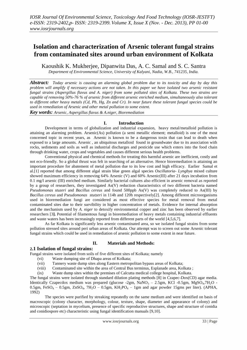
IOSR Journal Of Environmental Science, Toxicology And Food Technology (IOSR-JESTFT)
e-ISSN: 2319-2402,p- ISSN: 2319-2399. Volume X, Issue X (Nov. - Dec. 2013), PP 01-00
www.iosrjournals.org
www.iosrjournals.org 33 | Page
Isolation and characterization of Arsenic tolerant fungal strains
from contaminated sites around urban environment of Kolkata
Kaoushik K. Mukherjee, Dipanwita Das, A. C. Samal and S. C. Santra Department of Environmental Science, University of Kalyani, Nadia, W.B., 741235, India.
Abstract: Today arsenic is causing an alarming global problem due to its toxicity and day by day this
problem will amplify if necessary actions are not taken. In this paper we have isolated two arsenic resistant
fungal strains (Aspergillus flavus and A. niger) from some polluted sites of Kolkata. These two strains are
capable of removing 50%-76 % of arsenic from different arsenic enriched medium, simultaneously also tolerant
to different other heavy metals (Cd, Pb, Hg, Zn and Cr). In near future these tolerant fungal species could be
used in remediation of Arsenic and other metal pollution to some extent.
Key words: Arsenic, Aspergillus flavus & A.niger, Bioremediation
I. Introduction Development in terms of globalization and industrial expansion, heavy metal/metalloid pollution is
attaining an alarming problem. Arsenic(As) pollution (a semi metallic element; metalloid) is one of the most
concerned topic in recent years, as Arsenic is known to be a dangerous toxin that can lead to death when
exposed to a large amounts. Arsenic , an ubiquitous metalloid found in groundwater due to its association with
rocks, sediments and soils as well as industrial discharges and pesticide use which enters into the food chain
through drinking water, crops and vegetables and causes different serious health problems.
Conventional physical and chemical methods for treating this harmful arsenic are inefficient, costly and
not eco-friendly. So a global thrust was felt in searching of an alternative. Hence bioremediation is attaining an
important procedure for abatement of metal pollution due to its low cost and high efficacy. Earlier Samal et
al.[1] reported that among different algal strain blue green algal species Oscillatoria- Lyngbya mixed culture
showed maximum efficiency in removing 64% Arsenic (V) and 60% Arsenic(III) after 21 days incubation from
0.1 mg/l arsenic (III) enriched medium. Similarly bacterial cultures also efficient in arsenic removal as reported
by a group of researchers, they investigated As(V) reduction characteristics of two different bacteria named
Pseudomonas stuzeri and Bacillus cereus and found 500ppb As(V) was completely reduced to As(III) by
Bacillus cereus and Pseudomonas stutzeri in 114h and 120h respectively[2]. Among different microorganisms
used in bioremediation fungi are considered as most effective species for metal removal from metal
contaminated sites due to their survibility in higher concentration of metals. Evidence for internal absorption
and the mechanism used by A. niger to detoxify environmental copper and zinc has been observed by earlier
researchers [3]. Potential of filamentous fungi in bioremediation of heavy metals containing industrial effluents
and waste waters has been increasingly reported from different parts of the world [4,5,6,7].
As far Kolkata is significantly less arsenic contaminated area, so we isolated fungal strains from some
pollution stressed sites around peri urban areas of Kolkata. Our attempt was to screen out some Arsenic tolerant
fungal strains which could be used in remediation of arsenic pollution to some extent in near future.
II. Materials and Methods:
2.1 Isolation of fungal strains:
Fungal strains were isolated from soils of five different sites of Kolkata; namely
(vi) Waste dumping site of Dhapa areas of Kolkata;
(vii) Tannery waste dump sites along Eastern metropolitan bypass areas of Kolkata;
(viii) Contaminated site within the area of Central Bus terminus, Esplanade area, Kolkata ;
(ix) Waste dump sites within the premises of Calcutta medical college hospital, Kolkata.
The fungal strains were isolated through standard dilution plating methods [8] in Czapec-Dox(CD) agar media.
Identically Czapecdox medium was prepared (glucose -2gm, NaNO3 – 2.5gm, KCl -0.5gm, MgSO4,7H2O -
0.5gm, FeSO4 – 0.5gm, ZnSO4, 7H2O – 0.5gm, KH2PO4 – 1gm and agar powder 15gms per liter). (APHA,
1992)
The species were purified by streaking repeatedly on the same medium and were identified on basis of
macroscopic (colony character, morphology, colour, texture, shape, diameter and appearance of colony) and
microscopic (septation in mycelium, presence of specific reproductive structures, shape and structure of conidia
and conidiospore etc) characteristic using fungal identification manuals [9,10].
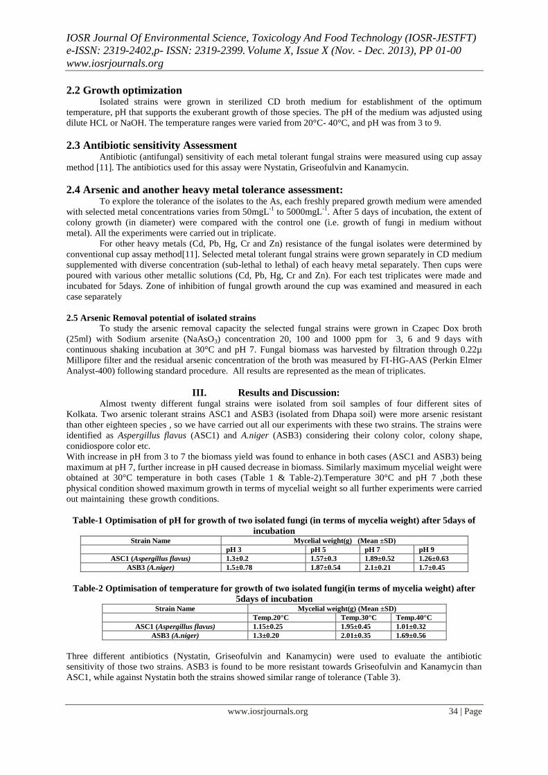
IOSR Journal Of Environmental Science, Toxicology And Food Technology (IOSR-JESTFT)
e-ISSN: 2319-2402,p- ISSN: 2319-2399. Volume X, Issue X (Nov. - Dec. 2013), PP 01-00
www.iosrjournals.org
www.iosrjournals.org 34 | Page
2.2 Growth optimization Isolated strains were grown in sterilized CD broth medium for establishment of the optimum
temperature, pH that supports the exuberant growth of those species. The pH of the medium was adjusted using
dilute HCL or NaOH. The temperature ranges were varied from 20°C- 40°C, and pH was from 3 to 9.
2.3 Antibiotic sensitivity Assessment Antibiotic (antifungal) sensitivity of each metal tolerant fungal strains were measured using cup assay
method [11]. The antibiotics used for this assay were Nystatin, Griseofulvin and Kanamycin.
2.4 Arsenic and another heavy metal tolerance assessment: To explore the tolerance of the isolates to the As, each freshly prepared growth medium were amended
with selected metal concentrations varies from 50mgL-1
to 5000mgL-1
. After 5 days of incubation, the extent of
colony growth (in diameter) were compared with the control one (i.e. growth of fungi in medium without
metal). All the experiments were carried out in triplicate.
For other heavy metals (Cd, Pb, Hg, Cr and Zn) resistance of the fungal isolates were determined by
conventional cup assay method[11]. Selected metal tolerant fungal strains were grown separately in CD medium
supplemented with diverse concentration (sub-lethal to lethal) of each heavy metal separately. Then cups were
poured with various other metallic solutions (Cd, Pb, Hg, Cr and Zn). For each test triplicates were made and
incubated for 5days. Zone of inhibition of fungal growth around the cup was examined and measured in each
case separately
2.5 Arsenic Removal potential of isolated strains
To study the arsenic removal capacity the selected fungal strains were grown in Czapec Dox broth
(25ml) with Sodium arsenite (NaAsO3) concentration 20, 100 and 1000 ppm for 3, 6 and 9 days with
continuous shaking incubation at 30°C and pH 7. Fungal biomass was harvested by filtration through 0.22µ
Millipore filter and the residual arsenic concentration of the broth was measured by FI-HG-AAS (Perkin Elmer
Analyst-400) following standard procedure. All results are represented as the mean of triplicates.
III. Results and Discussion: Almost twenty different fungal strains were isolated from soil samples of four different sites of
Kolkata. Two arsenic tolerant strains ASC1 and ASB3 (isolated from Dhapa soil) were more arsenic resistant
than other eighteen species , so we have carried out all our experiments with these two strains. The strains were
identified as Aspergillus flavus (ASC1) and A.niger (ASB3) considering their colony color, colony shape,
conidiospore color etc.
With increase in pH from 3 to 7 the biomass yield was found to enhance in both cases (ASC1 and ASB3) being
maximum at pH 7, further increase in pH caused decrease in biomass. Similarly maximum mycelial weight were
obtained at 30°C temperature in both cases (Table 1 & Table-2).Temperature 30°C and pH 7 ,both these
physical condition showed maximum growth in terms of mycelial weight so all further experiments were carried
out maintaining these growth conditions.
Table-1 Optimisation of pH for growth of two isolated fungi (in terms of mycelia weight) after 5days of
incubation Strain Name Mycelial weight(g) (Mean ±SD)
pH 3 pH 5 pH 7 pH 9
ASC1 (Aspergillus flavus) 1.3±0.2 1.57±0.3 1.89±0.52 1.26±0.63
ASB3 (A.niger) 1.5±0.78 1.87±0.54 2.1±0.21 1.7±0.45
Table-2 Optimisation of temperature for growth of two isolated fungi(in terms of mycelia weight) after
5days of incubation Strain Name Mycelial weight(g) (Mean ±SD)
Temp.20°C Temp.30°C Temp.40°C
ASC1 (Aspergillus flavus) 1.15±0.25 1.95±0.45 1.01±0.32
ASB3 (A.niger) 1.3±0.20 2.01±0.35 1.69±0.56
Three different antibiotics (Nystatin, Griseofulvin and Kanamycin) were used to evaluate the antibiotic
sensitivity of those two strains. ASB3 is found to be more resistant towards Griseofulvin and Kanamycin than
ASC1, while against Nystatin both the strains showed similar range of tolerance (Table 3).
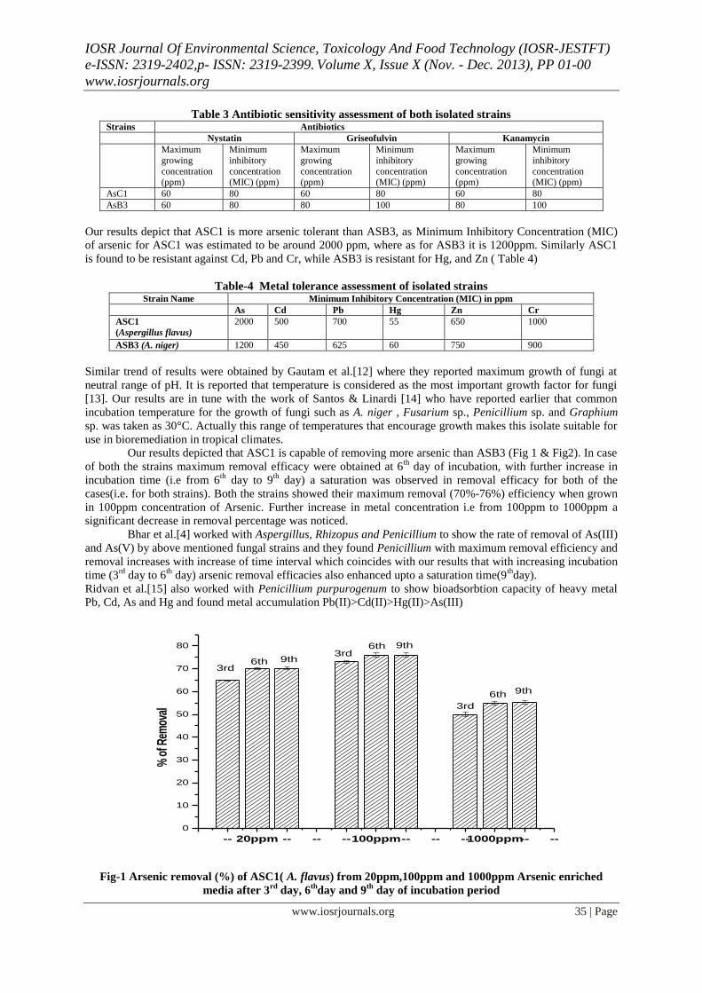
IOSR Journal Of Environmental Science, Toxicology And Food Technology (IOSR-JESTFT)
e-ISSN: 2319-2402,p- ISSN: 2319-2399. Volume X, Issue X (Nov. - Dec. 2013), PP 01-00
www.iosrjournals.org
www.iosrjournals.org 35 | Page
Table 3 Antibiotic sensitivity assessment of both isolated strains Strains Antibiotics
Nystatin Griseofulvin Kanamycin
Maximum
growing
concentration (ppm)
Minimum
inhibitory
concentration (MIC) (ppm)
Maximum
growing
concentration (ppm)
Minimum
inhibitory
concentration (MIC) (ppm)
Maximum
growing
concentration (ppm)
Minimum
inhibitory
concentration (MIC) (ppm)
AsC1 60 80 60 80 60 80
AsB3 60 80 80 100 80 100
Our results depict that ASC1 is more arsenic tolerant than ASB3, as Minimum Inhibitory Concentration (MIC)
of arsenic for ASC1 was estimated to be around 2000 ppm, where as for ASB3 it is 1200ppm. Similarly ASC1
is found to be resistant against Cd, Pb and Cr, while ASB3 is resistant for Hg, and Zn ( Table 4)
Table-4 Metal tolerance assessment of isolated strains Strain Name Minimum Inhibitory Concentration (MIC) in ppm
As Cd Pb Hg Zn Cr
ASC1
(Aspergillus flavus)
2000 500 700 55 650 1000
ASB3 (A. niger) 1200 450 625 60 750 900
Similar trend of results were obtained by Gautam et al.[12] where they reported maximum growth of fungi at
neutral range of pH. It is reported that temperature is considered as the most important growth factor for fungi
[13]. Our results are in tune with the work of Santos & Linardi [14] who have reported earlier that common
incubation temperature for the growth of fungi such as A. niger , Fusarium sp., Penicillium sp. and Graphium
sp. was taken as 30°C. Actually this range of temperatures that encourage growth makes this isolate suitable for
use in bioremediation in tropical climates.
Our results depicted that ASC1 is capable of removing more arsenic than ASB3 (Fig 1 & Fig2). In case
of both the strains maximum removal efficacy were obtained at 6th
day of incubation, with further increase in
incubation time (i.e from 6th
day to 9th
day) a saturation was observed in removal efficacy for both of the
cases(i.e. for both strains). Both the strains showed their maximum removal (70%-76%) efficiency when grown
in 100ppm concentration of Arsenic. Further increase in metal concentration i.e from 100ppm to 1000ppm a
significant decrease in removal percentage was noticed.
Bhar et al.[4] worked with Aspergillus, Rhizopus and Penicillium to show the rate of removal of As(III)
and As(V) by above mentioned fungal strains and they found Penicillium with maximum removal efficiency and
removal increases with increase of time interval which coincides with our results that with increasing incubation
time (3rd
day to 6th
day) arsenic removal efficacies also enhanced upto a saturation time(9th
day).
Ridvan et al.[15] also worked with Penicillium purpurogenum to show bioadsorbtion capacity of heavy metal
Pb, Cd, As and Hg and found metal accumulation Pb(II)>Cd(II)>Hg(II)>As(III)
Fig-1 Arsenic removal (%) of ASC1( A. flavus) from 20ppm,100ppm and 1000ppm Arsenic enriched
media after 3rd
day, 6th
day and 9th
day of incubation period
-- 20ppm -- -- --100ppm-- -- --1000ppm-- --
0
10
20
30
40
50
60
70
80
9th
9th
9th
6th
6th
6th
3rd
3rd
3rd
% o
f Rem
oval
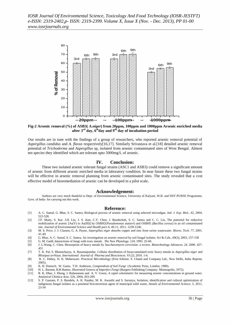
IOSR Journal Of Environmental Science, Toxicology And Food Technology (IOSR-JESTFT)
e-ISSN: 2319-2402,p- ISSN: 2319-2399. Volume X, Issue X (Nov. - Dec. 2013), PP 01-00
www.iosrjournals.org
www.iosrjournals.org 36 | Page
Fig-2 Arsenic removal (%) of ASB3( A.niger) from 20ppm, 100ppm and 1000ppm Arsenic enriched media
after 3rd
day, 6th
day and 9th
day of incubation period
Our results are in tune with the findings of a group of researchers, who reported arsenic removal potential of
Aspergillus candidus and A. flavus respectively[16,17]. Similarly Srivastava et al.[18] detailed arsenic removal
potential of Trichoderma and Aspergillus sp, isolated from arsenic contaminated sites of West Bengal. Almost
ten species they identified which are tolerant upto 5000mg/L of arsenic.
IV. Conclusion: These two isolated arsenic tolerant fungal strains (ASC1 and ASB3) could remove a significant amount
of arsenic from different arsenic enriched media in laboratory condition. In near future these two fungal strains
will be effective in arsenic removal planning from arsenic contaminated sites. The study revealed that a cost
effective model of bioremediation of arsenic can be developed in a pilot scale.
Acknowledgement: Authors are very much thankful to Dept. of Environmental Science, University of Kalyani, W.B. and DST-PURSE Programme,
Govt. of India for carrying out this work.
Reference: [1] A. C. Samal, G. Bhar, S. C. Santra, Biological process of arsenic removal using selected microalgae, Ind. J. Exp. Biol., 42, 2004,
522-528.
[2] J.P. Maity, S. Kar, J.H. Liu, J. S. Jean, C.Y. Chen, J. Bundschuh, S. C. Santra and C. C. Liu, The potential for reductive mobilization of arsenic [As(V) to As(III)] by OSBH2(Pseudomonas stutzeri) and OSBH5 (Bacillus cereus) in an oil contaminated
site. Journal of Environmental Science and Health part A, 46:11, 2011, 1239-1246.
[3] M. S. Price, J. J. Classen, G. A. Payne, Aspergillus niger absorbs copper and zinc from swine wastewater. Biores. Tech. 77, 2001. 41-49.
[4] G. Bhar, A. C. Samal, S. C. Santra, An investigation on arsenic removal by soil fungal isolates. Sci & Cult., 69(3), 2003, 157-158
[5] G. M. Gadd, Interactions of fungi with toxic metals. The New Phytology. 124, 1993. 25-60. [6] J. L.Wang, C. Chen. Biosorption of heavy metals by Saccharomyces cerevisiae: a review. Biotechnology Advances. 24. 2006. 427-
451
[7] T. K. Pal, S. Bhattacharya, A. Basumajumdar. Cellular distribution of bioaccumulated toxic heavy metals in Aspergillus niger and Rhizopus arrhizus. International Journal of Pharma and Biosciences. VI (2), 2010. 1-6.
[8] R. C. Dubey, D. K. Maheswari. Practical Microbiology (First Edition. S. Chand and Company Ltd., New Delhi, India Reprint,
2005). [9] K. H. Domsch, W. Gams, T.H. Anderson, Compendium of Soil Fungi (Academic Press, London, 1980).
[10] H. L. Barnett, B.B Hunter, Illustrated Genera of Imperfect Fungi (Burgess Publishing Company. Minneapolis, 1972).
[11] R. K. Dhar, J. Zheng, J. Rubenstone and A. V. Green, A rapid colorimetric for measuring arsenic concentrations in ground water. Analytical Chimica Acta. 526, 2004, 203-209.
[12] S. P. Gautam, P. S. Bundela, A. K. Pandey, M. K. Awasthi and S. Sarsaiya, Isolation, identification and cultural optimization of
indigenous fungal isolates as a potential bioconversion agent of municipal solid waste, Annals of Environmental Science, 5, 2011, 23-34
--20ppm-- -- --100ppm-- -- --1000ppm--
0
10
20
30
40
50
60
70
80
9th
9th
9th
6th
6th
6th
3rd
3rd
3rd%
of
Rem
ova
l
IOSR Journal Of Environmental Science, Toxicology And Food Technology (IOSR-JESTFT)
e-ISSN: 2319-2402,p- ISSN: 2319-2399. Volume X, Issue X (Nov. - Dec. 2013), PP 01-00
www.iosrjournals.org
www.iosrjournals.org 37 | Page
[13] D. Delille, F. Coulan and E. Pelletier, Effects of temperature warming during a bioremediation study of natural and nutrient-amended hydrocarbon-contaminated sub-Antarctic soils. Cold Reg. Sci. Technol. 40, 2004, 61-70.
[14] V. L. Santos & V. R. Linardi, Biodegradation of phenol by a filamentous fungi isolated from industrial effluents-identification and
degradation potential. Process. Biochem. 39, 2004, 1001-1006. [15] S. Ridvan, Biosorption of cadmium, lead, mercury and arsenic ions by the fungus Penicilium purpurogenum, Seperation Sc. and
Technology, 38(9), 2003, 2039-2053.
[16] A.K. Vala, Tolerance and removal of arsenic by a facultative marine fungus Aspergillus candidus. Bioresour. Technol. 101(7), 2010, 2565-7.
[17] S. Maheswari and A. G. Murugesan, Removal of arsenic(III) from aqueous solutions using Aspergillus flavus isolated from arsenic
contaminated sites. Ind. J. Chem. Technol., 18, 2011, 45-52. [18] P. K. Srivastava, A. Vaish, S. Dwivedi, D .Chakrabarty, N. Singh, R. D. Tripathi, Biological removal of arsenic pollution by soil
fungi. Sci Total Environ., 409(12), 2011, 2430-42.