Embed Size (px)
Citation preview

Structure of the Human Body (2018/2019) Amy Kule, MD
11/2/2018
Abdominal Aorta and Inferior Vena Cava Ultrasound
Date: Friday, November 02, 2018
Time: 10:00 AM
Location: ONLINE and SMALL GROUP LABORATORY
Watch:
➢ Abdominal Aorta Ultrasound Scanning Protocol: (6:05)
https://youtu.be/08fF1OUcecM
➢ Inferior Vena Cava Ultrasound Scanning Protocol: (4:59)
https://www.youtube.com/watch?v=Q6VlG3kv28Y&list=PLGEKJJ3ekUkzFqY2SFfAod
P_NJUPV0qqF&index=2
LEARNING OBJECTIVES
➢ Correlate anatomic structures identified during live-dissection with findings on
ultrasound
➢ Describe normal ultrasound anatomy of the abdominal great vessels, including the aorta,
inferior vena cava, its branches, as well as its relationship with surrounding structures
➢ Select the appropriate transducer and optimizing image capture by adjusting function
keys
HANDS-ON OBJECTIVES
➢ Identify Abdominal Aorta and Correlating Structures
o Transverse View
▪ Vertebral body with posterior shadow
▪ Inferior vena cava
▪ Aorta
▪ Proximal aorta
• Celiac trunk
• Hepatic artery
• Splenic artery
▪ Mid aorta
• Superior mesenteric artery
• Splenic vein
• Left renal vein
▪ Distal aorta
• Bifurcation into common iliac arteries
o Longitudinal View
▪ Celiac trunk
▪ Superior mesenteric artery

Structure of the Human Body (2018/2019) Amy Kule, MD
11/2/2018
➢ Identify Inferior Vena Cava structures
o Longitudinal View
▪ Inferior vena cava
▪ Right atrium
▪ Hepatic vein (may not be visible)

Structure of the Human Body (2018/2019) Amy Kule, MD
11/2/2018
AORTA ULTRASOUND
Gross Anatomy
Credit: https://www.youtube.com/watch?v=uiTsFtanyzM
Credit: https://quizlet.com/14095296/anatomy-lab-1415-checklist-flash-cards/

Structure of the Human Body (2018/2019) Amy Kule, MD
11/2/2018
Ultrasound Anatomy
Probe Selection:
➢ Curvilinear
➢ Phased array
Patient Positioning and Preparation:
➢ Supine
➢ Ideally, fasting prior to exam (avoid bowel gas, which causes air artifact and decreased
visualization of the structures beneath)
1. Technique for scanning the abdominal aorta:
➢ Place probe in the subxiphoid region – transverse orientation, probe indicator oriented
toward patient’s right.
➢ Identify the vertebral shadow, which is a hyper-echoic rim with a posterior shadow, that
appears like an upsidedown “U” or “horseshoe sign”. The aorta will be found anterior to
vertebral body and appear circular and pulsatile. The aorta in relation to the inferior vena
cava (IVC) will appear on the right side of the screen, as it is oriented in the left side of
the patient’s body.
➢ Without removing the probe from the skin, continue to scan in the transverse orientation
by sliding the probe towards the umbilicus.
➢ A complete exam of the abdominal aorta entails visualization of the proximal, mid, and
distal aorta. Normal diameter of the aorta is 3 cm.
➢ The proximal and mid-aorta can also be evaluated in longitudinal orientation.
Credit: https://www.youtube.com/watch?v=8EB0Au3l4AM&index=1&list=PLGEKJJ3ekUkzFqY2SFfAodP_NJUPV0qqF

Structure of the Human Body (2018/2019) Amy Kule, MD
11/2/2018
Credit: reference.medscape.com
Proximal abdominal aorta (below the diaphragm)
➢ The celiac trunk is the 1st major branch of the abdominal aorta.
➢ Look for the “seagull sign” – the wings are the hepatic (screen left) and splenic (screen
right) arteries.
➢ The left gastric artery, the third component of the celiac trunk is usually not visualized on
ultrasound.
Credit: http://www.scielo.cl/scielo.php?script=sci_arttext&pid=S0717-95022007000200002&lng=en&

Structure of the Human Body (2018/2019) Amy Kule, MD
11/2/2018
Credit: http://radiology-anatomy.blogspot.com/2014/03/celiac-trunk-ultrasound-anatomy.html
Credit: https://vimeo.com/41791516

Structure of the Human Body (2018/2019) Amy Kule, MD
11/2/2018
Mid abdominal aorta (near the level of the renal arteries)
➢ The superior mesenteric artery (SMA) is the second major branch of the abdominal aorta.
➢ The splenic vein passes anterior and the left renal vein runs posterior to the SMA.
➢ The SMA view has been called the “mantle clock” sign given its resemblance to one.
Credit: http://www.omicsonline.org/pentafurcation-of-the-celiac-trunk-2161-0940.S7-001.php?aid=4737
Credit: https://vimeo.com/41791516

Structure of the Human Body (2018/2019) Amy Kule, MD
11/2/2018
Distal (Above and at the Iliac Bifurcation)
➢ The aorta will bifurcate at the level of the umbilicus into the common iliac arteries.
➢ Slowly fan up and down to view the distal aorta branch off into the two branches of the
iliac arteries.
Credit: http://www.jcdr.net/ReadXMLFile.aspx?id=4559
Credit: Source: https://www.youtube.com/watch?v=uiTsFtanyzM

Structure of the Human Body (2018/2019) Amy Kule, MD
11/2/2018
Longitudinal Evaluation of Abdominal Aorta
➢ Turning the transducer clock-wise 90 degrees allows for the longitudinal view of the
aorta to appear.
➢ The patient’s head will be to the left of the screen, and the feet to the right.
➢ The celiac trunk (proximal aorta) and the SMA (mid-aorta) can be seen exiting the aorta,
with the SMA running parallel to the aorta distally.
Source: www.emergencyultrasoundteaching.com
Structures to Identify:
➢ Transverse View
✓ Vertebral body with posterior shadow
✓ Inferior vena cava
✓ Aorta
✓ Proximal aorta
• Celiac trunk
• Hepatic artery
• Splenic artery
✓ Mid aorta
• Superior mesenteric artery
• Splenic vein
• Left renal vein
✓ Distal aorta
• Bifurcation into common iliac arteries
➢ Longitudinal View
✓ Celiac trunk
✓ Superior mesenteric artery

Structure of the Human Body (2018/2019) Amy Kule, MD
11/2/2018
INFERIOR VENA CAVA ULTRASOUND
Gross Anatomy
Credit: anatomytopics.wordpress.com
Ultrasound Anatomy
Probe Selection:
➢ Curvilinear
➢ Phased array
Patient Positioning and Preparation:
➢ Supine
➢ Ideally, fasting prior to exam (avoid bowel gas, which causes air artifact and decreased
visualization of the structures beneath)
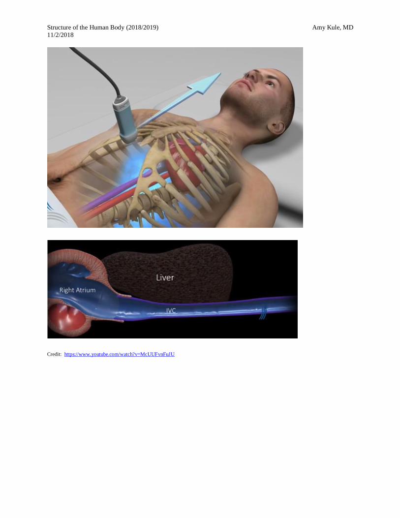
1. Technique:
➢ Place the probe in longitudinal orientation to the subxyphoid region.
➢ Slide probe 1-2 cm to the patient’s right and slightly rock the probe towards the head and
under the rib margin.
➢ The inferior vena cava (IVC) can be seen entering the right atrium of the heart, which
will help differentiate it from the aorta.
➢ The IVC can also be differentiated from the aorta by seeing diameter variability on
patient inspiration or sniffing.
Structure of the Human Body (2018/2019) Amy Kule, MD
11/2/2018
Credit: https://www.youtube.com/watch?v=McUUFvnFuJU

Structure of the Human Body (2018/2019) Amy Kule, MD
11/2/2018
Credit: https://www.ahcmedia.com/articles/136856-ultrasound-for-trauma
Structures to Identify:
➢ Inferior vena cava
➢ Right atrium
➢ Hepatic vein (may not be visible)