Embed Size (px)
Citation preview
A well-controlled nucleus pulposus tissue culture systemwith injection port for evaluating regenerative therapiesCitation for published version (APA):Arkesteijn, I. T. M., Mouser, W. H. M., Mwale, F., Dijk, van, B. G. M., & Ito, K. (2016). A well-controlled nucleuspulposus tissue culture system with injection port for evaluating regenerative therapies. Annals of BiomedicalEngineering, 44(5), 1798-1807. DOI: 10.1007/s10439-015-1428-y
DOI:10.1007/s10439-015-1428-y
Document status and date:Published: 01/05/2016
Document Version:Publisher’s PDF, also known as Version of Record (includes final page, issue and volume numbers)
Please check the document version of this publication:
• A submitted manuscript is the version of the article upon submission and before peer-review. There can beimportant differences between the submitted version and the official published version of record. Peopleinterested in the research are advised to contact the author for the final version of the publication, or visit theDOI to the publisher's website.• The final author version and the galley proof are versions of the publication after peer review.• The final published version features the final layout of the paper including the volume, issue and pagenumbers.Link to publication
General rightsCopyright and moral rights for the publications made accessible in the public portal are retained by the authors and/or other copyright ownersand it is a condition of accessing publications that users recognise and abide by the legal requirements associated with these rights.
• Users may download and print one copy of any publication from the public portal for the purpose of private study or research. • You may not further distribute the material or use it for any profit-making activity or commercial gain • You may freely distribute the URL identifying the publication in the public portal.
If the publication is distributed under the terms of Article 25fa of the Dutch Copyright Act, indicated by the “Taverne” license above, pleasefollow below link for the End User Agreement:
www.tue.nl/taverne
Take down policyIf you believe that this document breaches copyright please contact us at:
providing details and we will investigate your claim.
Download date: 26. Mar. 2019
A Well-Controlled Nucleus Pulposus Tissue Culture System with Injection
Port for Evaluating Regenerative Therapies
IRENE T. M. ARKESTEIJN,1 VIVIAN H. M. MOUSER,1,2 FACKSON MWALE,3 BART G. M. VAN DIJK,1
and KEITA ITO1,2
1Orthopaedic Biomechanics, Department of Biomedical Engineering, Eindhoven University of Technology, PO Box 513, 5600MB Eindhoven, The Netherlands; 2Department of Orthopaedics, University Medical Center Utrecht, Utrecht, The Netherlands;and 3Division of Orthopaedic Surgery, Lady Davis Institute for Medical Research, McGill University, Montreal, Canada
(Received 30 April 2015; accepted 11 August 2015; published online 21 August 2015)
Associate Editor Amit Gefen oversaw the review of this article.
Abstract—In vitro evaluation of nucleus pulposus (NP) tissueregeneration would be useful, but current systems for NPculture are not ideal for injections. The aim of this study wasto develop a long-term culture system for NP tissue thatallows injections of regenerative agents. Bovine caudal NPswere harvested and placed in the newly designed culturesystem. After equilibration of the tissue to 0.3 MPa thevolume was fixed and the tissue was cultured for 28 days. Thecell viability and extracellular matrix composition remainedunchanged during the culture period and gene expressionprofiles were similar to those obtained in earlier studies.Furthermore, to test the responsiveness of bovine caudal NPsin the system, samples were cultured for 4 days and injectedtwice (day 1 and 3) with (1) PBS, (2) Link-N, for regener-ation, and (3) TNF-a, for degeneration. It was shown thatTNF-a increased COX2 gene expression, whereas no effect ofLink-N was detected. In conclusion, the newly designedsystem allows long-term culture of NP tissue, wherein tissuereactions to injected stimulants can be observed.
Keywords—Intervertebral disc, Regeneration, In vitro, Link-
N, TNF-a.
INTRODUCTION
Intervertebral discs support high magnitude loadsand allow multi-directional flexibility in the spine, dueto an ingenious interplay of the tissues within the disc.The highly hydrated nucleus pulposus (NP) evenlydistributes compressive stresses to the organized
annulus fibrosus (AF), which balances these withinternal tensile stresses.31 The human NP starts todegenerate early in adulthood, resulting in a non-uni-form stress distribution, subsequent disorganizationand disruption of the AF, and potentially pain.36 It hasbeen suggested that early stage NP regeneration couldhalt this degenerative cascade.12 Biological therapiesfor NP regeneration aim at restoring the water andproteoglycan content of the tissue. For this purposeexogenous cells, to produce extracellular matrix, andmolecular agents or gene therapy, to increase the ma-trix production by resident cells, can be introducedinto the tissue.12 However promising, these methodsstill face challenges and require more extensive testing.
Currently, the most widely used model to testregenerative therapies is the in vivo animal model. Itsmain advantage is the presence of all natural interac-tions of the organism. However in most test cases,rodents or other small animals are used in whichdegeneration is artificially induced. First of all, suchsmall animals have a mature nuclear cell type, which isdifferent than that of humans.1 Second, the artificiallyinduced degeneration is different from the humanpathophysiological degenerative cascade. Thus, trans-lation of results obtained in studies with artificialdegeneration to naturally degenerated human NPs isdifficult.2 Testing regenerative therapies on in vitrocultured cells is another standard method. However,cell metabolism and phenotype outside of the nativematrix and environment change.9 Hence, tissue culturemodels are of great interest.
Although both organ andNP tissue culture models areavailable, long-term organ cultures are still under devel-opment.4,10 As the evaluation of regenerative therapies
Address correspondence to Bart G. M. van Dijk, Orthopaedic
Biomechanics, Department of Biomedical Engineering, Eindhoven
University of Technology, PO Box 513, 5600 MB Eindhoven, The
Netherlands. Electronic mails: [email protected], v.h.m.
[email protected], [email protected], b.g.m.v.dijk@
tue.nl, [email protected]
Annals of Biomedical Engineering, Vol. 44, No. 5, May 2016 (� 2015) pp. 1798–1807
DOI: 10.1007/s10439-015-1428-y
0090-6964/16/0500-1798/0 � 2015 The Author(s). This article is published with open access at Springerlink.com
1798

often requires long-term studies, we focused on optimizingcurrently available NP culture systems. The advantage ofthe NP culture system is the presence of cell–matrixinteractions in a controlled culture environment. In stan-dard culture conditions NP tissue does not thrive in thelong-term, because it swells tremendously. Previously de-signed culture systems forNP explants have demonstratedthat the tissue matrix can be maintained when it is physi-cally constrained.11,30 Furthermore, when explants werecultured at physiological oxygen, pHand glucose levels for42 days, matrix protein gene expression was nearly pre-served.30 However, our existing system has two majordrawbacks: the swelling pressure cannot be standardized,limiting the reproducibility, and repeated administrationof injections is not feasible, as these damage the culturesystem.
Therefore, the aim of this study is to design and de-velop a novelNP culture system,which allows preventionof tissue swelling in a controlled manner and repeatedinjections of regenerative agents. Itwas hypothesized thatbovine NP tissue, when first equilibrated to a physiolog-ical pressure of 0.3 MPa33 in this new system, is respon-sive to injected stimuli. In a first experiment, NP tissuewas cultured long-term to test the maintenance of theextracellular matrix and gene expression in the system.Then, the response to injections of regenerative peptideLink-N or inflammatory cytokine TNF-a was evaluatedin a short-term experiment.
MATERIALS AND METHODS
Culture System Design
To limit tissue swelling, a system was designed tophysically constrain NP tissue in a custom-made hol-
low cylinder (polyether ether ketone; PEEK) with twoporous stainless steel plates (sintered 316L stainlesssteel; pores 0.5 lm) that allow for molecular transport(Fig. 1). Prior to culture, the position of the upperporous plate, and herewith the tissue’s volume, was setby applying an axial load until equilibrium wasreached (Fig. 2). In these experiments, a pressure of0.3 MPa was applied, and after equilibration (24 h),the position of the upper porous plate was locked.Furthermore, the cylindrical container has an injectionport on the side, which can be (re-)closed with a screw.
Culture System Validation
Fresh bovine tails (24 months old) were obtainedfrom the abattoir according to local regulations.Adjacent to the endplates, the discs (Cy1–Cy5) wereopened transversally, and a biopsy punch (ø 8 mm;273693, Kruuse, Sherburn, UK) was used to harvesttissue from the center of the disc (180–220 mg). NPtissue biopsies were placed in the culture system andequilibrated. After equilibration and subsequent fixa-tion of the tissue volume, the system was placed in aplastic container (60 mL medium) with a lid made of agas permeable silicone membrane (MED82-5010-10,NuSil, Carpinteria, USA) and cultured at 37 �C, 5%O2 and 5% CO2. Culture medium consisted of ad-vanced DMEM (12491-015, Invitrogen, Bleiswijk, theNetherlands) supplemented with 10% fetal bovineserum (758093, Greiner Bio-one, Alphen a/d Rijn, theNetherlands) and 1% penicillin/streptomycin (DE17-602E, Lonza, Basel, Switzerland) and was changedtwice a week.
To assess long-term culture potential, bovine tails ofeight donors were used to obtain eighteen NP samples,
FIGURE 1. Culture system design.
Nucleus Pulposus Culture System for Regeneration 1799

which were randomly distributed (disc level and donor)over the following groups:
a. Day 0, collected immediately after harvesting,b. Day 1, collected after equilibration, andc. Day 28, collected after 28 days of culture.
For each group, 6 samples (corresponding to 6 do-nors) were used. The biochemical and water content(¼ sample), histology and cell viability (½ sample)and gene expression (¼ sample) were analyzed.
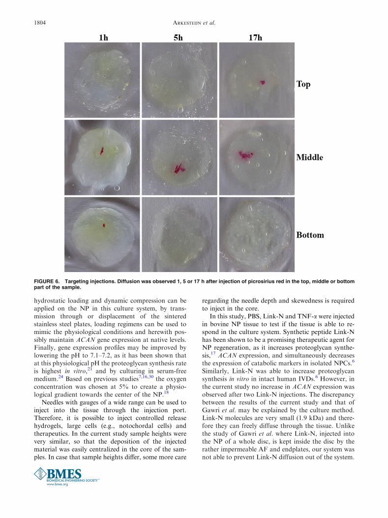
To visualize injections, bovine tails of three donorswere used to obtain six NP samples, which were in-jected with 10 lL 0.1% Picrosirius red (36.554-8, Sig-ma, Zwijndrecht, the Netherlands) with a 32 gaugeneedle (7635-01/00, Hamilton, Bonaduz, Switzerland).Samples were collected 1, 5 or 17 h after injection(n = 2/group) to assess diffusion of the dye throughoutthe tissue.
To assess tissue responsiveness to injected agentsbovine tails of nine donors were used to obtain four-teen NP samples, which were randomly distributed(disc level and donor) over the following groups:
a. Sham, injected with 10 lL of phosphate buf-fered saline (PBS; n = 4),
b. Link-N, injected with 10 lL of 20 lg/lL Link-N peptide (DHLSDNYTLDHDRAIH;CanPeptide Inc; Pointe Claire, QC, Canada;n = 5)
c. TNF-a, injected with 10 lL of 0.5 ng/lLrecombinant human tumor necrosis factor-a(TNF-a; PHC3016, Invitrogen; n = 5),
Injections were administered on day 1, after equili-bration, and on day 3. Samples were harvested on day4. As injections were administered in the center of thesamples, the core of each sample was obtained with a5 mm biopsy punch (Kruuse), and analyzed for geneexpression (½ sample).
Analysis
To assess the biochemical content, samples wereweighed (wet weight), lyophilized (Freezone 2.4, Lab-conco, Kansas City, MO, USA) overnight and then thedry weight was determined. The water content wascalculated by dividing the difference between wet anddry weight by the wet weight. Subsequently, the sam-ples were digested overnight in a papain digestionbuffer (100 mM phosphate buffer, 5 mM L-cystein,5 mM ethylenediaminetetraacetic acid and 140 lg/mLpapain, all from Sigma) at 60 �C. The DNA content ofthe digested sample was measured with a Hoechst dyeassay3 and calf thymus DNA as reference (D3664,Sigma). The glycosaminoglycan (GAG) content wasused as a measure for the amount of proteoglycans.The GAG content was determined with a DMMBassay,5 using shark cartilage chondroitin sulfate(C4384, Sigma) as reference. The fixed charge density(FCD) was calculated from the GAG per wet weight,as described by Narmoneva et al.19 The hydroxypro-line (HYP) content was measured with a chloramin-Tassay8 using trans-4-hydroxyproline (H5534, Sigma) asreference.
For histology, samples were snap-frozen in Tissue-Tek O.C.T. compound (4583, Sakura Finetek,Zoeterwoude, the Netherlands) in isopentane in liquidN2 and stored at 230 �C until further use. 10 lm thicksections were cut (Microm, Thermo Fisher Scientific,Kalamazoo, MI, USA) to assess cell viability andhistology. Lactate dehydrogenase (LDH) staining(N5514, Sigma) to stain living cells dark, as describedby Stoddart et al.28 was used in combination withpropidium iodide staining (P3566, Invitrogen) to markall DNA red-fluorescent to assess the cell viability anddistribution. Extracellular matrix was stained withWeigert’s hematoxylin for nuclei, Safranin-O (84120,Fluka, Sigma) for proteoglycans and Fast Green(1.00056.2500, Merck, Darmstadt, Germany) for col-lagen. Bright field images were made of Safranin-O/Fast Green and LDH sections, while fluorescentimages were taken for the LDH staining (Observer,Zeiss, Sliedrecht, the Netherlands).
To measure the gene expression, samples were snap-frozen and stored at 280 �C until further use. Mes-senger RNA (mRNA) extraction was done as de-scribed by van Dijk et al.30 The concentration of RNAwas determined with a spectrophotometer (ND-1000,
FIGURE 2. Axial load system design. The axial load systemconsists of the following components: (a) (replaceable)weight, (b) transmission of load to the upper sintered stain-less steel plate, (c) culture system as described in Fig. 1, (d)reservoir for culture medium.
ARKESTEIJN et al.1800

Isogen, de Meern, the Netherlands). In total, 63 ng ofmRNA was used for cDNA synthesis (VILO kit,Invitrogen). Gene expression was analyzed in a realtime polymerase chain reaction (qPCR, CFX384, Bio-Rad, Veenendaal, the Netherlands) using the SYBRGreen Supermix (Bio-Rad). Three reference genes [18s(PrimerDesign, Southampton, UK), glyceraldehyde-3-phosphate dehydrogenase (GAPDH), and RPL13a]were evaluated in each experiment, and the most stableone was selected as reference gene. For the long-termculture 18s was selected and for the short-term tissueresponse experiment GAPDH. The genes of interestwere aggrecan (ACAN), collagen type I (COL1), col-lagen type II (COL2), matrix metalloprotease 13(MMP13), tissue inhibitor of metalloproteinase 1 and2 (TIMP1, TIMP2) for both experiments. Addition-ally, cyclooxygenase 2 (COX2) was tested in the short-term tissue response experiment. Primer sequences areprovided in Table 1. Levels of expression were deter-mined using the DCt method13 and Ct values werenormalized to the selected reference gene.
To visualize picrosirius injections, samples wereentirely snap-frozen in Tissue-Tek (Sakura) after cul-ture. 100 lm-thick transversal slices were cut at dif-ferent heights (top, middle and bottom) of the sampleto visualize (Observer, Zeiss) the distribution of thestaining inside the sample.
Statistics
Statistical analysis was performed using R-projectsoftware (version 3.0.2).23 At first, all data sets were
tested for group homogeneity and normal distributionwith Levene’s and Shapiro-Wilks’ tests respectively.When homogeneous and normally distributed, a one-way ANOVA was performed with Bonferroni’s cor-rected post hoc independent t test. If data were notnormal, a Kruskal–Wallis test was done with Bonfer-roni’s corrected post hoc Mann–Whitney test. Statisti-cal significance was assumed for p< 0.05.
RESULTS
NP Tissue is Maintained in Long-Term Culture
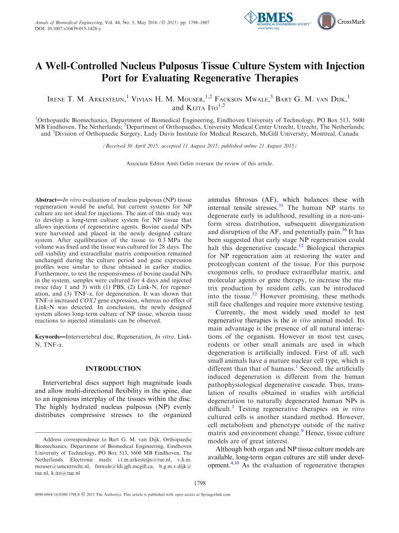
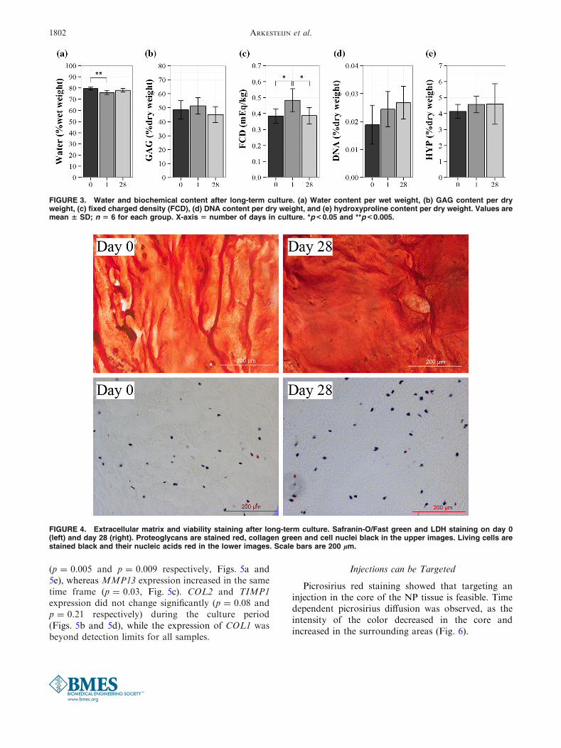
The water content decreased between day 0 and day1 (p = 0.003), which indicates that water was removedfrom the tissue during the equilibration phase. Bytracking the change in sample height, it could be seenthat the equilibrium was reached within 15 h (data notshown). Related to the change in water content, theFCD was higher on day 1 than on day 0 (p = 0.02,Fig. 3c). However, by day 28 of culture, the watercontent and FCD were not significantly different fromday 0 (p = 0.30 and p = 0.95 respectively, Fig. 3a).The total GAG content did not change (p = 0.27,Fig. 3b), which was corroborated with the Safranin-O/Fast Green staining (Fig. 4). The collagen content,DNA content (p = 0.55 and p = 0.15 respectively,Figs. 3d and 3e), and cell viability (Fig. 4) were notdifferent between days of culture. The mRNAexpression of ACAN, the main extracellular matrixprotein, and TIMP2 decreased between day 0 and 28
TABLE 1. List of primers for gene expression analysis.
Gene Accession number Oligonucleotide sequence (5¢ fi 3¢)
ACAN NM_173981 F: CCAACGAAACCTATGACGTGTACT
R: GCACTCGTTGGCTGCCTC
COL1 NM_001034039 F: TGAGAGAGGGGTTGTTGGAC
R: GGGAGACCATTGAGTCCATC
COL2 NM_001113224 F: TGGCTGACCTGACCTGAC
R: GGGCGTTTGACTCACTCC
MMP13 NM_174389 F: CCTTGATGCCATAACCAGTCTCC
R: ATCAATACGGTTGGGAAGTTCTGG
TIMP1 NM_174471 F: GTCAATGAAACTGCCTTATACC
R: TTCTGGGACCTGTGGAAG
TIMP2 NM_174412 F: GCAACGACATCTACGGCAACC
R: CCCACACACGGCAGAGGAG
COX2 NM_174445 F: TCCACCAACTTATAATGTGCAC
R: GGCAGTCATCAGGCACAGGA
GAPDH* NM_001034034 F: GGCGTGAACCACGAGAAGTATAA
R: CCCTCCACGATGCCAAAGT
18s* (Primer Design Ltd)
*Reference gene.
ACAN = aggrecan; COL1/COL2 = collagen type I/II; MMP13 = matrix metalloprotease 13; TIMP1/TIMP2 = tissue inhibitor of metallopro-
teinase 1/2; COX2 = cyclooxygenase 2; GAPDH = glyceraldehydes-3-phosphate dehydrogenase.
Nucleus Pulposus Culture System for Regeneration 1801
(p = 0.005 and p = 0.009 respectively, Figs. 5a and5e), whereas MMP13 expression increased in the sametime frame (p = 0.03, Fig. 5c). COL2 and TIMP1expression did not change significantly (p = 0.08 andp = 0.21 respectively) during the culture period(Figs. 5b and 5d), while the expression of COL1 wasbeyond detection limits for all samples.
Injections can be Targeted
Picrosirius red staining showed that targeting aninjection in the core of the NP tissue is feasible. Timedependent picrosirius diffusion was observed, as theintensity of the color decreased in the core andincreased in the surrounding areas (Fig. 6).
FIGURE 4. Extracellular matrix and viability staining after long-term culture. Safranin-O/Fast green and LDH staining on day 0(left) and day 28 (right). Proteoglycans are stained red, collagen green and cell nuclei black in the upper images. Living cells arestained black and their nucleic acids red in the lower images. Scale bars are 200 lm.
FIGURE 3. Water and biochemical content after long-term culture. (a) Water content per wet weight, (b) GAG content per dryweight, (c) fixed charged density (FCD), (d) DNA content per dry weight, and (e) hydroxyproline content per dry weight. Values aremean 6 SD; n 5 6 for each group. X-axis 5 number of days in culture. *p< 0.05 and **p< 0.005.
ARKESTEIJN et al.1802

NP Tissue is Responsive to TNF-a
COX2 expression was significantly higher in thesamples injected with TNF-a than in the sham andLink-N groups (p = 0.02 and p = 0.04 respectively,Fig. 7f), which shows that NP tissue is responsive inthis system. Injections of Link-N did not result in achange in gene expression when compared to the shamgroup (all genes, p> 0.40; Figs. 7a–e). The expressionof COL1 was beyond detection limits for all samples.
DISCUSSION
It was previously shown that constraining NP tissuemaintains the extracellular matrix and mostly pre-serves native gene expression profiles in long-termculture.30 However, constraining of the tissue was donemanually and reproducibility was less than desired.Furthermore, the actual osmolarity and hydraulicpressure in the NP were unknown.30 By controlling theinitial osmotic pressure, a novel and very reproducibleNP culture system was designed where a fixed tissuevolume could be created as a function of the initialosmotic pressure.
Previous studies have shown adverse effects on geneexpression and matrix composition if static compres-sion of 0.4 MPa or higher was applied.14,32 Therefore,the axial load for equilibration in this study was chosento reach an osmotic pressure of approximately0.3 MPa, as the osmotic pressure range at rest inhumans is 0.1–0.3 MPa in vitro.33 Equilibration with0.3 MPa caused some water loss initially, resulting inan increased FCD and osmotic pressure at day 1.Nevertheless, after 28 days of culture the water contentand FCD returned to that in freshly harvested tissue.While no statistically significant changes in the GAGcontent and histology were observed compared to day
1, it is possible that a small (quantitatively insignifi-cant) amount of GAG was lost. As aggrecan is partlyfragmented, even in healthy IVD tissue,26 fragmentscould have diffused out of the tissue between day 0 and28. This GAG loss could have caused a decrease inFCD and osmotic pressure. Without a change in hy-draulic pressure this would have resulted in a loss ofwater, but with a reduction of the tissue’s volumeaccompanying the loss of GAGs, the drop in hydraulicpressure could have been equal or greater than thedrop in osmotic pressure. This caused either no ex-change of water or a slight influx into the tissue untilosmotic and hydraulic pressure were again equal.Therefore, in hindsight 0.3 MPa may have been asuboptimal equilibrium load, as self-regulation wasnecessary to maintain the tissue. Nevertheless, theextracellular matrix composition and cell viability re-mained statistically unchanged in long-term culture,thus the decrease in osmotic pressure was small andchanges in tissue content were statistically insignifi-cant.
However, the expression of some genes did change.The expression of main matrix component ACANdecreased, like in some previous long-term NP cul-tures.30 Also in this respect, the 0.3 MPa during equi-libration may have been suboptimal, and an optimalpressure, at which both extracellular matrix and geneexpression are maintained, may exist and should beexplored. However, additional influential factors mayalso be considered. Previously, it has been shown thatanabolic gene expression of isolated NP cells (NPCs) inhydrogels increases after dynamic hydrostatic load-ing,15,20 that mildly degenerated rat NPs showincreased ACAN expression after dynamic axial com-pression,34 and that unloaded caprine discs show asignificantly lower ACAN expression in their NPs thanthe NPs of loaded discs,22 if the applied forces arewithin the physiological range. Since both dynamic
FIGURE 5. Gene expression after long-term culture. The expression of (a) aggrecan, (b) collagen type II, (c) matrix metallopro-tease 13, (d) tissue inhibitor of metalloprotease (TIMP) 1, and (e) TIMP2 relative to reference gene 18s. Please note the logarithmicy-axis and error bars. X-axis 5 number of days in culture. Values are mean 6 SD; n 5 5 for day 0 and 1; n 5 3 for day 28. *p< 0.05.
Nucleus Pulposus Culture System for Regeneration 1803
hydrostatic loading and dynamic compression can beapplied on the NP in this culture system, by trans-mission through or displacement of the sinteredstainless steel plates, loading regimens can be used tomimic the physiological conditions and herewith pos-sibly maintain ACAN gene expression at native levels.Finally, gene expression profiles may be improved bylowering the pH to 7.1–7.2, as it has been shown thatat this physiological pH the proteoglycan synthesis rateis highest in vitro,21 and by culturing in serum-freemedium.24 Based on previous studies7,16,30 the oxygenconcentration was chosen at 5% to create a physio-logical gradient towards the center of the NP.18
Needles with gauges of a wide range can be used toinject into the tissue through the injection port.Therefore, it is possible to inject controlled releasehydrogels, large cells (e.g., notochordal cells) andtherapeutics. In the current study sample heights werevery similar, so that the deposition of the injectedmaterial was easily centralized in the core of the sam-ples. In case that sample heights differ, some more care
regarding the needle depth and skewedness is requiredto inject in the core.
In this study, PBS, Link-N and TNF-a were injectedin bovine NP tissue to test if the tissue is able to re-spond in the culture system. Synthetic peptide Link-Nhas been shown to be a promising therapeutic agent forNP regeneration, as it increases proteoglycan synthe-sis,17 ACAN expression, and simultaneously decreasesthe expression of catabolic markers in isolated NPCs.6
Similarly, Link-N was able to increase proteoglycansynthesis in vitro in intact human IVDs.6 However, inthe current study no increase in ACAN expression wasobserved after two Link-N injections. The discrepancybetween the results of the current study and that ofGawri et al. may be explained by the culture method.Link-N molecules are very small (1.9 kDa) and there-fore they can freely diffuse through the tissue. Unlikethe study of Gawri et al. where Link-N, injected intothe NP of a whole disc, is kept inside the disc by therather impermeable AF and endplates, our system wasnot able to prevent Link-N diffusion out of the system.
FIGURE 6. Targeting injections. Diffusion was observed 1, 5 or 17 h after injection of picrosirius red in the top, middle or bottompart of the sample.
ARKESTEIJN et al.1804

Furthermore, in the current study, the injected amountof Link-N was not corrected for the (very large) vol-ume of culture medium, in which Link-N diffused.Picrosirius red, used for testing the injection port, has asimilar molecular weight as Link-N and diffusedthroughout the whole tissue within 17 h. In this re-spect, it is likely that the final tissue concentration ofLink-N in the current study was below the effectivenesslevel. For future studies, it is recommended to inject acontrolled release hydrogel containing Link-N, in or-der to keep Link-N available to the cells during the fullperiod of culture. Alternatively, a larger growth factorlike osteogenic protein-1 (OP-1; a.k.a. BMP-7) may beused. Due to their size, the diffusion of these moleculesis expected to be slower.
TNF-a (17.5 kDa) is one of the major inflammatorycytokines measured during herniation and degenera-tion of the NP.25,35 In this study, TNF-a injectionsresulted in an increased COX2 expression compared tothe sham and Link-N groups. Up regulation of COX2expression in response to TNF-a stimulation is inaccordance with literature that describes the effect of
TNF-a on isolated NPCs29 or in tissue engineeredNP.27 In addition to COX2 up regulation, the latterstudy showed down regulation of ACAN expressionafter TNF-a supplementation, which was not observedin the current study.27 This discrepancy could be ex-plained by the difference in experimental set-upbetween the studies: cells isolated from their nativeenvironment may not fully maintain their phenotypeduring the isolation procedure and/or they may behavedifferently when no native extracellular matrix is pre-sent.9 The differences between the studies emphasizethe added value of culturing NP tissue compared toisolated NPC culture only. In these culture systems theNP tissue can be maintained and responsive underphysiological conditions. IVD organ culture systemsadditionally include interactions with the other tissuesof the IVD, but maintaining the tissue for longerperiods is difficult.4,10 The in vivo testing of therapeu-tics is most attractive, but expensive and not ethical inearly stages of research.1
In conclusion, the native biochemical content andcell viability of NP tissue were maintained when
FIGURE 7. Gene expression after injections of Link-N and TNF-a. The expression of (a) aggrecan, (b) collagen type II, (c) matrixmetalloprotease 13, (d) tissue inhibitor of metalloprotease (TIMP) 1, (e) TIMP2, and (f) cyclooxygenase 2 relative to reference geneglyceraldehyde-3-phosphate dehydrogenase. Please note the logarithmic y-axis and error bars. X-axis 5 injection group. Valuesare mean 6 SD; n 5 3 for sham; n 5 4 for Link-N; n 5 5 for TNF-a. *p<0.05.
Nucleus Pulposus Culture System for Regeneration 1805
cultured at a fixed tissue volume after equilibrationwith an osmotic pressure of 0.3 MPa for 28 days. Inaddition, injections of cytokine TNF-a showed that thetissue is responsive. This novel culture system can beused to culture NP explants in a very reproduciblemanner and to test potential regenerative therapies.
ACKNOWLEDGEMENTS
Financial support: This project was internally fun-ded.
OPEN ACCESS
This article is distributed under the terms of theCreative Commons Attribution 4.0 InternationalLicense (http://creativecommons.org/licenses/by/4.0/),which permits unrestricted use, distribution, and re-production in any medium, provided you give appro-priate credit to the original author(s) and the source,provide a link to the Creative Commons license, andindicate if changes were made.
REFERENCES
1Alini, M., S. M. Eisenstein, K. Ito, C. Little, A. A. Kettler,K. Masuda, J. Melrose, J. Ralphs, I. Stokes, and H. J.Wilke. Are animal models useful for studying human discdisorders/degeneration? Eur. Spine J. 17:2–19, 2008.2Bergknut, N., J. P. H. J. Rutges, H.-J. C. Kranenburg, L.A. Smolders, R. Hagman, H.-J. Smidt, A.-S. Lagerstedt, L.C. Penning, G. Voorhout, H. A. W. Hazewinkel, G. C. M.Grinwis, L. B. Creemers, B. P. Meij, and W. J. A. Dhert.The dog as an animal model for intervertebral discdegeneration? Spine (Phila Pa. 1976) 37(351–8):2012,2012.3Cesarone, C. F., C. Bolognesi, and L. Santi. Improvedmicrofluorometric DNA determination in biologicalmaterial using 33258 Hoechst. Anal. Biochem. 100:188–197, 1979.4Chan, S. C. W., B. Gantenbein-Ritter, V. Y. L. Leung, D.Chan, K. M. C. Cheung, and K. Ito. Cryopreservedintervertebral disc with injected bone marrow-derivedstromal cells: a feasibility study using organ culture. SpineJ. 10:486–496, 2010.5Farndale, R., D. Buttle, and A. Barrett. Improved quan-titation and discrimination of sulphated glycosaminogly-cans by use of dimethylmethylene blue. Biochim. Biophys.Acta 883:173–177, 1986.6Gawri, R., J. Antoniou, J. Ouellet, W. Awwad, T. Steffen,P. Roughley, L. Haglund, and F. Mwale. Best paper NASS2013: Link-N can stimulate proteoglycan synthesis in thedegenerated human intervertebral discs. Eur. Cells Mater.26:107–119, 2013.7Gorth, D. J., K. E. Lothstein, J. A. Chiaro, M. J. Farrell,G. R. Dodge, D. M. Elliott, N. R. Malhotra, R. L. Mauck,
and L. J. Smith. Hypoxic regulation of functional extra-cellular matrix elaboration by nucleus pulposus cells inlong-Term agarose culture. J. Orthop. Res. 33:747–754,2015.8Huszar, G., J. Maiocco, and F. Naftolin. Monitoring ofcollagen and collagen fragments in chromatography ofprotein mixtures. Anal. Biochem. 105:424–429, 1980.9Ishihara, H., and K. Warensjo. Proteoglycan synthesis inthe intervertebral disk nucleus: the role of extracellularosmolality. Am. J. Physiol. 272:c1499–c1506, 1997.
10Korecki, C. L., J. J. MacLean, and J. C. Iatridis. Charac-terization of an in vitro intervertebral disc organ culturesystem. Eur. Spine J. 16:1029–1037, 2007.
11Le Maitre, C. L., J. A. Hoyland, and A. J. Freemont.Studies of human intervertebral disc cell function in aconstrained in vitro tissue culture system. Spine (Phila. Pa.1976) 29:1187–1195, 2004.
12Lewis, G. Nucleus pulposus replacement and regeneration/repair technologies: present status and future prospects. J.Biomed.Mater. Res. B. Appl. Biomater. 100:1702–1720, 2012.
13Livak, K. J., and T. D. Schmittgen. Analysis of relativegene expression data using real-time quantitative PCR andthe 2(-Delta Delta C(T)) Method. Methods 25:402–408,2001.
14Lotz, J. C., A. H. Hsieh, A. L. Walsh, E. I. Palmer, and J.R. Chin. Mechanobiology of the intervertebral disc. Bio-chem. Soc. Trans. 30:853–858, 2002.
15Lyn, C., L. Maitre, J. Frain, A. P. Fotheringham, and A. J.Freemont. Human cells derived from degenerate interver-tebral discs respond differently to those derived from non-degenerate intervertebral discs following application ofdynamic hydrostatic pressure. Biorheology 45:563–575,2008.
16Mwale, F., I. Ciobanu, D. Giannitsios, P. Roughley, T.Steffen, and J. Antoniou. Effect of oxygen levels on pro-teoglycan synthesis by intervertebral disc cells. Spine(Phila. Pa. 1976) 36:E131–E138, 2011.
17Mwale, F., C. N. Demers, A. Petit, P. Roughley, A. R.Poole, T. Steffen, M. Aebi, and J. Antoniou. A syntheticpeptide of link protein stimulates the biosynthesis of col-lagens II, IX and proteoglycan by cells of the intervertebraldisc. J. Cell. Biochem. 88:1202–1213, 2003.
18Naqvi, S. M., and C. T. Buckley. Extracellular matrixproduction by nucleus pulposus and bone marrow stemcells in response to altered oxygen and glucose microenvi-ronments. J. Anat. Epub, 2015.
19Narmoneva, D. A., H. S. Cheung, J. Y. Wang, D. S.Howell, and L. A. Setton. Altered swelling behavior offemoral cartilage following joint immobilization in a caninemodel. J. Orthop. Res. 20:83–91, 2002.
20Neidlinger-Wilke, C., K. Wurtz, A. Liedert, C. Schmidt, W.Borm, A. Ignatius, H.-J. Wilke, and L. Claes. A three-dimensional collagen matrix as a suitable culture system forthe comparison of cyclic strain and hydrostatic pressureeffects on intervertebral disc cells. J. Neurosurg. Spine2:457–465, 2005.
21Ohshima, H., and J. Urban. The effect of lactate and pH onproteoglycan and protein synthesis rates in the interverte-bral disc. Spine (Phila. Pa. 1976) 17:1079–1082, 1992.
22Paul, C. P. L., H. A. Zuiderbaan, B. Z. Doulabi, A. J. vander Veen, P. M. van de Ven, T. H. Smit, M. N. Helder, B. J.van Royen, and M. G. Mullender. Simulated-physiologicalloading conditions preserve biological and mechanicalproperties of caprine lumbar intervertebral discs in ex vivoculture. PLoS One 7:29–34, 2012.
ARKESTEIJN et al.1806
23R Core Team. R: A Language and Environment for Sta-tistical Computing. 2014.
24Reza, A. T., and S. B. Nicoll. Serum-free, chemically definedmedium with TGF-beta3 enhances functional properties ofnucleus pulposus cell-laden carboxymethylcellulose hydrogelconstructs. Biotechnol. Bioeng. 105:384–395, 2010.
25Risbud, M. V., and I. M. Shapiro. Role of cytokines inintervertebral disc degeneration: pain and disc content.Nat. Rev. Rheumatol. 10:44–56, 2014.
26Roughley, P. J., L. I. Melching, T. F. Heathfield, R. H.Pearce, and J. S. Mort. The structure and degradation ofaggrecan in human intervertebral disc. Eur. Spine J.15(Suppl 3):S326–S332, 2006.
27Seguin, C. A., M. Bojarski, R. M. Pilliar, P. J. Roughley,and R. A. Kandel. Differential regulation of matrixdegrading enzymes in a TNFalpha-induced model of nu-cleus pulposus tissue degeneration. Matrix Biol. 25:409–418, 2006.
28Stoddart, M. J., P. I. Furlong, A. Simpson, C. M. Davies,and R. G. Richards. A comparison of non-radioactivemethods for assessing viability in ex vivo cultured cancel-lous bone: technical note. Eur. Cells Mater. 12:16–25, 2006.
29Studer, R. K., N. Vo, G. Sowa, C. Ondeck, and J. Kang.Human nucleus pulposus cells react to IL-6: independentactions and amplification of response to IL-1 and TNF-a.Spine (Phila. Pa. 1976) 36:593–599, 2011.
30Van Dijk, B. G. M., E. Potier, and K. Ito. Long-termculture of bovine nucleus pulposus explants in a nativeenvironment. Spine J. 13:454–463, 2013.
31Walker, M. H., and D. G. Anderson. Molecular basis ofintervertebral disc degeneration. Spine J. 4:158S–166S,2004.
32Wang, D. L., S.-D. Jiang, and L.-Y. Dai. Biologic responseof the intervertebral disc to static and dynamic compressionin vitro. Spine (Phila. Pa. 1976) 32:2521–2528, 2007.
33Wilke, H., P. Neef, M. Caimi, T. Hoogland, and L. Claes.New in vivo measurements of pressures in the interverte-bral disc in daily life. Spine (Phila. Pa. 1976) 24:755–762,1999.
34Wuertz, K., K. Godburn, J. J. MacLean, A. Barbir, J. S.Donnelly, P. J. Roughley, M. Alini, and J. C. Iatridis. Invivo remodeling of intervertebral discs in response to short-and long-term dynamic compression. J. Orthop. Res.27:1235–1242, 2009.
35Wuertz, K., and L. Haglund. Inflammatory mediators inintervertebral disk degeneration and discogenic pain. Glob.Spine J. 3:175–184, 2013.
36Zhao, C.-Q., L.-M. Wang, L.-S. Jiang, and L.-Y. Dai. Thecell biology of intervertebral disc aging and degeneration.Ageing Res. Rev. 6:247–261, 2007.
Nucleus Pulposus Culture System for Regeneration 1807


![Puerarin Relieved Compression-Induced Apoptosis and ...downloads.hindawi.com/journals/sci/2020/7126914.pdfimpaired nucleus pulposus cell (NPC) proliferation [4]. Nucleus pulposus mesenchymal](https://img.pdfslide.us/doc/110x75/5f7fa24ee6184370f175b23e/puerarin-relieved-compression-induced-apoptosis-and-impaired-nucleus-pulposus.jpg)