Embed Size (px)
Citation preview
IN VITRO INVESTIGATION OF CYTOKINE-MEDIATED NUCLEUS PULPOSUS
DEGENERATION
NSF Summer Undergraduate Fellowship in Sensor Technologies
Sarena Horava, Chemical Engineering, University of Massachusetts Amherst
Advisor: Dawn Elliott
Post-Doctoral Fellow: Lachlan Smith
ABSTRACT
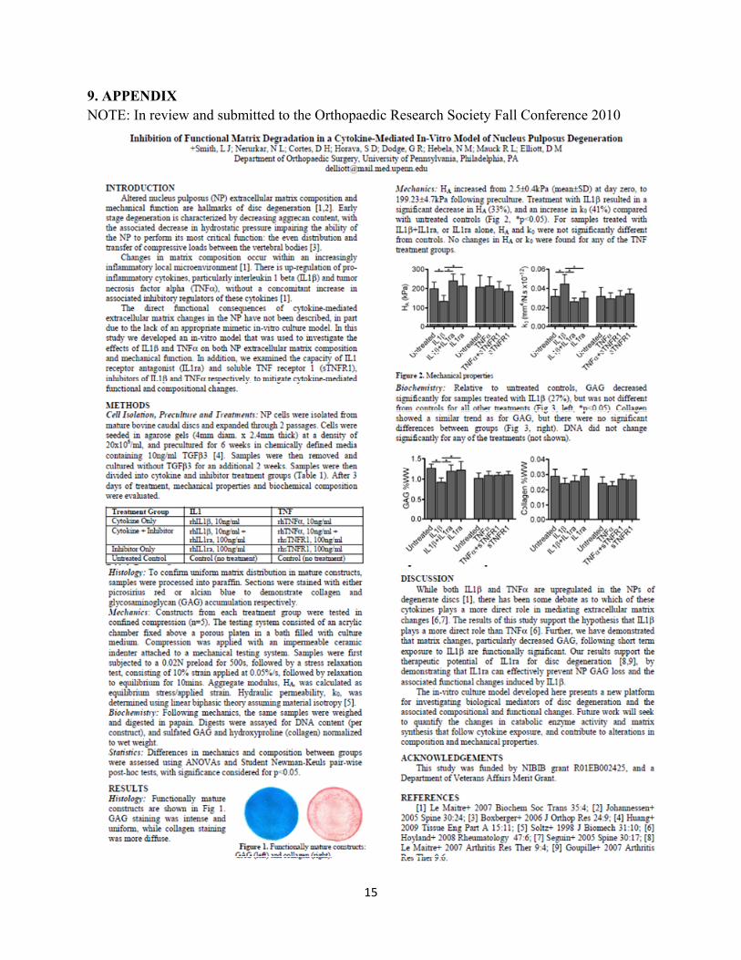
Degeneration of the lumbar intervertebral discs is strongly implicated as a cause of low back pain, and may also lead to impaired mobility. A lack of understanding of the pathomechanisms that underlie degeneration limits our ability to develop biological treatments that both alleviate painful symptoms and restore function. The process of degeneration is characterized by up-regulation of pro-inflammatory cytokines—particularly interleukin 1 beta (IL1β) and tumor necrosis factor alpha (TNFα)—within the central nucleus pulposus. Furthermore, the increased production of these cytokines is not matched by increasing amounts of their inhibitory regulators, resulting in an imbalance of catabolic and anabolic activity. In this study, we developed an in-vitro model of the nucleus pulposus that was used to investigate the effects of IL1β and TNFα on composition and mechanical function. In addition, we examined the capacity of IL1 receptor antagonist (IL1ra) and soluble TNF receptor 1 (sTNFR1), inhibitors of IL1β and TNFα respectively, to mitigate cytokine-mediated functional and compositional changes. Our results demonstrated that short-term exposure to IL1β, but not TNFα, causes loss of matrix components that significantly compromises mechanical function, suggesting that IL1β plays a more direct role than TNFα in driving matrix degradation in the nucleus pulposus. Our results also demonstrated that IL1ra can effectively prevent compositional and functional changes induced by IL1β, highlighting its therapeutic potential.
2
Table of Contents
1. INTRODUCTION ...................................................................................................................... 3
2. BACKGROUND ........................................................................................................................ 3
2.2 Intervertebral Discs ............................................................................................................... 3
2.2.1 Disc Anatomy ................................................................................................................. 3
2.2.2 Nucleus Pulposus ............................................................................................................ 4
2.3 Human Disc Degeneration .................................................................................................... 4
2.3.1 Alterations in NP Structure, Composition, and Mechanics ............................................ 4
2.3.2 Cytokine-Mediated Matrix Degradation ........................................................................ 5
2.4 Pro-Inflammatory Cytokines ................................................................................................. 6
2.4.1 Association of IL1 with IVD .......................................................................................... 6
2.4.2 Association of TNFα with IVD ...................................................................................... 6
2.4.3 Roles of Cytokines ......................................................................................................... 7
3. METHODS ................................................................................................................................. 7
3.3 Histology ............................................................................................................................... 7
3.4 Mechanics.............................................................................................................................. 8
3.5 Biochemical Analysis ............................................................................................................ 8
3.6 Statistical Analysis ................................................................................................................ 8
4. RESULTS ................................................................................................................................... 8
4.1 Histology ............................................................................................................................... 8
4.2 Mechanics.............................................................................................................................. 9
4.3 Biochemical Analysis ............................................................................................................ 9
5. DISCUSSION ........................................................................................................................... 10
6. RECOMMENDATIONS .......................................................................................................... 11
7. ACKNOWLEDGMENTS ........................................................................................................ 11
8. REFERENCES ......................................................................................................................... 11
9. APPENDIX ............................................................................................................................... 15
3
1. INTRODUCTION
Disorders of the lower back impact our society, both physically through individual impairment and financially through medical expenses. The most common cause of musculoskeletal impairment is low back pain. The prevalence of low back pain among the general U.S. population is very high, reaching 25 percent [1]. From a financial viewpoint, back disorders result in lost productivity and increased health care costs. Since low back pain is a common disorder negatively affecting society, much medical research has focused on the causes and treatment of low back pain.
Intervertebral discs (IVDs) are a critical component of the spine. These discs are pads of fibrocartilage that both transfer and distribute compressive loads between vertebral bodies, and permit spinal movement. Intervertebral disc degeneration is strongly implicated as a cause of low back pain [4-7]. Current treatments for painful disc degeneration, such as spinal fusion, are aimed at allaying painful symptoms, without restoring function. The limit of these treatment options is in part due to an incomplete understanding of the biological mechanisms involved. The central nucleus pulposus (NP) is crucial to disc function and health, and is the focus of this research [8,9]. Our objective was to investigate the initiation and progression of IVD degeneration and potential therapies using a new in vitro NP model. A complete understanding of the fundamental mechanisms associated with disc degeneration is necessary for developing novel treatments of low back pain.
2. BACKGROUND
2.2 Intervertebral Discs
2.2.1 Disc Anatomy
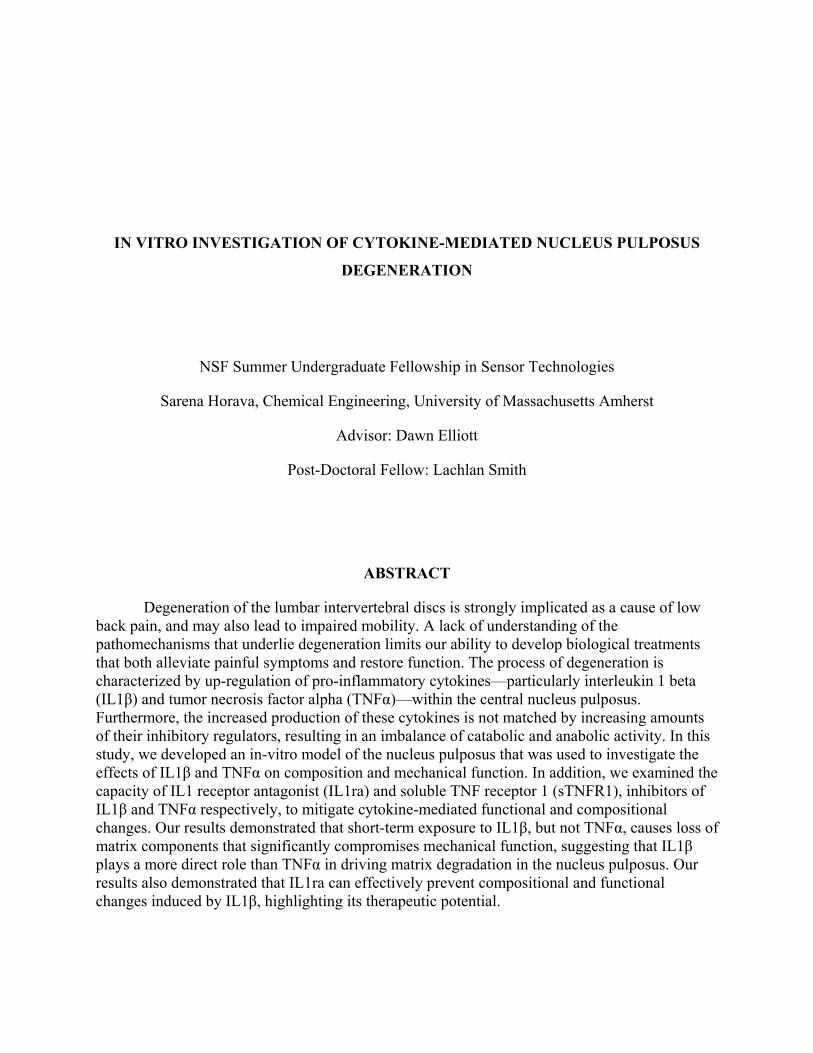
The spine consists of vertebral bones interconnected with intervertebral discs. The discs are soft tissue structures that provide flexibility and integrity to the spine. IVDs both transfer and distribute compressive loads between vertebrae, and permit spinal movement. Proper function is based on IVD structure. Three regions comprise each disc: an outer anulus fibrosus (AF); superior and inferior cartilaginous end plates; and the central NP [8, 9] (Figure 1).
A B
4
Figure 1. The structure of the intervertebral disc: (A) sagittal and (B) transverse sections. (Sources: http://www.medscape.com/viewarticle/405642_2 and http://www.chiropractic-help.com/L4-Lumbar-Spine.html)
2.2.2 Nucleus Pulposus
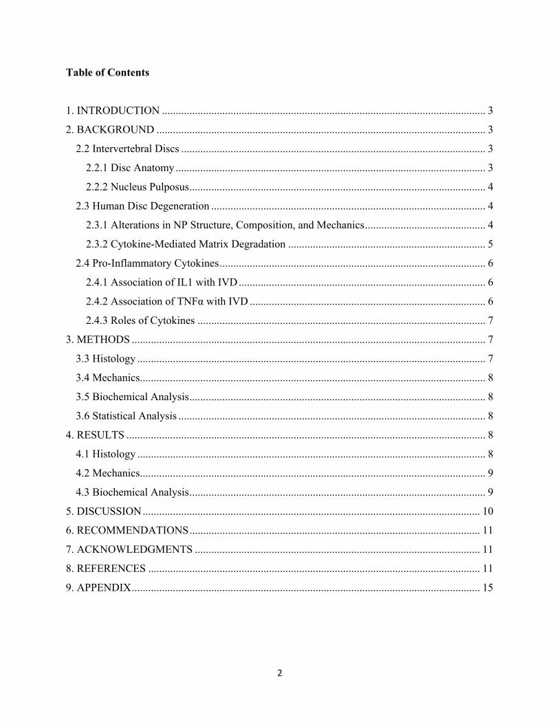
The NP is a pressurized gel of randomly distributed collagen II fibrils in a hydrated extrafibrillar matrix rich in proteoglycans [10, 11] (Figure 2). In compression under spinal axial loads, the NP is confined peripherally by the AF, which generates a region of uniform hydrostatic pressure in the disc. The NP pressure further enables the even distribution of the axial compressive loads between vertebral bodies [8, 9].
Figure 2. This NP matrix schematic shows collagen fibers interwoven with proteoglycans. GAG are the side chains found on proteoglycans. The negative charges are associated with GAG amd the positive charges are the sodium ions in water. (Source: Mow et al, 1998, Int. J. Solids Structures)
2.3 Human Disc Degeneration
2.3.1 Alterations in NP Structure, Composition, and Mechanics
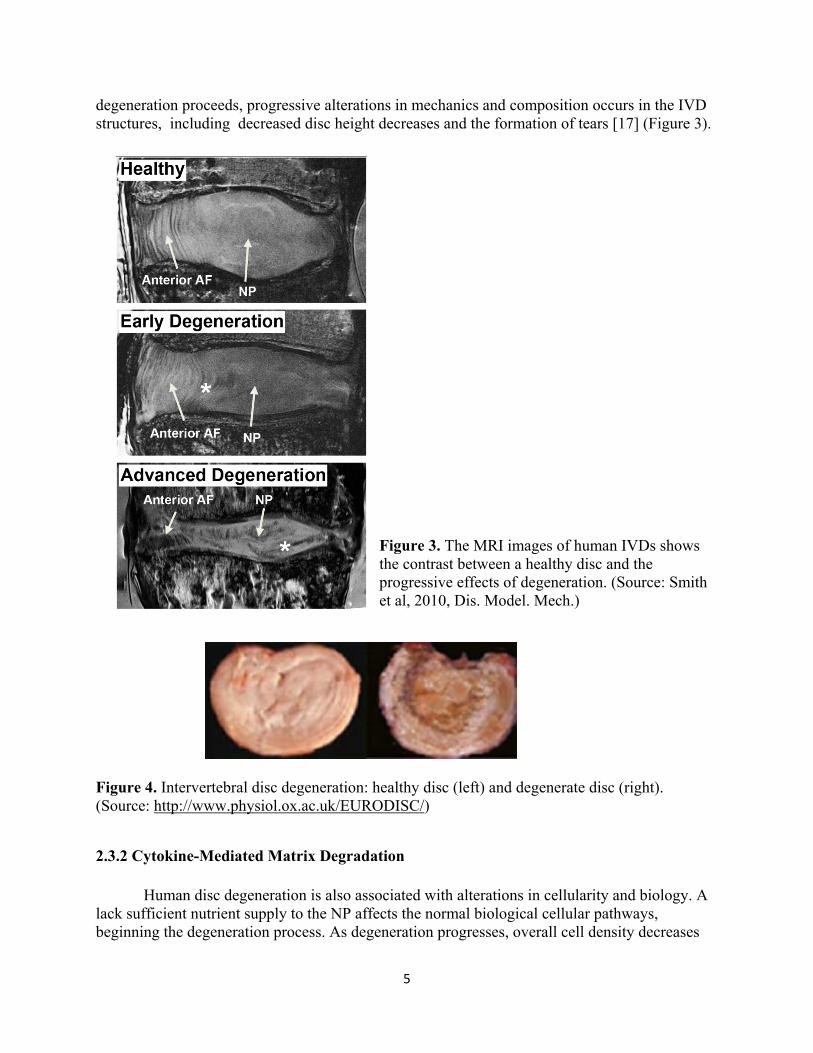
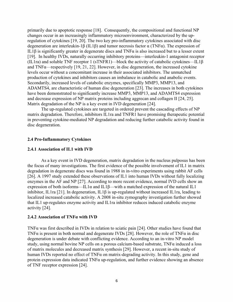
Human disc degeneration starts in the NP with associated alterations in structure, composition, and mechanics. One of the earliest indicators of disc degeneration is loss of glycosaminoglycan (GAG) content in the NP, followed by water loss [14-16]. Changes in matrix composition impair mechanical function. Such comprised functional changes result in reduction in NP pressure and alterations in motion segment stiffness, predisposing the tissue to injury and stress [12, 13]. These NP changes initiate a cascade expanding to other disc structures, particularly the AF. The reduced NP pressure causes an inward bulging of the AF, which is distinctly visible in the middle image of Figure 3 [12]. A loss of distinction between the nucleus pulposus and anulus fibrosus is also characteristic of disc degeneration, as seen in Figure 4. As
5
degeneration proceeds, progressive alterations in mechanics and composition occurs in the IVD structures, including decreased disc height decreases and the formation of tears [17] (Figure 3).
Figure 3. The MRI images of human IVDs shows the contrast between a healthy disc and the progressive effects of degeneration. (Source: Smith et al, 2010, Dis. Model. Mech.)
Figure 4. Intervertebral disc degeneration: healthy disc (left) and degenerate disc (right). (Source: http://www.physiol.ox.ac.uk/EURODISC/)
2.3.2 Cytokine-Mediated Matrix Degradation
Human disc degeneration is also associated with alterations in cellularity and biology. A lack sufficient nutrient supply to the NP affects the normal biological cellular pathways, beginning the degeneration process. As degeneration progresses, overall cell density decreases
6
primarily due to apoptotic response [18]. Consequently, the compositional and functional NP changes occur in an increasingly inflammatory microenvironment, characterized by the up-regulation of cytokines [19, 20]. The two key pro-inflammatory cytokines associated with disc degeneration are interleukin-1β (IL1β) and tumor necrosis factor α (TNFα). The expression of IL1β is significantly greater in degenerate discs and TNFα is also increased but to a lesser extent [19]. In healthy IVDs, naturally occurring inhibitory proteins—interleukin-1 antagonist receptor (IL1ra) and soluble TNF receptor 1 (sTNFR1)—block the activity of catabolic cytokines—IL1β and TNFα—respectively [19, 21, 22]. However, in disc degeneration, the increased cytokine levels occur without a concomitant increase in their associated inhibitors. The unmatched production of cytokines and inhibitors causes an imbalance in catabolic and anabolic events. Secondarily, increased levels of catabolic enzymes, specifically MMP3, MMP13, and ADAMTS4, are characteristic of human disc degeneration [23]. The increases in both cytokines have been demonstrated to significantly increase MMP3, MMP13, and ADAMTS4 expression and decrease expression of NP matrix proteins including aggrecan and collagen II [24, 25]. Matrix degradation of the NP is a key event in IVD degeneration [24].
The up-regulated cytokines are targeted in ordered prevent the cascading effects of NP matrix degradation. Therefore, inhibitors IL1ra and TNFR1 have promising therapeutic potential in preventing cytokine-mediated NP degradation and reducing further catabolic activity found in disc degeneration.
2.4 Pro-Inflammatory Cytokines
2.4.1 Association of IL1 with IVD As a key event in IVD degeneration, matrix degradation in the nucleus pulposus has been the focus of many investigations. The first evidence of the possible involvement of IL1 in matrix degradation in degenerate discs was found in 1988 in in-vitro experiments using rabbit AF cells [26]. A 1997 study extended these observations of IL1 into human IVDs without fully localizing enzymes in the AF and NP [27]. According to more recent evidence, normal IVD cells show an expression of both isoforms—IL1α and IL1β—with a matched expression of the natural IL1 inhibitor, IL1ra [21]. In degeneration, IL1β is up-regulated without increased IL1ra, leading to localized increased catabolic activity. A 2008 in-situ zymography investigation further showed that IL1 up-regulates enzyme activity and IL1ra inhibitor reduces induced catabolic enzyme activity [24].
2.4.2 Association of TNFα with IVD TNFα was first described in IVDs in relation to sciatic pain [24]. Other studies have found that TNFα is present in both normal and degenerate IVDs [28]. However, the role of TNFα in disc degeneration is under debate with conflicting evidence. According to an in-vitro NP model study, using normal bovine NP cells on a porous calcium-based substrate, TNFα induced a loss of matrix molecules and decreased matrix synthesis [29]. However, a recent in-situ study of human IVDs reported no effect of TNFα on matrix-degrading activity. In this study, gene and protein expression data indicated TNFα up-regulation, and further evidence showing an absence of TNF receptor expression [24].
7
2.4.3 Roles of Cytokines
Previous studies have established an association of both cytokines IL1β and TNFα in normal and degenerate IVDs. However, the roles of these cytokines in initiating NP matrix changes in under debate. Previous studies have also shown matrix changes due to increases in these cytokines, but not the functional significance of NP compositional changes.
3. METHODS 3.1 Cell Isolation and Preculture
Mature NP cells were isolated from bovine caudal discs. Isolated cells were expanded in monolayer, in Dulbeccos Modified Eagly Medium (DMEM) containing 10% fetal bovine serum and 1% penicillin/streptomycin/Fungizone (PSF). Cells were expanded through two passages for this study.
3.2 NP Constructs and Treatment
NP cells were suspended in chemically defined media (CM) and combined 1:1 with
sterile type VII agarose in phosphate buffered saline (PBS) at room temperature. Chemically defined media consisted of DMEM supplemented with 1x PSF; 0.1µM dexamethasone; 50mg/mL ascorbate 2-phosphate; 40mg/mL L-proline; 100mg/mL sodium pyruvate; and 1X6.25µg/mL insulin, 6.25µg/mL transferring, 6.25ng/mL selenious acid, 1.25mg/mL bovine serum albumin (BSA), and 5.35µg/mL linoleic acid. NP Cells were seeded in agarose gels (4mm diameter X 2.254mm thick) at a density of 20X106/ml and allowed to gel for 20 minutes. NP constructs were precultured for 6 weeks in CM containing 10ng/ml transforming growth factor beta 3 (TGFβ3). The preculture period allowed for the development of a functional matrix. Samples were then removed and cultured for an additional two weeks in media without TGFβ3. Samples were divided into treatment groups (n=7) designated for IL1 treatment or TNF treatment (Table 1). After 3 days of treatment, samples were evaluated for histology, mechanics, and biochemistry. IL1 Treatment Groups (n=7) TNF Treatment Groups (n=7) rh IL1β, 10ng/ml rh TNFα, 10ng/ml rh IL1β, 10ng/ml + rh IL1ra, 100ng/ml rh TNFα, 10ng/ml + rh TNFR1, 100ng/ml rh IL1ra, 100ng/ml rh TNFR1, 100ng/ml Control (no treatment) Control (no treatment) Table 1. Treatment groups for both IL1 and TNF with dosages of cytokine and/or inhibitor.
3.3 Histology
For histology, samples were fixed in 4% paraformaldehyde, dehydrated in a graded series of ethanol, and embedded in paraffin. Samples were sectioned at 7 µm-thickness from the
8
middle of the construct. Sections were stained with Alcian Blue (pH 1.0) for sulfated proteoglycans and Picrosirius Red (0.1% w/v in saturated picric acid) for collagens.
3.4 Mechanics
Samples from each treatment group were mechanically tested in confined compression (n=5). The confined compression test was used to replicate the physiological conditions of the NP. The mechanical testing device consisted of an acrylic chamber fixed above a porous platen, in a bath filled with culture medium. The in-vitro NP construct was peripherally confined by the chamber in order to model the in-vivo NP confined by the AF. An impermeable ceramic indenter attached to a mechanical testing device applied compression. First, samples were equilibrated under a static preload of 0.02 N held for 500 seconds. Samples were subjected to a stress relaxation test, consisting of 10% strain (calculated from post-creep thickness values) applied at 0.05%/s, followed by relaxation to equilibrium for 10 minutes. Aggregate modulus, HA, was calculated as the final equilibrium stress divided by the applied strain. The aggregate modulus was used as a measure of stiffness of the tissue at equilibrium. Hydraulic permeability, k0, was calculated using a linear biphasic theory, assuming material isotropy [30]. The hydraulic permeability models the rate at which fluid flows out of the construct.
3.5 Biochemical Analysis
For the biochemical analysis, the glycosaminoglycans, collagen, and DNA content were determined using three assays. First, the samples were weighed for wet weight, dried overnight, and weighed again for dry weight. Dried samples were digested for 16 hours in papain (0.56U/mL in 0.1M sodium acetate, 10M cysteine hydrochloric acid, 0.05M ethylenediaminetetraacetic acid, pH 6.0) at 60C. After digestion, the PicoGreen dsDNA assay was used to determine the DNA content. Digested samples were evaluated for GAG content using the 1,9-dimethylmethylene blue dye-binding (DMMB) assay against a standard curve of chondroitin-6-sulfate. After acid hydrolysis of the sample digests, the collagen content was determined using the orthohydroxyproline (OHP) assay. DNA content was reported as amount per construct, and GAG and collagen values are reported as percentages of wet weight.
3.6 Statistical Analysis Differences in mechanics and composition between groups were assessed using ANOVAs and Student Newman-Keuls pair-wise post-hoc tests, with significance considered for p<0.05.
4. RESULTS
4.1 Histology
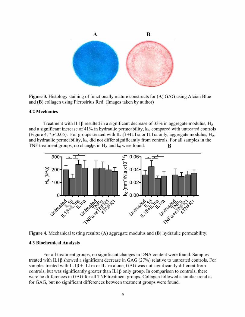
Histological staining results were used to confirm the uniform distribution, particularly of GAG, in the extracellular matrix. Functionally mature constructs are shown in Figure 3. Alcian Blue staining for GAG was uniformly distributed and intense. Picrosirius Red staining for collagen was a more diffuse distribution, showing both pericellular and intercellular staining.
9
Figure 3. Histology staining of functionally mature constructs for (A) GAG using Alcian Blue and (B) collagen using Picrosirius Red. (Images taken by author)
4.2 Mechanics
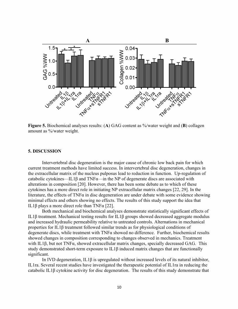
Treatment with IL1β resulted in a significant decrease of 33% in aggregate modulus, HA, and a significant increase of 41% in hydraulic permeability, k0, compared with untreated controls (Figure 4, *p<0.05). For groups treated with IL1β +IL1ra or IL1ra only, aggregate modulus, HA, and hydraulic permeability, k0, did not differ significantly from controls. For all samples in the TNF treatment groups, no changes in HA and k0 were found.
Figure 4. Mechanical testing results: (A) aggregate modulus and (B) hydraulic permeability.
4.3 Biochemical Analysis
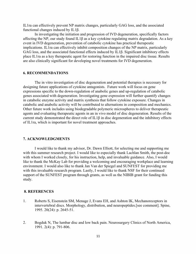
For all treatment groups, no significant changes in DNA content were found. Samples treated with IL1β showed a significant decrease in GAG (27%) relative to untreated controls. For samples treated with IL1β + IL1ra or IL1ra alone, GAG was not significantly different from controls, but was significantly greater than IL1β only group. In comparison to controls, there were no differences in GAG for all TNF treatment groups. Collagen followed a similar trend as for GAG, but no significant differences between treatment groups were found.
A B
A B
10
Figure 5. Biochemical analyses results: (A) GAG content as %/water weight and (B) collagen amount as %/water weight.
5. DISCUSSION
Intervertebral disc degeneration is the major cause of chronic low back pain for which current treatment methods have limited success. In intervertebral disc degeneration, changes in the extracellular matrix of the nucleus pulposus lead to reduction in function. Up-regulation of catabolic cytokines—IL1β and TNFα—in the NP of degenerate discs are associated with alterations in composition [20]. However, there has been some debate as to which of these cytokines has a more direct role in initiating NP extracellular matrix changes [22, 29]. In the literature, the effects of TNFα in disc degeneration are under debate with some evidence showing minimal effects and others showing no effects. The results of this study support the idea that IL1β plays a more direct role than TNFα [22].
Both mechanical and biochemical analyses demonstrate statistically significant effects of IL1β treatment. Mechanical testing results for IL1β groups showed decreased aggregate modulus and increased hydraulic permeability relative to untreated controls. Alternations in mechanical properties for IL1β treatment followed similar trends as for physiological conditions of degenerate discs, while treatment with TNFα showed no difference. Further, biochemical results showed changes in composition corresponding to changes observed in mechanics. Treatment with IL1β, but not TNFα, showed extracellular matrix changes, specially decreased GAG. This study demonstrated short-term exposure to IL1β induced matrix changes that are functionally significant.
In IVD degeneration, IL1β is upregulated without increased levels of its natural inhibitor, IL1ra. Several recent studies have investigated the therapeutic potential of IL1ra in reducing the catabolic IL1β cytokine activity for disc degeneration. The results of this study demonstrate that
A B
11
IL1ra can effectively prevent NP matrix changes, particularly GAG loss, and the associated functional changes induced by IL1β.
In investigating the initiation and progression of IVD degeneration, specifically factors affecting the NP, our study found IL1β as a key cytokine regulating matrix degradation. As a key event in IVD degeneration, prevention of catabolic cytokine has practical therapeutic implications. IL1ra can effectively inhibit composition changes of the NP matrix, particularly GAG loss, and the associated functional effects induced by IL1β. Significant inhibitory effects place IL1ra as a key therapeutic agent for restoring function in the impaired disc tissue. Results are also clinically significant for developing novel treatments for IVD degeneration.
6. RECOMMENDATIONS
The in vitro investigation of disc degeneration and potential therapies is necessary for designing future applications of cytokine antagonists. Future work will focus on gene expressions specific to the down-regulation of anabolic genes and up-regulation of catabolic genes associated with degeneration. Investigating gene expression will further quantify changes in catabolic enzyme activity and matrix synthesis that follow cytokine exposure. Changes in catabolic and anabolic activity will be contributed to alternations in composition and mechanics. Other future work includes using biodegradable polymeric microspheres to deliver therapeutic agents and evaluating therapeutic agents in an in vivo model of disc degeneration. Results of the current study demonstrated the direct role of IL1β in disc degeneration and the inhibitory effects of IL1ra, which is important for novel treatment approaches.
7. ACKNOWLEDGMENTS
I would like to thank my advisor, Dr. Dawn Elliott, for selecting me and supporting me with this summer research project. I would like to especially thank Lachlan Smith, the post-doc with whom I worked closely, for his instruction, help, and invaluable guidance. Also, I would like to thank the McKay Lab for providing a welcoming and encouraging workplace and learning environment. I would also like to thank Jan Van der Spiegel and SUNFEST for providing me with this invaluable research program. Lastly, I would like to thank NSF for their continued support of the SUNFEST program through grants, as well as the NIBIB grant for funding this study.
8. REFERENCES 1. Roberts S, Eisenstein SM, Menage J, Evans EH, and Ashton IK, Mechanoreceptors in
intervertebral discs. Morphology, distribution, and neuropeptides.[see comment]. Spine, 1995. 20(24): p. 2645-51.
2. Bogduk N, The lumbar disc and low back pain. Neurosurgery Clinics of North America, 1991. 2(4): p. 791-806.
12
3. Andersson GB, Epidemiological features of chronic low-back pain. Lancet, 1999.
354(9178): p. 581-5. 4. Deyo RA and Tsui-Wu YJ, Descriptive epidemiology of low-back pain and its related
medical care in the United States. Spine, 1987. 12(3): p. 264-8. 5. Frymoyer JW, Back pain and sciatica. New England Journal of Medicine, 1988. 318(5):
p. 291-300. 6. Luo X, Pietrobon R, Sun SX, Liu GG, and Hey L, Estimates and patterns of direct health
care expenditures among individuals with back pain in the United States. Spine, 2004. 29(1): p. 79-86.
7. Knoke JD, Smith TC, Gray GC, Kaiser KS, and Hawksworth AW, Factor analysis of
self-reported symptoms: does it identify a Gulf War syndrome? Am J Epidemiol, 2000. 152(4): p. 379-88.
8. Ishihara H, McNally DS, Urban JP, and Hall AC, Effects of hydrostatic pressure on
matrix synthesis in different regions of the intervertebral disk. Journal of Applied Physiology, 1996. 80(3): p. 839-46.
9. Hunter CJ, Matyas JR, and Duncan NA, The three-dimensional architecture of the
notochordal nucleus pulposus: novel observations on cell structures in the canine intervertebral disc. Journal of Anatomy, 2003. 202(Pt 3): p. 279-91.
10. Cassidy JJ, Hiltner A, and Baer E, Hierarchical structure of the intervertebral disc.
Connective Tissue Research, 1989. 23(1): p. 75-88. 11. Marchand F and Ahmed AM, Investigation of the laminate structure of lumbar disc
anulus fibrosus. Spine, 1990. 15(5): p. 402-10. 12. Thompson JP, Pearce RH, Schechter MT, Adams ME, et al., Preliminary evaluation of a
scheme for grading the gross morphology of the human intervertebral disc. Spine, 1990. 15(5): p. 411-5.
13. Adams MA, Freeman BJ, Morrison HP, Nelson IW, and Dolan P, Mechanical initiation
of intervertebral disc degeneration. Spine, 2000. 25(13): p. 1625-36. 14. Antoniou J, Steffen T, Nelson F, Winterbottom N, et al., The human lumbar
intervertebral disc: evidence for changes in the biosynthesis and denaturation of the extracellular matrix with growth, maturation, ageing, and degeneration. J Clin Invest, 1996. 98(4): p. 996-1003.
13
15. Frobin W, Brinckmann P, Kramer M, and Hartwig E, Height of lumbar discs measured from radiographs compared with degeneration and height classified from MR images. European Radiology, 2001. 11(2): p. 263-9.
16. Olczyk K, Age-related changes in proteoglycans of human intervertebral discs.
Zeitschrift fur Rheumatologie, 1994. 53(1): p. 19-25. 17. Stokes IA and Iatridis JC, Mechanical Conditions That Accelerate Intervertebral Disc
Degeneration: Overload Versus Immobilization. Spine, 2004. 29(23): p. 2724-32. 18. Keshari KR, Lotz JC, Kurhanewicz J, and Majumdar S, Correlation of HR-MAS
spectroscopy derived metabolite concentrations with collagen and proteoglycan levels and Thompson grade in the degenerative disc. Spine, 2005. 30(23): p. 2683-8.
19. Gruber HE, Ingram JA, Norton HJ, and Hanley EN, Jr., Senescence in cells of the aging
and degenerating intervertebral disc: immunolocalization of senescence-associated beta-galactosidase in human and sand rat discs. Spine, 2007. 32(3): p. 321-7.
20. Le Maitre CL, Hoyland JA, and Freemont AJ, Catabolic cytokine expression in
degenerate and herniated human intervertebral discs: IL-1beta and TNFalpha expression profile. Arthritis Res Ther, 2007. 9(4): p. R77.
21. Lotz JC, Colliou OK, Chin JR, Duncan NA, and Liebenberg E, Compression-induced
degeneration of the intervertebral disc: an in vivo mouse model and finite-element study. Spine, 1998. 23(23): p. 2493-506.
22. Hutton WC, Elmer WA, Boden SD, Hyon S, et al., The effect of hydrostatic pressure on
intervertebral disc metabolism. Spine, 1999. 24(15): p. 1507-15. 23. Le Maitre CL, Freemont AJ, and Hoyland JA, The role of interleukin-1 in the
pathogenesis of human intervertebral disc degeneration. Arthritis Res Ther, 2005. 7(4): p. R732-45.
24. Hoyland JA, Le Maitre CL, and Freemont AJ, Investigation of the role of IL-1 and TNF
in matrix degradation in the intervertebral disc, 2008 Rheumatology 47:6 25. Ohshima H, Urban JP, and Bergel DH, Effect of static load on matrix synthesis rates in
the intervertebral disc measured in vitro by a new perfusion technique. Journal of Orthopaedic Research, 1995. 13(1): p. 22-9.
26. Shinmei M, Kikuchi T, Yamagishi M, Shimomura Y. The role of interleukin-1 on
proteoglycan metabolism of rabbit annulus fibrosus cells cultured in vitro. Spine 1988; 13: 1284-90.
14
27. Kang JD, Stefanovic-Racic M, McIntyre LA, Georgescu HI, Evans CH. Toward a biochemical understanding of human intervertebral disc degeneration and herniation. Contributions of nitric oxide, interleukins, prostaglandin E2, and matrix metalloproteinases. Spine 1997; 22: 1065-73. 28. Weiler C, Nerlich AG, Bachmeier BE, Boos N. Expression and distribution of tumor necrosis factor alpha in human lumbar intervertebral discs: a study in surgical specimen and autopsy controls. Spine 2005; 30: 44-54. 29. Seguin CA, Pilliar RM, Roughley PJ, Kandel RA. Tumor necrosis factor α modulates matrix production and catabolism in nucleus pulposus tissue. Spine 2005: 30: 1940-8. 30. Soltz MA and Ateshian GA. Experimental verification and theoretical prediction of cartilage interstitial fluid pressurization at an impermeable contact interface in confined compression. Journal of Biomechanics 1998: 31:10: 927-34.
15
9. APPENDIX NOTE: In review and submitted to the Orthopaedic Research Society Fall Conference 2010
![The protective effects of PI3K/Akt pathway on human nucleus pulposus … · 2020. 1. 28. · nucleus pulposus cells and nucleus pulposus progenitor cells [14]. Previous studies have](https://img.pdfslide.us/doc/110x75/60b265dd0d8b8040e758b496/the-protective-effects-of-pi3kakt-pathway-on-human-nucleus-pulposus-2020-1-28.jpg)


![Puerarin Relieved Compression-Induced Apoptosis and ...downloads.hindawi.com/journals/sci/2020/7126914.pdfimpaired nucleus pulposus cell (NPC) proliferation [4]. Nucleus pulposus mesenchymal](https://img.pdfslide.us/doc/110x75/5f7fa24ee6184370f175b23e/puerarin-relieved-compression-induced-apoptosis-and-impaired-nucleus-pulposus.jpg)