Embed Size (px)
Citation preview

THE JOURNAL OF BIOLOGICAL CHEMISTRY Vol. 242, No. 20, Issue of October 25,~~. 4691-4701,1967
Printed in U.S.A.
The Proteinpolysaccharides of Bovine Nucleus Pulposus*
(Received for publication, May 26, 1967)
LAWRENCE ROSENBERG~ AND MAXWELL SCHUBERT
From the Departments of Medicine and Orthopedics and the Study Group for Rheumatic Diseases, New York University School of Medicine, New York, New York 10016
JOHN SANDSON
From the Department of Medicine, Albert Einstein College of Medicine, New York, New York 10461
SUMMARY
Bovine nucleus pulposus has been found to be a good source of a series of proteinpolysaccharides containing chondroitin 6-sulfate and keratan sulfate. Six distinct fractions have been separated, totaling 40 % of the dry weight of the tissue. Each fraction contains protein, chondroitin B-sulfate, and keratan sulfate, but the proportions of these components are different in each fraction. Of these six fractions, the one called PPL3 is obtained in largest yield (17%), has the lowest content of protein (14%) and the highest content of chondroitin sulfate (45%), and also con- tains keratan sulfate (35%). On sedimentation velocity studies in the analytical ultracentrifuge under a wide range of conditions, it appears to be homogeneous. Sedimentation equilibrium studies at low initial concentrations indicate some degree of polydispersity and a weight average molecular weight of 171,000. On zone electrophoresis the chondroitin sulfate, keratan sulfate, and protein move together. Fraction PPW has almost the same amino acid profile as the corre- sponding fraction from bovine nasal cartilage, although these fractions differ greatly in protein and keratan sulfate contents. The data provide reasons to believe that chondroitin sulfate and keratan sulfate are parts of the same molecule. Three other fractions, called PPL4, PPL5, and PPL6, show a regular increase in sedimentation velocity, a regular decrease in speed of migration on zone electrophoresis, a regular increase in protein content, and a ratio of chondroitin sulfate to keratan sulfate which is constant (0.62) and differs from that of PPL3 (1.3). For each of these fractions, on zone electrophoresis, protein, chondroitin sulfate, and keratan sulfate stand or move together. There is no evidence that in this tissue any independent kinetic component exists which contains only chondroitin sulfate or only keratan sulfate.
*This work was supported by United States Public Health Service Grant AM-00028 from the National Institute of Arthritis and Metabolic Diseases, and United St(ates Public Health Service Research Career Program Award 5-K6-AM-18,434.
2 Fellow of The Helen Hay Whitney Foundation.
Human articular and costal cartilages contain roughly equal amounts of chondroitin sulfate and keratan sulfate, both of which are attached to protein as compounds called protein- polysaccharides. From these cartilages, proteinpolysaccharides can be extracted only in small part as the water-soluble product called PPLl (0.07 g per g of dry cartilage). PPL from human articular cartilage has been fractionated to give three products, each of which contains chondroitin sulfate and keratan sulfate in roughly equal amounts (1). There appeared to be no tendency to segregate products containing only chondroitin sulfate at- tached to protein from products containing only keratan sulfat.e attached to protein. Because of the difficulty of fractionation in this group of compounds, it is not safe to conclude from these results that chondroitin sulfate and keratan sulfate are parts of the same molecules.
In order to learn whether chondroitin sulfate and keratan sulfate may be parts of a single molecule, a more searching study of fractionation is desirable, and for this purpose PPL
1 Abbreviations and Terminology-The products isolated from cartilage and other connective tissues that contain chondroitin sulfate and keratan sulfate covalently bound to protein have been called proteinpolysaccharides to emphasize that their carbo- hydrate is polysaccharide rather than oligosaccharide, as in the case of glycoproteins. Proteinpolysaccharides have now been isolated from several different forms of connective tissue, and as far as possible a uniform system of designating them will be used. The initial product isolated from whole cartilage or other connec- tive tissue by homogenization of the tissue with water followed by removal of the insoluble residue by centrifugation is called crude proteinpolysaccharide. PPL designates the light fraction of the crude proteinpolysaccharide; it is that fraction isolated from the supernatant solution after ultracentrifugation of crude nroteinpolvsaccharide in 0.15 M KCI; PPH. the heavv fraction. is that-pa& which sediments. PPL .is further separable into $ series of fractions, called PPL3, PPL4, PPL5, and PPL6 in the order of increasing sedimentability. These are operationally defined by the conditions of their separation indicated in the diagram attached to Table IV and specified in the detailed de- scription of each step. Intermediate products, PPLR and PPLRF, are also defined by this Diagram, as is PPL2. From different cartilages PPH and the different forms of PPL are ob- tained in different vields and have different comnositions. vet there are common “trends in composition and sedimentability within each set of forms from a given tissue.
4691
by guest on May 16, 2018
http://ww
w.jbc.org/
Dow
nloaded from

4692 Proteinpolysaccharides of Nucleus Pulposus Vol. 242, Ko. 20
from human cartilages is not w-e11 suited. This is because of the low yields of PPL, the still lower yields of each of the frac- tions, and t,he variation in proportions of these fractions in material from different individuals. This makes impossible a comparison of different methods of fractionation on cartilage samples from different individuals.
Bovine nasal cartilage, rich in chondroitin sulfate and poor in keratan sulfate, gives high yields of the water-soluble PPL (0.37 g per g of dry cartilage). Bovine nasal PPL has been separated into four fractions, each high in chondroitin sulfate and poor in kerat’an sulfate (2). But each of these fractions has so low a content of keratan sulfate that even if fract’ions containing only this polysaccharide existed they would be dif- ficult to isolate in more than minute amounts.
To pursue the problem suggested, whether chondroitin sulfate and keratan sulfate may both be parts of the same molecule, a more suitable starting material has been found in bovine nucleus pulposus. hlthough it is not usually classed as a car- tilage, nucleus pulposus has a high polysaccharide content, consisting of nearly equal amounts of chondroitin sulfate and keratan sulfate.
Examination of the polysaccharides of nucleus pulposus has been largely confined to material from human discs. Malmgren and Sylven (3) isolated chondroitin sulfate. From an enzymatic digest of human disc, Garde11 and Rastgeldi (4) isolated a crude polysaccharide which contained nearly equal amounts of gluco- samine and galactosamine. Since Meyer et al. (5) had just discovered keratan sulfate in cornea, Garde11 suggested that the crude polysaccharide might be a mixture of chondroitin sulfate and keratan sulfate, and showed this by fractionation (6). Davidson and Woodhall (7) reported a separation of chondroitin sulfate and keratan sulfate, apparently without the use of en- zymes. Lyons et al. (8) isolated a product which contained chondroitin sulfate, keratan sulfate, and protein. The method they used would yield a mixture of PPL and PPH.
The present work is a report in quantitative terms of the isolation of crude proteinpolysaccharide from bovine nucleus pulposus, its fractionation into five main water-soluble frac- tions, and the characterization of each of these fractions analyt- ically, as well as by sedimentation and zone electrophoresis. Neither chondroitin sulfate nor keratan sulfate exists free of prot,ein, no fractions containing only chondroitin sulfate or keratan sulfate have been found, and some evidence is given that at least one fraction, called PPL3, behaves as a single com- pound. This set of fractions shows a striking parallel to the set isolated from bovine nasal cartilage, although the two sets differ markedly in composition.
EXPERIMENTS AND RESULTS
An&&al Methods
The methods used were uranic acid (9), total hexosamine (lo), hexose (ll), sialate (la), protein (13), hydroxyproline (14), and sulfate (15).
Chondroitin 6-sulfate was estimated by the procedure de- scribed by Mathews and Inouye (16) with chromatographically purified testicular hyaluronidase (3000 U.S.P. units per mg, Worthington). The acetylhexosamine liberated was deter- mined by the modification of the Morgan-Elson reaction de- scribed by Dische (17) and by Aminoff, Morgan, and Watkins
(18), with N-acetylgalactosamine (Nutritional Biochemicals) as a standard.
Glucosamine and galactosamine were separately determined by the following procedure. Samples of proteinpolysaccharide, chondroitin sulfate, or keratan sulfate were hydrolyzed in 4 N HCl (1 ml per mg) in sealed, evacuated tubes at 100” for 8 hours. The hydrolysate was filtered through sintered glass to remove humins, and HCl and water uere removed under reduced pres- sure at 40”. The dried residue was dissolved in 0.1 M acetate- citrate buffer, pH 2.5, to give an approximate concentration of 0.5 mM total hexosamine. The sample (about 2 ml) was applied to a 150.cm Amberlite IRC-50 column and chromatographed with citrate buffers, first at pH 3.25, then at pH 4.25, the change to pH 4.25 buffer being made immediately after the emergence of glycine to give good separation between phenylalanine and glucosamine. Glucosamine and galactosamine were estimated by comparison with glucosamine and galactosamine standards chromatographed in the same manner. This procedure, partic- ularly the milder hydrolytic conditions, gives a much better recovery of glucosamine and galactosamine than their simul- taneous determination with total amino acid analysis by the usual, more vigorous hydrolysis conditions (6 N HCl, lOO-llO”, 24 hours). The values given below for glucosamine and galac- tosamine are not corrected for hydrolysis losses.
Preparative Procedures
Preparative work was carried out in a cold room at 5”. In this room a VirTis 45 homogenizer was operated with the baffled flask set in an ice bath. An International refrigerated cen- trifuge, model PR, was used for speeds below 5000 rpm; a Spinco model L centrifuge, for speeds above 5000 rpm.
The diagram attached to Table IV, below, shows the steps to be described and the designation of all intermediate and final products isolated. At each step where a separation occurs, the product that stays in solution is put to the left side of the fork, and the product that is insoluble or is sedimented is put to the right side of the fork.
Step I-Fresh wet bovine nucleus pulposus, collected from the lumbar region within + hour after slaughter, was dried by homogenization in a VirTis 45 homogenizer at 5” with ethanol (50 ml per g) for 5 to 10 min; the finely divided product was separated by centrifugation at 2000 rpm, washed with ethanol, then ether, and dried in a vacuum. The fine, white, dry powder weighed 0.28 g per g of t,issue, fresh wet. This is the form best suited for storage and extraction.
Step g-Dry, powdered nucleus pulposus (4 g) was homog- enized with water (250 ml) in the VirTis’ 45 (5”; 30 min, top speed). Cold ethanol (600 ml) was added, and the suspension was centrifuged at 4000 rpm for 30 min at 5”. From the slightly opalescent supernatant solut’ion, filtered through glass wool to remove stray floccules, the crude proteinpolysaccharide was precipitated by adding potassium acetate (10 g). After standing at 5” overnight, the product was separated by centrifugation, washed with ethanol, then with ether, and dried in a vacuum.
In one experiment the residual tissue, resuspended each time in water (250), was successively extracted three times more by the entire procedure described in the preceding paragraph. The final residue remaining after the fourth extraction was washed with ethanol and ether, and dried as usual. Table I shows the yields and compositions of the four successive crude
by guest on May 16, 2018
http://ww
w.jbc.org/
Dow
nloaded from
Issue of October 25, 1967 L. Rosenberg, M. Schubert, and J. Xandson 4693
TABLE I Yields and compositions of crude proteinpolysaccharide obtained
from dry bovine nucleus pulposus powder by each of four successive extractions with water as described for Step 2
The composition of the residue left after the four extractions with water is also shown.
Extraction Yie’d from “~:ii” HCtOS-
dry powder Hexose amine Protein Hydroxy- proline
g/g YO % % % %
1 0.389 14.2 15.4 17.3 27.1 0.12
2 0.115 9.9 14.0 16.8 31.9 0.27
3 0.018 8.1 12.2 14.2 45.7 1.06 4 0.003 5.8 10.8 10.3 56.3 2.47
Residue 0.397 0.8 4.6 1.9 77.8 10.8
proteinpolysaccharides and the residue, as well as data on dry, powdered nucleus pulposus. The yields of crude proteinpolysac- charide from the third and fourth extractions not only were low, but their content of protein and collagen was high. In all subsequent work, therefore, only two extractions with water were made in Step 2.
Step S-Crude proteinpolysaccharide (1 g) was stirred with KC1 solution (0.15 M, 200 ml, 6 hours, So) and centrifuged at 40,000 rpm for 1 hour, and the clear supernatant solution was poured from the gelatinous sediment. Addition of ethanol (400 ml) to the supernatant solution precipitated PPL. The gelatinous sediment left in the centrifuge cups was dissolved in water (50 ml), and potassium acetate (0.5 g) and ethanol (100 ml) were added to precipitate PPH. The two products, PPL and PPH, were sedimented, washed with ethanol and ether, and dried in a vacuum. By this procedure each of the four successive crude proteinpolysaccharides of Step 2, shown in Table I, were separated into PPH and PPL. The yields and analytical data are summarized in Table II.
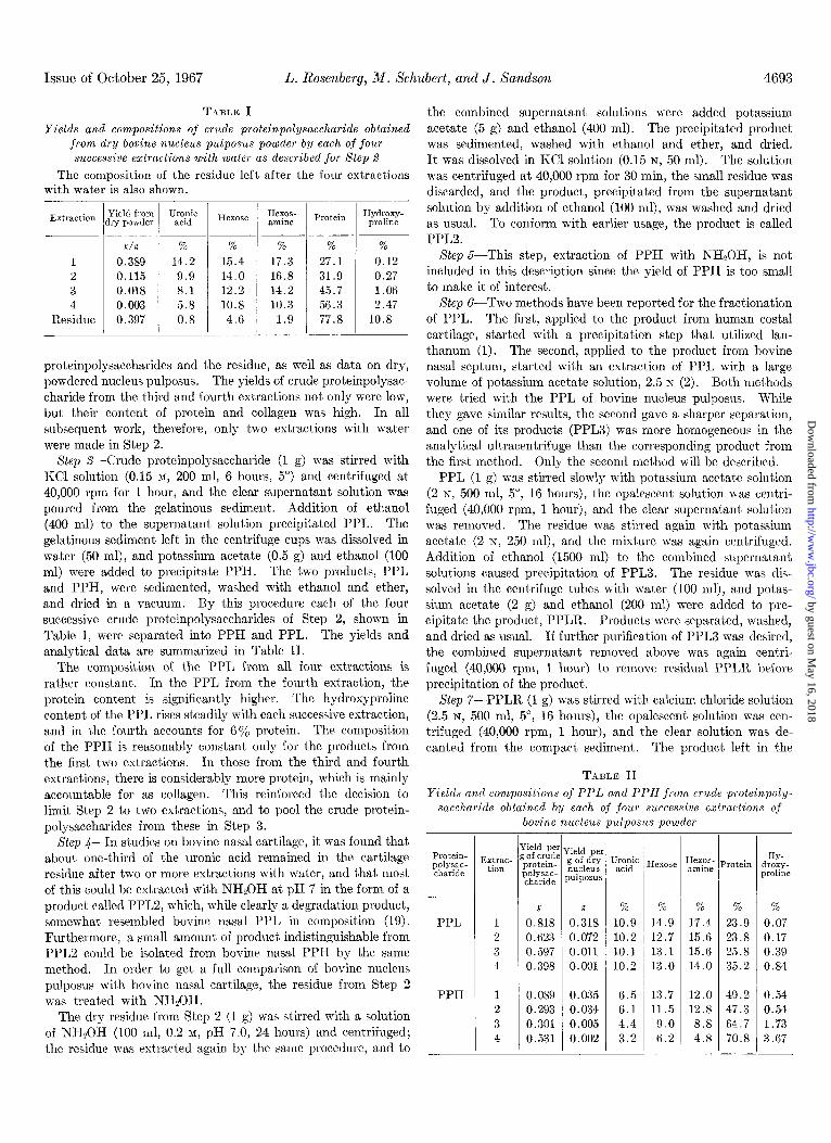
The composition of the PPL from all four extractions is rather constant. In the PPL from the fourth extraction, the protein content is significantly higher. The hydroxyproline content of the PPL rises steadily with each successive extraction, and in the fourth accounts for 6% protein. The composition of the PPH is reasonably constant only for the products from the first two extractions. In those from the third and fourth extractions, there is considerably more protein, which is mainly accountable for as collagen. This reinforced the decision to limit Step 2 to two extractions, and to pool the crude protein- polysaccharides from these in Step 3.
Step C-In studies on bovine nasal cartilage, it was found that about one-third of the uranic acid remained in the cartilage residue after two or more extractions with water, and that most of this could be extracted with NH,OH at pH 7 in the form of a product called PPLS, which, while clearly a degradation product, somewhat resembled bovine nasal PPL in composition (19). Furthermore, a small amount of product indistinguishable from PPL2 could be isolated from bovine nasal PPH by the same method. In order to get a full comparison of bovine nucleus pulposus with bovine nasal cartilage, the residue from Step 2 was treated with NHQOH.
The dry residue from Step 2 (1 g) was stirred with a solution of NHzOH (100 ml, 0.2 M, pH 7.0, 24 hours) and centrifuged; the residue was extracted again by the same procedure, and to
the combined supernatant solutions were added potassium acetate (5 g) and ethanol (400 ml). The precipitated product was sedimented, washed with ethanol and ether, and dried. It was dissolved in KC1 solution (0.15 N, 50 ml). The solution was centrifuged at 40,000 rpm for 30 min, the small residue was discarded, and the product, precipitated from the supernatant solution by addition of ethanol (100 ml), was washed and dried as usual. To conform with earlier usage, the product is called PPLS.
Step 5-This step, extraction of PPH with NH,OH, is not included in this description since the yield of PPH is too small to make it of interest.
Step ~-TWO methods have been reported for the fractionation of PPL. The first, applied to the product from human costal cartilage, started with a precipitation step that utilized lan- thanum (1). The second, applied to the product from bovine nasal septum, started with an extraction of PPL with a large volume of potassium acetate solution, 2.5 N (2). Both methods were tried with the PPL of bovine nucleus pulposus. While they gave similar results, the second gave a sharper separation, and one of its products (PPL3) was more homogeneous in the analytical ultracentrifuge than the corresponding product from the first method. Only the second method will be described.
PPL (1 g) was stirred slowly with potassium acetate solution (2 N, 500 ml, 5”, 16 hours), the opalescent solution was centri- fuged (40,000 rpm, 1 hour), and the clear supernatant solution was removed. The residue was stirred again with potassium acetate (2 N, 250 ml), and the mixture was again centrifuged. Addition of ethanol (1500 ml) to the combined supernatant solutions caused precipitation of PPL3. The residue was dis- solved in the centrifuge tubes with water (100 ml), and potas- sium acetate (2 g) and ethanol (200 ml) were added to pre- cipitate the product, PPLR. Products were separated, washed, and dried as usual. I f further purification of PPL3 was desired, the combined supernatant removed above was again centri- fuged (40,000 rpm, 1 hour) to remove residual PPLR before precipitation of the product.
Step 7-PPLR (1 g) was stirred with calcium chloride solution (2.5 N, 500 ml, 5”, 16 hours), the opalescent solution was cen- trifuged (40,000 rpm, 1 hour), and the clear solution was de- canted from the compact sediment. The product left in the
TABLE II Yields and compositions of PPL and PPH from crude proteinpoly-
saccharide obtained by each of four successive extractions of
bovine nucleus pulposus powder
PPI,
PPH
Yield pe Extrac- g of crud’
tion protem- pryd;
g
1 0.818 2 0.623
3 0.597 4 0.398
1 0.089 2 0.293 3 0.301 4 0.531
-
r7 e Yield pe
Pn~Ef: pulposus
g 70 Y* YO % %
0.318 10.9 14.9 17.4 23.9 0.07 0.072 10.2 12.7 15.6 23.8 0.17
0.011 10.1 13.1 15.6 25.8 0.39 0.001 10.2 13.0 14.0 35.2 0.84
0.035 6.5 13.7 12.0 49.2 0.54
0.034 6.1 11.5 12.8 47.3 0.54 0.005 4.4 9.0 8.8 64.7 1.73 0.002 3.2 6.2 4.8 70.8 3.67
r I : I hose Hexes- P
amine
-
HY- rotein droxy-
proline
by guest on May 16, 2018
http://ww
w.jbc.org/
Dow
nloaded from

4694 Proteinpolysaccharides of Nucleus Pulposus Vol. 242, No. 20
solut’ion was precipitated as a calcium salt by addition of ethanol (1000 ml), sedimented, and dissolved in water (50 ml); the clear solution was stirred with Dowex 50 in its potassium form to remove calcium and was precipitated as a potassium salt by addition of potassium acetate (1 g) and ethanol (100 ml). This product, PPLRF, was used in Step 8. The compact sediment
left in the tubes after the high speed centrifugation was dis- solved in water (50 ml), the solution was stirred with Dowex 50 in its potassium form to remove calcium, andthe product, PPL6, was precipitated by addition of potassium acetate (1 g) and ethanol (100 ml).
Step 8-Since the product PPLRF is part of the product PPLR, which in Step 6 had sedimented in 2 N potassium acetate, one might expect that PPLRF would sediment in this solvent. It sediments only in part. In this respect it behaves like the corresponding product from bovine nasal cartilage, and a further step of fractionation becomes possible. This step is carried out exactly as Step 6 and yields PPL4 from the supernatant solution and PPL5 from the material scdimented.
TABLE III
Comparison of yields of PPH, PPL, and PPL2 from several different cartilages
Kind of cartilage PPH 1 PPL I PPL2
gig dry powdered cartilage
Bovine nucleus pulposus Bovine nasal. Bovine tracheal. Bovine costal. Bovine articular Human costal..
0.037 0.450 0.040 0.372
0.034 0.129 0.026 0.090 0.041 0.044 0.143 0.064
0.014 0.120
0.06G Yields of PPL from Diferent Cartilages
0.063 0.038
Bovine nucleus pulposus gives a higher yield of PPL than
0.025 any form of cartilage that has yet been examined. Table III gives a comparison of the yields of PPL, PPH, and PPL2 for
TABLE IV Outline of entire extraction procedure used to wparatefcve forms of PPL and one of PPH from bovine nucleus pulposus, together with yields
and analytical data on these products
Wet nucleus pulposus
1. I
Ethanol; VirTis 45
Dry powder (1.000 g)
2. H20; VirTis 45
Crude pr$teinpolysaccharide (0.531 g) 4
Residue
4. NHzOH
3. 0.15 N KCI; Centrifuge
1
PPL (0.450 g)
6. 1 2 N polassium acetate
PPLR’(O.214 g)
Analysis Whole powder PPL3
+ PPT.4 PPL6 PPH PPL2 Final residue
0.177 0.060 0.047 0.066 14.7 9.0 7.8 6.0 14.4 9.3 6.9 6.7 15.8 15.8 15.0 12.5 11.4 13.8 13.0 10.7 20.2 18.3 17.4 14.0 16.4 16.5 15.2 13.6
2.8 3.4 3.7 3.0 14.4 18.9 31.8 48.0 0.03 0.07 0.21 0.18
0.037 6.5
6.7 13.7 10.4
12.0
2.4
49.2 0.44
0.014 10.8
9.3 12.8
7.2
17.0 14.3
2.1 24.4
2.0
0.347
0.5
4.6
0.9
0.07 69.0 10.3
12.9 8.1 6.8 7.2 6.5 9.2
Y -
Yield, g/g dry powder. .............. Hexnronate, %. ..................... Galactosamine,% ................... Hexose,% ...........................
Glucosamine, %. .................... Hexosamine, %. ..................... Sulfate, %. ..........................
Sialate,7, ........................... Protein, % .......................... Hydroxyproline, %. . Acetylgalactosamine end groups, %,
aft)er hyaluronidase ................
5.7 5.1
11.6
5.8 10.1
15.0 2.2
45.0
3.9
by guest on May 16, 2018
http://ww
w.jbc.org/
Dow
nloaded from
Issue of October 25, 1967 L. Rosenberg, M. Schubert, and J. Xandson 4695
five forms of bovine cartilage and one form of human cartilage. Yields of PPH are usually small except for human costal cartilage; yields of PPL2 are usually small except for bovine nasal cartilage. A striking feature of the behavior of bovine nucleus pulposus is the ease with which virtually all the proteinpolysaccharide is removed by homogenization with water, leaving a residue almost free of polysaccharide. This behavior stands in contrast to bovine nasal cartilage, from which only about two-thirds of the polysaccharide is extracted with water, leaving one-third associated with the cartilage residue. Most of this remaining third can be extracted as PPL2 by treatment with NAzOH (19). In the case of bovine nucleus pulposus, the PPL2 fraction is very small.
Compositions of Proteinpolysaccharides
Table IV and Diagram 1 summarize the procedure for extrac- tion and fractionation of the six forms of proteinpolysaccharide isolated from the dry bovine nucleus pulposus, and give data on the yields and the analyses of each of the products for a number of components. The six proteinpolysaccharides account for 0.40 g per g of dry nucleus pulposus powder. PPL3 is the largest single fraction. The fractions PPLS, PPL4, PPL5, and PPL6, all derived from PPL, constitute, in the order given, a series which shows a regular increase in sedimentability and in protein content, and a regular decrease in uranic acid and total hexosa- mine. In this series there seems also to be a small decrease in the contents of hexose and sulfate, but no significant change in sialate. PPLB, the fraction obtained in smallest yield, is not a member of this series since it was extracted from the residue with NHzOH and is probably a degraded form. Analytically it falls approximately between PPL3 and PPL5, but seems not identical with PPL4. PPH and PPL6 are analytically very similar.
The most interesting feature of the proteinpolysaccharides of bovine nucleus pulposus is their high content of hexose and glucosamine, a reflection of their high content of keratan sulfate. This is in strong contrast to the corresponding fractions from bovine nasal cartilage, all of which have only about 3% hexose. In some other respects the main series of products, PPLS, PPL4, PPL5, and PPLG, from bovine nucleus pulposus show parallels to the corresponding series from bovine nasal cartilage; in both series the protein contents increase regularly and the hexuronate and hexosamine contents decrease regularly. Each member of the bovine nucleus pulposus series has 20 to 80% more protein than the corresponding member of the bovine nasal cartilage. The proteinpolysaccharides of bovine nucleus pulposus contain chondroitin B-sulfate, while those of bovine nasal cartilage contain mainly chondroitin 4-sulfate, as judged by the values for acetylgalactosamine liberated by testicular hyaluronidase and measured by the Morgan-Elson reaction (16).z
2 The figures found for percentage of acetylgalactosamine end groups after hyaluronidase treatment are almost identical with the figures found for percentage of galactosamine. This is proba- bly an accidental result of a balancing of two factors: (a) digestion by hyaluronidase probably liberates about half the galactosamine as end groups, and (h) the use of galactosamine as a standard in the procedure of Mathews and Inouye gives a lower standard absorbance per mole than would the use of a 3-O-substituted galactosamine, and consequently a higher percentage of acetyl- galactosamine is estimated in the sample which generatIes more color per mole of acetylgalactosamine end groups that are 3- substituted (20).
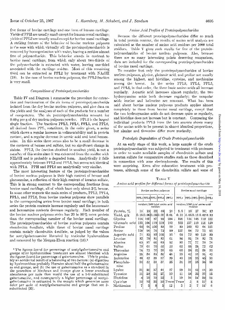
Amino Acid Profiles of Proteinpolysaccharides
Because the different proteinpolysaccharides differ so much in total protein content, the results of amino acid analysis are calculated as the number of amino acid residues per 1000 total residues. Table V gives such results for five of the protein- polysaccharides of bovine nucleus pulposus. Also, because there are so many interesting points deserving comparison, data are included for the corresponding proteinpolysaccharides of bovine nasal cartilage.
To consider first only the proteinpolysaccharides of bovine nucleus pulposus, glycine, glutamic acid, and proline are usually among the highest, and histidine, cysteine, and methionine among the lowest. In the series PPL3, PPL4, PPL5, and PPLG, in that order, the three basic amino acids all increase regularly. Aspartic acid increases almost regularly; the two hydroxyamino acids both decrease regularly; the nonpolar acids leucine and isoleucine are constant. What has been said about bovine nucleus pulposus products applies almost completely to those from bovine nasal cartilage, except that the two hydroxyamino acids do not decrease quite so regularly, and histidine does not increase but is constant. Comparing the individual products PPL3 from the two sources shows many of the amino acids to be present in almost identical proportions, but alanine and threonine differ more markedly.
Proteolytic Degradation of Crude Proteinpolysaccharide
At an early stage of this work, a large sample of the crude proteinpolysaccharide was subjected to treatment with proteases in order to make available samples of chondroitin sulfate and keratan sulfate for comparative studies such as those described in connection with zone electrophoresis. The results of this were surprising. Even after exhaustive treatment with pro- teases, although some of the chondroitin sulfate and some of
TABLE V Amino acid pro3les for different forms of proteinpolysaccharide
Protein, y0 Yield, g/g Glvcine Gl;tamic acid Proline Serine Aspartic acid Leucine Alanine Valine Threonine Arginine Isoleucine Phenylala-
nine Lysine Tyrosine Histidine Cysteine Methionine
-
P
0
L
Bovine nucleus pulposus
qYPL4~PPLSIPPL6~ PPL2
residues/1000 tote1 amino acid residues
Bovine nasal cartilage
PPL3 IPPL4JPPL51PPL6/PPL2 __
14 19 32 48 24 .180.060.050.07 0.01
124 107 87 92 234 133 106 119 119 122 90 96 100 88 99
128 96 76 72 90 71 83 82 108 35 82 78 81 83 61
65 81 66 65 92 70 64 70 53 52 74 72 70 59 63 29 50 55 56 46
40 42 39 37 29 34 42 39 37 25
8.5 10 27 36 12 0.11 .050.110.020.12
126 126 148 110 133 132 137 119 111 129 89 100 92 66 121
127 98 72 75 93 68 72 89 110 80 84 81 76 82 71 83 72 77 70 74 63 66 58 72 62 60 58 51 55 53
33 42 51 56 43
40 38 35 45 36 34 37 35 36 35
21 26 37 44 27 29 31 43 44 30 11 29 31 27 10 11 18 25 23 15
8 13 19 22 3 19 16 14 19 13 13 12 22 33 Trace rrace 5 8 11 8
7 3 8 5 12 3 3 7 14 6
by guest on May 16, 2018
http://ww
w.jbc.org/
Dow
nloaded from
4696 Proteinpolysaccharides of Nucleus Pulposus
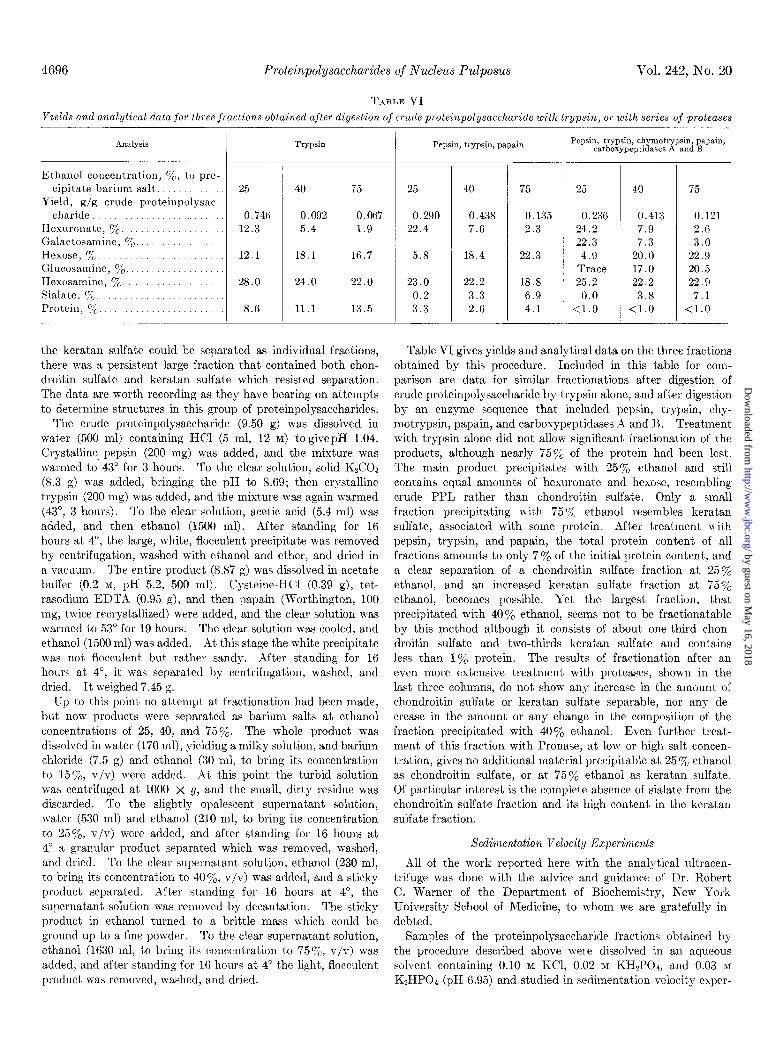
TABLE VI
Vol. 242, Ko. 20
Yields and analytical data jor three fractions obtained after digestion of crude proteinpolysaccharide with frypsin, or with series of proteases
Analysis Trypsin
Ethanol concentration, 7c, to pre- cipit,ate barium salt. .......
Yield, g/g crude proteinpolgsac- charide ........ ......
Hexuronate, 70 ............ Galactosamine, yo ............... Hexose, 7 o .....................
Glrlcosamine, %. ................ Hexosamine, %. ............. Sialate, yO. ...................... Protein, yo .................
25 40
0.746 0.092 12.3 5.4
12.1
28.0
8 .6
18.1
24.0
11.1
75
0.067 1.9
16.7
22.0
13.5
the keratan sulfate could be separated as individual fractions, there was a persistent large fraction that contained both chon- droitin sulfate and keratan sulfate which resisted separation. The data are worth recording as they have bearing on attempts to determine structures in this group of proteinpolysaccharides.
The crude proteinpolysaccharide (9.50 g) m-as dissolved in water (500 ml) containing HCl (5 ml, 12 M) to givepH 1.04. Crystalline pepsin (200 mg) was added, and the mixture was warmed to 43” for 3 hours. To the clear solution, solid Ii&O3 (8.3 g) was added, bringing the pH to 8.69; then crystalline trypsin (200 mg) was added, and the mixture was again warmed (43”, 3 hours). To the clear solution, acetic acid (5.4 ml) was added, and then ethanol (1500 ml). After standing for 16 hours at 4”, the large, white, flocculent precipitate was removed by centrifugation, washed with ethanol and ether, and dried in a vacuum. The entire product (8.87 g) was dissolved in acetate buffer (0.2 M, pH 5.2, 500 ml). Cysteine-HCl (0.39 g), tet- rasodium EDTA (0.95 g), and then papain (Worthington, 100 mg, twice recrystallized) were added, and the clear solution was warmed to 53“ for 19 hours. The clear solution was cooled, and ethanol (1500 ml) was added. Xt this stage the white precipitate was not flocculent but rather sandy. After standing for 16 hours at 4”, it w-as separated by centrifugat,ion, washed, and dried. It weighed 7.45 g.
I’p to this point no attempt at fractionation had been made, but now products were separated as barium salts at ethanol concentrations of 25, 40, and 75%. The whole product was dissolved in water (170 ml), yielding a milky solution, and barium chloride (7.5 g) and ethanol (30 ml, to bring its concentration to 15%, v/v) were added. At this point the turbid solution was centrifuged at 1000 x g, and the small, dirt’y residue was discarded. To the slightly opalescent supernatant solution, water (530 ml) and ethanol (210 ml, to bring its concentration to 25%, v/v) were added, and after standing for 16 hours at 4” a granular product separated which was removed, washed, and dried. To the clear supernatant solution, ethanol (230 ml, to bring its concentration to 407,, v/v) was added, and a sticky product separated. After standing for 16 hours at 4”, the supernatant solution was removed by decantation. The sticky product in ethanol t,urned to a brittle mass \\hich could be ground up to a fine powder. To the clear supernatant solution, ethanol (1630 ml, to bring its concentration to 7570, v/v) was added, and after standing for 16 hours at 4” the light, flocculent product w-as removed, washed, and dried.
Pepsin, trypsin, papain
25 40 75 25 40 75
0.290 0.438 0.135 22.4 7.6 2.3
5.8 18.4 22.3
23.0 22.2 18.8
0.2 3.3 6.9 3.3 2.6 4.1
0.236 0.413 0.121 24.2 7.9 2.6 22.3 7.3 3.0
4.9 20.0 22.9 Trace 17.0 20.5 25.2 22.2 22.9
0.0 3.8 7.1 <l.O <l.O <l.O
Pepsin, trypsin, chymotrypsin, papain, carboxypeptidases A and B
Table VI gives yields and analytical data on the three fractions obtained by this procedure. Included in this table for com- parison are data for similar fractionations after digestion of crude proteinpolysaccharide by trypsin alone, and after digestion by an enzyme sequence that included pepsin, trypsin, chy- motrypsin, papain, and carboxypeptidases A and B. Treatment with trypsin alone did not allow significant fractionation of the products, although nearly 75% of the protein had been lost. The main product precipit’ates with 25% ethanol and still contains equal amounts of hexuronate and hexose, resembling crude PPL rather than chondroitin sulfate. Only a small fraction precipitating with 75 7. ethanol resembles keratan sulfate, associated with some protein. After treatment n ith pepsin, trypsin, and papain, the total protein content of all fractions amounts to only 7% of the initial protein content, and a clear separation of a chondroitin sulfate fraction at 25% ethanol, and an increased keratan sulfate fraction at 75% ethanol, becomes possible. Yet the largest fraction, that precipitated with 40% ethanol, seems not to be fractionatable by this method although it consists of about one-third chon- droitin sulfate and two-thirds keratan sulfate and contains less than 1% protein. The results of fractionation after an even more extensive treatment with proteases, shown in the last three columns, do not show any increase in the amount of chondroitin sulfate or keratan sulfate separable, nor any de- crease in the amount or any change in the composition of the fraction precipitated with 40y0 ethanol. Even further treat- ment of this fraction with Pronase, at low or high salt concen- tration, gives no additional material precipitable at 25 70 ethanol as chondroitin sulfate, or at 75% ethanol as keratan sulfate. Of particular interest is the complete absence of sialate from the chondroitin sulfate fraction and its high content in the keratan sulfate fraction.
Sedimentation Velocity Experiments
All of the work reported here with the analytical ultracen- trifuge was done with the advice and guidance of Dr. Robert C. Warner of the Department of Biochemistry, New York University School of Medicine, to whom we are gratefully in- debted.
Samples of the proteinpolysaccharide fractions obtained by the procedure described above were dissolved in an aqueous solvent containing 0.10 M KCl, 0.02 M KH2P04, and 0.03 M
K,HPOI (pH 6.95) and studied in sedimentation velocity esper-
by guest on May 16, 2018
http://ww
w.jbc.org/
Dow
nloaded from

Issue of October 25, 1967 L. Rosenberg, n/r. Schubert, and J. Xandson 4697
iments in the Spinco model E analytical ultracentrifuge operated at 56,100 rpm and 20”. Photographs were taken with the schlie- ren optical system at 8-min intervals. Examples of sedimenta- tion patterns obtained are shown in Fig. 1, for which the con- centration of each fraction is 5 mg per ml. Strip a shows PPL3 (top) and PPL4 (bottom) examined together in two double sector cells, one with a wedge window. PPL3 appeared homogeneous over a concentration range of 2 to 6 mg per ml in the potassium chloride-phosphate buffer; with Si,,, = 6.6 S. The same sedimentation pattern was obtained for PPL3 at pH 4.5 and pH 9.0.
PPL4 (Strip a, bottom) showed an apparently homogeneous major component contaminated by a second component with a higher sedimentation velocity. The major component of PPL4 showed a higher sedimentation velocity than PPL3, with s:~,~ = 11.1 s.
Strip b compares the sedimentation patterns of PPL4 (top)
and PPL5 (bottom) examined together in double sector cells. PPL5 shows two boundaries, (Y slower and p faster. Comparison of the appearance of the small contaminating component of PPL4 with a! and @ of PPL5 over a wide range of concentrations indicates that this component in PPL4 is residual PPL5 (o( and 0) still present in PPL4. An interesting feature of the be- havior of PPL5 (cu and p) is the variation of sedimentation pattern with concentration shown in Fig. 2, Strips a, b, c, and d (bottom patterns), run at 4, 5, 6, and 8 mg per ml, respectively. With increasing concentrations of PPL5, the slower /?-peak progressively rises and the faster P-peak falls, so that their relative heights become reversed. Because of this accumulation of increased amounts of LY component behind the faster /3 bound- ary (Johnston-Ogston effect), the determination of valid sedi- mentation coefficients for PPL5 (a and /3) was not possible. Yet comparison of the sedimentation patterns of PPL5 (01 and 0) with the major component of PPL4 (Fig. 2, upper patterns
FIG. 1. Sedimentation velocity patterns of several forms of PPL (5 mg per ml; 0.05 M ph0sphat.e buffer-O.10 M KCl, pH 6.95) at 20.0” and 56,100 rpm; bar angle, 60”. Strip a: PPL3, upper pattern; PPL4, lower pattern. Strip b: PPL4, upper pattern; PPLS, lower pattern.
Strip c: PPLG, upper pattern, PPLS, lower pattern. In each strip, patterns were recorded at 16, 24, 32, and 40 min, from left to right.
FIG. 2. Changing proportions of CX- and P-peaks in sedimentation velocit,y patterns at different concentrations of PPLB. In each strip, PPL4 is the upper pattern and PPL5 is the lower pattern. Strip a: PPL5,4 mg per ml; b, PPL5, 5 mg per ml; c, PPL5, 6 mg per ml; cl, PPL5, 8 mg per ml. PPL5 shows a marked variation of sedimentation velocity with concentration. In each strip, patterns
were recorded at time intervals such that the relative heights of OL and p could most clearly be compared.
by guest on May 16, 2018
http://ww
w.jbc.org/
Dow
nloaded from

4698 Proteinpolysaccharides of Nucleus Pulposus Vol. 242, No. 20
of Strips a, 6, c, and d) indicates that both CY and /3 of PPL5 are of faster sedimentation velocity than the major component of PPL4.
PPL6 dissolves slowly and poorly in the phosphate buffer, to form opalescent solutions. It sediments rapidly to the base of the cell during acceleration to 12,000 rpm, and appears, in the upper pattern of Strip c, Fig. 1, as a broad band at the bottom of the cell which becomes progressively narrower with longer sedimentation time.
The sedimentation pattern of PPLS, the degradation product obtained from the cartilage residue by treatment with hydroxyl- amine, is shown in the lower pattern of Strip c, Fig. 1. The excessive spreading of the boundary with time indicates heter- ogeneity. The sedimentation velocities of the molecular species included in PPL2 are lower than all the other forms of PPL.
Sedimentation Equilibrium Experiments
For the further evaluation of the homogeneity of PPL3, sedimentation equilibrium experiments were carried out ac- cording to the following procedure. PPL3 was dissolved in and dialyzed against the same solvent used in the sedimentation velocity experiments. With the use of a six-channeled center- piece, three solution-solvent pairs, including three different concentrations of PPL3, were simultaneously studied in a single experiment. Conditions were established (16,200 rpm, 20 hours) such that, at equilibrium, the concentration distribution of solute near the base of the cell resulted in satisfactory re- fractive index gradients with relatively dilute initial solutions of PPL3, 0.2 to 0.5 mg per ml, while the concentration of solute throughout the region from the meniscus to the middle of the cell was essentially zero (21). Rayleigh interference patterns were recorded at 19 and 20 hours and measured with a Nikon two-dimensional microcomparator.
In Fig. 3, the natural logarithms of the fringe displacements (centimeters) are plotted against the squares of the distances from the axis of rotation. Plots A, B, and C represent the points (with net fringe displacement greater than 0.010 cm) from experiments at initial concentrations of 0.4, 0.2, and 0.5 mg per ml of PPL3, respectively. The straight lines are plotted from averaged values of each slope (d In c/d+) and y intercept cal- culated from a least squares treatment of the experimental
-2.5c /A
A B C -3.0
r
2
” -5
-35 I/‘1 __jl: -4.0 *
-4.5 .
(A ond 8)- 374 3:6 j , , ,
(C) 43.9 44.1 44.3
r2 (cm*)
FIG. 3. Results of sedimentation equilibrium study of PPL3 at three concentrations: A, 0.4; B, 0.2; C, 0.5 mg per ml. The abscissas show square of the distance from the axis of rotation (centimeters) ; ordinates, natural logarithm of the fringe displace- ment (centimeters).
TABLE VII Weight average molecular weights of PPLS calculated from
sedimentation equilibrium data. at several concentrations of PPLS
Concentration
m/ml
0.2 0.4 0.4 0.5
Average
Molecular weight
186,000 149,000 166,000 181,000 170,500
points. While plots at 0.5 mg per ml appear linear, at lower concentrations (0.2 mg per ml) upward curvature is revealed, indicating some degree of polydispersity. The weight average molecular weights in four cases calculated from the averaged value of d In c/&2 obtained by the least squares treatment are given in Table VII. The partial specific volume of PPL3 (0.56) was calculated from the densities of PPL3 solutions measured in a lo-ml pycnometer.
Zone Electrophoresis
The chondroitin sulfate and keratan sulfate, the preparations of which have been described above, show characteristic mo- bilities on zone electrophoresis on polyvinyl chloride under standardized conditions. Chondroitin sulfate separates from kerat*an sulfate, moving more rapidly toward the anode; keratan sulfate follows as a slower, second peak. Since all of the protein- polysaccharide fractions, PPLX, PPL4, PPL5, PPLG, and PPL2, contain both chondroitin sulfate (as indicated by uronate and galactosamine) and keratan sulfate (as indicated by hexose and glucosamine), we were interested in determining whether there was any tendency for the chondroitin sulfate and keratan sulfate in any fraction to separate on zone electrophoresis, or whether in all fractions chondroitin sulfate, keratan sulfate, and protein would move together. Since PPL3 appeared reasonably homogeneous with respect to molecular weight according to the analysis of sedimentation equilibrium data, its behavior was of particular interest. In addition, since PPL3, PPL4, PPLS, and PPL6 showed increasing sedimentation velocities, sug- gesting in a rough way that they were fractions of increasing size, we wanted to know whether zone electrophoresis would give a similar regularity in results. Each fraction was therefore subjected to zone electrophoresis.
Polyvinyl chloride (Geon 435, B. F. Goodrich) was washed three times with phosphate buffer, 0.034 M, pH 7.4, following which sufficient buffer was added to made a thick slurry, which was poured into a plastic block containing two chambers 1 x 8 x 40 cm. The sample, 15 mg, dissolved in 1.5 ml of distilled water, was applied to the block. Zone electrophoresis was carried out at a voltage gradient of 9 volts per cm for 16 hours at 4” in the same phosphate buffer. After electrophoresis, the block was immediat’ely blotted with filter paper, and the poly- vinyl chloride was cut into l-cm segments. Each segment was thoroughly mixed wit’h 3 ml of water, the polyvinyl chloride was removed by centrifugation at 2000 rpm for 15 min, and the supernatant solution from each segment was analyzed for uronate (as an index of chondroitin sulfate), hexose (as an index of keratan sulfate), and protein.
by guest on May 16, 2018
http://ww
w.jbc.org/
Dow
nloaded from

Issue of Oct’ober 25, 1967 L. Rosenberg, M. Schubert, and J. Xandson 4699
The behavior of purified samples of chondroitin sulfate and keratan sulfate is shown in Fig. 4A. Chondroitin sulfate (28 segments/l6 hours) and keratan sulfate (18 segments/l6 hours) move toward the anode at distinctly different rates..
Fig. 4, B and C, and Fig. 5, n and B, show the behavior of
1.0
c -- t-m. URONATE A
0.8 -HEXOSE /
B
I I I I I I I I I I I I I
PPL4 C
-A 0 5 10 15 20 25 30 35
SEGMENT NUMBER
FIG. 4. Results of zone electrophoresis for 16 hours on poly- vinyl chloride and analysis of successive l-cm segments for uronate, hexose, and protein. -4, chondroitin sulfate (CS) and keratan sulfate (KS), each isolated from a proteolytic digest of the crude protein polysaccharide; B, PPL3; C, PPL4.
PPL.5
---. URONATE - HEXOSE *..---+ PROTEIN
, , / , , , , , , , , , , , , ,
“ . ”
0.6
0.4
0.2
0
SEGMEI\!T NUMBER
FIG. 5. Same as Fig. 4. A, PPL5; B, PPLG; C, unfractionated PPL.
l’PL3, 4, 5, and 6. In each one of these proteinpolysaccharidc fractions chondroitin sulfate, keratan sulfate, and protein all migrated at the same rate, and this rate was characteristic for each particular fraction. In the case of PPL3, closer scrutiny of Fig. 4B shows the proportion of chondroitin sulfate was consistently higher id the leading edge of the curve than in the trailing edge of the curve. PPL4 (Fig. 4C) migrated more slowly (8 segments/l6 hours) than PPL3 (10 segments/l6 hours). PPL5 (Fig. 5A) separated into two components, the faster migrating’at a rate of 4 segments/l6 hours, and the slower component, at 2 segments/l6 hours. It seemed likely that the faster component represented PPL5. (a! + ,B) identified in the sedimentation velocity studies. PPL6 (Fig. 5B) migrated at a rate of 2 to 3 se,ments/l6 hours.
UISCURSION
A major aim of this laboratory has been to develop effective methods for the separation of the various macromolecular species present in the mixtures of proteinpolysaccharides ex- tractable from the ground substance of connective tissues. The fractionation of proteinpolysaccharides from bovine nasal cartilage has been previously reported (2), and by a different method some fractionation of those from human costal cartilage has been achieved (1).
l3ovine nucleus pulposus is a tissue rich in polysaccharide. It contains both chondroitin B-sulfate’ and keratan sulfate in amounts that are nearly equal. Practically the entire poly- saccharide content can be extracted with water, and of the poly- saccharide extractable with water nearly all is of the kind called PPL, with only a small amount of the kind called PPH. There is no evidence that any free chondroitin sulfate or keratan sulfate is present. The PPL can be fractionated by several methods. The one recommended, and the only one described in detail, is the same as that used to fractionate the PPL of bovine nasal cartilage (2). The PPL of bovine nucleus pulposus yields four distinct fractions: PPL3, PPL4, PPL5, and PPLG, in the order of increasing sedimentability. This is also the order of in- creasing protein content, increasing viscosity, decreasing hexu- ronate content, and decreasing mobility on zone electrophoresis. The fractionation procedure recommended shows a high degree of reproducibility in regard to yield, composition, sedimentation pattern, and electrophoretic characteristics of each fraction.
It seems worth mentioning, however, that although ‘the method outlined in the diagram of Table IV is the same as that described for the’ fractionation of bovine nasal PPL, it was not simply a blind application of the latter method to a new tissue, but was reached after an independent set of trials and errors. The work leading to the resdlts described here was begun at the time of reporting a method to fractionate human costal PPL (l), a method that involves as its first step ,addition of a solution of lanthanum chloride to a dilute cold solution of PPL. This precipitates a part of the PPL and leaves in solution a part which was similar analytically to the fraction now called PPL3 in the diagram, but which was not quite so homogeneous in the analytical ultracentrifuge. For this reason the lanthanum method was dropped. Step 6 of the diagram is similar to the corresponding step in the fractionation of bovine dasal PPL (2), but it was found that a cleaner separation of products results if potassium acetate is used at 2 N instead of 2.5 N. A method of fractionation of PPLR different from that of Step 7 of the diagram was also tried. Stirred with 1 N potassium
by guest on May 16, 2018
http://ww
w.jbc.org/
Dow
nloaded from

Proteinpol~saccharides of Nucleus Pulposus Vol. 242, n-o. 20
Protein poly- saccharide
% % % % % % % PPL2 24.4 33.4 22.3 5.2 5.6 2.1 93.0 1.50 PPL3 14.4 45.0 35.4 3.1 4.4 2.8 105.7 1.29
PPL4 18.9 27.9 42.8 4.7 2.0 3.4 99.7 0.65 PPL5 31.8 24.2 40.3 4.4 2.0 3.7 106.4 0.60 PP1.B 48.0 20.5 33.2 4.9 1.8 3 .o 111.4 0.62
PPN 49.2 20.2 33.2 3.3 2.4 0.63
T.WLE VIII
Swnmary oj” contents of three major and lhree minor components of six proteinpolysaccharides of bovine nwleus pulposua
a Extra sulfate, beyond the amounts contained in chondroitin and keratan sIllfates, assuming 1 mole of sIllfate per mole of hexos- amine in each. This has frequently been described (22).
* Extra hexose, beyond the amount contained in keratan sul- fate. This is probably analogous to t.he galactose and xylose which link polysaccharide to protein in bovine nasal PPL as
described by Roden and Smith (23). c The sllrn of the preceding six columns.
acetate and then centrifuged at 40,000 rpm, PPLR yielded in the sediment a product similar to PPLG, and in the supernatant solution a product similar to PPL4 + PPL5. The outcome of such trials showed that several variant methods could be worked out, and choice of that shown in the diagram rests on the fact that its products, in the analytical ultracentrifuge, behaved least like complicated mixtures. In addition, it would be desir- able if a single fractionat.ion method could be applied to PPL from different cartilages to make significant comparison of results possible. For example, the data of Table III show how widely the yields of crude PPH, PPL, and PPL2 vary for a few cartilages which have been examined by identical extraction methods. Not only the yields but the compositions of PPL from these cartilages differ.
The present results and the results (2) from bovine nasal PPL give the first opportunity for a detailed comparison of analytical values in the series of products from l’PL3 to PPL6 from two different cartilages. ..4lthough each of the fractions from the PPL of bovine nucleus pulposus differs in composition from the corresponding fraction from the PPL of bovine nasal septum, the trends of values in each series from PPL3 to PPL6 are regular and parallel. In each series sedimentability and limiting viscosity numbers increase, protein contents increase about 4-fold, hexuronate contents decrease to about half, hex- osamine contents decrease somewhat less, and sialate, hexose, and sulfate contents do not vary significantly. Amino acid contents show a number of regularities. Although the protein contents of PPL3 from these two sources differ, their amino acid profiles are nearly identical. Amino acid profiles of the other corresponding pairs of fractions are also similar but not so nearly identical. another way of comparing the products is by t,he trends in contents of each amino acid in each series from PPLY to PPLG. Most of the nonpolar amino acids of both series are nearly constant. Serine and threonine generally decrease; aspartic acid and the basic amino acids generally in- crease. These tendencies of some amino acids to increase or decrease in the series PPL3 to PPL6 are sufficiently regular to raise a suspicion that the trends are due to the presence of two
different proteins in each series, the ratio of which varies regularly as the total protein content increases.
To see whether the total composition of each of the protein- polysaccharides can be accounted for by the known components, the calculations summarized in Table VIII were made. Chon- droitin sulfate was calculated as 3.1 x the percentage of uronate; keratan sulfate, as 3.1 x the percentage of glucosamine; extra sulfate, as the difference between tot’al sulfate and the amount of sulfate in chondroitin sulfate and keratan sulfate, and extra hexose, as the difference between total hexose and the percentage of glucosamine. The total of these components lies in the range 93 to 111%. The last column of Table VIII shows that the proteinpolysaccharides of bovine nucleus pulposus have a ratio of chondroitin sulfate to keratan sulfate either close to 1.4, as PPL2 and PPL3, or to 0.6, as all the other forms.
There was no evidence that from bovine nucleus pulposus any native fraction could be obtained containing only chon- droitin sulfate or only keratan sulfate. Treatment with several proteases was found necessary before a part of the polysaccharide could be separated as chondroitin sulfate and as keratan sulfate (Table VI). Even then, much of the chondroitin sulfate and keratan sulfate remain in an intermediate fraction and are not readily separable. In contrast, Davidson and Woodhall (7) have reported isolation of chondroitin sulfate and keratan sulfate in good yields after ext’raction of normal human nucleus pulposus with KC1 (0.15 M) and fractionation.
A fundamental question is how significant the fractionation procedure is, whether an arbitrary selection is being made among a continuously variable set of molecules, or whether there exist within the total set of proteinpolysaccharide molecules subsets that can be separated and that have nonoverlapping characteristics. Are any of the fractions homogeneous? Does each fraction represent only a somewhat narrow sampling of a completely heterogeneous population?
In this regard PPL3 is of particular interest, since it contains roughly equal amounts of keratan sulfate and chondroitin sulfate in addition to protein and sialic acid, and yet it appears homogeneous in sedimentation velocity experiments under a variety of conditions, suggesting that it might be a single molec- ular species in which both keratan sulfate and chondroitin sulfate are bound to the same protein core. Based upon the appearance of the sedimenting boundary alone, however, this conclusion would be unwarranted. Macromolecules such as proteinpolysaccharides, which consist of a number of flexible, threadlike polysaccharide chains bound to protein, and certain glycoproteins may form a kind of molecular network at the solute concentrations (0.2 to 0.6%) generally used in sedi- mentation velocity experiments with the schlieren optical system. A mixture of different molecular species entangled in such a network at these relatively high concentrations may not separate cleanly during the course of a sedimentation velocity experi- ment to produce discrete boundaries. The appearance of the boundary might suggest a single homogeneous species while a mixture of diffuse, entangled macromolecules is actually present. Second, this class of macromolecules may show significant bound- ary sharpening because of marked dependence of sedimentation coefficient on solute concentration, resulting in apparently single symmetrical boundaries with heterogeneous materials. While the appearance of the sedimentation velocity patterns of PPL3 and the extent to which the sedimentation coefficient decreases with increasing solute concentration indicated that
by guest on May 16, 2018
http://ww
w.jbc.org/
Dow
nloaded from
Issue of October 25, 1967 L. Rosenberg, M. Xchubert, and J. Xandson 4701
the above cffectn are not prominent features of the hchavior of this particular molecular species, we wished to test the homo- geneity of PPL3 by more sensitive methods. Sedimentation equilibrium experiments mere carried out at initial concen- trations of PPL3 from 0.2 to 0.5 mg per ml. At the lowest concentrations (0.2 mg per ml), plots of In c/r” revealed some degree of polydispersity. On zone electrophoresis of PPLS, prot’ein, keratan sulfate, and chondroitin sulfate traveled to- gether with maxima in the same segment. It was consistently noted with every preparation of PPL3, however, that the leading edge of the curve showed a slightly higher concentration of chondroitin sulfate than the trailing edge. PPL3 may represent a molecular species with individual members that are relatively close in molecular weight and similar but not identical in detailed chemical structure. Variations in the numbers of chondroitin sulfate and keratan sulfate chains in each molecule, or in their chain lengths, might account for these observations. Yet it seems likely that each member of this species is a compound containing roughly equal amounts of chondroitin sulfate and keratan sulfate bound to the same protein core or to each other.
The sedimentation velocity experiments indicate that PPL4 and PPL5 (a and /3) are also relatively discrete molecular species, and not segments of a continuously varying set of molecules. PPL6, however, may be grossly heterogeneous since no single component is present in sufficient concentration to produce a discrete boundary in sedimentation velocity experiments. The parallel gradation from PPL3 to PPL6 in regard to increasing sedimentation velocity and decreasing zone electrophoretic mobility suggests that t’he molecular weights of the fractions increase from PPL3, the smallest molecular weight species, to PPLG.
Sialic acid is absent from purified samples of chondroitin sulfate. Its content (7 %) in purified samples of keratan sulfate containing less t’han 1% protein is twice that of all the native proteinpolysaccharide fractions (3 %). The sialic acid associated with purified samples of keratan sulfate moves with it on zone electrophoresis, is eluted with it on exclusion chromatography, and is liberated by neuraminidase, indicating that it is terminally bound to kerat,an sulfate and not a part of a separate glyco- protein. If there were only one terminal sialate per keratan sulfate chain, the chain weight would be about 4000, corre- sponding to six or seven periods per chain.
However, such estimates are complicated by the fact that polysaccharide preparations from the same or different tissue sources may show variations in composition and polydispersity in chain weight. This has been particularly true of keratan sulfate. Anseth and Laurent (24) isolated from bovine cornea1 stroma, after digestion with collagenase and trypsin, a series of fractions of keratan sulfate of constant composition, by elution from Ecteola cellulose with increasing concentrations of NaCl. This series of fractions was found, from sedimentation and diffusion data, to have average molecular weights increasing progressively from 4,200 to 19,000 (25, 26). Not only variations in molecular size with constant composition, but variations in composition of keratan sulfate depending on its source, have been found to occur. Indeed, Meyer (27) considers one of the most outstanding properties of the mucopolysaccharides to be their occurrence in major and minor variations.: f Variations occur in the type of linkage of keratan sulfate to protein, leading to a distinction between cornea1 keratan sulfate, in which the linkage is mainly N-glycosidic, to asparagine and glutamine, and car-
tilage keratan sulfate, in which the linkage is mainly 0.glycosidic to scrine and threonine (28). The amount of sialate associated wit,h keratan sulfate may vary from 0.2 to 1.0% for bovine cornea to 7.5% for kerat,an sulfate from calf epiphysial plate. While cornea1 keratan sulfate contains roughly 1 mole of sulfate per repeating unit, cartilage keratan sulfate is usually over- sulfated (22, 28). Both galactose 6-sulfate and N-acetylglucosa- mine sulfate have been obtained from cornea1 and cartilage keratan sulfates (29). ,4s indicated by 1Jeyer (27, 30), al- though polymorphism and microheterogeneity appear to dis- tinguish mucopolysaccharides as a class of substances sharply from the proteins in which the number of permissible variations seems very limited, the biolcgical basis and biological conse- quences of these variations in the mucopolysaccharides are unknown. A comparison of the keratan sulfates associated with the series of proteinpolysaccharides from bovine nucleus pulposus may provide information regarding the significance of these variations.
1. ROSENBERG, L., JOHNSON, B., AND SCHUBERT, M., J. Gin. Invest., 44, 1647 (1965).
2. PAL, S., DOGANGES, P. T., AND SCIKJBERT, M., J. Biol. Chem., 241, 4261 (1966).
3. MALMGREN, H., AND SYLVEN, B., Biochim. Biophys. Acta, 9, 706 (1952).
4. GARD~LL, s., AND RASTGELDI, S., Acta Chem. Stand., 8, 362 (1954).
5. MEYER, K., LINKER, A., DAVIDSON, E. A., AND WEISSM~NN, B., J. Biol. Chem., 206, 611 (1953).
6. 7.
8.
GARDELL, S., Acta Chem. Stand., 9, 1035 (1955). DAVIDSON, E. A., AND WOODHALL, B., J. Biol. Chem., 234,
2951 (1959). LYONS, H., JONES, E., QUINN, F. E., AND SPRUNT, D. H.,
Proc. Sot. EXD. Biol. Med.. 115. 610 (1964). 9.
10. 11. 12. 13.
14. 15.
DISCHE, Z., J. biol. Chem., i67, i89 (1947). SCHLOS~, B., Anal. Chem., i3, 1321 (1951): YEMM. E. W.. AND WILI~S. A. J.. Biochem. J.. 57.508 11954). WARREN, L.,‘J. Biol. Chek, 234, 1971 (1959). ’ ’ ’ LOWRY, 0. H., ROSEBROUGH, N. J., FARR, A. L., AND RANDALL,
R. J., J. Biol. Chem., 193, 265 (1951). WOESSNER, J. F., Arch. Biochem. Biophys., 93, 440 (1961). GREEN, J. P., AND ROBINSON, J. I>., JR., J. Biol. Chem., 235,
1621 (1960). 16. MATHEWS, M. B., AND INOUYE, M., Biochim. Biophys. Acta,
63, 509 (1961). 17. 18.
DISCHE, Z., Method. Carbohyd. Chem., 1, 511 (1962). AMINOFF, D., MORGAN, W. T. J., AND WATKINS, W. M., Bio-
them. J., 51, 379 (1952). 19. 20.
21. 22.
23. 24. 25. 26. 27.
PAL, S., AND SCHUBERT, M., J. Bio7. Chem., 240, 3245 (1965). NEUBERGER, A., MARSHALL, R. D., AND GOTTSCHALK, A., in
A. GOTTSCHALK (Editor), Glucoproteins, Elsevier Publishing Company, New York, 1966,-p. i66.
YPHANTIS. D. A.. Biochemistru. 3, 297 (1964). MATHEWS; M. B.; AND CIFONE~L;, J. A:, J.‘Biol. Chem., 240,
4140 (1965). ROD&N, L., AND SMITH, R., J. Biol. Chem., 241, 5949 (1966). ANSETH, A., AND LAURENT, T. C., Exp. Eve Res., 1, 25 (1961). LAURENT, ‘I!. C., AND ANSETH, A.; E&p. Eye Res.; 1, 99 (1961). LA.URENT, T. C., AND SCOTT, J. E., Nature, 202, 661 (1964). MEYER, K., Fed. Proc., 25, 1032 (1966).
28. SENO, N., MEYER, K., ANDERSON, B., AND HOFFMAN, P., J. Biol. Chem.. 240. 1005 (1965).
REFERENCES
29. BHAVANANDA~, VI P., ~KD MEYER, K., Science, 151, 1404 (1966).
30. MEYER, K., in I>. BERGSMA (Editor), Birth defects, Original Article Series, Structural organization of the skeleton, Vol. II, No. 1, Nat,ional Foundation, March of Dimes, New York, 1966, p. 21.
by guest on May 16, 2018
http://ww
w.jbc.org/
Dow
nloaded from
Lawrence Rosenberg, Maxwell Schubert and John SandsonThe Proteinpolysaccharides of Bovine Nucleus Pulposus
1967, 242:4691-4701.J. Biol. Chem.
http://www.jbc.org/content/242/20/4691Access the most updated version of this article at
Alerts:
When a correction for this article is posted•
When this article is cited•
to choose from all of JBC's e-mail alertsClick here
http://www.jbc.org/content/242/20/4691.full.html#ref-list-1
This article cites 0 references, 0 of which can be accessed free at
by guest on May 16, 2018
http://ww
w.jbc.org/
Dow
nloaded from


![Puerarin Relieved Compression-Induced Apoptosis and ...downloads.hindawi.com/journals/sci/2020/7126914.pdfimpaired nucleus pulposus cell (NPC) proliferation [4]. Nucleus pulposus mesenchymal](https://img.pdfslide.us/doc/110x75/5f7fa24ee6184370f175b23e/puerarin-relieved-compression-induced-apoptosis-and-impaired-nucleus-pulposus.jpg)


![The protective effects of PI3K/Akt pathway on human nucleus pulposus … · 2020. 1. 28. · nucleus pulposus cells and nucleus pulposus progenitor cells [14]. Previous studies have](https://img.pdfslide.us/doc/110x75/60b265dd0d8b8040e758b496/the-protective-effects-of-pi3kakt-pathway-on-human-nucleus-pulposus-2020-1-28.jpg)