Embed Size (px)
Citation preview
ORIGINAL PAPER
A Comparison of Floating-Electrode DBD and kINPenJet: Plasma Parameters to Achieve Similar GrowthReduction in Colon Cancer Cells Under StandardizedConditions
Sander Bekeschus1 • Abraham Lin2 • Alexander Fridman2 •
Kristian Wende1 • Klaus-Dieter Weltmann1 • Vandana Miller2
Received: 16 June 2017 / Accepted: 31 August 2017 / Published online: 6 September 2017� The Author(s) 2017. This article is an open access publication
Abstract A comparative study of two plasma sources (floating-electrode dielectric barrier
discharge, DBD, Drexel University; atmospheric pressure argon plasma jet, kINPen, INP
Greifswald) on cancer cell toxicity was performed. Cell culture protocols, cytotoxicity
assays, and procedures for assessment of hydrogen peroxide (H2O2) were standardized
between both labs. The inhibitory concentration 50 (IC50) and its corresponding H2O2
deposition was determined for both devices. For the DBD, IC50 and H2O2 generation were
largely dependent on the total energy input but not pulsing frequency, treatment time, or
total number of cells. DBD cytotoxicity could not be replicated by addition of H2O2 alone
and was inhibited by larger amounts of liquid present during the treatment. Jet plasma
toxicity depended on peroxide generation as well as total cell number and amount of liquid.
Thus, the amount of liquid present during plasma treatment in vitro is key in attenuating
short-lived species or other physical effects from plasmas. These in vitro results suggest a
role of liquids in or on tissues during plasma treatment in a clinical setting. Additionally,
we provide a platform for correlation between different plasma sources for a predefined
cellular response.
Keywords Atmospheric pressure argon plasma jet � Dielectric barrier
discharge � Hydrogen peroxide � kINPen � Plasma medicine
& Sander [email protected]
Vandana [email protected]
1 Leibniz-Institute for Plasma Science and Technology (INP Greifswald), ZIK Plasmatis, Felix-Hausdorff-Str. 2, 17489 Greifswald, Germany
2 Nyheim Plasma Institute, Drexel University, 200 Federal Street, Camden, NJ 08103, USA
123
Plasma Chem Plasma Process (2018) 38:1–12https://doi.org/10.1007/s11090-017-9845-3
Introduction
In the study of plasma medicine, partially ionized gases and their physico-chemical
effectors are investigated for beneficial biological responses [1–3]. The observed beneficial
effects in wound healing [4–6] and cancer [7–9] have significantly spurred research efforts
and novel findings in recent years. Promising in vitro research investigations are an ethical
and scientific necessity for translation of plasma applications to in vivo models and
eventually to humans. However, there are technical and methodological challenges for
direct plasma applications, especially with regard to different types of plasma sources and
comparison of results. Major among them is the extent to which in vitro plasma effects
depend on long-lived species or other effectors of the multicomponent system plasma, such
as UV-radiation or electrical fields. Therefore, two main types of plasma sources, a
floating-electrode dielectric barriers discharge [10] and an atmospheric pressure argon
plasma jet [11] were compared with regard to cell growth reduction and its dependence on
main plasma active components. The sources were chosen because they have been of
relevance in plasma medical research for more than a decade and thus were subject to
extensive physical characterization [12]. To compare plasma effects across labs which is
easy to perform, cheap and could be applicable for clinical device calibration, a simple
biological read-out was chosen.
Plasma medical research inevitably involves experiments on reactive species because
these were found to be central effectors in a number of biological targets exposed to
plasma, such as skin cells [13–15], immune cells [16–18], and cancer cells [19–52]. In
many instances, hydrogen peroxide (H2O2) was an important mediator in these in vitro
experiments [53–55]. H2O2 is not necessarily toxic by itself but rather exerts its biological
effects through secondary processes, for example, Fenton reaction [56], by acting as
substrate for oxygenases [57], and in redox signaling events enabling the translation of
redox events into distinct biological responses [58]. We selected CT26 murine colon
cancer cells for this work because H2O2 has been previously identified as inducing
apoptotic but not the necroptotic cell death pathway in these cells [59, 60]. CT26 colon
cancer monolayer cultures were exposed to either DBD or argon jet plasma. Plasma source
dependent, our results demonstrate that H2O2 correlates with inhibition of CT26 metabolic
activity. It plays a central but not exclusive role in plasma-induced cell toxicity.
Materials and Methods
Cell Culture
Murine CT26 colon cancer cells were maintained in cell culture flasks in Rosswell Park
Memorial Institute 1640 (RPMI1640) cell culture medium supplemented with 10% fetal
bovine serum (FCS), 2% glutamine, and 1% penicillin/streptomycin (all Sigma). For
culturing and experiments, cells were maintained in standard incubation conditions at
37 �C with 95% humidified atmosphere and 5% carbon dioxide.
DBD Plasma Treatment
Treatment of cells with the DBD plasma system was performed in the absence of liquid
unless otherwise specified. The DBD electrode used was 1.3 cm in diameter and fit into the
wells of a 24-well plate. Cells were treated with plasma as previously described [61].
2 Plasma Chem Plasma Process (2018) 38:1–12
123
Briefly, CT26 cells were seeded at 1.5 9 105 cells per well in 0.5 ml of fully supplemented
cell culture medium and incubated overnight at 37 �C with 5% CO2. Prior to plasma
treatment, cells were washed twice with PBS, and PBS from the second wash was removed
immediately before plasma exposure. Plasma was generated by applying a high voltage
pulse to the DBD electrode 1 mm above the cells in the well. The pulse was generated with
a nanosecond pulser (FPB-20-05NM, FID GmbH, Burbach, Germany) and the frequency
of pulses was controlled with an external function generator (TTi, TG5011 LXT,
Philadelphia, PA, USA). Treatment time was fixed at 10 s unless otherwise specified. In
some experiments, either treatment time or pulse frequency was altered to deliver a fixed
plasma treatment energy over different times. For the DBD comparative study, a
microsecond pulse (0.07 mJ/pulse) was also used. The energy per pulse from both system
was measured as previously described [62, 63] and total plasma energy delivered to the
cells for both systems were calculated from treatment time, pulse frequency and energy per
pulse. Complete media was added to each well after plasma treatment and cells were
incubated overnight before viability was measured. Pulse parameters of both the
nanosecond- and the microsecond-pulsed DBD system are summarized in Table 1.
Jet Plasma Treatment
Treatment with the genotoxically-safe [64–66] atmospheric pressure argon plasma jet
kINPen 11 (similar in construction to the kINPen MED that has received accreditation as
medical device in Germany; neoplas control, Germany) operated at a feed gas flux of three
standard liters per minute (SLPM) of Argon gas (Air Liquide, France) was performed as
described previously [67]. It is a DBD-like jet, with a central pin electrode shielded against
an outer electrode by a dielectric, powered by 2–6 kV at 1 MHz. Briefly, 1 9 105 CT26
cells in 1 ml of fully supplemented cell culture medium were added to each well of a
24-well plate, and incubated overnight. In some experiments, 5 lg/ml of the H2O2-scav-
enging enzyme catalase (Sigma) was added prior to plasma treatment. A layout was
programmed for a computer-driven xyz-table (CNC, Germany) hovering the plasma jet
over the center of each well at a predetermined distance for the indicated treatment time.
Table 1 Technical parametersof the two DBD plasma settingsapplied to cells
Nanosecond-pulsed DBD plasma parameters
Voltage 29 kV
Energy per Pulse 0.9 mJ/pulse
Pulse Width 20 ns
Gap Distance 1 mm
Pulse Frequency 0, 5, 15, 30, 75, 150 Hz
Treatment Time 10 s
Plasma Energy 0, 50, 100, 300, 700, 1000 mJ
Microsecond-pulsed DBD plasma parameters
Voltage 30 kV
Energy per Pulse 0.07 mJ/pulse
Pulse Width 1.65 ls
Gap Distance 1 mm
Pulse Frequency 50 Hz
Treatment Time 0, 15, 30, 90, 200, 290 s
Plasma Energy 0, 50, 100, 300, 700, 1000 mJ
Plasma Chem Plasma Process (2018) 38:1–12 3
123
To compensate for evaporation of liquid, a predetermined amount of double-distilled water
was added after plasma treatment.
Cytotoxicity
Cytotoxicity was assessed as a measure of total metabolic activity. After treatment, cells
were incubated for 21 h at 37 �C. The medium was replaced with 1 ml of fully supple-
mented cell culture medium without phenol red and containing 100 lM of resazurin (Alfa
Aesar, USA). Metabolically active cells generate NADPH that is used by intracellular
enzymes to create highly fluorescent resorufin from resazurin. Both compounds freely
diffuse through cell membranes for a convenient readout in cell culture supernatants. After
incubation for 3 h at 37 �C, 4 9 200 ll of each well were transferred to a flat-bottom
96-well plate. Fluorescence was read at kex 535 nm and kem 590 nm using a microplate
reader. Background fluorescence of cell culture medium alone containing resazurin was
subtracted from all sample and control readings. Relative fluorescence of samples was then
normalized to that of untreated controls cells.
Hydrogen Peroxide
H2O2 was quantified in double-distilled water (Drexel) or PBS (INP) using the amplex red
detection reagent (Thermo, USA). If plasma does not acidify the solution, double-distilled
water and PBS are in principle interchangeable. The pH of distilled water did not change
under any of the treatment conditions used in this study for DBD therefore peroxide
measurements were made in water. Plasma treated water or PBS was diluted 1:20 in
amplex red reagent (5 lM) in PBS supplemented with horseradish peroxide (10U/ml).
After incubation for 15 min in dark, fluorescence was read at kex 535 nm and kem 590 nm
using a microplate reader. Relative fluorescence units were quantified against a linear
regression delineated from a H2O2 standard curve (5000–78 nM), and multiplied by
dilution factors to retrieve actual concentrations.
Software
Graphing, calculation of mean and standard deviation (S.D.), and linear regressions were
done using prism software (Graphpad, USA). One-way analysis of variances was per-
formed using prism as well.
Results
DBD and Jet Growth Inhibition of CT26 Cells and Relationship to H2O2
Deposition in Liquids
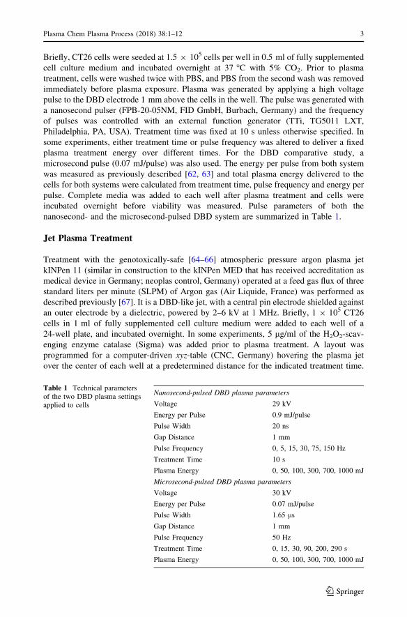
CT26 cells were treated with DBD (Fig. 1a) or jet (Fig. 1b) plasma, and metabolic activity
was assessed after 24 h. At constant treatment times of 10 s, DBD toxicity increased with
higher energy doses, giving a 50% inhibitory concentration (IC50) at about 740 mJ
(Fig. 2a). The kINPen plasma cannot be tuned electrically as settings are fixed. Hence,
dosimetry and increase in ‘energy’ can only be achieved by extending the treatment time.
As expected, the latter was proportional to cellular toxicity with an IC50 of 100 s (Fig. 2b).
4 Plasma Chem Plasma Process (2018) 38:1–12
123
At IC50 treatment conditions, H2O2 deposition by DBD and jet plasma was 50 lM(Fig. 2c) and 115 lM (Fig. 2d), respectively. In both cases, H2O2 levels increased with
energy or time of exposure. Therefore, toxicity increased proportionally with increased
energy and/or plasma treatment time, and concentrations of plasma-generated H2O2 cor-
related with that.
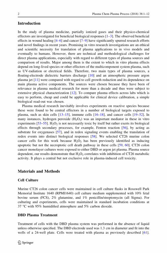
Fig. 1 DBD and jet plasma. a DBD treatment of cells in a 24-well plate. b Atmospheric pressure argonplasma jet treatment of cells in a 24-well plate
Fig. 2 IC50 and generation of H2O2. a DBD treatment of CT26 cells at different energy dosages.b Atmospheric pressure argon plasma jet treatment of CT26 cells at different treatment times. c H2O2
deposition of DBD plasma at different energy dosages. d H2O2 deposition of jet plasma at differenttreatment times. In (a) and (b), metabolic activity was assessed after 24 h and normalized to that ofuntreated control cells. Data are presented as mean with SD
Plasma Chem Plasma Process (2018) 38:1–12 5
123
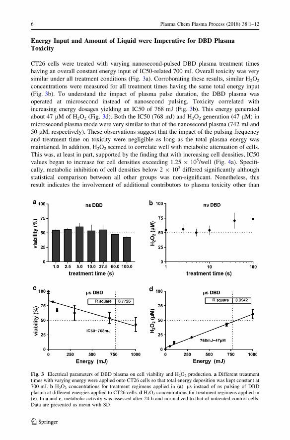
Energy Input and Amount of Liquid were Imperative for DBD PlasmaToxicity
CT26 cells were treated with varying nanosecond-pulsed DBD plasma treatment times
having an overall constant energy input of IC50-related 700 mJ. Overall toxicity was very
similar under all treatment conditions (Fig. 3a). Corroborating these results, similar H2O2
concentrations were measured for all treatment times having the same total energy input
(Fig. 3b). To understand the impact of plasma pulse duration, the DBD plasma was
operated at microsecond instead of nanosecond pulsing. Toxicity correlated with
increasing energy dosages yielding an IC50 of 768 mJ (Fig. 3b). This energy generated
about 47 lM of H2O2 (Fig. 3d). Both the IC50 (768 mJ) and H2O2 generation (47 lM) in
microsecond plasma mode were very similar to that of the nanosecond plasma (742 mJ and
50 lM, respectively). These observations suggest that the impact of the pulsing frequency
and treatment time on toxicity were negligible as long as the total plasma energy was
maintained. In addition, H2O2 seemed to correlate well with metabolic attenuation of cells.
This was, at least in part, supported by the finding that with increasing cell densities, IC50
values began to increase for cell densities exceeding 1.25 9 105/well (Fig. 4a). Specifi-
cally, metabolic inhibition of cell densities below 2 9 105 differed significantly although
statistical comparison between all other groups was non-significant. Nonetheless, this
result indicates the involvement of additional contributors to plasma toxicity other than
Fig. 3 Electrical parameters of DBD plasma on cell viability and H2O2 production. a Different treatmenttimes with varying energy were applied onto CT26 cells so that total energy deposition was kept constant at700 mJ. b H2O2 concentrations for treatment regimens applied in (a). ls instead of ns pulsing of DBDplasma at different energies applied to CT26 cells. d H2O2 concentrations for treatment regimens applied in(c). In a and c, metabolic activity was assessed after 24 h and normalized to that of untreated control cells.Data are presented as mean with SD
6 Plasma Chem Plasma Process (2018) 38:1–12
123
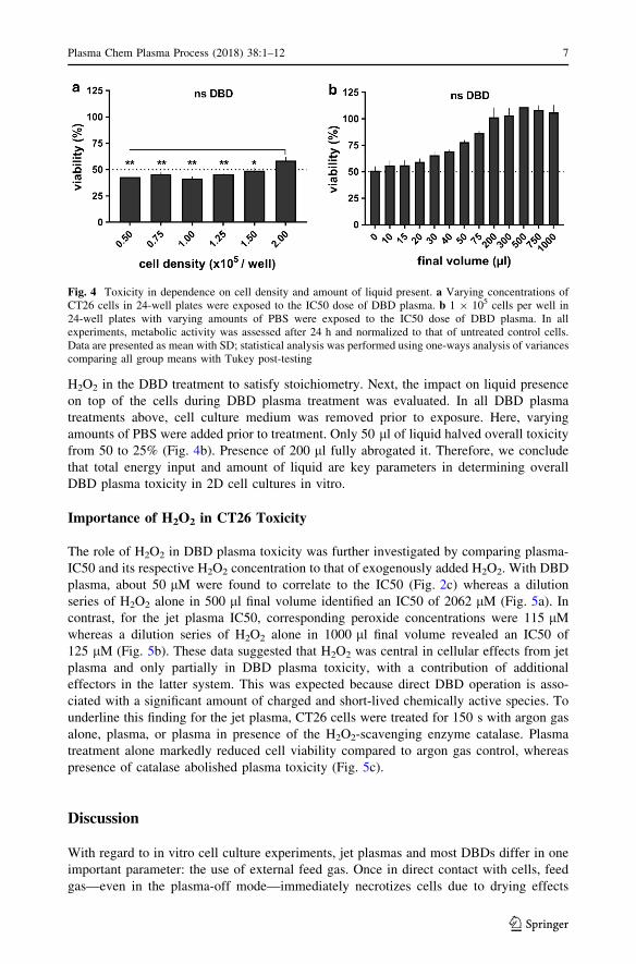
H2O2 in the DBD treatment to satisfy stoichiometry. Next, the impact on liquid presence
on top of the cells during DBD plasma treatment was evaluated. In all DBD plasma
treatments above, cell culture medium was removed prior to exposure. Here, varying
amounts of PBS were added prior to treatment. Only 50 ll of liquid halved overall toxicity
from 50 to 25% (Fig. 4b). Presence of 200 ll fully abrogated it. Therefore, we conclude
that total energy input and amount of liquid are key parameters in determining overall
DBD plasma toxicity in 2D cell cultures in vitro.
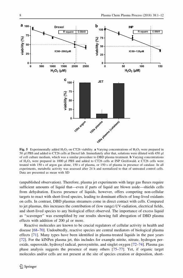
Importance of H2O2 in CT26 Toxicity
The role of H2O2 in DBD plasma toxicity was further investigated by comparing plasma-
IC50 and its respective H2O2 concentration to that of exogenously added H2O2. With DBD
plasma, about 50 lM were found to correlate to the IC50 (Fig. 2c) whereas a dilution
series of H2O2 alone in 500 ll final volume identified an IC50 of 2062 lM (Fig. 5a). In
contrast, for the jet plasma IC50, corresponding peroxide concentrations were 115 lMwhereas a dilution series of H2O2 alone in 1000 ll final volume revealed an IC50 of
125 lM (Fig. 5b). These data suggested that H2O2 was central in cellular effects from jet
plasma and only partially in DBD plasma toxicity, with a contribution of additional
effectors in the latter system. This was expected because direct DBD operation is asso-
ciated with a significant amount of charged and short-lived chemically active species. To
underline this finding for the jet plasma, CT26 cells were treated for 150 s with argon gas
alone, plasma, or plasma in presence of the H2O2-scavenging enzyme catalase. Plasma
treatment alone markedly reduced cell viability compared to argon gas control, whereas
presence of catalase abolished plasma toxicity (Fig. 5c).
Discussion
With regard to in vitro cell culture experiments, jet plasmas and most DBDs differ in one
important parameter: the use of external feed gas. Once in direct contact with cells, feed
gas—even in the plasma-off mode—immediately necrotizes cells due to drying effects
Fig. 4 Toxicity in dependence on cell density and amount of liquid present. a Varying concentrations ofCT26 cells in 24-well plates were exposed to the IC50 dose of DBD plasma. b 1 9 105 cells per well in24-well plates with varying amounts of PBS were exposed to the IC50 dose of DBD plasma. In allexperiments, metabolic activity was assessed after 24 h and normalized to that of untreated control cells.Data are presented as mean with SD; statistical analysis was performed using one-ways analysis of variancescomparing all group means with Tukey post-testing
Plasma Chem Plasma Process (2018) 38:1–12 7
123
(unpublished observation). Therefore, plasma jet experiments with large gas fluxes require
sufficient amounts of liquid that—even if parts of liquid are blown aside—shields cells
from dehydration. Excess presence of liquids, however, offers competing non-cellular
targets to react with short-lived species, leading to dominant effects of long-lived oxidants
on cells. In contrast, DBD plasmas streamers come in direct contact with cells. Compared
to jet plasmas, this increases the contribution of (low range) UV-radiation, electrical fields,
and short-lived species to any biological effect observed. The importance of excess liquid
as ‘‘scavenger’’ was exemplified by our results showing full abrogation of DBD plasma
effects with addition of 200 ll or more.
Reactive molecules are known to be crucial regulators of cellular activity in health and
disease [68–70]. Undoubtedly, reactive species are central mediators of biological plasma
effects [71]. Many types have been identified in plasma-treated liquids in the past years
[72]. For the kINPen plasma jet, this includes for example nitrite, nitrate, hydrogen per-
oxide, superoxide, hydroxyl radical, peroxynitrite, and singlet oxygen [72–74]. Plasma gas
phase analysis suggests the presence of many others [75–77]. Yet, if organic target
molecules and/or cells are not present at the site of species creation or deposition, short-
Fig. 5 Experimentally added H2O2 on CT26 viability. a Varying concentrations of H2O2 were prepared in50 ll PBS and added to CT26 cells at Drexel lab. Immediately after that, solutions were diluted with 450 llof cell culture medium, which was a similar procedure to DBD plasma treatment. b Varying concentrationsof H2O2 were prepared in 1000 ll PBS and added to CT26 cells at INP Greifswald. c CT26 cells weretreated with 150 s of argon gas alone, 150 s of plasma, or 150 s of plasma in presence of catalase. In allexperiments, metabolic activity was assessed after 24 h and normalized to that of untreated control cells.Data are presented as mean with SD
8 Plasma Chem Plasma Process (2018) 38:1–12
123
lived species yield more stable products such as hydrogen peroxide or hypochlorous acid
[73].
Our results indicated a dominant and a partial role of peroxide for the jet and DBD
toxicity, respectively. The latter was especially illustrated by a lower IC50 H2O2 deposition
by the DBD compared to the jet, and the non-linear correlation between cell number and
DBD cytotoxicity. For the DBD, this suggested additional cytotoxic effectors at work. As
200 ll of liquid on top of cells fully abrogated DBD plasma toxicity, these effectors may
be for instance poration, UV-radiation, charged particles or short-lived species being
decomposed in absence of target cells. For example, the DBD plasma may create nano-
pores [78] allowing the entry of species into cells by a process similar to aquaporins [79], a
route potentially counteracted by excess liquid. Also, short wavelength UV radiation is
efficiently scavenged by a few hundred nanometers of liquid layer [80]. A combination of
different plasma properties is also possible as seen with bacteria [81–83]. Corroborating
results of the jet plasma in the present work, presence and concentration of H2O2 strongly
correlated with cytotoxic effects of the kINPen [84–86] and other jet and DBD plasma
sources [87–90], especially plasma-treated liquids [91–93]. Yet, in a helium/oxygen
plasma jet, we previously demonstrated cytotoxicity correlating with short-lived species
supporting the generation of hypochlorous acid in aqueous media [94].
H2O2 itself is a relatively non-reactive molecule with low reaction-constants with
biomolecules [95]. Its effects mainly depend on two factors: the presence of catalysts and/
or other oxidants, and the enzymatic profile of the target cells handling oxidants. It is well
known the reaction of H2O2 and iron generates highly toxic hydroxyl radicals, by a process
known as the Fenton reaction [96]. Hence, iron close to cell membranes, intracellular iron,
and/or shuttling of H2O2 through membranes would be important denominators for plasma
effects. Moreover, synergistic effects of plasma-generated H2O2 with nitrite have been
proposed [97] that may act in concert with membrane-based oxidases to form peroxynitrite
and other toxic species [98]. Extracellular trap formation following kINPen plasma
treatment was not replicated by addition of hydrogen peroxide alone either [99]. In
addition, redox enzymes and antioxidant defenses guide diverse cell fates when two dif-
ferent cell types are subjected to the same plasma treatment [100].
Nonetheless, in vitro cell cultures are only model systems that may guide in vivo
studies. It is important to considered that the biomolecule to liquid ratio in tissues is much
lower than in vitro systems. There are several challenges before the translation of in vitro
plasma effects to in vivo effects becomes intuitive. The species variety is unique to cold
plasmas with their concentration depending on the plasma source. Methods to directly
detect short-lived species in tissues are currently unavailable. Modeling studies suggest
that plasma in direct contact with a target alters species deposition and cell membrane
oxidation [101–103]. Experimentally, plasma-derived oxidants have been shown to be
deposited on cells and liquids through plasma-treated agarose membranes [104] and
micropores [105], which may facilitate the deciphering effects of jet plasma short-lived
species in cells.
While this study was not comprehensive and has limitations, it is the first attempt to find
common and divergent themes between different plasma sources intended for future
medical applications. Only one type of DBD and jet plasma source was investigated. A
straightforward biological read-out of metabolic/growth inhibition was chosen for com-
parison because its no-wash-one-step approach minimizes lab-to-lab variation. We offer
protocols for testing other plasma sources to ‘‘standardize’’ their biological effects by
relatively easy means.
Plasma Chem Plasma Process (2018) 38:1–12 9
123
In conclusion, we provide a biological response model (IC50 of CT26 colorectal cancer
cells) that allows for easy comparison between very different plasma discharges.
Acknowledgements Funding at INP Greifswald was received from the German Federal Ministry of Edu-cation and Research (BMBF, Grant Numbers 03Z22DN11 and 03Z22DN12).
Open Access This article is distributed under the terms of the Creative Commons Attribution 4.0 Inter-national License (http://creativecommons.org/licenses/by/4.0/), which permits unrestricted use, distribution,and reproduction in any medium, provided you give appropriate credit to the original author(s) and thesource, provide a link to the Creative Commons license, and indicate if changes were made.
References
1. Fridman G, Friedman G, Gutsol A, Shekhter AB, Vasilets VN, Fridman A (2008) Plasma ProcessPolym 5:503
2. Weltmann KD, von Woedtke T (2017) Plasma Phys Control Fusion 59:0140313. Laroussi M (2009) IEEE Trans Plasma Sci 37:7144. Heinlin J, Isbary G, Stolz W, Morfill G, Landthaler M, Shimizu T, Steffes B, Nosenko T, Zimmer-
mann J, Karrer S (2011) J Eur Acad Dermatol Venereol 25:15. Bekeschus S, Schmidt A, Weltmann K-D, von Woedtke T (2016) Clin Plasma Med 4:196. Emmert S, Brehmer F, Hanßle H, Helmke A, Mertens N, Ahmed R, Simon D, Wandke D, Maus-
Friedrichs W, Daschlein G (2013) Clin Plasma Med 1:247. Hirst AM, Frame FM, Arya M, Maitland NJ, O’Connell D (2016) Tumour Biol 37:70218. Yan D, Sherman JH, Keidar M (2016) Oncotarget 2016, 59. Miller V, Lin A, Fridman A (2016) Plasma Chem Plasma Process 36:25910. Dobrynin D, Fridman G, Friedman G, Fridman A (2009) New J Phys 11:11502011. Weltmann KD, Kindel E, Brandenburg R, Meyer C, Bussiahn R, Wilke C, von Woedtke T (2009)
Contrib Plasma Phys 49:63112. Fridman G, Shereshevsky A, Jost MM, Brooks AD, Fridman A, Gutsol A, Vasilets V, Friedman G
(2007) Plasma Chem Plasma Process 27:16313. Arndt S, Landthaler M, Zimmermann JL, Unger P, Wacker E, Shimizu T, Li YF, Morfill GE,
Bosserhoff AK, Karrer S (2015) PLoS ONE 10:e012004114. Kim KC, Piao MJ, Madduma Hewage SR, Han X, Kang KA, Jo JO, Mok YS, Shin JH, Park Y, Yoo
SJ, Hyun JW (2016) Int J Mol Med 37:2915. Schmidt A, von Woedtke T, Bekeschus S (2016) Oxid. Med. Cell. Longev. 2016:981607216. Bekeschus S, Rodder K, Schmidt A, Stope MB, von Woedtke T, Miller V, Fridman A, Weltmann K-D,
Masur K, Metelmann H-R, Wende K, Hasse S (2016) Plasma Process. Polym 13:114417. Kaushik NK, Kaushik N, Min B, Choi KH, Hong YJ, Miller V, Fridman A, Choi EH (2016) J Phys D
Appl Phys 49:08400118. Miller V, Lin A, Fridman G, Dobrynin D, Fridman A (2014) Plasma Process Polym 11:119319. Ishaq M, Kumar S, Varinli H, Han ZJ, Rider AE, Evans MD, Murphy AB, Ostrikov K (2014) Mol Biol
Cell 25:152320. Brulle L, Vandamme M, Ries D, Martel E, Robert E, Lerondel S, Trichet V, Richard S, Pouvesle JM,
Le Pape A (2012) PLoS ONE 7:e5265321. Bekeschus S, Rodder K, Fregin B, Otto O, Lippert M, Weltmann KD, Wende K, Schmidt A,
Gandhirajan RK (2017) Oxid Med Cell Longev 2017:439646722. Ahn HJ, Kim KI, Hoan NN, Kim CH, Moon E, Choi KS, Yang SS, Lee JS (2014) PLoS ONE
9:e8617323. Attri P, Kumar N, Uhm HS, Choi EH, RSC Adv 201424. Binenbaum Y, Ben-David G, Gil Z, Slutsker YZ, Ryzhkov MA, Felsteiner J, Krasik YE, Cohen JT
(2017) PLoS ONE 12:e016945725. Chang JW, Kang SU, Shin YS, Kim KI, Seo SJ, Yang SS, Lee J-S, Moon E, Baek SJ, Lee K (2014)
Arch Biochem Biophys 545:13326. Chang JW, Kang SU, Shin YS, Seo SJ, Kim YS, Yang SS, Lee JS, Moon E, Lee K, Kim CH (2015)
Sci Rep 5:1820827. Chernets N, Kurpad DS, Alexeev V, Rodrigues DB, Freeman TA (2015) Plasma Process Polym
12:1400
10 Plasma Chem Plasma Process (2018) 38:1–12
123
28. Conway GE, Casey A, Milosavljevic V, Liu Y, Howe O, Cullen PJ, Curtin JF (2016) Br J Cancer114:435
29. Duan J, Lu X, He G (2017) J Appl Phys 121:01330230. Han D, Cho JH, Lee RH, Bang W, Park K, Kim MS, Shim JH, Chae JI, Moon SY (2017) Sci Rep
7:4308131. Hou J, Ma J, Yu KN, Li W, Cheng C, Bao L, Han W (2015) BMC Genom 16:43532. Ikeda J, Tsuruta Y, Nojima S, Sakakita H, Hori M, Ikehara Y (2015) Plasma Process Polym 12:137033. Kang SU, Cho JH, Chang JW, Shin YS, Kim KI, Park JK, Yang SS, Lee JS, Moon E, Lee K, Kim CH
(2014) Cell Death Dis 5:e105634. Kim SJ, Chung TH (2015) Appl Phys Lett 107:06370235. Kumar N, Attri P, Yadav DK, Choi J, Choi EH, Uhm HS (2014) Sci Rep 4:758936. Lee HJ, Shon CH, Kim YS, Kim S, Kim GC, Kong MG (2009) New J Phys 11:11502637. Lee S, Lee H, Bae H, Choi EH, Kim SJ (2016) Sci Rep 6:3000538. Ma Y, Ha CS, Hwang SW, Lee HJ, Kim GC, Lee KW, Song K (2014) PLoS ONE 9:e9194739. Mizuno K, Yonetamari K, Shirakawa Y, Akiyama T, Ono R (2017) J Phys D Appl Phys 50(12L):T0140. Nguyen NH, Park HJ, Yang SS, Choi KS, Lee JS (2016) Sci Rep 6:2902041. Panngom K, Baik KY, Nam MK, Han JH, Rhim H, Choi EH (2013) Cell Death Dis 4:e64242. Park SB, Kim B, Bae H, Lee H, Lee S, Choi EH, Kim SJ (2015) PLoS ONE 10:e012993143. Schmidt A, Bekeschus S, von Woedtke T, Hasse S (2015) Clin Plasma Med 3:2444. Siu A, Volotskova O, Cheng X, Khalsa SS, Bian K, Murad F, Keidar M, Sherman JH (2015) PLoS
ONE 10:e012631345. Suzuki-Karasaki Y (2016) Int J Mol Med 38:S5046. Tabuchi Y, Uchiyama H, Zhao QL, Yunoki T, Andocs G, Nojima N, Takeda K, Ishikawa K, Hori M,
Kondo T (2016) Int J Mol Med 37:170647. Tanaka H, Mizuno M, Toyokuni S, Maruyama S, Kodera Y, Terasaki H, Adachi T, Kato M, Kikkawa
F, Hori M (2015) Phys Plasmas 22:12200448. Torii K, Yamada S, Nakamura K, Tanaka H, Kajiyama H, Tanahashi K, Iwata N, Kanda M, Kobayashi
D, Tanaka C, Fujii T, Nakayama G, Koike M, Sugimoto H, Nomoto S, Natsume A, Fujiwara M,Mizuno M, Hori M, Saya H, Kodera Y (2015) Gastric Cancer 18:635
49. Utsumi F, Kajiyama H, Nakamura K, Tanaka H, Hori M, Kikkawa F (2014) Springerplus 3:39850. Yajima I, Iida M, Kumasaka MY, Omata Y, Ohgami N, Chang J, Ichihara S, Hori M, Kato M (2014)
Exp Dermatol 23:42451. Yan DY, Nourmohammadi N, Talbot A, Sherman JH, Keidar M (2016) J Phys D Appl Phys 49:27400152. Yang H, Lu R, Xian Y, Gan L, Lu X, Yang X (2015) Phys Plasmas 22:12200653. Golkowski M, Golkowski C, Leszczynski J, Plimpton SR, Maslowski P, Foltynowicz A, Ye J,
McCollister B (1984) IEEE Trans Plasma Sci 2012:4054. Kalghatgi S, Kelly CM, Cerchar E, Torabi B, Alekseev O, Fridman A, Friedman G, Azizkhan-Clifford
J (2011) PLoS ONE 6:e1627055. Girard PM, Arbabian A, Fleury M, Bauville G, Puech V, Dutreix M, Sousa JS (2016) Sci Rep 6:2909856. Winterbourn CC (1995) Toxicol Lett 82–83:96957. Winterbourn CC (1985) Biochim Biophys Acta 840:20458. Hanschmann EM, Godoy JR, Berndt C, Hudemann C, Lillig CH (2013) Antioxid Redox Signal
19:153959. Peng X, Gandhi V (2012) Ther Deliv 3:82360. Aaes TL, Kaczmarek A, Delvaeye T, De Craene B, De Koker S, Heyndrickx L, Delrue I, Taminau J,
Wiernicki B, De Groote P, Garg AD, Leybaert L, Grooten J, Bertrand MJ, Agostinis P, Berx G,Declercq W, Vandenabeele P, Krysko DV (2016) Cell Rep 15:274
61. Lin A, Truong B, Pappas A, Kirifides L, Oubarri A, Chen S, Lin S, Dobrynin D, Fridman G, FridmanA, Sang N, Miller V (2015) Plasma Process Polym 12:1392
62. Ayan H, Fridman G, Gutsol AF, Vasilets VN, Fridman A, Friedman G (2008) IEEE Trans Plasma Sci36:504
63. H. Ayan, D. Staack, G. Fridman, A. Gutsol, Y. Mukhin, A. Starikovskii, A. Fridman, G. Friedman(2009) J Phys D-Appl Phys 42:125202
64. Kluge S, Bekeschus S, Bender C, Benkhai H, Sckell A, Below H, Stope MB, Kramer A (2016) PLoSONE 11:e0160667
65. Wende K, Bekeschus S, Schmidt A, Jatsch L, Hasse S, Weltmann KD, Masur K, von Woedtke T(2016) Mutat Res, Genet Toxicol Environ Mutagen 798–799:48
66. Schmidt A, Woedtke TV, Stenzel J, Lindner T, Polei S, Vollmar B, Bekeschus S (2017) Int J Mol Sci18:868
Plasma Chem Plasma Process (2018) 38:1–12 11
123
67. Bekeschus S, Kolata J, Muller A, Kramer A, Weltmann K-D, Broker B, Masur K (2013) Plasma Med3:1
68. Quijano C, Trujillo M, Castro L, Trostchansky A (2016) Redox Biol 8:2869. Cortese-Krott MM, Koning A, Kuhnle GG, Nagy P, Bianco C, Pasch A, Wink DA, Fukuto J, Jackson
AA, van Goor H, Olson KR, Feelisch M, Antioxid Redox Signal 201770. Prescott C, Bottle SE (2016) Cell Biochem Biophys 2016, 171. Graves DB (2012) J Phys D Appl Phys 45:26300172. Jablonowski H, von Woedtke Th (2015) Clin Plasma Med 3:4273. Wende K, Williams P, Dalluge J, Gaens WV, Aboubakr H, Bischof J, von Woedtke T, Goyal SM,
Weltmann KD, Bogaerts A, Masur K, Bruggeman PJ (2015) Biointerphases 10:02951874. Bekeschus S, Kolata J, Winterbourn C, Kramer A, Turner R, Weltmann KD, Broker B, Masur K
(2014) Free Radic Res 48:54275. Iseni S, Zhang S, van Gessel AFH, Hofmann S, van Ham BTJ, Reuter S, Weltmann KD, Bruggeman
PJ (2014) New J Phys 16:12301176. Schmidt-Bleker A, Winter J, Iseni S, Dunnbier M, Weltmann KD, Reuter S (2014) J Phys D Appl Phys
47:14520177. Dunnbier M, Schmidt-Bleker A, Winter J, Wolfram M, Hippler R, Weltmann KD, Reuter S (2013) J
Phys D Appl Phys 46:43520378. Zerrouki A, Yousfi M, Rhallabi A, Motomura H, Jinno M (2016) Plasma Process Polym 13:63379. Yan D, Talbot A, Nourmohammadi N, Sherman JH, Cheng X, Keidar M (2015) Biointerphases
10:04080180. Jablonowski H, Bussiahn R, Hammer MU, Weltmann KD, von Woedtke T, Reuter S (2015) Phys
Plasmas 22:12200881. Lackmann JW, Schneider S, Edengeiser E, Jarzina F, Brinckmann S, Steinborn E, Havenith M,
Benedikt J, Bandow JE (2013) J R Soc Interface 10:2013059182. Pavlovich MJ, Sakiyama Y, Clark DS, Graves DB (1051) Plasma Process Polym 2013:1083. Shimizu T, Nosenko T, Morfill GE, Sato T, Schmidt HU, Urayama T (2010) Plasma Process Polym
7:28884. Winter J, Tresp H, Hammer MU, Iseni S, Kupsch S, Schmidt-Bleker A, Wende K, Dunnbier M, Masur
K, Weltmannan KD, Reuter S (2014) J Phys D Appl Phys 47:28540185. Bekeschus S, Iseni S, Reuter S, Masur K, Weltmann K-D (2015) IEEE Trans Plasma Sci 43:77686. Winter J, Wende K, Masur K, Iseni S, Dunnbier M, Hammer MU, Tresp H, Weltmann KD, Reuter S
(2013) J Phys D Appl Phys 46:29540187. Brun P, Pathak S, Castagliuolo I, Palu G, Brun P, Zuin M, Cavazzana R, Martines E (2014) PLoS ONE
9:e10439788. Priya Arjunan K, Morss Clyne A, Plasma Process Polym 2011, 8, 115489. Saito K, Asai T, Fujiwara K, Sahara J, Koguchi H, Fukuda N, Suzuki-Karasaki M, Soma M, Suzuki-
Karasaki Y (2016) Oncotarget 7:1991090. Balzer J, Heuer K, Demir E, Hoffmanns MA, Baldus S, Fuchs PC, Awakowicz P, Suschek CV,
Oplander C (2015) PLoS ONE 10:e014496891. Adachi T, Kano A, Nonomura S, Kamiya T, Hara H (2016) Arch Biochem Biophys 606:12092. Boehm D, Heslin C, Cullen PJ, Bourke P (2016) Sci Rep 6:2146493. Adachi T, Tanaka H, Nonomura S, Hara H, Kondo S, Hori M (2015) Free Radic Biol Med 79:2894. Bekeschus S, Wende K, Hefny MM, Rodder K, Jablonowski H, Schmidt A, Woedtke TV, Weltmann
KD, Benedikt J (2017) Sci Rep 7:279195. Winterbourn CC (2008) Nat Chem Biol 4:27896. Neyens E, Baeyens J (2003) J Hazard Mater 98:3397. Girard F, Badets V, Blanc S, Gazeli K, Marlin L, Authier L, Svarnas P, Sojic N, Clement F, Arbault S
(2016) Rsc Adv 6:7845798. Bauer G, Graves DB (2016) Plasma Process Polym 201699. Bekeschus S, Winterbourn CC, Kolata J, Masur K, Hasse S, Broker BM, Parker HA (2016) J Leukoc
Biol 100:791100. Schmidt A, Rodder K, Hasse S, Masur K, Toups L, Lillig CH, von Woedtke T, Wende K, Bekeschus S
(2016) Plasma Process Polym 13:1179101. Norberg SA, Tian W, Johnsen E, Kushner MJ (2014) J Phys D Appl Phys 47:475203102. Van der Paal J, Verlackt C, Yusupov M, Neyts E, Bogaerts A (2015) J Phys D Appl Phys 48:155202103. Stoffels E, Sakiyama Y, Graves DB (2008) IEEE Trans Plasma Sci 36:1441104. Oh JS, Szili EJ, Gaur N, Hong SH, Furuta H, Kurita H, Mizuno A, Hatta A, Short RD (2016) J Phys D
Appl Phys 49:304005105. Oh JS, Kojima S, Sasaki M, Hatta A, Kumagai S (2017) Sci Rep 7:41953
12 Plasma Chem Plasma Process (2018) 38:1–12
123

![DBD plasma microbubble reactor for pre-treatment of … · DBD plasma microbubble reactor for pre-treatment of lignocellulosic biomass [poster] ... DBD plasma microbubble reactor](https://img.pdfslide.us/doc/110x75/5e4523a0e85b14090f08d100/dbd-plasma-microbubble-reactor-for-pre-treatment-of-dbd-plasma-microbubble-reactor.jpg)