Embed Size (px)
Citation preview
A CASE OF OSTEOPETROSIS (ALBERS-SCHONBERG)WITH INTERCURRENT PNEUMOCYSTIS PNEUMONITIS
BY
W. AHERNEFrom the Department ofPathology, Radcliffe Infirmary, Oxford
(RECEIVED FOR PUBLICATION JANUARY 26, 1960)
Albers-Schonberg (1904, 1907) defined the clinicaland radiological features of a sclerosing disease ofbone for which Karshner (1926) suggested the termosteopetrosis. As published cases of the diseaseaccumulated, two types came to be distinguished(McCune and Bradley, 1934; Harnapp, 1937;Nussey, 1938; Zetterstrom, 1958). The first, orclinically 'malignant' type may be characterized bya refractory and often leucoerythroblastic anaemia,enlargement of liver, spleen and lymph nodes,a liability to multiple fractures, and death in infancyor early childhood. In the second or clinically'benign' type, more commonly seen in adults,anaemia is mild and the osteosclerosis, thoughoften severe, may yet be symptomless.
In the more severe forms of osteopetrosis, inter-current infection, particularly of the respiratorytract, is a serious hazard. The case to be reportedhere, an example of the 'malignant' type, succumbedto an interstitial 'plasma cell' pneumonitis.Though this unusual pneumonitis has been
recognized for many years in central Europe itsaetiology had been obscure until 1951. In that yearVanek, in Czechoslovakia, identified Pneumocystiscarinii in the lungs of all of a series of 16 fatalcases of this disease. In a series of papers (Vanek,1951; Vanek and Jirovec, 1952; Vanek, Jirovec andLukes, 1953) evidence was presented that Pneumo-cystis carinii was the causal agent of interstitialplasma cell pneumonitis. Nevertheless, Deamerand Zollinger (1953), who first drew Americanattention to the disease, concluded that it wasprobably a viral one; they dismissed a fungal orprotozoal agent as unlikely. Baar (1955) reportedthe first case to be observed in England and sub-scribed to the hypothesis that Pneumocystis carinjiwas the pathogen. The subject is comprehensivelyreviewed by Gajdusek (1957).
Pneumocystis carinii pneumonitis has been foundassociated with congenital agammaglobulinaemia(Hutchison, 1955; Bird and Thomson, 1957;
Russell, 1959), cytomegalic inclusion disease (Baar,1955; Hamperl, 1956), and neoplastic disease of thelympho-reticular tissue in both children and adults(Vanek, 1952; Jirovec and Vanek, 1954; Le TanVinh, 1954; Hamperl, 1956). Association withAlbers-Schonberg's osteopetrosis has apparentlynot been reported before.
Case ReportW.T. (Reg. No. R.I. 250922), was a male infant, the
first-born and only child of healthy unrelated parents.Pregnancy and labour had been uneventful. In the first24 hours bruises were noticed on his upper lip and onseveral toes. These bruises disappeared within a fewdays. On the second day he regurgitated blood-fleckedmilk. Nothing further happened until the 13th day whenanother bruise appeared on his right leg and a smallhaematoma-like swelling was found on his umbilicus.As these subsided others appeared near the anus andover the chest wall. He became progressively paler.On admission to hospital at 3 weeks of age, he was
pale but reasonably vigorous and not acutely ill. Heweighed 5 lb. 14 oz. There were bruises on the leftchest, right thigh, right groin and umbilicus. A smallabscess cavity was found close to the anus posteriorly.The liver was palpated about a finger breadth belowthe costal margin, and though the spleen was not pal-pable at first it could be tipped after four days and readilypalpated within a week. Apart from these there were nounusual clinical findings.
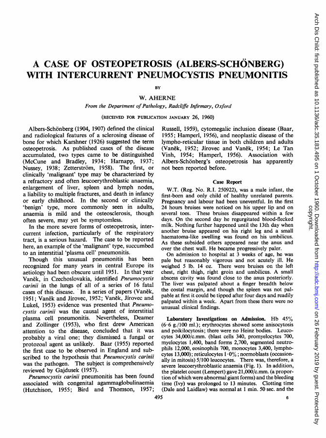
Laboratory Investigations on Admission. Hb 45%(6-6 g./100 ml.); erythrocytes showed some anisocytosisand poikilocytosis; there were no Heinz bodies. Leuco-cytes 34,000/c.mm. (blast cells 340, promyelocytes 700,myelocytes 1,400, band forms 2,700, segmented neutro-phils 12,000, eosinophils 700, monocytes 3,400, lympho-cytes 13,000); reticulocytes 1 -0% ; normoblasts (occasion-ally in mitosis) 5/100 leucocytes. There was, therefore, asevere leucoerythroblastic anaemia (Fig. 1). In addition,the platelet count (Lempert) gave 21,000/c.mm. (a propor-tion of which were abnormal giant forms) and the bleedingtime (Ivy) was prolonged to 13 minutes. Clotting time(Dale and Laidlaw) was normal at 1 min. 50 sec. and the
495 6
copyright. on 26 F
ebruary 2019 by guest. Protected by
http://adc.bmj.com
/A
rch Dis C
hild: first published as 10.1136/adc.35.183.495 on 1 October 1960. D
ownloaded from
ARCHIVES OF DISEASE IN CHILDHOOD
FIG. 1.-Peripheral blood film showing immature erythroid andmyeloid cells. May-Gruenwald-Giemsa x 584.
tourniquet test was negative. The blood group wasO Rh positive. The direct Coombs test was negativeand the mother's serum was found to be compatiblewith the patient's erythrocytes. No platelet agglutininscould be demonstrated in the mother's serum. The
lomi. toomi.
70.
z
i002 40I
I
IIso ml.
I III la
toxoplasma dye test dilution was 1/6 and the toxoplasmacomplement fixation test was negative. Wassermann andKahn reactions were negative in both mother and infant.Puncture of the tibial marrow was attempted unsuccess-fully.
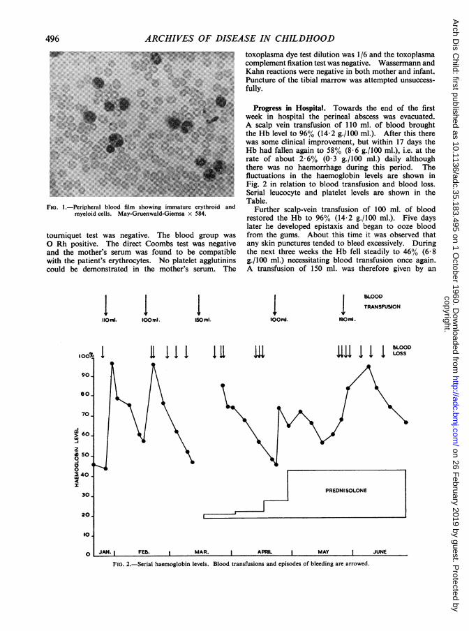
Progress in Hospital. Towards the end of the firstweek in hospital the perineal abscess was evacuated.A scalp vein transfusion of 110 ml. of blood broughtthe Hb level to 96% (14-2 g./100 ml.). After this therewas some clinical improvement, but within 17 days theHb had fallen again to 58% (8-6 g./100 ml.), i.e. at therate of about 2-6% (03 g./100 ml.) daily althoughthere was no haemorrhage during this period. Thefluctuations in the haemoglobin levels are shown inFig. 2 in relation to blood transfusion and blood loss.Serial leucocyte and platelet levels are shown in theTable.
Further scalp-vein transfusion of 100 ml. of bloodrestored the Hb to 96% (14-2 g./100 ml.). Five dayslater he developed epistaxis and began to ooze bloodfrom the gums. About this time it was observed thatany skin punctures tended to bleed excessively. Duringthe next three weeks the Hb fell steadily to 46% (6- 8g./100 ml.) necessitating blood transfusion once again.A transfusion of 150 ml. was therefore given by an
4lOOmi. solfid.
BLOOD
TRANSFUSION
11 1111 bLOOD
FIG. 2.-Serial haemoglobin levels. Blood transfusions and episodes of bleeding are arrowed.
496
copyright. on 26 F
ebruary 2019 by guest. Protected by
http://adc.bmj.com
/A
rch Dis C
hild: first published as 10.1136/adc.35.183.495 on 1 October 1960. D
ownloaded from
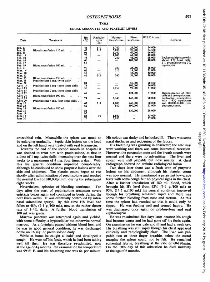
497OSTEOPETROSISTABLE
SERIAL LEUCOCYTE AND PLATELET LEVELS
Hb Reticulo- Normo- Plate- W.B.C./c.mm.Date Treatment (%) cytes blasts/c.mm. lets/c.mm. Remarks
(%
Jan. 23 45 10 1,700 21,000 34,000Jan. 28 Blood transfusion 110 ml. 43 2-2 5,800 20,000 64,500Jan. 31 96 - 1,300 67,000 66,000Feb. 5 76 - 1,000 61,000 48,000Feb. 10 75 - 280 48,000 28,000 LeukaemoidpicturewithFeb. 14 60 - - 102,000 40,000 about 1 % blast cells;Feb. 17 58 - - - - 2% promyelocytes; 5%Feb. 19 Blood transfusion 100 ml. myelocytesFeb. 21 96 - 380 25,000 38,000Mar. 4 62 - 310 14,000 31,000Mar. 10 52 - - 45,000 50,000Mar. 13 46 2-2 360 51,000 36,000Mar. 15 Blood transfusion 150 ml.Mar. 18 Prednisolone 1 mg. twice dailyMar. 28 84 - - 50,000 28,500April 3 Prednisolone 1 mg three times daily 74 - - 102,000 36,000April 15 56 - 2,850 91,000 57,000April 17 Prednisolone 2 mg. three times dailyApril 22 48 - - 124,000 57,000 Disappearance of blastApril 24 Blood transfusion 100 ml. cells and promyelocytes;April 25 74 - 2,940 145,000 98,000 neutrophil leucocytosisApril 29 Prednisolone 4 mg. four times daily with 1-3 ° myelocytesMay 12 67 3-8 4,000 240,000 80,000 and 20,000-30,000 lym-May 23 62 - 880 190,000 22,000 phocytes/c.mm.May 28 Blood transfusion 180 ml.May 31 84 - - 130,000 28,000June 9 95June 13 84 10 1,800 + 45,000June 20 75 3,050 + 77,000
antecubital vein. Meanwhile the spleen was noted tobe enlarging gradually. Septic skin lesions on the headand on the left hand were treated with oral terramycin.Towards the end of the second month in hospital it
was decided to treat him with prednisolone, at first ina dose of 1 mg. twice daily, increasing over the next fourweeks to a maximum of 4 mg. four times a day. Withthis his general condition improved considerably,although he continued to show purpuric lesions over theskin and abdomen. The platelet count began to riseshortly after administration of prednisolone and reachedthe normal level of 240,000/c.mm. during the subsequenteight weeks.
Nevertheless, episodes of bleeding continued. Tendays after the start of prednisolone treatment severeepistaxis began again and continued in bouts during thenext three weeks. It was eventually controlled by intra-nasal adrenaline sprays. By this time Hb level hadfallen to 48% (7-1 g./100 ml.), now at the rather slowerrate of 1 *4% daily. A further blood transfusion of100 ml. was given.Marrow puncture was attempted again and yielded,
with some difficulty, a hypocellular but otherwise normalmarrow. As a fortnight passed without blood loss, andhe was in good general condition, he was dischargedhome on 16 mg. of prednisolone daily.While at home he caught a 'cold' and developed a
cough. He went off his feeds, which he had been takingwell till then. He was therefore re-admitted, nowat the age of 4i months. On examination his temperaturewas 99- 60 F. and his breathing rate was 64 per minute.
His colour was dusky and he looked ill. There was somenasal discharge and reddening of the fauces.
His breathing was grunting in character; the alae nasiwere working and there was some intercostal recession.However, the percussion note and the breath sounds werenormal and there were no adventitiae. The liver andspleen were still palpable but now smaller. A chestradiograph showed no definite radiological lesion.Two days later there was a fresh crop of purpuric
lesions on his abdomen, although his platelet countwas now normal. He maintained a persistent low-gradefever with some cough but no physical signs in the chest.After a further transfusion of 180 ml. blood, whichbrought his Hb level from 62% (9 1 g./100 ml.) to95% (14-1 g./100 ml.) his general condition improvedthough his breathing remained rapid and there wassome further bleeding from nose and rectum. At thistime the spleen had receded so that it could only betipped. He was feeding well and seemed happy. Hewas discharged once again on prednisolone and oralerythromycin.He was re-admitted five days later because his cough
had become worse and he had gone off his feeds again.On examination he was pale and ill and had a weak cry.His breathing was still rapid though his chest appearedclinically and radiologically clear. The liver was pal-pable two or three finger breadths below the costalmargin; the spleen could not be felt. He remainedsomewhat febrile, breathing at the rate of 60-120/min.On the 10th day of this admission he died suddenlyat the age of 6 months.
copyright. on 26 F
ebruary 2019 by guest. Protected by
http://adc.bmj.com
/A
rch Dis C
hild: first published as 10.1136/adc.35.183.495 on 1 October 1960. D
ownloaded from
ARCHIVES OF DISEASE IN CHILDHOODMorbid Anatomy. At autopsy (P.M. 512/58) the
following were the main macroscopical findings:
MACROSCOPICAL FINDINGSLungs. The lungs failed to retract as the chest was
opened. Both were of a mottled cafe-au-lait colour.They presented a curious, vaguely nodular consolidationwhich was more marked in the lower lobes. The cutsurfaces were dry and showed small scattered yellowish-white areas. The tracheobronchial tree was normal.
Liver. The liver (196 g.) appeared normal in size,consistency and colour.
Spleen and lymphoid tissue. The spleen (16 g.) wasrather firm, but not appreciably enlarged. The cutsurfaces appeared normal. Lymph nodes in generalwere inconspicuous. The thymus was involuted.
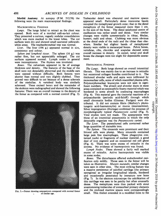
Bones. The calvarium appeared to be of averagethickness and density. The features of the base of theskull were not detectably abnormal and the middle earswere opened without difficulty. Both femora wereshorter than normal and very slightly clubbed. Theyproved very difficult to cut because of a dense sclerosisof the medullae. A vertebral body was similarlysclerosed. These excised bones and the remainder ofthe skeleton were radiographed and showed the followingfeatures: There was an overall increase in the density ofthe femur as compared with a normal control (Fig. 3).
FIG. 3.-Femur showing osteopetrosis compared with normal femurof similar age.
Trabecular detail was obscured and marrow spacesappeared small. Particularly dense transverse bandsoccupied the metaphyseal growth zone; that in the distalmetaphysis of the femur measured more than 1 cm.in the axis of the bone. The distal epiphyseal centre ofossification was rather small and dense. Very similarchanges were visible symmetrically in tibiae, fibulae,humeri, radii and ulnae. Clubbing was more obviousat the distal ends of the radii and the tibiae than else-where. Similar but slighter bands of metaphysealdensity were visible in metacarpal bones. Pelvic bones,vertebrae, ribs, clavicles and scapulae showed someoverall increase in density. In carpal, tarsal and skullbones the changes were too slight for dependable assess-ment.
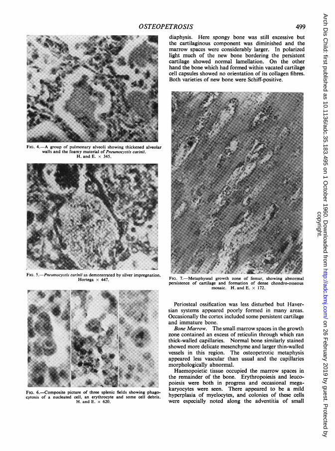
HISTOLOGICAL FINDINGSThe Lungs. Both lungs showed an overall interstitial
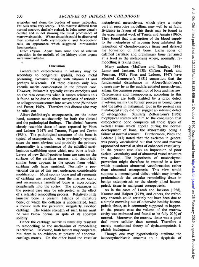
fibrosis. This was mainly due to an increase in reticulinbut occasional collagen bundles contributed to it. Thethickened alveolar walls and septa were infiltrated bymononuclear cells, some of which resembled plasma cells.Polymorphonuclear leucocytes were not seen in signi-ficant numbers. The alveoli and alveolar ducts in mostareas contained an eosinophilic foamy material which wasbordered in some alveoli by coalescing macrophages(Fig. 4). This material gave the tinctorial reactions of apolysaccharide (periodic acid-Schiff) through whichsmall chromatin bodies (Giemsa: Feulgen) were dis-tributed. It did not contain fibrin (Mallory's photo-tungstic acid-haematoxylin) or mucus (mucicarmine).Silver impregnation (Hortega) confirmed the presence ofmorphologically typical Pneumocystis carinii (Fig. 5).Viral studies were not made. The appearances werethose of an interstitial pneumonitis in which the onlydetectable organism was the Pneumocystis carinii.
The Liver. The parenchymal cells contained ratherlarge quantities of stainable iron.
The Spleen. The sinusoids were prominent and theirlittoral cells were plump. Many sinusoids containedlarge pale free macrophages. These phagocytes con-tained nuclei or fragments of nuclear material, a gooddeal of iron pigment and an occasional red cell ghost(Fig. 6). There was some excess of reticulin in thestroma. No evidence of haemopoiesis was found.Lymph Nodes. These showed a fairly well marked
excess of reticulin. Active haemopoiesis was visiblein some nodes.
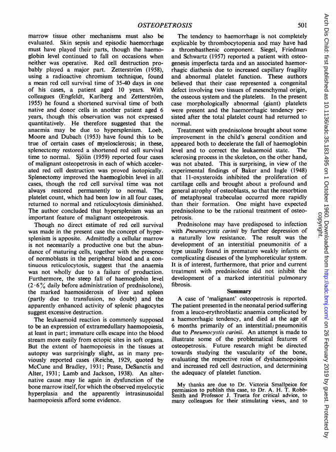
Bones. The disturbances affected endochondral ossi-fication only mildly. Those seen in the femur will betaken as illustrative. Mutatis mutandis, similar changeswere seen in other sites. The most striking feature wasa massive persistence of calcified cartilage (the primaryspongiosa) as irregular longitudinal islands, borderedand occasionally penetrated by immature new bone(Fig. 7). In the electron microscope the calcification ofthis cartilage was seen to be patchy but often unusuallydense. The metaphyses were thus packed with stoutanastomosing trabeculae of unresorbed primary elementsand the enclosed marrow spaces were correspondinglysmall. This thicket extended in a modified form to the
498
copyright. on 26 F
ebruary 2019 by guest. Protected by
http://adc.bmj.com
/A
rch Dis C
hild: first published as 10.1136/adc.35.183.495 on 1 October 1960. D
ownloaded from
OSTEOPit -
ETROSIS 499diaphysis. Here spongy bone was still excessive butthe cartilaginous component was diminished and themarrow spaces were considerably larger. In polarizedlight much of the new bone bordering the persistentcartilage showed normal lamellation. On the otherhand the bone which had formed within vacated cartilagecell capsules showed no orientation of its collagen fibres.Both varieties of new bone were Schiff-positive.
FiG. 4.-A group of pulmonary alveoli showing thickened alveolarwalls and the foamy material of Pneumocystis carinii.
H. and E. x 345.
FIG. 5.-Pneumocystis carinii as demonstrated by silver impregnation.Hortega x 447.
--<---'UW-
FIG. 6.-Composite picture of three splenic fields showing phago-cytosis of a nucleated cell, an erythrocyte and some cell debris.
H. and E. x 620.
FIG. 7.-Metaphyseal growth zone of femur, showing abnormalpersistence of cartilage and formation of dense chondro-osseous
mosaic. H. and E. x 172.
Periosteal ossification was less disturbed but Haver-sian systems appeared poorly formed in many areas.Occasionally the cortex included some persistent cartilageand immature bone.Bone Marrow. The small marrow spaces in the growth
zone contained an excess of reticulin through which ranthick-walled capillaries. Normal bone similarly stainedshowed more delicate mesenchyme and larger thin-walledvessels in this region. The osteopetrotic metaphysisappeared less vascular than usual and the capillariesmorphologically abnormal.
Haemopoietic tissue occupied the marrow spaces inthe remainder of the bone. Erythropoiesis and leuco-poiesis were both in progress and occasional mega-karyocytes were seen. There appeared to be a mildhyperplasia of myelocytes, and colonies of these cellswere especially noted along the adventitia of small
copyright. on 26 F
ebruary 2019 by guest. Protected by
http://adc.bmj.com
/A
rch Dis C
hild: first published as 10.1136/adc.35.183.495 on 1 October 1960. D
ownloaded from
ARCHIVES OF DISEASE IN CHILDHOODarterioles and along the borders of many trabeculae.Fat cells were very scanty. This marrow differed fromnormal marrow, similarly stained, in being more denselycellular and in not showing the usual prominence ofmarrow sinusoids. Where sinusoids could be discoveredthey contained both erythropoietic and leucopoieticcells, an appearance which suggested intravascularhaemopoiesis.Other Organs. Apart from some foci of calcium
deposition in the medulla of the kidneys other organswere unremarkable.
DiscussionGeneralized osteosclerosis in infancy may be
secondary to congenital syphilis, heavy metalpoisoning, excessive dosage with vitamin D andperhaps leukaemia. Of these diseases only leu-kaemia merits consideration in the present case.However, leukaemia typically causes osteolysis andon the rare occasions when it causes sclerosis thisis found to be due to direct metaplasia of reticularor collagenous structures into woven bone (Windholzand Foster, 1949). Therefore this disease also mayte ruled out.
Albers-Schonberg's osteopetrosis, on the otherhand, accounts satisfactorily for both the clinicaland the pathological findings. The clinical featuresclosely resemble those of cases reported by Pinesand Lederer (1947) and Turano, Fagan and Corbo(1954). The pathological structure of the bone istypical of osteopetrosis. As in previously reportedcases the most obvious and probably the primaryabnormality is a persistence of the calcified carti-laginous scaffolding upon which new bone is built.Layers of new Schiff-positive bone are seen on thesurfaces of the cartilage masses, and tinctoriallysimilar bone appears in the spaces from whichcartilage cells have vanished. Normally a pro-visional design of this sort undergoes considerablemodification. Most spongy bone and all remnantsof cartilage are resorbed from the marrow cavityand increasingly lamellated bone is incorporatedperipherally into the cortex. The appearances inthe present case may be interpreted as the effectof a retarded remodelling process. Relatively littlelamellar bone is present. Islands of immaturebone, of which the collagen is unorientated, forma mosaic with the persistent irregularly calcifiedcartilage. The tensile strength of such tissue mustbe well below normal in spite of its apparentsolidity.
Either the cartilage matrix is unusually resistantto remodelling or the mechanism of remodellingis defective. Of course, both factors may cooperate,but there is no evidence at present of abnormalcartilage matrix. On the other hand the vascular
metaphyseal mesenchyme, which plays a majorpart in resorptive modelling, may well be at fault.Evidence in favour of this thesis may be found inthe experimental work of Trueta and Amato (1960).They found that interruption of the blood supplyto the metaphysis of growing bone inhibited theresorption of chondro-osseous tissue and delayedthe formation of final bone. Large zones ofcalcified cartilage and preliminary bone remainedat a level in the metaphysis where, normally, re-modelling is taking place.Many authors (McCune and Bradley, 1934;
Lamb and Jackson, 1938; Clifton, Frank andFreeman, 1938; Pines and Lederer, 1947) haveadopted Klemperer's (1931) suggestion that thefundamental disturbance in Albers-Schonberg'sdisease may lie in the undifferentiated mesenchymalanlage, the common progenitor of bone and marrow.Osteogenesis and haemopoiesis, according to thishypothesis, are both blighted by a prior defectinvolving mainly the former process in benign casesand the latter in malignant. But in the present casehistological study did not suggest an intrinsic defectof osteogenesis. Similarly, Zetterstrom's (1958)biophysical studies led him to the conclusion thatosteopetrotic bone comprises all those types ofskeletal tissue which appear during the normaldevelopment of bone, the abnormality being afailure of normal removal. Furthermore, Pines andLederer (1947) noted that the skeleton in generalwas poorly vascularized but that bone and marrowapproached normal at sites of enhanced vascularity.In the present case also an impression of poorskeletal vascularity and of abnormal vessel structurewas gained. The hypothesis of mesenchymalperversion might therefore be restated in a formwhich postulates abnormal vasoformation ratherthan abnormal osteogenesis. This view wouldsuppose a mesenchymal defect which may involvepredominantly the vascular remodelling tissue inbenign osteopetrosis or the closely allied haemo-poietic tissue in malignant osteopetrosis.As in the cases of Lamb and Jackson (1938),
Kramer and Halpert (1939), and others, the refrac-tory anaemia could certainly not have been due toa simple crowding out of otherwise healthy haemo-poietic tissue, as is commonly supposed to happen.In the present case the volume of the marrowcavity was estimated and found to be fully 70% ofnormal. Moreover, the marrow tissue was a gooddeal more cellular than normal. Therefore amerely mechanical theory of dyshaemopoiesis isplainly inadequate.Though one may hypothetically attribute the
leucoerythroblastic anaemia to a dysplasia of
500
copyright. on 26 F
ebruary 2019 by guest. Protected by
http://adc.bmj.com
/A
rch Dis C
hild: first published as 10.1136/adc.35.183.495 on 1 October 1960. D
ownloaded from
OSTEOPETROSISmarrow tissue other mechanisms must also beevaluated. Skin sepsis and episodic haemorrhagemust have played their parts, though the haemo-globin level continued to fall on occasions whenneither was operative. Red cell destruction pro-bably played a major part. Zetterstrom (1958),using a radioactive chromium technique, founda mean red cell survival time of 35-40 days in oneof his cases, a patient aged 10 years. Withcolleagues (Engfeldt, Karlberg and Zetterstrom,1955) he found a shortened survival time of bothnative and donor cells in another patient aged 6years, though this observation was not expressedquantitatively. He therefore suggested that theanaemia may be due to hypersplenism. Loeb,Moore and Dubach (1953) have found this to betrue of certain cases of myelosclerosis; in these,splenectomy restored a shortened red cell survivaltime to normal. Sjolin (1959) reported four casesof malignant osteopetrosis in each of which acceler-ated red cell destruction was proved isotopically.Splenectomy improved the haemoglobin level in allcases, though the red cell survival time was notalways restored permanently to normal. Theplatelet count, which had been low in all four cases,returned to normal and reticulocytosis diminished.The author concluded that hypersplenism was animportant feature of malignant osteopetrosis.Though no direct estimate of red cell survival
was made in the present case the concept of hyper-splenism is apposite. Admittedly a cellular marrowis not necessarily a productive one but the abun-dance of maturing cells, together with the presenceof normoblasts in the peripheral blood and a con-tinuous reticulocytosis, suggest that the anaemiawas not wholly due to a failure of production.Furthermore, the steep fall of haemoglobin level(2 * 6% daily before administration of prednisolone),the marked haemosiderosis of liver and spleen(partly due to transfusion, no doubt) and theapparently enhanced activity of splenic phagocytessuggest excessive destruction.The leukaemoid reaction is commonly supposed
to be an expression of extramedullary haemopoiesis,at least in part; immature cells escape into the bloodstream more easily from ectopic sites in soft organs.But the extent of haemopoiesis in the tissues atautopsy was surprisingly slight, as in many pre-viously reported cases (Reiche, 1929, quoted byMcCune and Bradley, 1931; Pease, DeSanctis andAlter, 1931; Lamb and Jackson, 1938). An alter-native cause may lie again in dysfunction of thebone marrow itself, for which the observed myelocytichyperplasia and the apparently intrasinusoidalhaemopoiesis afford some evidence.
The tendency to haemorrhage is not completelyexplicable by thrombocytopenia and may have hada thrombasthenic component. Siegel, Friedmanand Schwartz (1957) reported a patient with osteo-genesis imperfecta tarda and an associated haemor-rhagic diathesis due to increased capillary fragilityand abnormal platelet function. These authorsbelieved that their case represented a congenitaldefect involving two tissues of mesenchymal origin,the osseous system and the platelets. In the presentcase morphologically abnormal (giant) plateletswere present and the haemorrhagic tendency per-sisted after the total platelet count had returned tonormal.
Treatment with prednisolone brought about someimprovement in the child's general condition andappeared both to decelerate the fall of haemoglobinlevel and to correct the leukaemoid state. Thesclerosing process in the skeleton, on the other hand,was not abated. This is surprising, in view of theexperimental findings of Baker and Ingle (1948)that 1 1-oxysteroids inhibited the proliferation ofcartilage cells and brought about a profound andgeneral atrophy of osteoblasts, so that the resorbtionof metaphyseal trabeculae occurred more rapidlythan their formation. One might have expectedprednisolone to be the rational treatment of osteo-petrosis.
Prednisolone may have predisposed to infectionwith Pneumocystis carinii by further depression ofa naturally low resistance. The result was thedevelopment of an interstitial pneumonitis of atype usually found in premature weakly infants orcomplicating diseases of the lymphoreticular system.It is of interest, furthermore, that prior and currenttreatment with prednisolone did not inhibit thedevelopment of a marked interstitial pulmonaryfibrosis.
SummaryA case of 'malignant' osteopetrosis is reported.
The patient presented in the neonatal period sufferingfrom a leuco-erythroblastic anaemia complicated bya haemorrhagic tendency, and died at the age of6 months primarily of an interstitial Ipneumonitisdue to Pneumocystis carinii. An attempt is made toillustrate some of the problematical features ofosteopetrosis. Future research might be directedtowards studying the vascularity of the bone,evaluating the respective roles of dyshaemopoiesisand increased red cell destruction, and determiningthe adequacy of platelet function.
My thanks are due to Dr. Victoria Smallpeice forpermission to publish this case, to Dr. A. H. T. Robb-Smith and Professor J. Trueta for critical advice, tomany colleagues for their stimulating views, and to
501copyright.
on 26 February 2019 by guest. P
rotected byhttp://adc.bm
j.com/
Arch D
is Child: first published as 10.1136/adc.35.183.495 on 1 O
ctober 1960. Dow
nloaded from
502 ARCHIVES OF DISEASE IN CHILDHOODMrs. D. Jackson and Mr. E. L. Tugwell for the photo-graphs.
REFERENCESAlbers-Schonberg, H. (1904). Rontgenbildereiner seltenen Knochen-
erkrankung, Aertzl. Verein., Hamburg, February 9, 1904.Munch. med. Wschr., 51, 365.
-- (1907). Eine bisher nicht beschriebene Allgemeinerkrankungdes Skelettes im Rontgenbild. Fortschr. Rontgenstr., 11, 261.
Baar, H. S. (1955). Interstitial plasmacellular pneumonia due topneumocystis carinii. J. clin. Path., 8, 19.
Baker, B. L. and Ingle, D. J. (1948). Growth inhibition in bone andbone marrow following treatment with adrenocorticotrophin(ACTH). Endocrinology, 43, 422.
Bird, T. and Thomson, J. (1957). 'Pneumocystis carinii' pneumonia.Lancet, 1, 59.
Clifton, W. M., Frank, A. and Freeman, S. (1938). Osteopetrosis(marble bones). Amer. J. Dis. Child., 56, 1020.
Deamer, W. C. and Zollinger, H. U. (1953). Interstitial 'plasmacell' pneumonia of premature and young infants. Pediatrics,12, 11.
Engfeldt, B., Karlberg, P. and Zetterstrom, R. (1955). (Quoted byZetterstrom.) Studies on the skeletal changes and on the etiologyof the anaemia in osteopetrosis. Acta path. microbiol. scand.,36, 10.
Gajdusek, D. C. (1957). Pneumocystis carinii-etiologic agent ofinterstitial plasma cell pneumonia of premature and younginfants. Pediatrics, 19, 543.
Hamperl, H. (1956). (Quoted by Bird and Thomson.) Proceedingsof the Pathological Society of Great Britain and Ireland.Manchester, July, 1956.
Harnapp, G. 0. (1937). (Quoted by Nussey.) Zum Bilde derMarmorknochenkrankheit. Die familiare, gutartige Form derdiffusen Osteosklerose. Mschr. Kinderheilk., 69, 1.
Hutchison, J. H. (1955). Congenital agammaglobulinaemia. Lancet,2, 844.
Jirovec, O., and Vanek, J. (1954). (Quoted by Bird and Thomson.)Zur Morphologie dcr Pnetimocystis carinii und zur Pathogeneseder Pneumocystis-Pneumonie. Zbl. allg. Path. path. Anat.,92, 424.
Karshner, R. G. (1926). Osteopetrosis. Amer. J. Rcentgenol.,16, 405.
Klemperer, P. (1931). In discussion of paper by Pease, Desanctisand Alter, q.v.
Kramer, B. and Halpert, B. (1939). Marble bones. I. Clinico-pathologic observations. Amer. J. Dis. Child., 57, 795.
Lamb, F. H. and Jackson, R. L. (1938). Osteopetrosis (marble bonedisease). Amer. J. clin. Path., 8, 255.
Le Tan Vinh (1954). La pneumonie i pneumocystis; ses rapportsavec la pneumonie interstitielle i plasmocytes. Arch. franC.Pediat., 11, 1035.
Loeb, V., Moore, C. V. and Dubach, R. (1953). The physiologicevaluation and management of chronic bone marrow failure.Amer. J. Med., 15, 499.
McCune, D. J. and Bradley, C. (1934). Osteopetrosis (marblebones) in an infant. Review of the literature and report of acase. Amer. J. Dis. Child., 48, 949.
Nussey, A. M. (1938). Osteopetrosis. Arch. Dis. Childh., 13, 161.Pease, M. C., DeSanctis, A. G. and Alter, N. M. (1931). Albers-
Schoenberg's Disease. 'Marble-Bones'. Arch. Path. (Chicago),11, 509.
Pines, B. and Lederer, M. (1947). Osteopetrosis: Albers-Schonbergdisease (marble bones). Report of a case and morphologic study.Amer. J. Path., 23, 755.
Reiche, F. (1929). Quoted by McCune and Bradley (1931).Russell, J. G. B. (1959). Pneumocystis pneumonia associated with
agammaglobulinaemia. Arch. Dis. Childh., 34, 338.Siegel, B. M., Friedman, I. A. and Schwartz, S. 0. (1957). Haemor-
rhagic disease in osteogenesis imperfecta. Study of plateletfunctional defect. Amer. J. Med., 22, 315.
Sjolin, S. (1959). Studies on osteopetrosis. II. Investigationsconcerning the nature of the anaemia. Acta Paediat. (Uppsala)48, 529.
Trueta, J. and Amato, V. (1960). The vascular contribution toosteogenesis: IIl. Changes in the growth cartilage caused byexperimentally-induced ischaemia. J. Bone Jt Surg., (in thepress).
Turano, A. F., Fagan, K. A. and Corbo, P. A. (1954). Variationsin clinical manifestations of osteopetrosis. Report of two cases.J. Pediat., 44, 688.
Vanek, J. (1951). (Quoted by Gajdusek.) Atypical interstitialpneumonia of infants produced by pneumocystis carpinii.Cas. Lek. ees., 90, 1121.
- (1952). (Quoted by Gadjusek and by Bird and Thomson.)Parasitic pneumonia due to pneumocystis carinii infection ina 60-year-old woman. Ibid., 91, 1260.
- and Jirovec, 0. (1952). (Quoted by Gajdusek.) ParasitarePneumonie. 'Interstitielle' plasmazellenpneumonie de Fruhge-borenen, verursacht durch Pneumocystis Carinii. Zbl. Bakt.,158 (1 Abt.: Orig.), 120.
and Lukes, J. (1953). Interstitial plasma cell pneumoniain infants. Ann. paediat., 180, 1.
Windholz, F. and Foster, S. E. (1949). Bone sclerosis in leukemiaand in non-leukemic myelosis. Amer. J. Roentgenol., 61, 61.
Zetterstrom, R. (1958). Osteopetrosis (marble bones disease).Clinical and pathological review. Mod. Probl. Paediat., 3, 488.
copyright. on 26 F
ebruary 2019 by guest. Protected by
http://adc.bmj.com
/A
rch Dis C
hild: first published as 10.1136/adc.35.183.495 on 1 October 1960. D
ownloaded from