Embed Size (px)
Citation preview
J Int Adv Otol 2015; 11(2): 173-5 • DOI: 10.5152/iao.2015.449
Case Report
Osteopetrosis is a heterogeneous group of skeletal disorders. It is a rare genetic disease caused by osteoclast dysfunction, leading to invalid bone desorption and remodeling and an increase in skeletal mass and density. We present the case of a 52-year-old female with osteopetrosis of the temporal bone. She reported loss of hearing in her left ear 14 years ago because of a head trauma. Four months ago, she was conservatively treated because of sudden sensorineural hearing loss in her right ear with no improvement. Her pure tone average audiogram was bilaterally 90 dB with 10% speech recognition. The patient was implanted with a cochlear implant. Except for the extremely thick and dense cortical bone of the mastoid, surgery was uneventful. Speech recognition 6 months after the surgery showed 75%. The results were stable for 3 years follow-up. Patients with profound hearing loss caused by osteopetrosis may benefit from cochlear implantation.
KEYWORDS: Osteopetrosis, cochlear implant, hearing loss
INTRODUCTIONOsteopetrosis is a heterogeneous group of skeletal disorders. It is a rare genetic disease caused by osteoclast dysfunction, leading to invalid bone desorption and remodeling and an increase in skeletal mass and density. Osteopetrosis may be inherited in an au-tosomal recessive or a dominant fashion. The incidence of recessive (malignant) osteopetrosis is estimated to be 1:250 000 and that of autosomal (non-malignant) disease is 1: 20 000. The malignant type occurs in infants and is lethal. The autosomal dominant type of osteopetrosis has a heterogeneous course from an asymptomatic to a lethal disease [1]. Patients with this type can be accidentally diagnosed. Osteopetrosis can cause pathological bone fractures, bone marrow defects, cranium overgrowth leading to cranial nerve weakness, hydrocephalus, or nasal cavity narrowing [1, 2].
We present a case of osteopetrosis of the temporal bone treated with cochlear implant surgery with good results. To our knowl-edge, this is the first case of osteopetrosis described where a cochlear implant was used. The local institutional review board ap-proved the study.
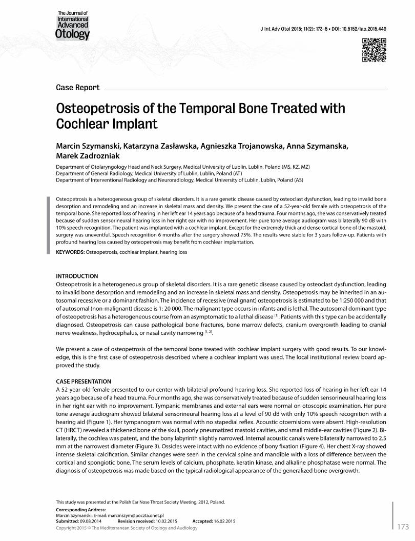
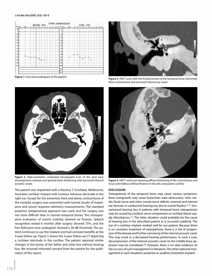
CASE PRESENTATIONA 52-year-old female presented to our center with bilateral profound hearing loss. She reported loss of hearing in her left ear 14 years ago because of a head trauma. Four months ago, she was conservatively treated because of sudden sensorineural hearing loss in her right ear with no improvement. Tympanic membranes and external ears were normal on otoscopic examination. Her pure tone average audiogram showed bilateral sensorineural hearing loss at a level of 90 dB with only 10% speech recognition with a hearing aid (Figure 1). Her tympanogram was normal with no stapedial reflex. Acoustic otoemisions were absent. High-resolution CT (HRCT) revealed a thickened bone of the skull, poorly pneumatized mastoid cavities, and small middle-ear cavities (Figure 2). Bi-laterally, the cochlea was patent, and the bony labyrinth slightly narrowed. Internal acoustic canals were bilaterally narrowed to 2.5 mm at the narrowest diameter (Figure 3). Ossicles were intact with no evidence of bony fixation (Figure 4). Her chest X-ray showed intense skeletal calcification. Similar changes were seen in the cervical spine and mandible with a loss of difference between the cortical and spongiotic bone. The serum levels of calcium, phosphate, keratin kinase, and alkaline phosphatase were normal. The diagnosis of osteopetrosis was made based on the typical radiological appearance of the generalized bone overgrowth.
This study was presented at the Polish Ear Nose Throat Society Meeting, 2012, Poland.
Corresponding Address:Marcin Szymanski, E-mail: [email protected]: 09.08.2014 Revision received: 10.02.2015 Accepted: 16.02.2015Copyright 2015 © The Mediterranean Society of Otology and Audiology 173
Osteopetrosis of the Temporal Bone Treated with Cochlear Implant
Marcin Szymanski, Katarzyna Zasławska, Agnieszka Trojanowska, Anna Szymanska, Marek ZadrozniakDepartment of Otolaryngology Head and Neck Surgery, Medical University of Lublin, Lublin, Poland (MS, KZ, MZ)Department of General Radiology, Medical University of Lublin, Lublin, Poland (AT)Department of Interventional Radiology and Neuroradiology, Medical University of Lublin, Lublin, Poland (AS)
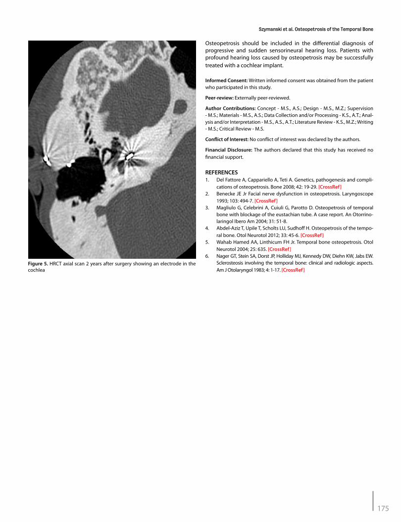
The patient was implanted with a Nucleus 5 (Cochlear, Melbourne, Australia) cochlear implant with Contour Advance electrode in the right ear. Except for the extremely thick and dense cortical bone of the mastoid, surgery was uneventful with normal results of imped-ance and neural response telemetry measurements. The standard posterior tympanotomy approach was used, and the surgery was not more difficult than in normal temporal bones. The intraoper-ative evaluation of ossicle mobility showed no fixation. Speech recognition tested 6 months after surgery showed 75%, and the free field pure tone audiogram showed a 30 dB threshold. The pa-tient continues to use the implant and had constant benefits at the 3-year follow-up. Figure 5 shows the 3-year follow-up CT depicting a cochlear electrode in the cochlea. The patient reported similar changes in the bones of her father and sister but without hearing loss. We received informed consent from the patient for the publi-cation of this report.
DISCUSSIONOsteopetrosis of the temporal bone may cause various symptoms. Bone overgrowth may cause Eustachian tube obstruction, otitis me-dia, facial nerve and other cranial nerve deficits, external and internal ear stenosis, or conductive hearing loss due to ossicle fixation [3, 4]. Sen-sorineural hearing loss in patients with temporal bone osteopetrosis may be caused by cochlear nerve compression or cochlear blood sup-ply disturbances [5]. The latter situation could probably be the cause of hearing loss in the described patient as it occurred suddenly. The use of a cochlear implant worked well for our patient. Because there is no causative treatment of osteopetrosis, there is a risk of progres-sion of the disease and further narrowing of the internal acoustic canal. This may result in a decreased hearing performance. In such a case, decompression of the internal acoustic canal via the middle fossa ap-proach may be considered [6]; however, there is no clear evidence on the success of such management in literature. The other optional man-agement in such situations would be an auditory brainstem implant.
174
J Int Adv Otol 2015; 11(2): 173-5
Figure 1. Pure tone audiogram of the patient
Figure 2. High-resolution computed tomography scan of the skull base showing bone sclerosis and general bone thickening with narrowed internal acoustic canals
Figure 3. HRCT scan with the frontal section of the temporal bone. Extremely thick cortical bone and narrowed internal ear canal
Figure 4. HRCT axial scan showing diffuse thickening of the cortical bone and incus and malleus without fixation in the attic and patent cochlea
Osteopetrosis should be included in the differential diagnosis of progressive and sudden sensorineural hearing loss. Patients with profound hearing loss caused by osteopetrosis may be successfully treated with a cochlear implant.
Informed Consent: Written informed consent was obtained from the patient who participated in this study.
Peer-review: Externally peer-reviewed.
Author Contributions: Concept - M.S., A.S.; Design - M.S., M.Z.; Supervision - M.S.; Materials - M.S., A.S.; Data Collection and/or Processing - K.S., A.T.; Anal-ysis and/or Interpretation - M.S., A.S., A.T.; Literature Review - K.S., M.Z.; Writing - M.S.; Critical Review - M.S.
Conflict of Interest: No conflict of interest was declared by the authors.
Financial Disclosure: The authors declared that this study has received no financial support.
REFERENCES1. Del Fattore A, Cappariello A, Teti A. Genetics, pathogenesis and compli-
cations of osteopetrosis. Bone 2008; 42: 19-29. [CrossRef]2. Benecke JE Jr Facial nerve dysfunction in osteopetrosis. Laryngoscope
1993; 103: 494-7. [CrossRef]3. Magliulo G, Celebrini A, Cuiuli G, Parotto D. Osteopetrosis of temporal
bone with blockage of the eustachian tube. A case report. An Otorrino-laringol Ibero Am 2004; 31: 51-8.
4. Abdel-Aziz T, Upile T, Scholts LU, Sudhoff H. Osteopetrosis of the tempo-ral bone. Otol Neurotol 2012; 33: 45-6. [CrossRef]
5. Wahab Hamed AA, Linthicum FH Jr. Temporal bone osteopetrosis. Otol Neurotol 2004; 25: 635. [CrossRef]
6. Nager GT, Stein SA, Dorst JP, Holliday MJ, Kennedy DW, Diehn KW, Jabs EW. Sclerosteosis involving the temporal bone: clinical and radiologic aspects. Am J Otolaryngol 1983; 4: 1-17. [CrossRef]
175
Szymanski et al. Osteopetrosis of the Temporal Bone
Figure 5. HRCT axial scan 2 years after surgery showing an electrode in the cochlea