Embed Size (px)
Citation preview
1
Sustained Attention in the Face of Distractors: A Study of Children with Rett Syndrome
Susan A. Rose, Ph.Da*, Sam Wass, Ph.D,b* Ph.D, Jeffery J. Jankowski, Ph.D,ac
Judith F. Feldman, Ph.Da, and Aleksandra Djukic, MD,da*
aDepartment of Pediatrics, Montefiore Medical Center, Albert Einstein College of
Medicine/Children’s Hospital at Montefiore, Bronx, NY
b Department of Psychology, University of East London, London, England
cDepartment of Social Sciences, Queensborough Community College/CUNY, United States
dRett Syndrome Center, Department of Neurology, Montefiore Medical Center, Albert
Einstein College of Medicine/Children’s Hospital at Montefiore, Bronx, NY
Corresponding author: Susan A. Rose, Departments of Pediatrics, Van Etten Building, Albert
Einstein College of Medicine/Children’s Hospital at Montefiore, 1300 Morris Park Avenue,
Bronx, NY 10461. Tel: 718-839-7230. email: [email protected]
ACKNOWLEDGMENTS
We thank the participants and their families for their cooperation and effort. This research was
funded by a grant from the International Rett Syndrome Foundation (IRSF).
2
Abstract
Objective. The object of the present study is to advance our understanding of the cognitive
profile of Rett Syndrome (RTT), an x-linked neurodevelopmental disorder caused by mutations
in the MECP2 gene. We focus on sustained attention, which plays a critical role in driving
cognitive growth, and use an innovative, gaze-based task that minimizes demands on the limited
verbal and motor abilities associated with RTT.
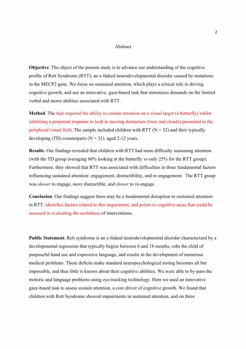
Method. The task required the ability to sustain attention on a visual target (a butterfly) whilst
inhibiting a prepotent response to look to moving distractors (trees and clouds) presented in the
peripheral visual field. The sample included children with RTT (N = 32) and their typically
developing (TD) counterparts (N = 32), aged 2-12 years.
Results. Our findings revealed that children with RTT had more difficulty sustaining attention
(with the TD group averaging 60% looking at the butterfly vs only 25% for the RTT group).
Furthermore, they showed that RTT was associated with difficulties in three fundamental factors
influencing sustained attention: engagement, distractibility, and re-engagement. The RTT group
was slower to engage, more distractible, and slower to re-engage.
Conclusion. Our findings suggest there may be a fundamental disruption to sustained attention
in RTT, identifies factors related to this impairment, and points to cognitive areas that could be
assessed in evaluating the usefulness of interventions.
Public Statement. Rett syndrome is an x-linked neurodevelopmental disorder characterized by a
developmental regression that typically begins between 6 and 18 months, robs the child of
purposeful hand use and expressive language, and results in the development of numerous
medical problems. These deficits make standard neuropsychological testing becomes all but
impossible, and thus little is known about their cognitive abilities. We were able to by-pass the
motoric and language problems using eye-tracking technology. Here we used an innovative
gaze-based task to assess sustain attention, a core driver of cognitive growth. We found that
children with Rett Syndrome showed impairments in sustained attention, and on three
3
fundamental factors influencing sustained attention: engagement, distractibility, and re-
engagement. Children with Rett Syndrome were slower to engage, more distractible, and slower
to re-engage than their age-matched peers. This work not only begins to elucidate the nature of
the cognitive problems associated with Rett syndrome, but is essential for designing markers to
assess the effects of pharmacological interventions.
Key Words: Rett syndrome; sustained attention; gaze-based task; eye-tracking; cognition
4
Sustained Attention in the Face of Distractors: A Study of Children with Rett Syndrome
Rett syndrome (Rett, 1966) is a severely disabling, x-linked neurodevelopmental disorder
characterized by apparently normal early development followed by developmental regression
between 6 and 18 months in which purposeful hand use and expressive language are lost and
impaired gait and hand stereotypies appear (Chahrour & Zoghbi, 2007). Other symptoms include
the development of seizures, apraxia, spasticity and scoliosis, breathing irregularities
(hyperventilation, breath holding, apnea), and a slowing of brain and head growth (Neul et al.,
2010).
This disorder, which affects about 1 in 10,000 females, is caused by spontaneous
mutations in the MECP2 gene, located on the long arm of the X chromosome – Xq28 (Amir et
al., 1999). The MECP2 gene encodes methyl-CpG-binding protein 2 (MeCP2), which is involved
in regulating the transcription of other genes, synaptic development and maintenance (Guy, Gan,
Selfridge, Cobb, & Bird, 2007), and is required for learning and memory (Moretti et al., 2006.).
Mutations lead to a significant reduction in long-term potentiation after symptom onset in
MECP2+/– females, with the magnitude of the defect similar to that reported in MeCP2-null
mice (Guy et al., 2007).
The severe limitations in language and purposeful hand use associated with Rett
syndrome (RTT) have precluded most neuropsychological testing of these children, with the
result that little is known about the cognitive phenotype of the disorder. However, recent studies
using eye tracking technology have shown progress in characterizing the behavioral and
cognitive profile of RTT. These studies found that children with RTT showed a preference for
socially weighted stimuli, as well as selective attention to salient areas and novel elements
5
(Djukic & Valicenti McDermott, 2012; Djukic, Valicenti McDermott, Mavrommatis, & Martins,
2012 ). While they were able to recognize simple patterns, faces and some emotional
expressions, their performance was significantly poorer than that of typically developing (TD)
children, and appeared to be related to attentional difficulties (Djukic, Rose, Jankowski, &
Feldman, 2014; Rose et al., 2013; Rose, Djukic, Jankowski, Feldman, & Rimler, 2016). These
difficulties included less looking at the targets and frequent failure to look at critical aspects.
These problems in attention are of particular concern because attention is a core
dimension of cognitive growth that has a cascading effect on subsequent learning and
development. Recent studies have shown that attention plays a pivotal role in gating the
development of working memory (Astle & Scerif, 2009) as well as in driving the development of
more complex outcomes, including IQ (Rose, Feldman, Jankowski, & Van Rossem, 2005, 2008),
language (Rose, Feldman, & Jankowski, 2009; Whedon, Perry, Calkins, & Bell, 2016), executive
functions (Rose, Feldman, & Jankowski, 2012), academic achievement (Bornstein, Hahn, &
Wolke, 2013), and eventual employment status (Kalechstein, Newton, & van Gorp, 2003). In
our own lab, we identified a developmental cascade in which elementary abilities evidenced in
infancy (attention and speed) influenced more complex abilities (memory and representational
competence) that, in turn, influenced general cognition in toddlerhood and early adolescence
(Cornish, Cole, Longhi, Karmiloff-Smith, & Scerif, 2012; Rose, Feldman, Jankowski, & Van
Rossem, 2012; Rose et al., 2005, 2008; Rose, Feldman, Jankowski, & Van Rossem, 2011; Scerif,
Longhi, Cole, Karmiloff-Smith, & Cornish, 2012).
To understand the role of attention, we need to recognize that it is a multi-dimensional
construct that includes a number of different processes, with different attentional functions
subserved by distinct, but overlapping neural systems (Fan, McCandliss, Fossella, Flombaum, &
6
Posner, 2005; Posner & Petersen, 1990). Posner distinguished three specialized brain networks
underlying attention – alerting, orienting, and executive attention (Petersen & Posner, 2012).
Alerting, which involves the thalamus, as well as right frontal and parietal cortical sites, and is
mediated primarily by the neuromodulator norepinephrine, achieves and maintains high
sensitivity to stimuli (Aston-Jones & Cohen, 2005; Petersen & Posner, 2012). Orienting, which
involves a dorsal network (including the frontal eye fields and superior parietal lobe), as well as
a more ventral network (including the parietal-temporal junction), and is thought to be subserved
primarily by cholinergic networks (Davidson & Marrocco, 2000), is important for the selection
of stimuli from sensory input. Although it was previously thought that the dorsal network was
endogenously driven, and the ventral network exogenously driven (Corbetta & Shulman, 2002),
more recent evidence indicates that both networks are involved in re-orienting, showing that this
process is endogenously as well as exogenously driven (Corbetta, Patel, & Shulman, 2008).
Executive attention, which involves the anterior cingulate cortex and prefrontal areas, is
important for situations involving conflict, where inhibition is necessary.
The tasks used in our earlier work involved several aspects of attention in combination.
One that figured prominently was sustained attention -- the ability to focus or concentrate
attention on a task or maintain vigilance in the face of distractors. The present study attempts to
better understand the difficulty Rett children have with this aspect of attention and identify
factors influencing it. Sustained attention, which is thought to involve top-down connectivity
extending from the anterior attention system, particularly prefrontal and parietal regions in the
right hemisphere, right down into V1 (Grahn & Manly, 2012; Sarter, Givens, & Bruno, 2001;
Silver, Ress, & Heeger, 2007), has repeatedly been found to be compromised across a wide
range of neurological and psychiatric disorders, e.g., ADHD, autism, bipolar disorder and Fragile
7
X (Cornish, Scerif, & Karmiloff-Smith, 2007; Cornish, Turk, & Levitas, 2007; Fortenbaugh et
al., 2015; O'Connell, Bellgrove, Dockree, & Robertson, 2004)
While sustained attention is often tested in adults with the continuous performance test,
the verbal instructions and motoric requirements preclude using this task in children with RTT.
To overcome these limitations, sustained attention was assessed here by building on tasks that
have assessed how well children can visually concentrate on a target while ignoring distractors
(Oakes, Kannass, & Shaddy, 2002; Richards, 1987; Ruff & Rothbart, 1996). We used an
innovative, gaze-based task modeled after Wass and colleagues (Wass, Porayska-Pomsta, &
Johnson, 2011). A target (a butterfly) was presented on the screen. When the child fixated on the
target it moved from left to right and distractors (trees and clouds) scrolled in the opposite
direction. When the child looked to any of the distractors, the display froze. The task has three
key features. First, the movement of the butterfly is gaze-contingent (it moves only when fixated)
and thus there is a reward component for sustaining attention. Second, the necessity for motoric
and verbal abilities is minimized. Third, the task allows us to assess not only sustained attention,
but also factors that impact it, including time to engage the target, distractibility, and re-
engagement. This new task thus targets executive attention and the orienting network. Executive
attention is involved in inhibiting attention to the distractors, and the orienting network when the
child initially directs attention to the target at the outset of a trial or re-directs attention from the
distractor to the butterfly during a trial.
We hypothesize that the Rett children will show less sustained attention and more
distractibility (time off task) than typically developing children, particularly as the number of
distractors increases. This hypothesis is based on brain imaging studies of children with Rett
showing global decreases in brain volume (Carter et al., 2008), selective reductions in frontal
8
white matter (Mahmood et al., 2010), and selective vulnerability of the frontal lobes (Naidu et
al., 2001), all areas involved in inhibiting attention to distractors. We also hypothesized that
group differences in orienting and re-orienting might be less marked, given data showing
selective preservation of the occipital cortex, although selective reductions in dorsal parietal grey
matter, an area involved in re-orienting, makes this hypothesis more tenuous (Carter et al., 2008).
Method
Participants
This study was conducted on 32 females with clinically diagnosed classical Rett
syndrome (Neul et al., 2010), consecutively recruited from the Rett Center at the Children’s
Hospital of Montefiore (M=7.92 years; SD=2.89, range=2-12) and a comparison group of 32
typically developing (TD) females (M=7.66 years; SD=2.83, range=2-12), t(62)=.35. The TD
group, recruited from Outpatient Clinics of the same hospital, was drawn from children who
were family members of patients with appointments at pediatric specialty clinics. The TD
group was screened to exclude any children with significant neurological disorders (e.g.,
epilepsy, brain tumor), sensory impairment, neurodevelopmental disorders (e.g., autism,
ADHD) or first-degree relatives with neurodevelopmental disorders.
RTT was genetically confirmed in all Rett participants. Testing was attempted, but
terminated, for an additional 3 Rett patients who could not successfully complete the
calibration procedure (described below) and 5 who were too overactive/restless to complete
the testing procedure; these eight did not differ in clinical/background factors from the rest of
the RTT group (falling in the moderate range of the RSSS scale described below).
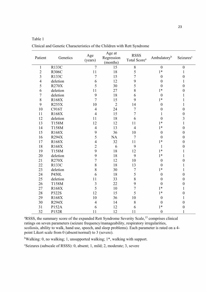
Clinical characteristics of the Rett sample. Table 1 shows the genetic mutation, age at
test, age at regression, scores on the Rett Syndrome Severity Scale (RSSS) (Kaufmann et al.,
9
2012) and notes their status on two subscales of the RSSS – walking and seizures. Composite
scores on the RSSS averaged 8.25 (SD =2.43), with 14 patients (43.8) scoring in the mild range
(0–7), and the remainder (56.2%) in the moderate range (8–14). Many (46.9%) were ambulatory
(able to walk unaided or with support); 43.8% of the group had a history of seizures.
The protocol was approved by the institutional review board and written consent was
obtained for all participants.
__________________________
Table 1 goes about here.
__________________________
Apparatus
Stimuli were presented on a 23-inch flat panel monitor (resolution, 1024 768 pixels)
and integrated with a Tobii X2-60 eye-tracker, using Matlab and Psychtoolbox. Talk2Tobii
software was used to allow for a live gaze-contingent interface via Matlab during stimulus
presentation. Manufacturer-supplied algorithms for pupil, corneal reflection, and face
identification were used during eye-tracking; gaze data were sampled at 60 Hz.
Calibration
At the beginning of the session each participant’s point-of-gaze- was calibrated using a 5-
point calibration procedure. The calibration stimuli, five pulsing colored blocks (1° to 1.5°) were
presented sequentially, at different locations on the screen, accompanied by a sound (‘Whee’).
Point-of-gaze was calibrated by comparing each look to the known coordinates of the target, and
results were inspected graphically. The quality of the calibration data was determined by the
closeness of the fixation points to the calibration points. If the points did not cluster, or any
10
targets were missed, the calibration was repeated until a satisfactory calibration was achieved.
Each calibration attempt took less than a minute.
Stimuli and Procedure
Testing was conducted in a quiet room, with participants seated approximately 45 cm from
the monitor. Ambient light levels were reduced to diminish distraction. Verbal instructions, limited
to ‘Look at the TV,’ were used at the beginning of the session. To minimize body and head
movement, all participants with RTT (and all TD participants < 5 years) were seated on their
parent’s lap. Parents kept their eyes closed during testing.
Trials started with a target, a butterfly (subtending 6°), presented on the screen (Wass et
al., 2011). When the child fixated the target, it moved, fluttering its wings and ‘flying”
horizontally from left to right across the screen. Distractors, consisting of a house, a tree, and
clouds (subtending 5-15°), scrolled in the opposite direction. The butterfly travelled at a rate of
2.5 cm/s, while the distractors moved in the opposite direction at the same rate. When the child
looked at any of the distractors they disappeared, with only the butterfly target remaining. On re-
fixating the target, it recommenced moving across the screen and fluttering its wings, and the
distractors re-appeared and continued scrolling. Trials lasted 15 s and an engaging sound track
(the melody, Zip-a-Dee-Doo-Dah) played throughout each trial. There were two blocks of 9
trials; each block contained three trials each with 1, 2, and 3 distractors, presented in pseudo-
random order. The two blocks of trials, each lasting less than 2.5 minutes, were interleaved with
two other attention tasks. The entire testing session took about 10 min.
Data Analyses
All measures were examined for normality and outliers and analyzed using a mixed
model 2 (Group: RTT vs TD) x 2 (Age: younger vs older) x 3 (number of distractors: 1, 2, or 3)
11
ANOVA, with repeated measures on the last factor. Age was dichotomized for these analyses
using a median split (< 8 years vs ≥ 8 years, for both groups). Where necessary, measures were
transformed to achieve normality, using a log or square root transform (see below). All effects
were evaluated at a .05 level of significance; SPSS (version 24) was used in all analyses;
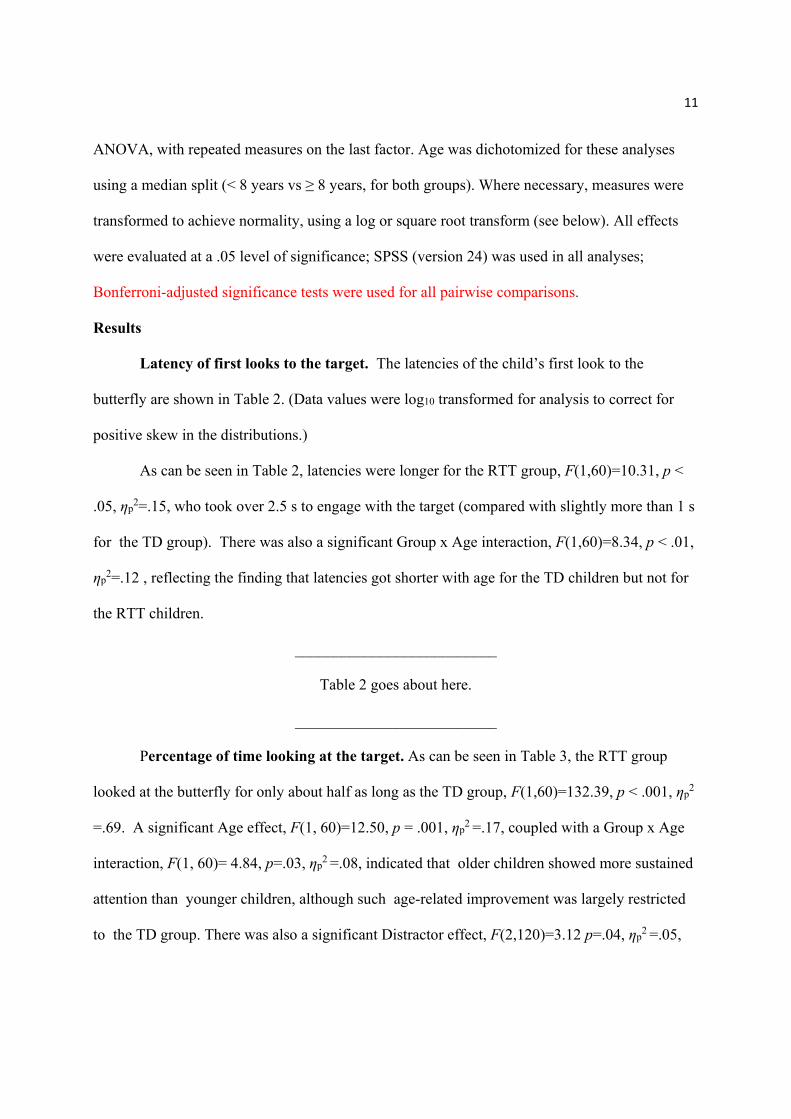
Bonferroni-adjusted significance tests were used for all pairwise comparisons.
Results
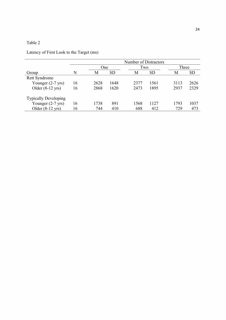
Latency of first looks to the target. The latencies of the child’s first look to the
butterfly are shown in Table 2. (Data values were log10 transformed for analysis to correct for
positive skew in the distributions.)
As can be seen in Table 2, latencies were longer for the RTT group, F(1,60)=10.31, p <
.05, ηp2=.15, who took over 2.5 s to engage with the target (compared with slightly more than 1 s
for the TD group). There was also a significant Group x Age interaction, F(1,60)=8.34, p < .01,
ηp2=.12 , reflecting the finding that latencies got shorter with age for the TD children but not for
the RTT children.
__________________________
Table 2 goes about here.
__________________________
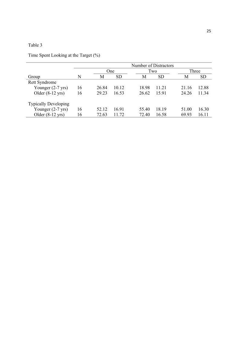
Percentage of time looking at the target. As can be seen in Table 3, the RTT group
looked at the butterfly for only about half as long as the TD group, F(1,60)=132.39, p < .001, ηp2
=.69. A significant Age effect, F(1, 60)=12.50, p = .001, ηp2 =.17, coupled with a Group x Age
interaction, F(1, 60)= 4.84, p=.03, ηp2 =.08, indicated that older children showed more sustained
attention than younger children, although such age-related improvement was largely restricted
to the TD group. There was also a significant Distractor effect, F(2,120)=3.12 p=.04, ηp2 =.05,
12
due to looking at the target decreasing as the number of distractors increased; a marginally
significant Group x Distractor interaction, F(2,120)=2.72, p=.07, ηp2 =.04, indicated that the fall-
off in performance was most pronounced for the RTT children.
__________________________
Table 3 goes about here.
__________________________
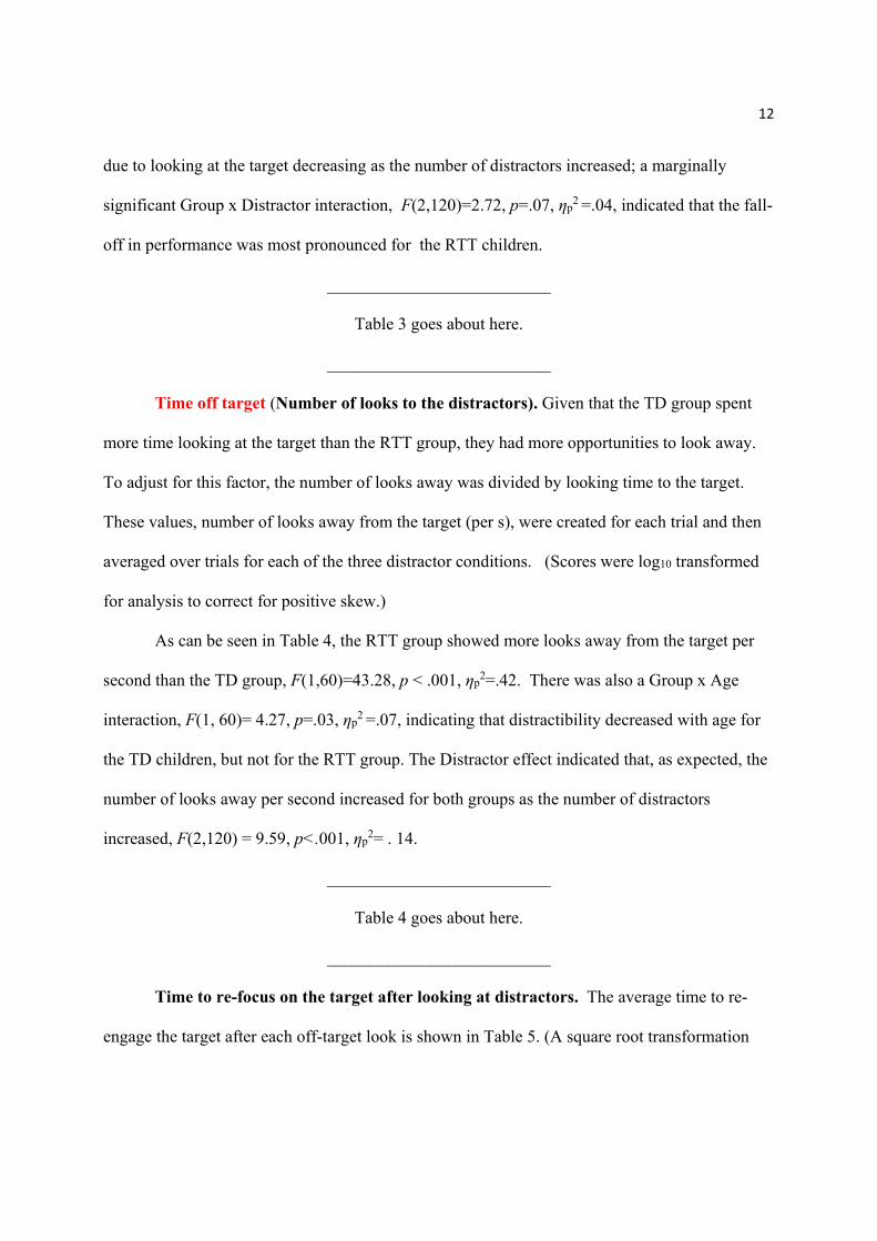
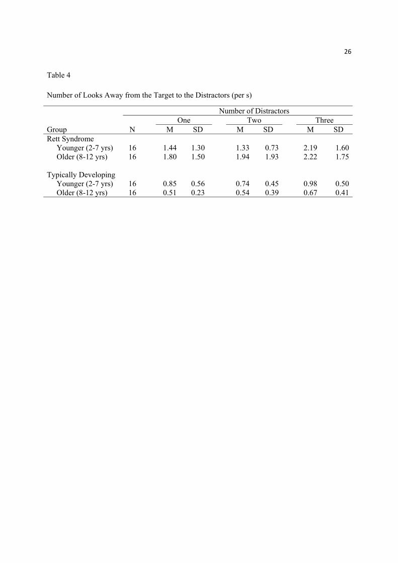
Time off target (Number of looks to the distractors). Given that the TD group spent
more time looking at the target than the RTT group, they had more opportunities to look away.
To adjust for this factor, the number of looks away was divided by looking time to the target.
These values, number of looks away from the target (per s), were created for each trial and then
averaged over trials for each of the three distractor conditions. (Scores were log10 transformed
for analysis to correct for positive skew.)
As can be seen in Table 4, the RTT group showed more looks away from the target per
second than the TD group, F(1,60)=43.28, p < .001, ηp2=.42. There was also a Group x Age
interaction, F(1, 60)= 4.27, p=.03, ηp2 =.07, indicating that distractibility decreased with age for
the TD children, but not for the RTT group. The Distractor effect indicated that, as expected, the
number of looks away per second increased for both groups as the number of distractors
increased, F(2,120) = 9.59, p<.001, ηp2= . 14.
__________________________
Table 4 goes about here.
__________________________
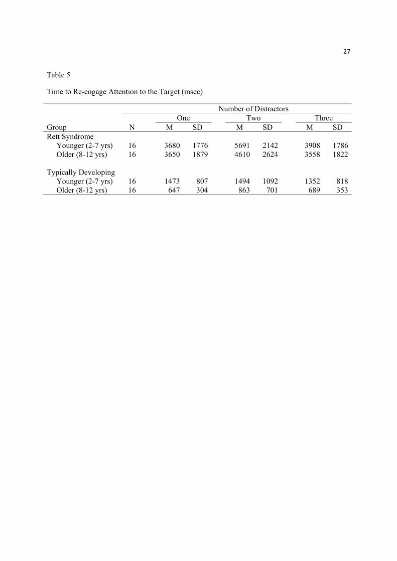
Time to re-focus on the target after looking at distractors. The average time to re-
engage the target after each off-target look is shown in Table 5. (A square root transformation
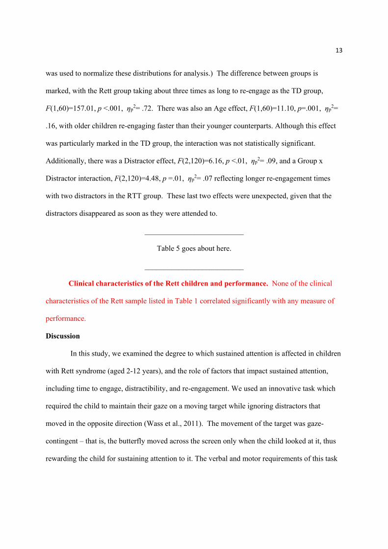
13
was used to normalize these distributions for analysis.) The difference between groups is
marked, with the Rett group taking about three times as long to re-engage as the TD group,
F(1,60)=157.01, p <.001, ηp2= .72. There was also an Age effect, F(1,60)=11.10, p=.001, ηp
2=
.16, with older children re-engaging faster than their younger counterparts. Although this effect
was particularly marked in the TD group, the interaction was not statistically significant.
Additionally, there was a Distractor effect, F(2,120)=6.16, p <.01, ηp2= .09, and a Group x
Distractor interaction, F(2,120)=4.48, p =.01, ηp2= .07 reflecting longer re-engagement times
with two distractors in the RTT group. These last two effects were unexpected, given that the
distractors disappeared as soon as they were attended to.
__________________________
Table 5 goes about here.
__________________________
Clinical characteristics of the Rett children and performance. None of the clinical
characteristics of the Rett sample listed in Table 1 correlated significantly with any measure of
performance.
Discussion
In this study, we examined the degree to which sustained attention is affected in children
with Rett syndrome (aged 2-12 years), and the role of factors that impact sustained attention,
including time to engage, distractibility, and re-engagement. We used an innovative task which
required the child to maintain their gaze on a moving target while ignoring distractors that
moved in the opposite direction (Wass et al., 2011). The movement of the target was gaze-
contingent – that is, the butterfly moved across the screen only when the child looked at it, thus
rewarding the child for sustaining attention to it. The verbal and motor requirements of this task
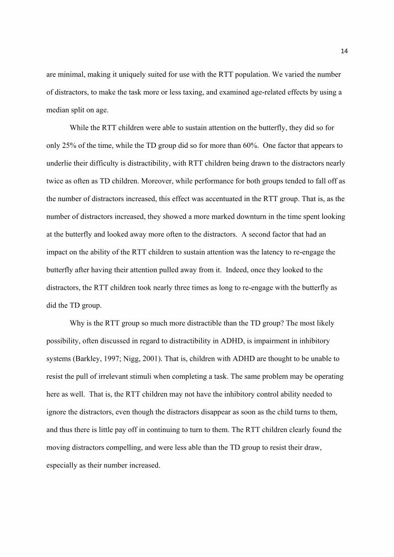
14
are minimal, making it uniquely suited for use with the RTT population. We varied the number
of distractors, to make the task more or less taxing, and examined age-related effects by using a
median split on age.
While the RTT children were able to sustain attention on the butterfly, they did so for
only 25% of the time, while the TD group did so for more than 60%. One factor that appears to
underlie their difficulty is distractibility, with RTT children being drawn to the distractors nearly
twice as often as TD children. Moreover, while performance for both groups tended to fall off as
the number of distractors increased, this effect was accentuated in the RTT group. That is, as the
number of distractors increased, they showed a more marked downturn in the time spent looking
at the butterfly and looked away more often to the distractors. A second factor that had an
impact on the ability of the RTT children to sustain attention was the latency to re-engage the
butterfly after having their attention pulled away from it. Indeed, once they looked to the
distractors, the RTT children took nearly three times as long to re-engage with the butterfly as
did the TD group.
Why is the RTT group so much more distractible than the TD group? The most likely
possibility, often discussed in regard to distractibility in ADHD, is impairment in inhibitory
systems (Barkley, 1997; Nigg, 2001). That is, children with ADHD are thought to be unable to
resist the pull of irrelevant stimuli when completing a task. The same problem may be operating
here as well. That is, the RTT children may not have the inhibitory control ability needed to
ignore the distractors, even though the distractors disappear as soon as the child turns to them,
and thus there is little pay off in continuing to turn to them. The RTT children clearly found the
moving distractors compelling, and were less able than the TD group to resist their draw,
especially as their number increased.
15
Why are the RTT children so much slower than the TD group to re-engage the butterfly
after being distracted? This finding is more difficult to explain. After all, the distractors have
disappeared. Oculomotor factors cannot fully account for this difference given that, despite
similar oculomotor demands, the re-engagement latencies in the RTT group were substantially
longer than their initial latencies to engage the target, t(31) = 3.88, p = .001, d = .69. One
possibility is that the RTT children have more difficulty making the complex set of adjustments
involved in the interaction of the dorsal and ventral frontoparietal systems involved in re-
orienting (Corbetta & Shulman, 2008). This possibility receives some support from a recent
study which showed that states of high global integration of neural networks are associated with
better performance (Shine, 2016). Any differences that exist between groups in arousal level may
also have affected re-orienting, since the integration of networks tracks with fluctuations in
arousal (Shine, 2016), and attention has been shown to be modulated by arousal (Aston-Jones &
Cohen, 2005; de Barbaro, Clackson, & Wass, in press). Another possibility is that the RTT
children are slower to re-calibrate, and in appraising the situation realize that there is no pay-off
in continuing to look to locations where the distractors had been. This possibility is consistent
with earlier findings where children with RTT had difficulty learning the rule underlying event
sequences (Rose et al., 2016).
The effects of age were examined using a median split for both groups. Children in the
TD group showed improvement over age for all measures, significantly so for latency to first
look and sustained attention (time spent looking at the target) and marginally so for re-
engagement. There were no age effects for the RTT group, a finding consistent with previous
work (Rose et al., 2013). It is probable that any tendency to improve over age is counteracted by
the progressive nature of the disorder.
16
The gaze-based task would appear to be a useful way for testing sustained attention in
other populations where verbal and motoric impairments preclude using other tasks, such as the
continuous performance test. In the latter, often treated as the ‘gold standard’ for assessing
sustained attention, a button is to be pressed as quickly as possible each time a target appears,
while distractors are to be ignored; the critical measure is errors of omission (failures to press
when the target appears). Two of the typical effects found with this task -- a strong negative
effect of distractors and age-related improvement in sustained attention (Conners, Epstein,
Angold, & Klaric, 2003) -- also prominent effects for the TD children on the gaze-based task
used in the present study. This agreement in findings supports the usefulness of the gaze-based
task for assessing sustained attention.
In summary, the present work identified difficulties in sustained attention associated with
RTT and determined at least two factors implicated in these difficulties – distractibility and
slowness to re-engage after distraction. This work helps to elucidate the nature of the cognitive
problems associated with RTT, is essential for the design of intervention, and begins to indicate
functions and tasks that could serve as markers for the effects of pharmacological interventions.
17
References
Amir, R. E., Van den Veyver, I. B., Wan, M., Tran, C. Q., Francke, U., & Zoghbi, H. Y. (1999).
Rett syndrome is caused by mutations in X-linked MECP2, encoding methyl-CpG-
binding protein 2. Nature Genetics, 23(2), 185-188. doi: 10.1038/13810
Astle, D. E., & Scerif, G. (2009). Using developmental cognitive neuroscience to study
behavioral and attentional control. Dev Psychobiol, 51(2), 107-118. doi:
10.1002/dev.20350
Aston-Jones, G., & Cohen, J. D. (2005). An integrative theory of locus coeruleus-norepinephrine
function: adaptive gain and optimal performance. Annual review of neuroscience, 28,
403-450. doi: 10.1146/annurev.neuro.28.061604.135709
Barkley, R. A. (1997). Behavioral inhibition, sustained attention, and executive functions:
constructing a unifying theory of ADHD. Psychological Bulletin, 121(1), 65-94.
Bornstein, M.H., Hahn, C.S., & Wolke, D. (2013). Systems and cascades in cognitive
development and academic achievement. Child development, 84(1), 154-162. doi:
10.1111/j.1467-8624.2012.01849.x
Carter, J. C., Lanham, D. C., Pham, D., Bibat, G., Naidu, S., & Kaufmann, W. E. (2008).
Selective cerebral volume reduction in Rett syndrome: A multiple-approach MR imaging
study. American Journal of Neuroradiology, 29(3), 436-441. doi: 10.3174/ajnr.A0857
Chahrour, M., & Zoghbi, H. Y. (2007). The story of Rett syndrome: from clinic to neurobiology.
Neuron, 56(3), 422-437. doi: 10.1016/j.neuron.2007.10.001
Conners, C. K., Epstein, J. N., Angold, A., & Klaric, J. (2003). Continuous performance test
performance in a normative epidemiological sample. Journal of Abnormal Child
Psychology, 31(5), 555-562.
18
Corbetta, M., Patel, G., & Shulman, G. L. (2008). The reorienting system of the human brain:
from environment to theory of mind. Neuron, 58(3), 306-324. doi:
10.1016/j.neuron.2008.04.017
Corbetta, M., & Shulman, G. L. (2002). Control of goal-directed and stimulus-driven attention in
the brain. Nature reviews. Neuroscience, 3(3), 201-215. doi: 10.1038/nrn755
Cornish, K., Cole, V., Longhi, E., Karmiloff-Smith, A., & Scerif, G. (2012). Does attention
constrain developmental trajectories in fragile x syndrome? A 3-year prospective
longitudinal study. American Journal of Intellectual and Developmental Disabilities
117(2), 103-120. doi: 10.1352/1944-7558-117.2.103
Cornish, K., Scerif, G., & Karmiloff-Smith, A. (2007). Tracing syndrome-specific trajectories of
attention across the lifespan. Cortex, 43(6), 672-685.
Cornish, K., Turk, J., & Levitas, A. (2007). Fragile X syndrome and autism: common
developmental pathways? Current Pediatric Reviews, 3(1), 61-68.
Davidson, M. C., & Marrocco, R. T. (2000). Local infusion of scopolamine into intraparietal
cortex slows covert orienting in rhesus monkeys. J Neurophysiol, 83(3), 1536-1549.
de Barbaro, K., Clackson, K., & Wass, S..V. (in press). Infant attention is dynamically
modulated with changing arousal level Child Development.
Djukic, A., Rose, S. A., Jankowski, J. J., & Feldman, J. F. (2014). Rett syndrome: recognition of
facial expression and its relation to scanning patterns. Pediatric Neurology, 51(5), 650-
656. doi: 10.1016/j.pediatrneurol.2014.07.022
Djukic, A., & Valicenti McDermott, M. (2012). Social preferences in rett syndrome. Pediatric
Neurology, 46(4), 240-242. doi: 10.1016/j.pediatrneurol.2012.01.011
19
Djukic, A., Valicenti McDermott, M., Mavrommatis, K., & Martins, C.L. (2012 ). Rett
syndrome: Basic features of visual processing--a pilot study of eye-tracking. Pediatric
Neurology, 47, 25-29.
Fan, Jin, McCandliss, Bruce D., Fossella, John, Flombaum, Jonathan I., & Posner, Michael I.
(2005). The activation of attentional networks. Neuroimage, 26(2), 471-479.
Fortenbaugh, F. C., DeGutis, J., Germine, L., Wilmer, J. B., Grosso, M., Russo, K., & Esterman,
M. (2015). Sustained Attention Across the Life Span in a Sample of 10,000: Dissociating
Ability and Strategy. Psychological Science, 26(9), 1497-1510. doi:
10.1177/0956797615594896
Grahn, J. A., & Manly, T. (2012). Common neural recruitment across diverse sustained attention
tasks. PLoS One, 7(11), e49556. doi: 10.1371/journal.pone.0049556
Guy, J., Gan, J., Selfridge, J., Cobb, S., & Bird, A. (2007). Reversal of neurological defects in a
mouse model of Rett syndrome. Science, 315(5815), 1143-1147. doi:
10.1126/science.1138389
Kalechstein, A. D., Newton, T. F., & van Gorp, W. G. (2003). Neurocognitive functioning is
associated with employment status: a quantitative review. Journal of Clinical and
Experimental Neuropsychology, 25(8), 1186-1191. doi: 10.1076/jcen.25.8.1186.16723
Kaufmann, W. E., Tierney, E., Rohde, C. A., Suarez-Pedraza, M. C., Clarke, M. A., Salorio, C.
F., . . . Naidu, S. (2012). Social impairments in Rett syndrome: characteristics and
relationship with clinical severity. Journal of Intellectual Disabilities Research, 56(3),
233-247. doi: 10.1111/j.1365-2788.2011.01404.x
Mahmood, A., Bibat, G., Zham, A. L., Izbudak, I., Farage, L., Horska, A., . . . Naidu, S. (2010).
White Matter Impairment in Rett Syndrome: Diffusion Tensor Imaging Study with
20
Clinical Correlations. American Journal of Neuroradiology, 31(2), 295-299. doi:
10.3174/ajnr.A1792
Moretti, P., Levenson, J. M., Battaglia, F., Atkinson, R., Teague, R., Antalffy, B., . . . Zoghbi, H.
Y. (2006). Learning and memory and synaptic plasticity are impaired in a mouse model
of Rett syndrome. Journal of Neuroscience, 26(1), 319-327. doi:
10.1523/JNEUROSCI.2623-05.2006
Naidu, S., Kaufmann, W. E., Abrams, M. T., Pearlson, G. D., Lanham, D. C., Fredericksen, K.
A., . . . Johnston, M. V. (2001). Neuroimaging studies in Rett syndrome. Brain &
Development, 23, S62-S71. doi: Doi 10.1016/S0387-7604(01)00381-3
Neul, J. L., Kaufmann, W. E., Glaze, D. G., Christodoulou, J., Clarke, A. J., Bahi-Buisson, N., . .
. Percy, A. K. (2010). Rett syndrome: revised diagnostic criteria and nomenclature.
Annals of Neurology, 68(6), 944-950. doi: 10.1002/ana.22124
Nigg, J. T. (2001). Is ADHD a disinhibitory disorder? Psychological Bulletin, 127(5), 571-598.
O'Connell, R. G., Bellgrove, M. A., Dockree, P. M., & Robertson, I. H. . (2004). Reduced
electrodermal response to errors predicts poor sustained attention performance in
attention deficit hyperactivity disorder. Neuroreport, 15(16), 2535-2538.
Oakes, L. M., Kannass, K. N., & Shaddy, D. J. (2002). Developmental changes in endogenous
control of attention: the role of target familiarity on infants' distraction latency. Child
Development, 73(6), 1644-1655.
Petersen, S. E., & Posner, M. I. (2012). The attention system of the human brain: 20 years after.
Annual Review of Neuroscience, 35, 73-89. doi: 10.1146/annurev-neuro-062111-150525
Posner, M.I., & Petersen, S. (1990). The attention system of the human brain. Annual Review of
Neuroscience, 13, 25-42.
21
Rett, A. (1966). On a unusual brain atrophy syndrome in hyperammonemia in childhood. Wien
Med Wochenschr, 116(37), 723-726.
Richards, J. E. (1987). Infant visual sustained attention and respiratory sinus arrhythmia. Child
Dev, 58(2), 488-496.
Rose, S.A., Djukic, A., Jankowski, J. J., Feldman, J. F., Fishman, I., & Valicenti-Mcdermott, M.
(2013). Rett syndrome: an eye-tracking study of attention and recognition memory.
Developmental Medicine and Child Neurology, 55(4), 364-371. doi: Doi
10.1111/Dmcn.12085
Rose, S.A., Djukic, A., Jankowski, J. J., Feldman, J. F., & Rimler, M. (2016). Aspects of
Attention in Rett Syndrome. Pediatric Neurology, 57, 22-28. doi:
10.1016/j.pediatrneurol.2016.01.015
Rose, S.A., Feldman, J. F., & Jankowski, J. J. (2009). Information Processing in Toddlers:
Continuity from Infancy and Persistence of Preterm Deficits. Intelligence, 37(3), 311-
320. doi: 10.1016/j.intell.2009.02.002
Rose, S.A., Feldman, J. F., & Jankowski, J. J. (2012). Implications of infant cognition for
executive functions at age 11. Psychological Science, 23(11), 1345-1355. doi:
10.1177/0956797612444902
Rose, S.A., Feldman, J. F., Jankowski, J. J., & Van Rossem, R. (2012). Information processing
from infancy to 11 years: Continuities and prediction of IQ. Intelligence, 40(5), 445-457.
Rose, S.A., Feldman, J.F., Jankowski, J.J., & Van Rossem, R. (2005). Pathways from
prematurity and infant abilities to later cognition. Child Development, 76, 1172-1184.
22
Rose, S.A., Feldman, J.F., Jankowski, J.J., & Van Rossem, R. (2008). A cognitive cascade in
infancy: Pathways from prematurity to later mental development Intelligence, 36, 367-
378.
Rose, S.A., Feldman, J.F., Jankowski, J.J., & Van Rossem, R. (2011). Basic information
processing abilities at 11 years account for deficits in IQ associated with preterm birth.
Intelligence, 39, 198-209.
Ruff, H.A., & Rothbart, M.K. (1996). Attention in early development: themes and variations.
New York: Oxford University Press.
Sarter, M., Givens, B., & Bruno, J. P. (2001). The cognitive neuroscience of sustained attention:
where top-down meets bottom-up. Brain research. Brain research reviews, 35(2), 146-
160.
Scerif, G., Longhi, E., Cole, V., Karmiloff-Smith, A., & Cornish, K. (2012). Attention across
modalities as a longitudinal predictor of early outcomes: the case of fragile X syndrome.
J Child Psychol Psychiatry, 53(6), 641-650. doi: 10.1111/j.1469-7610.2011.02515.x
Silver, M. A., Ress, D., & Heeger, D. J. (2007). Neural correlates of sustained spatial attention in
human early visual cortex. J Neurophysiol, 97(1), 229-237. doi: 10.1152/jn.00677.2006
Wass, S., Porayska-Pomsta, K., & Johnson, M. H. (2011). Training attentional control in infancy.
Current Biology, 21(18), 1543-1547. doi: 10.1016/j.cub.2011.08.004
Whedon, M., Perry, N. B., Calkins, S. D., & Bell, M. A. (2016). Changes in Frontal EEG
Coherence Across Infancy Predict Cognitive Abilities at Age 3: The Mediating Role of
Attentional Control. Developmental Psychology. doi: 10.1037/dev0000149
23
Table 1
Clinical and Genetic Characteristics of the Children with Rett Syndrome
Patient Genetics Age
(years)
Age at Regression (months)
RSSS Total Scorea
Ambulatoryb Seizuresc
1 R133C 7 15 8 0 02 R306C 11 18 5 1* 1 3 R133C 7 15 7 0 0 4 deletion 6 12 9 0 15 R270X 5 30 5 0 0 6 deletion 11 27 8 1* 0 7 deletion 9 18 6 0 18 R168X 7 15 9 1* 1 9 R255X 10 2 14 0 1 10 C916T 4 24 7 0 011 R168X 4 15 7 1 0 12 deletion 11 18 6 0 3 13 T158M 12 12 11 1* 114 T158M 4 13 4 1* 0 15 R168X 9 36 10 0 0 16 R294X 5 NA 7 0 017 R168X 4 32 11 1* 0 18 R168X 2 6 9 1 0 19 T158M 9 18 12 1* 120 deletion 9 18 9 1* 1 21 R270X 7 12 10 0 0 22 R133C 8 18 13 0 123 deletion 8 30 7 1* 1 24 P450L 6 18 5 0 0 25 deletion 11 33 8 0 0 26 T158M 3 22 9 0 0 27 R168X 5 10 7 1* 1 28 P322S 12 15 5 1* 0 29 R168X 10 36 10 0 1 30 R294X 4 14 8 0 0 31 P152A 6 12 6 1* 0 32 P152R 11 12 11 0 1
aRSSS, the summary score of the expanded Rett Syndrome Severity Scale,15 comprises clinical ratings on seven parameters (seizure frequency/manageability, respiratory irregularities, scoliosis, ability to walk, hand use, speech, and sleep problems). Each parameter is rated on a 4-point Likert scale from 0 (absent/normal) to 3 (severe). bWalking: 0, no walking; 1, unsupported walking; 1*, walking with support. cSeizures (subscale of RSSS): 0, absent; 1, mild; 2, moderate; 3, severe
24
Table 2
Latency of First Look to the Target (ms)
Number of Distractors One Two Three Group N M SD M SD M SD Rett Syndrome Younger (2-7 yrs) 16 2628 1648 2377 1561 3113 2626 Older (8-12 yrs) 16 2868 1620 2473 1895 2937 2329 Typically Developing Younger (2-7 yrs) 16 1738 891 1568 1127 1793 1037 Older (8-12 yrs) 16 744 410 688 412 729 473
25
Table 3
Time Spent Looking at the Target (%)
Number of Distractors One Two Three Group N M SD M SD M SD Rett Syndrome Younger (2-7 yrs) 16 26.84 10.12 18.98 11.21 21.16 12.88 Older (8-12 yrs) 16 29.23 16.53 26.62 15.91 24.26 11.34 Typically Developing Younger (2-7 yrs) 16 52.12 16.91 55.40 18.19 51.00 16.30 Older (8-12 yrs) 16 72.63 11.72 72.40 16.58 69.93 16.11
26
Table 4
Number of Looks Away from the Target to the Distractors (per s)
Number of Distractors One Two Three Group N M SD M SD M SDRett Syndrome Younger (2-7 yrs) 16 1.44 1.30 1.33 0.73 2.19 1.60 Older (8-12 yrs) 16 1.80 1.50 1.94 1.93 2.22 1.75 Typically Developing Younger (2-7 yrs) 16 0.85 0.56 0.74 0.45 0.98 0.50 Older (8-12 yrs) 16 0.51 0.23 0.54 0.39 0.67 0.41
27
Table 5
Time to Re-engage Attention to the Target (msec)
Number of Distractors One Two Three Group N M SD M SD M SD Rett Syndrome Younger (2-7 yrs) 16 3680 1776 5691 2142 3908 1786 Older (8-12 yrs) 16 3650 1879 4610 2624 3558 1822 Typically Developing Younger (2-7 yrs) 16 1473 807 1494 1092 1352 818 Older (8-12 yrs) 16 647 304 863 701 689 353