Embed Size (px)
Citation preview
Chapter 33Obstetrics and Neonatal Care

National EMS Education Standard Competencies (1 of 7)
Special Patient PopulationsApplies a fundamental knowledge of growth, development, and aging and assessment findings to provide basic emergency care and transportation for a patient with special needs.

National EMS Education Standard Competencies (2 of 7)
Obstetrics• Recognition and management of
– Normal delivery– Vaginal bleeding in the pregnant patient
• Anatomy and physiology of normal pregnancy
• Pathophysiology of complications of pregnancy
National EMS Education Standard Competencies (3 of 7)
Obstetrics (cont’d)• Assessment of the pregnant patient• Management of
– Normal delivery– Abnormal delivery
• Nuchal cord• Prolapsed cord• Breech delivery

National EMS Education Standard Competencies (4 of 7)
• Management of (cont’d)– Third trimester bleeding
• Placenta previa• Abruptio placenta
– Spontaneous abortion/miscarriage– Ectopic pregnancy– Preeclampsia/eclampsia

National EMS Education Standard Competencies (5 of 7)
Neonatal Care• Assessment and management of
– Newborn care– Neonatal resuscitation

National EMS Education Standard Competencies (6 of 7)
TraumaApplies fundamental knowledge to provide basic emergency care and transportation based on assessment findings for an acutely injured patient.

National EMS Education Standard Competencies (7 of 7)
Special Considerations in Trauma• Recognition and management of trauma in
the– Pregnant patient
• Pathophysiology, assessment, and management of trauma in the– Pregnant patient

Introduction
• Most deliveries occur in a hospital.• Occasionally, the pregnant woman is
unable to get to a hospital.• You must then decide whether to:
– Assist the delivery on scene– Transport the patient to the hospital

Anatomy and Physiology of the Female Reproductive System (1 of 11)
• Female reproductive system includes:– Ovaries– Fallopian tubes– Uterus– Cervix– Vagina– Breasts

Anatomy and Physiology of the Female Reproductive System (2 of 11)
• The ovaries are two glands, one on each side of the uterus.– Similar in function to the male testes– Each ovary contains thousands of follicles, and
each follicle contains an egg.– Ovulation occurs approximately 2 weeks prior to
menstruation.

Anatomy and Physiology of the Female Reproductive System (3 of 11)
• The fallopian tubes extend out laterally from the uterus, with one tube associated with each ovary.– Fertilization usually occurs when a sperm meets
the egg inside the fallopian tube.– The fertilized egg continues to the uterus where,
if implantation occurs, it develops into an embryo and then a fetus and grows until the time of delivery.

Anatomy and Physiology of the Female Reproductive System (4 of 11)
• The uterus is a muscular organ that encloses and protects the developing fetus as it grows for approximately 9 months.– Produces contractions during labor– Helps to push the fetus through the birth canal– The birth canal is made up of the vagina and
the lower third of the uterus, called the cervix.

Anatomy and Physiology of the Female Reproductive System (5 of 11)
© Jones & Bartlett Learning.

Anatomy and Physiology of the Female Reproductive System (6 of 11)
• The vagina is the outermost cavity of the female reproductive system and forms the lower part of the birth canal.– Completes the passageway from the uterus to
the outside world– The perineum is the area of skin between the
vagina and the anus.

Anatomy and Physiology of the Female Reproductive System (7 of 11)
• The breasts produce milk that is carried through small ducts to the nipple to provide nourishment to the newborn once it is born.– Early signs of pregnancy in the breasts include
increased size and tenderness.

Anatomy and Physiology of the Female Reproductive System (8 of 11)
• The placenta attaches to the uterine wall and connects to the fetus by the umbilical cord.– The placental
barrier consists of two layers of cells.
© Jones & Bartlett Learning.

Anatomy and Physiology of the Female Reproductive System (9 of 11)
• Anything ingested by a pregnant woman has the potential to affect the fetus– Nutrients– Oxygen– Waste– Carbon dioxide– Many toxins– Most medications

Anatomy and Physiology of the Female Reproductive System
(10 of 11)
• After delivery, the placenta separates from the uterus and delivers.
• The umbilical cord is the lifeline of the fetus.– The umbilical vein carries oxygenated blood
from the placenta to the fetus.– The umbilical arteries carry deoxygenated blood
from the fetus to the placenta.

Anatomy and Physiology of the Female Reproductive System
(11 of 11)
• The fetus develops inside a fluid-filled, baglike membrane called the amniotic sac.– Contains about 500 to 1,000 mL of amniotic
fluid– Helps insulate and protect the fetus.– Fluid is released in a gush when the sac
ruptures, usually at the beginning of labor.

Normal Changes in Pregnancy (1 of 7)
• Many normal changes occur in the body that are not all directly related to the reproductive system.– Respiratory – Cardiovascular – Musculoskeletal
Normal Changes in Pregnancy (2 of 7)
• Hormone levels increase.– To support fetal development and prepare the
body for childbirth– As the fetus develops and grows, the uterus
also grows.– As the size of the uterus increases, so does the
amount of fluid it contains.– Uterus and organs are shifted from their normal
position.

Normal Changes in Pregnancy (3 of 7)
• Rapid uterine growth occurs during the second trimester.– As the uterus grows, it pushes up on the
diaphragm and displaces it.– Respiratory capacity changes, with increased
respiratory rates and decreased minute volumes.

Normal Changes in Pregnancy (4 of 7)
• Blood volume gradually increases to:– Allow for adequate perfusion of the uterus– Prepare for the blood loss during childbirth
• Number of red blood cells increases• Speed of clotting increases• Patient’s heart rate increases up to 20%.

Normal Changes in Pregnancy (5 of 7)
• In the third trimester, there is an increased risk of vomiting and potential aspiration following trauma.– Due to changes in gastrointestinal motility and
the displacement of the stomach upward
Normal Changes in Pregnancy (6 of 7)
• Changes in the cardiovascular system and the increased demands of supporting the fetus increase the workload of the heart.– Not all women are healthy when they begin
pregnancy.– Cardiac compromise is a life-threatening
possibility.

Normal Changes in Pregnancy (7 of 7)
• Weight gain during pregnancy is normal.– Weight gain will challenge the heart and impact
the musculoskeletal system.– The joints become “looser” or less stable.– Changes in the body’s center of gravity increase
the risk of slips and falls.
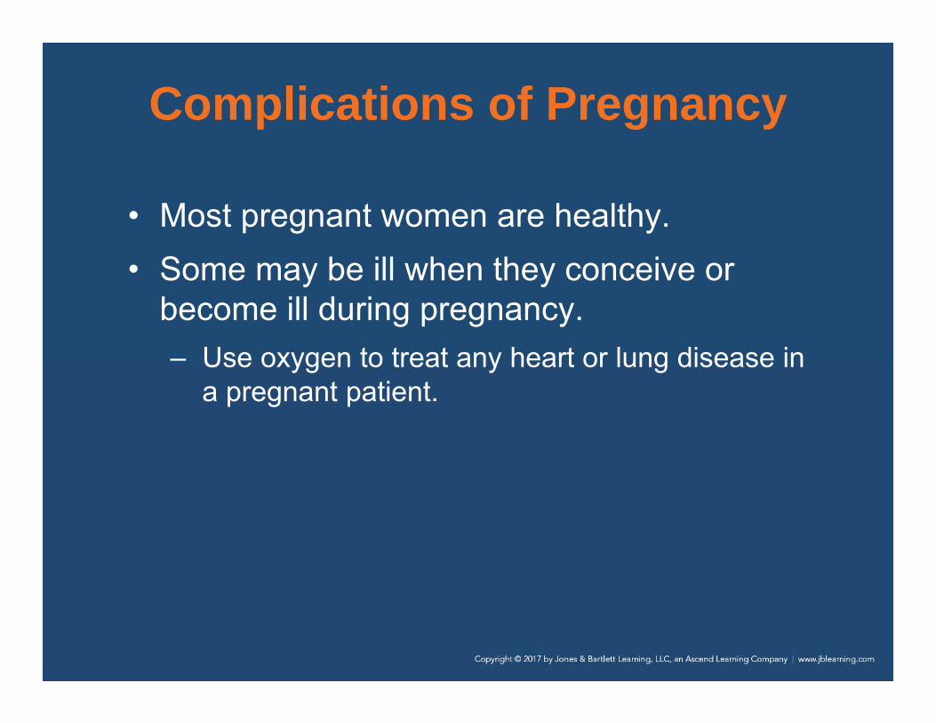
Complications of Pregnancy
• Most pregnant women are healthy.• Some may be ill when they conceive or
become ill during pregnancy.– Use oxygen to treat any heart or lung disease in
a pregnant patient.

Diabetes
• Develops during pregnancy in many women who have not had it previously
• Gestational diabetes usually resolves after delivery.
• Treatment is the same as for any other patient with diabetes.– Diet, exercise, or insulin injections

Hypertensive Disorders (1 of 3)
• Preeclampsia is pregnancy-induced hypertension– Can develop after the 20th week of gestation– Signs and symptoms include severe
hypertension, severe or persistent headache, visual abnormalities, swelling in the hands and feet, and anxiety.

Hypertensive Disorders (2 of 3)
• Eclampsia is characterized by seizures that occur as a result of hypertension.– To treat seizures caused by eclampsia:
• Lie the patient on her left side.• Maintain her airway.• Administer supplemental oxygen if
necessary.• If vomiting occurs, suction the airway.• Provide rapid transport and call for ALS.

Hypertensive Disorders (3 of 3)
• Transporting the patient on her left side can also prevent supine hypotensive syndrome.– Caused by compression of the descending
aorta and the inferior vena cava by the pregnant uterus when the patient lies supine
– Hypotension may result.
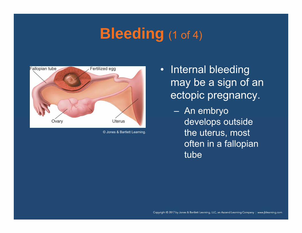
Bleeding (1 of 4)
• Internal bleeding may be a sign of an ectopic pregnancy.– An embryo
develops outside the uterus, most often in a fallopian tube
© Jones & Bartlett Learning.

Bleeding (2 of 4)
• Leading cause of maternal death in the first trimester is internal hemorrhage following rupture of an ectopic pregnancy.
• Consider the possibility in a woman who has missed a menstrual cycle and complains of sudden, severe pain in the lower abdomen.

Bleeding (3 of 4)
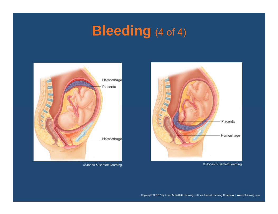
• Hemorrhage from the vagina that occurs before labor begins may be very serious.
• May be a sign of spontaneous abortion, or miscarriage.– In abruptio placenta, the placenta separates
prematurely from the wall of the uterus.– In placenta previa, the placenta develops over
and covers the cervix.
Bleeding (4 of 4)
© Jones & Bartlett Learning. © Jones & Bartlett Learning.
Abortion
• Passage of the fetus and placenta before 20 weeks
• May be spontaneous or induced• Most serious complications are bleeding
and infection• If the woman is in shock, treat and transport
her promptly to the hospital.

Abuse (1 of 2)
• Pregnant women have an increased chance of being victims of domestic violence and abuse.
• Abuse increases the chance of:– Spontaneous abortion– Premature delivery– Low birth weight

Abuse (2 of 2)
• The woman is at risk from bleeding, infection, and uterine rupture.
• Use a calm, professional approach.– Pay attention to the environment for any signs
of abuse.
• Talk to the patient in a private area, away from the potential abuser if possible.

Substance Abuse (1 of 2)
• Effects of addiction on the fetus include:– Prematurity– Low birth weight– Severe respiratory distress– Death
• Fetal alcohol syndrome describes the condition of infants born to women who have abused alcohol.

Substance Abuse (2 of 2)
• Pay special attention to your safety.• Wear eye protection, a face mask, and
gloves at all times.• Look for clues that you are dealing with an
addicted patient.• The newborn will probably need immediate
resuscitation.

Special Considerations for Trauma and Pregnancy (1 of 8)
• With a trauma call involving a pregnant woman, you have two patients:– The woman– The unborn fetus
• Trauma to a pregnant woman may have a direct effect on the fetus.

Special Considerations for Trauma and Pregnancy (2 of 8)
• Pregnant women may be victims of:– Assaults– Motor vehicle crashes– Shootings
• Pregnant women also have an increased risk of falling.

Special Considerations for Trauma and Pregnancy (3 of 8)
• Pregnant women have an increased amount of overall total blood volume and a 20% increase in heart rate.– May experience a significant amount of blood
loss before you will see signs of shock– Uterus is vulnerable to penetrating trauma and
blunt injuries.

Special Considerations for Trauma and Pregnancy (4 of 8)
• When a pregnant woman is involved in a motor vehicle crash, severe hemorrhage may occur from injuries to the uterus.– Trauma is one of the leading causes of abruptio
placenta.– Common symptoms include vaginal bleeding
and severe abdominal pain.

Special Considerations for Trauma and Pregnancy (5 of 8)
• Improper positioning of the seat belt can result in injury to a pregnant woman and the fetus. – Carefully assess a pregnant woman’s abdomen
and chest for seatbelt marks, bruising, and obvious trauma.

Special Considerations for Trauma and Pregnancy (6 of 8)
• Cardiac arrest– Your focus is the same as with other patients.– Perform CPR and provide transport.– Notify the receiving facility personnel that you
are en route with a pregnant trauma patient in cardiac arrest.

Special Considerations for Trauma and Pregnancy (7 of 8)
• Assessment and management– Your focus is on the woman.– Suspect shock based on the MOI.– Be prepared for vomiting and aspiration.– Attempt to determine the gestational age to
assist you with determining the size of the fetus and the position of the uterus.

Special Considerations for Trauma and Pregnancy (8 of 8)
• Follow these guidelines when treating a pregnant trauma patient:– Maintain an open airway.– Administer high-flow oxygen.– Ensure adequate ventilation.– Assess circulation.– Transport the patient on her left side.

Cultural Value Considerations (1 of 2)
• Cultural sensitivity is important. • Women of some cultures may have a value
system that will affect:– The choice of how they care for themselves
during pregnancy– How they have planned the childbirth process

Cultural Value Considerations (2 of 2)
• Some cultures may not permit a male health care provider to assess or examine a female patient.– Respect these differences and honor requests
from the patient.– A competent, rational adult has the right to
refuse all or any part of your assessment or care.

Teenage Pregnancy
• The United States has one of the highest teenage pregnancy rates.
• Pregnant teenagers may not know they are pregnant or may be in denial.– Respect the teenager’s privacy.– Assess and obtain her history away from her
parents.

Patient Assessment
• Childbirth is seldom an unexpected event, but there are occasions when it becomes an emergency.– Dispatcher usually asks simple questions to
determine whether birth is imminent.– Premature contractions may be caused by
trauma or medical conditions.

Scene Size-up (1 of 2)
• Scene safety– Take standard precautions.– Gloves and eye and face protection are a
minimum if delivery is already begun or is complete.
– If time allows, a gown should also be used.– Consider calling for additional resources.

Scene Size-up (2 of 2)
• Mechanism of injury/nature of illness– You will encounter pregnant patients who are
not in labor, so it is important to determine the MOI or NOI.
– Do not develop tunnel vision during a call.– Falls and necessity for spinal immobilization
must be considered.

Primary Assessment (1 of 5)
• Form a general impression.– The general impression should tell you whether
the patient is in active labor or whether you have time to assess and address other possible life threats.
– Perform a rapid examination.– When trauma or other medical problems
present, evaluate these first.

Primary Assessment (2 of 5)
• Airway and breathing– Life-threatening conditions with the woman’s
airway and breathing are usually not an issue during a birth.
– A motor vehicle crash, assault, or a medical condition may cause a life threat.
– Assess the airway and breathing to ensure they are adequate.

Primary Assessment (3 of 5)
• Circulation– External and internal bleeding are potential life
threats and should be assessed early.– Blood loss after delivery is expected, but
significant bleeding is not.– Assess for and treat life-threatening bleeding. – Assess the skin for color, temperature, and
moisture.– Check the pulse.

Primary Assessment (4 of 5)
• Transport decision– If delivery is imminent, prepare to deliver at the
scene.– Ideal place to deliver is in the ambulance or the
woman’s home.– If delivery is not imminent, prepare the patient
for transport.

Primary Assessment (5 of 5)
• Provide rapid transport for pregnant patients who:– Have significant bleeding and pain– Are hypertensive– Are having a seizure– Have an altered mental status

History Taking (1 of 2)
• Obtain a thorough obstetric history:– Her expected due date– Any complications that she is aware of– If she has been receiving prenatal care– A complete medical history

History Taking (2 of 2)
• Obtain a SAMPLE history.– Pertinent history should include questions
related specifically to prenatal care.– Determine the due date, frequency of
contractions, a history of previous pregnancies and deliveries, the possibility of multiples, and if she has taken any drugs or medications.
– If her water has broken, ask whether the fluid was green (due to meconium).

Secondary Assessment (1 of 2)
• Physical examinations– Assess the major body systems as needed.– Emphasis on the chief complaint– Assess for fetal movement.– If the patient is in labor, focus on contractions
and possible delivery.– If you suspect that delivery is imminent, check
for crowning.

Secondary Assessment (2 of 2)
• Vital signs – Pulse; respirations; skin color, temperature, and
condition; and BP– Be especially alert for tachycardia and hypo- or
hypertension.– Hypertension, even mild, may indicate more
serious problems.

Reassessment (1 of 3)
• Repeat the primary assessment.• Obtain another set of vital signs.• Check interventions and treatments
– In most cases, childbirth is a natural process that does not require your assistance.
– When childbirth is complicated by trauma, any interventions you provide the patient will benefit the fetus.

Reassessment (2 of 3)
• Communication and documentation– If delivery is imminent, notify staff at the
receiving hospital.– Provide an update on the status of the woman
and the newborn after delivery.– If delivery does not occur within 30 minutes,
provide rapid transport.
Reassessment (3 of 3)
• Communication and documentation (cont’d)– For a pregnant patient with a complaint
unrelated to childbirth, be sure to include the pregnancy status in your radio report.• The number of weeks of gestation• Her due date• Any known complications of the pregnancy
– If delivery occurs in the field, you will have two patient care reports to complete.

Stages of Labor
1. Dilation of the cervix2. Delivery of the fetus3. Delivery of the placenta

First Stage (1 of 4)
• Begins with the onset of contractions and ends when the cervix is fully dilated
• Usually the longest stage, lasting an average of 16 hours
• Uterine contractions become more regular and last about 30 to 60 seconds each.– Frequency and intensity increase

First Stage (2 of 4)
• Labor is generally longer in a primigravida (first pregnancy) than in a multigravida.
• A woman may experience preterm or false labor, or Braxton-Hicks contractions.– You should provide transport for the patient.

First Stage (3 of 4)
© Jones & Bartlett Learning.

First Stage (4 of 4)
• Some women experience a premature rupture of the amniotic sac.– Patient may or may not go into labor– Provide supportive care and transport.
• The head of the fetus descends into the woman’s pelvis as it positions for delivery.– This descent is called lightening.

Second Stage
• Begins when the fetus begins to encounter the birth canal– Ends when the newborn is born – Uterine contractions are usually closer together
and last longer.– The perineum will bulge significantly, and the
top of the fetus’s head will appear at the vaginal opening.• This is called crowning.

Third Stage
• Begins with the birth of the newborn and ends with the delivery of the placenta– The placenta must completely separate from
the uterine wall.– May take up to 30 minutes

Preparing for Delivery (1 of 10)
• Consider delivery at the scene when:– Delivery is imminent (will occur within a few
minutes)– A natural disaster, inclement weather, or other
environmental factor makes it impossible to reach the hospital
Preparing for Delivery (2 of 10)
• To determine if delivery is imminent, ask the patient:– How long have you been pregnant?– When are you due?– Is this your first baby?– Are you having contractions?
• How far apart?• How long do they last?

Preparing for Delivery (3 of 10)
• To determine if delivery is imminent, ask the patient (cont’d):– Have you had spotting or bleeding?– Has your water broken?– Do you feel as though you need to have a
bowel movement?– Do you feel the need to push?

Preparing for Delivery (4 of 10)
• To determine potential complications, ask:– Were any of your previous deliveries by
cesarean section? – Have you had problems in this or any previous
pregnancies? – Do you use drugs, drink alcohol, or take any
medications?– Is there a chance of multiple deliveries?– Does your physician expect complications?

Preparing for Delivery (5 of 10)
• If the patient says that she is about to deliver, she has to move her bowels, or feels the need to push, you should prepare for delivery.– Does she have an extremely firm abdomen?– Visually inspect the vagina to check for
crowning.

Preparing for Delivery (6 of 10)
• Once labor has begun, it cannot be slowed or stopped.– Never attempt to hold the patient’s legs
together.– Do not let her go to the bathroom.
• Remember, if you deliver at the scene, you are only assisting the woman with the delivery.

Preparing for Delivery (7 of 10)
• Your emergency vehicle should always be equipped with a sterile emergency obstetric (OB) kit.
Jones & Bartlett Learning.

Preparing for Delivery (8 of 10)
• Patient position– Preserve the patient’s privacy.– Place the patient on a firm surface padded with
blankets, sheets, and towels.– Elevate the hips about 2″ to 4″.– Support the head, neck, and upper back.– Have her keep her legs and hips flexed, with
her feet flat and her knees spread apart.

Preparing for Delivery (9 of 10)
• Preparing the delivery field– Place towels or sheets on the floor around the
delivery area.– Open the OB kit carefully.– Put on sterile gloves.– Use the sterile sheets and drapes from the OB
kit to make a sterile delivery field.

Preparing for Delivery (10 of 10)
Jones & Bartlett Learning. Jones & Bartlett Learning.

Delivery (1 of 6)
• Your partner should be at the patient’s head to comfort, soothe, and reassure.
• If the patient will allow it, apply oxygen.• Continually check for crowning.
– Some patients experience precipitous labor and birth.
– Position yourself so that you can see the perineal area at all times.

Delivery (2 of 6)
• Time the patient’s contractions.– Remind the patient to take quick, short breaths
during each contraction but not to strain.– Between contractions, encourage the patient to
rest and breathe deeply through her mouth.
• Delivering the head– Observe the head as it exits the vagina.– Support the head with your gloved hand as it
rotates.

Delivery (3 of 6)
• Delivering the head (cont’d)– Apply gentle pressure across the perineum with
a sterile gauze pad to reduce the risk of perineal tearing.
– Be prepared for the possibility of the patient having a bowel movement.
– Do not poke your fingers into the newborn’s eyes or fontanelles.
Delivery (4 of 6)
• Unruptured amniotic sac– If the amniotic sac does not rupture by the time
the head is crowning, it will appear as a fluid-filled sac emerging from the vagina.
– It will suffocate the fetus if not removed.– You may puncture the sac with a clamp or tear
it by twisting it between your fingers.– Clear the newborn’s mouth and nose
immediately.

Delivery (5 of 6)
• Umbilical cord around the neck– As soon as the head is delivered, use one finger
to feel whether the umbilical cord is wrapped around the neck.
– Usually, you can slip the cord gently over the delivered head.
– If not, you must cut it.– Once the cord is cut, attempt to speed delivery.

Delivery (6 of 6)
• Delivering the body– Once the head is born, the body usually delivers
easily.– Support the head and upper body as the
shoulders deliver.– Do not pull the fetus from the birth canal.– The newborn will be slippery and covered in
vernix caseosa.

Postdelivery Care (1 of 5)
• If the mother is able and willing, place the newborn on her abdomen so skin-to-skin contact can begin immediately.
• Dry off the newborn and wrap him or her in a blanket or towel.
• Wrap the newborn so only the face is exposed.

Postdelivery Care (2 of 5)
• You can pick up and cradle the newborn. – If local protocols specify, keep newborn at the
level of the woman’s vagina until the umbilical cord is cut.
– Always keep the head slightly downward to help prevent aspiration.
• Wipe the mouth with a sterile gauze pad as needed.

Postdelivery Care (3 of 5)
• Once the cord has stopped pulsing, clamp and cut the cord.
• Obtain the 1-minute Apgar score. • Delivery of the placenta
– Your job is only to assist.– The placenta delivers itself, usually within a few
minutes of the birth.– Never pull on the end of the umbilical cord.

Postdelivery Care (4 of 5)
• You can help to slow bleeding by gently massaging the woman’s abdomen with a firm, circular, “kneading” motion.
© University of Maryland Shock Trauma Center/MIEMSS.

Postdelivery Care (5 of 5)
• Record the time of birth in your patient care report.
• The following are emergency situations:– More than 30 minutes elapse and the placenta
has not delivered– There is more than 500 mL of bleeding before
delivery of the placenta.– There is significant bleeding after the delivery of
the placenta.

Neonatal Assessment and Resuscitation (1 of 4)
• Follow standard precautions.• Always put on gloves before handling a
newborn.– Newborn will usually begin breathing
spontaneously within 15 to 30 seconds after birth.
– Heart rate will be 120 beats/min or higher.

Neonatal Assessment and Resuscitation (2 of 4)
• If you do not observe these responses:– Gently tap or flick the soles of the feet or rub the
back.
• Many newborns require some form of stimulation, including:– Positioning the airway, drying, warming,
suctioning, or tactile stimulation

Neonatal Assessment and Resuscitation (3 of 4)
© Jones & Bartlett Learning.

Neonatal Assessment and Resuscitation (4 of 4)
• To maximize the effects of these measures:– Position the newborn on his or her back with the
head down and the neck slightly extended.– If necessary, suction the mouth and then the
nose.– In addition to drying the head, back, and body
with dry towels, rub the back and flick or slap the soles of the feet.

Additional Resuscitation Efforts (1 of 5)
• Observe the newborn for spontaneous respirations, skin color, and movement of the extremities.
• Evaluate the heart rate at the base of the umbilical cord or brachial artery or by listening to the newborn’s chest with a stethoscope.– The heart rate is the most important measure in
determining the need for further resuscitation.

Additional Resuscitation Efforts (2 of 5)
© Jones & Bartlett Learning.

Additional Resuscitation Efforts (3 of 5)
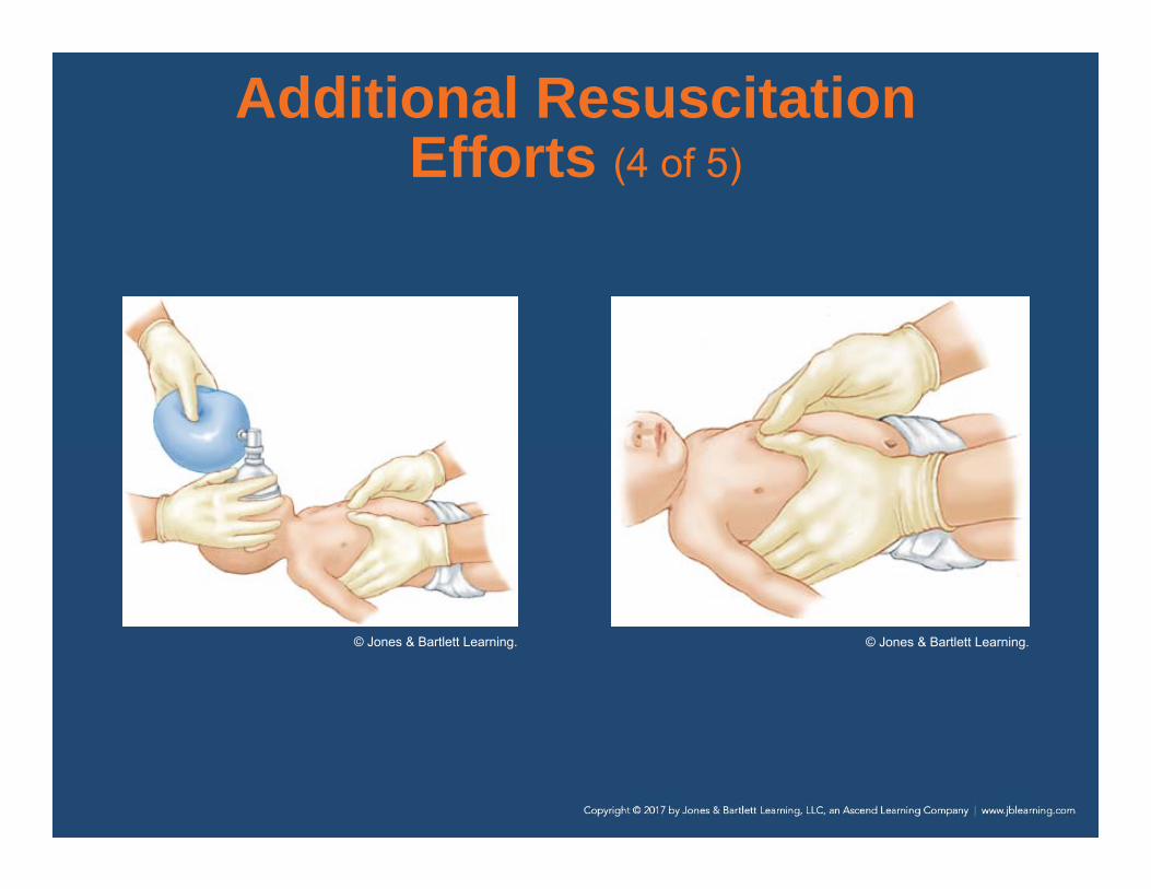
• If chest compressions are required, use the hand-encircling technique for two-person resuscitation.– Perform BVM ventilation during a pause after
every third compression, using a ratio of 3:1.– 120 actions per minute (90 compressions and
30 ventilations)
Additional Resuscitation Efforts (4 of 5)
© Jones & Bartlett Learning. © Jones & Bartlett Learning.

Additional Resuscitation Efforts (5 of 5)
• Any newborn that requires more than routine resuscitation requires transport to a hospital with a Level III neonatal ICU.
• About 12% to 16% of deliveries are complicated by the presence of meconium.– Consider quickly suctioning the newborn’s
mouth, then nose after delivery before providing rescue ventilations.
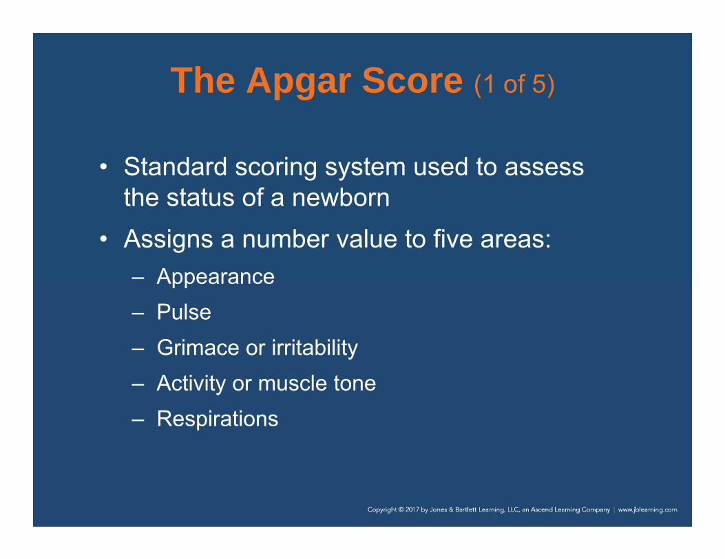
The Apgar Score (1 of 5)
• Standard scoring system used to assess the status of a newborn
• Assigns a number value to five areas:– Appearance– Pulse– Grimace or irritability– Activity or muscle tone– Respirations

The Apgar Score (2 of 5)
• The total of the five numbers is the Apgar score.– A perfect score is 10.– Calculate the Apgar score at 1 minute and 5
minutes after birth.

The Apgar Score (3 of 5)
© Jones & Bartlett Learning.

The Apgar Score (4 of 5)
• Assessing a newborn– Calculate the Apgar score.– Stimulation should result in an immediate
increase in respirations.– If the newborn is breathing well, assess the
pulse.– Assess oxygenation via pulse oximetry and
observe for central cyanosis.

The Apgar Score (5 of 5)
• Request a second unit if the newborn is in distress and will require resuscitation.
• In situations where assisted ventilation is required, use a newborn BVM.
• If the newborn does not begin breathing on his or her own or does not have an adequate heart rate, continue CPR and rapidly transport.

Breech Delivery (1 of 4)
• Most infants are born headfirst.
• Occasionally, the buttocks are delivered first.
• Called a breech presentation
© Jones & Bartlett Learning.

Breech Delivery (2 of 4)
• Breech deliveries usually take longer, so you will often have time to transport the pregnant woman to the hospital.– If the buttocks have passed through the vagina,
the delivery has begun.– Provide emergency care and call for ALS
backup.– Consult medical control to guide you.

Breech Delivery (3 of 4)
• Preparing for a breech delivery is the same as for a normal childbirth.– Position the pregnant woman.– Prepare the OB kit.– Place yourself and your partner as you would
normally.– Allow the buttocks and legs to deliver
spontaneously, supporting them with your hand.

Breech Delivery (4 of 4)
• Preparing for a breech delivery (cont’d)– The head is almost always facedown and
should be allowed to deliver spontaneously.– Make a “V” with your gloved fingers and position
them in the vagina to keep the walls from compressing the fetus’s airway.

Presentation Complications (1 of 4)
• On rare occasions, the presenting part of the fetus is a single arm, leg, or foot.– Called a limb
presentation
© Jones & Bartlett Learning.

Presentation Complications (2 of 4)
• An fetus with a limb presentation cannot be delivered in the field.– Usually surgery is needed.– Transport immediately.– If a limb is protruding, cover it with a sterile
towel.– Never try to push it in or pull on it.– Place the patient on her back, with her head
down and pelvis elevated.

Presentation Complications (3 of 4)
• Prolapse of the umbilical cord must be treated in the hospital.– The umbilical cord
comes out of the vagina before the fetus.
© Jones & Bartlett Learning.
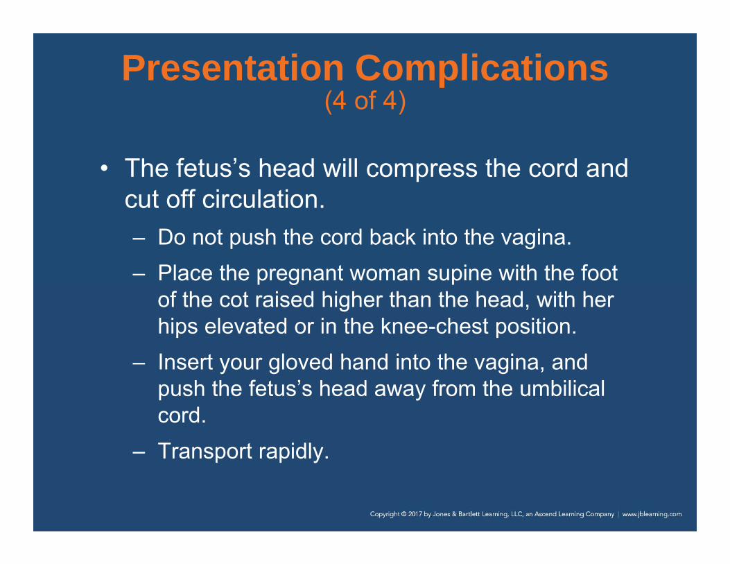
Presentation Complications (4 of 4)
• The fetus’s head will compress the cord and cut off circulation.– Do not push the cord back into the vagina.– Place the pregnant woman supine with the foot
of the cot raised higher than the head, with her hips elevated or in the knee-chest position.
– Insert your gloved hand into the vagina, and push the fetus’s head away from the umbilical cord.
– Transport rapidly.

Spina Bifida
• Developmental defect in which a portion of the spinal cord or meninges may protrude outside of the vertebrae– Seen on the newborn’s back– Cover the open area of the spinal cord with a
sterile, moist dressing.– Maintenance of body temperature is important
when applying moist dressings.

Multiple Gestation (1 of 2)
• Twins occur once in every 30 births.– Always be prepared for more than one
resuscitation, and call for assistance.
• Twins are smaller than single fetuses, and delivery is typically not difficult.– About 10 minutes after the first birth,
contractions will begin again, and the birth process will repeat itself.
– Second one is usually be born within 45 minutes of the first.

Multiple Gestation (2 of 2)
• The procedure is the same as that for a single fetus.– There may only be one placenta, or there may
be two.
• Record the time of birth of each twin separately.
• Twins may be so small that they look premature.– Handle carefully and keep them warm.

Premature Birth (1 of 3)
• A normal, full-term, single newborn will weigh about 7 lb at birth.
• Any newborn who delivers before 8 months (36 weeks) or weighs less than 5 lb at birth is considered premature.

Premature Birth (2 of 3)
• A premature newborn is smaller and thinner, and the head is proportionately larger.– The vernix caseosa
will be absent or minimal.
– There will be less body hair.
© American Academy of Orthopaedic Surgeons.

Premature Birth (3 of 3)
• Premature newborns require special care to survive.– Often require resuscitation efforts, which should
be performed unless it is physically impossible– With such care, premature newborns as small
as 1 lb have survived and developed normally.

Postterm Pregnancy (1 of 2)
• Pregnancies lasting longer than 42 weeks• Fetuses can be larger, sometimes weighing
10 lb or more.• Can lead to problems with the woman and
fetus– A more difficult labor and delivery

Postterm Pregnancy (2 of 2)
• Problems (cont’d):– Increased chance of injury to the fetus– Increased likelihood of cesarean section– Woman is at risk for perineal tears and
infection.– Postterm newborns have increased risks of
meconium aspirations, infection, and being stillborn.
– Newborns may not have developed normally.

Fetal Demise
• You may deliver an fetus who died in the woman’s uterus before labor.– Onset of labor may be premature, but labor will
progress normally in most cases.– If an intrauterine infection caused the demise,
you may note a foul odor.– Do not attempt to resuscitate an obviously dead
neonate.

Delivery Without Sterile Supplies (1 of 2)
• You may have to deliver an newborn without a sterile OB kit.
• You should always have eye protection, gloves, and a mask with you.
• Carry out the delivery as if sterile supplies were available.– Use freshly laundered sheets and towels.
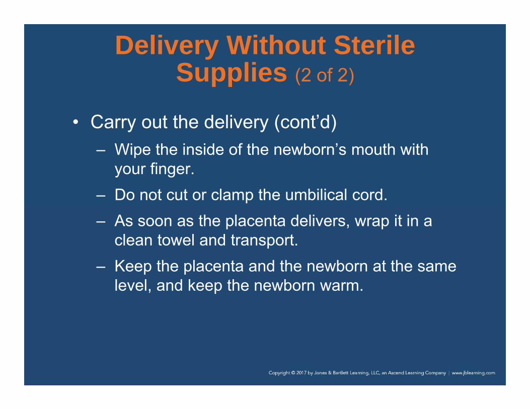
Delivery Without Sterile Supplies (2 of 2)
• Carry out the delivery (cont’d)– Wipe the inside of the newborn’s mouth with
your finger.– Do not cut or clamp the umbilical cord.– As soon as the placenta delivers, wrap it in a
clean towel and transport.– Keep the placenta and the newborn at the same
level, and keep the newborn warm.
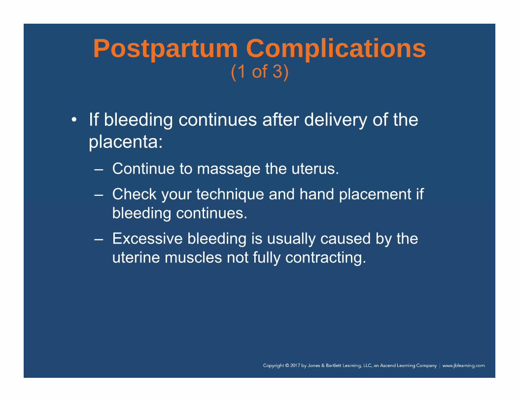
Postpartum Complications (1 of 3)
• If bleeding continues after delivery of the placenta:– Continue to massage the uterus.– Check your technique and hand placement if
bleeding continues.– Excessive bleeding is usually caused by the
uterine muscles not fully contracting.

Postpartum Complications (2 of 3)
• Cover the vagina with a sterile pad.– Change the pad as often as possible.– Do not discard any blood-soaked pads.
• Administer oxygen, monitor vital signs, and transport the patient immediately.

Postpartum Complications (3 of 3)
• Postpartum patients are at an increased risk of an embolism.– Most commonly a pulmonary embolism– Results from a clot that travels through the
bloodstream and becomes lodged in the pulmonary circulation
– Consider when a woman complains of sudden difficulty breathing or shortness of breath following delivery

Review
1. The first stage of labor ends when:A. the presenting part of the baby is visible.B. contractions are less than 10 minutes apart.C. the mother experiences her first contraction. D. the amniotic sac ruptures and labor pains
begin.

Review
Answer: ARationale: The first stage of labor begins with the onset of contractions and ends when the cervix is fully dilated. However, since cervical dilation cannot be assessed in the field, the first stage of labor is considered over when the presenting part of the baby is visible at the vaginal opening (crowning).

Review (1 of 2)
1. The first stage of labor ends when:A. the presenting part of the baby is visible.
Rationale: Correct answerB. contractions are less than 10 minutes apart.
Rationale: True labor is when the frequency and intensity of contractions increase and is part of the first stage of labor.
Review (2 of 2)
1. The first stage of labor ends when:C. the mother experiences her first contraction.
Rationale: This is the beginning of the first stage of labor.
D. the amniotic sac ruptures and labor pains begin. Rationale: This is considered to be a part of the first stage of labor.

Review
2. A 23-year-old woman, who is 24 weeks pregnant with her first baby, complains of edema to her hands, a headache, and visual disturbances. When you assess her vital signs, you note that her blood pressure is 160/94 mm Hg. She is MOST likely experiencing:A. eclampsia. B. preeclampsia. C. a hypertensive crisis. D. chronic water retention.

Review
Answer: BRationale: Preeclampsia—also called pregnancy-induced hypertension—usually develops after the 20th week of gestation and most commonly affects primagravida (first pregnancy) patients. It is characterized by a headache, visual disturbances, edema of the hands and feet, anxiety, and high blood pressure. Preeclampsia can lead to eclampsia, a life-threatening condition that is characterized by seizures.

Review (1 of 2)
2. A 23-year-old woman, who is 24 weeks pregnant with her first baby, complains of edema to her hands, a headache, and visual disturbances. When you assess her vital signs, you note that her blood pressure is 160/94 mm Hg. She is MOST likely experiencing:A. eclampsia.
Rationale: Eclampsia is a seizure that results from severe hypertension.
B. preeclampsia. Rationale: Correct answer
Review (2 of 2)
2. A 23-year-old woman, who is 24 weeks pregnant with her first baby, complains of edema to her hands, a headache, and visual disturbances. When you assess her vital signs, you note that her blood pressure is 160/94 mm Hg. She is MOST likely experiencing:
C. a hypertensive crisis. Rationale: This is a severe, sudden increase in blood pressure, typically greater than 110 diastolic, that can lead to a stroke.
D. chronic water retention. Rationale: This is a fluid imbalance usually caused by too much sodium in the body.

Review
3. You are transporting a woman who is 8 months pregnant. To prevent supine hypotensive syndrome, how should you position this patient?A. On her right sideB. SupineC. Semi-Fowler’sD. On her left side

Review
Answer: DRationale: To prevent supine hypotensive syndrome, the patient must be positioned on her left side. This stops the weight of the fetus from compressing the inferior vena cava, which can cause low blood pressure.

Review (1 of 2)
3. You are transporting a woman who is 8 months pregnant. To prevent supine hypotensive syndrome, how should you position this patient?A. On her right side
Rationale: The patient should be transported on her left side.
B. SupineRationale: Lying the patient supine will cause hypotension.

Review (2 of 2)
3. You are transporting a woman who is 8 months pregnant. To prevent supine hypotensive syndrome, how should you position this patient?C. Semi-Fowler’s
Rationale: The patient should be transported on her left side.
D. On her left sideRationale: Correct answer

Review
4. Immediately after delivery of the infant’s head, you should:A. suction the baby’s mouth and then nose.B. suction the baby’s nose and then mouth.C. assess the baby’s breathing effort and skin
color. D. check the position of the umbilical cord.

Review
Answer: DRationale: Immediately following delivery of the infant’s head, you should check the position of the umbilical cord to make sure it is not wrapped around the baby’s neck (nuchal cord). If a nuchal cord is not present, suction the infant’s mouth and nose.

Review (1 of 2)
4. Immediately after delivery of the infant’s head, you should:A. suction the baby’s mouth and then nose.
Rationale: After EMS has confirmed that the cord is not around the infant’s head, this should be performed.
B. suction the baby’s nose and then mouth.Rationale: After EMS has confirmed that the cord is not around the infant’s head, suctioning of the mouth and then the nose should be performed.

Review (2 of 2)
4. Immediately after delivery of the infant’s head, you should:C. assess the baby’s breathing effort and skin
color. Rationale: This cannot be performed until the entire infant has been delivered completely.
D. check the position of the umbilical cord. Rationale: Correct answer

Review
5. Upon delivery of the baby’s head, you note that the umbilical cord is wrapped around its neck. You should: A. immediately clamp and cut the cord.B. make one attempt to slide the cord over the
head. C. keep the cord moist and transport as soon as
possible. D. give the mother high-flow oxygen and
transport rapidly.

Review
Answer: BRationale: If the umbilical cord is wrapped around the baby’s neck (nuchal cord), you should make one attempt to gently remove the cord from around the baby’s neck. If this is not possible, the cord should be clamped and cut. Keep the cord moist, administer high-flow oxygen to the mother, and transport at once.

Review (1 of 2)
5. Upon delivery of the baby’s head, you note that the umbilical cord is wrapped around its neck. You should: A. immediately clamp and cut the cord.
Rationale: Do this only after an attempt is made to slide the cord over the infant’s head.
B. make one attempt to slide the cord over the head. Rationale: Correct answer

Review (2 of 2)
5. Upon delivery of the baby’s head, you note that the umbilical cord is wrapped around its neck. You should: C. keep the cord moist and transport as soon as
possible. Rationale: This is the treatment for deliveries where the cord presents and not the infant’s head.
D. give the mother high-flow oxygen and transport rapidly. Rationale: Do this only after an attempt to slide the cord over the infant’s head.

Review
6. The need for and extent of newborn resuscitation is based on:A. the 1-minute Apgar score. B. the gestational age of the newborn. C. the newborn’s response to oxygen. D. respiratory effort, heart rate, and color.

Review
Answer: DRationale: The need for and extent of newborn resuscitation is based on respiratory effort, heart rate, and skin color. The Apgar score is not used to determine if resuscitation is needed; the first score is not assigned until the newborn is 1 minute of age. Resuscitation, if needed, should commence immediately.

Review (1 of 2)
6. The need for and extent of newborn resuscitation is based on:A. the 1-minute Apgar score.
Rationale: The Apgar score is not used to determine if resuscitation is needed.
B. the gestational age of the newborn. Rationale: A premature gestational age may indicate a greater risk for the infant, but does not indicate if resuscitation is required.

Review (2 of 2)
6. The need for and extent of newborn resuscitation is based on:C. the newborn’s response to oxygen.
Rationale: Oxygen response is evaluated by respiratory rate, heart rate, and color.
D. respiratory effort, heart rate, and color. Rationale: Correct answer
Review
7. The 1-minute Apgar score of a newborn reveals that the baby has a heart rate of 90 beats/min, a pink body but blue hands and feet, and rapid respirations. The baby cries when the soles of its feet are flicked and resists attempts to straighten its legs. You should assign an Apgar score of: A. 4.B. 6.C. 8.D. 9.

Review
Answer: CRationale: The Apgar score, which is obtained at 1 and 5 minutes after birth, assigns a numeric value to the following five areas: appearance, pulse, grimace, activity, and respirations. A heart rate below 100 beats/min is assigned a 1; a pink body with blue hands and feet is a 1; rapid respirations is a 2; a strong cry in reaction to a painful stimulus is a 2; and resistance against an attempt to straighten the hips and knees is a 2. Added together, the Apgar score for this infant is 8.

Review (1 of 2)
7. The 1-minute Apgar score of a newborn reveals that the baby has a heart rate of 90 beats/min, a pink body but blue hands and feet, and rapid respirations. The baby cries when the soles of its feet are flicked and resists attempts to straighten its legs. You should assign an Apgar score of: A. 4
Rationale: The correct score is 8.B. 6
Rationale: The correct score is 8.

Review (2 of 2)
7. The 1-minute Apgar score of a newborn reveals that the baby has a heart rate of 90 beats/min, a pink body but blue hands and feet, and rapid respirations. The baby cries when the soles of its feet are flicked and resists attempts to straighten its legs. You should assign an Apgar score of: C. 8
Rationale: Correct answerD. 9
Rationale: The correct score is 8.

Review
8. The MOST effective way to prevent cardiopulmonary arrest in a newborn is to:A. rapidly increase its body temperature.B. allow it to remain slightly hypothermic. C. ensure adequate oxygenation and ventilation. D. start CPR if the heart rate is less than 100
beats/min.
Review
Answer: CRationale: Cardiopulmonary arrest in infants and children (including newborns) is most often the result of respiratory arrest. Therefore, ensuring adequate oxygenation and ventilation at all times is critical. It is also important to maintain the infant’s body temperature and to prevent hypothermia.

Review (1 of 2)
8. The MOST effective way to prevent cardiopulmonary arrest in a newborn is to:A. rapidly increase its body temperature.
Rationale: It is important to maintain the infant’s body temperature and prevent hypothermia.
B. allow it to remain slightly hypothermic. Rationale: Hypothermia and shivering will deplete the infant’s glucose and cause hypoglycemia.

Review (2 of 2)
8. The MOST effective way to prevent cardiopulmonary arrest in a newborn is to:C. ensure adequate oxygenation and ventilation.
Rationale: Correct answerD. start CPR if the heart rate is less than 100
beats/min. Rationale: Start CPR when the heart rate is less than 60 beats/min and not increasing with adequate ventilations.
Review
9. While assisting a woman in labor, you visualize her vaginal area and see an arm protruding from her vagina. She tells you that she feels the urge to push. You should: A. cover the arm with a sterile towel and
transport immediately. B. encourage her to keep pushing as you
prepare for rapid transport. C. insert your gloved fingers into the vagina and
try to turn the baby.D. instruct the mother to keep pushing and give
her high-flow oxygen.
Review
Answer: ARationale: Limb presentations do not deliver in the field—period! If the mother feels the urge to push, instruct her to stop; she should pant instead. Cover the protruding limb with a sterile towel, administer high-flow oxygen to the mother, and transport immediately. Delivery must take place in the hospital.
Review (1 of 2)
9. While assisting a woman in labor, you visualize her vaginal area and see an arm protruding from her vagina. She tells you that she feels the urge to push. You should: A. cover the arm with a sterile towel and
transport immediately. Rationale: Correct answer
B. encourage her to keep pushing as you prepare for rapid transport. Rationale: EMS cannot successfully deliver such a presentation in the field.
Review (2 of 2)
9. While assisting a woman in labor, you visualize her vaginal area and see an arm protruding from her vagina. She tells you that she feels the urge to push. You should: C. insert your gloved fingers into the vagina and try
to turn the baby.Rationale: You should only do this to create an airway for the infant in a breech presentation.
D. instruct the mother to keep pushing and give her high-flow oxygen. Rationale: EMS cannot successfully deliver such a presentation in the field.

Review
10. A newborn is considered to be “term” if it is born after ____ weeks and before ____ weeks. A. 34, 37 B. 37, 42 C. 38, 44 D. 39, 43
Review
Answer: BRationale: A term gestation ranges between 37 and 42 weeks. An infant who is born before 37 weeks gestation (or weighs less than 5 lb, regardless of gestational age) is considered premature. An infant born after 42 weeks is considered past due.
Review (1 of 2)
10. A newborn is considered to be “term” if it is born after ____ weeks and before ____ weeks. A. 34, 37
Rationale: A newborn is considered premature if he or she is born before 37 weeks.
B. 37, 42 Rationale: Correct answer

Review (2 of 2)
10. A newborn is considered to be “term” if it is born after ____ weeks and before ____ weeks. C. 38, 44
Rationale: A newborn is considered past due if he or she is born after 42 weeks.
D. 39, 43Rationale: A newborn is considered past due if he or she is born after 42 weeks.