Embed Size (px)
Citation preview

1STN E-Library 2012
5_Traumatic Brain Injury
2STN E-Library 2012
5_Traumatic Brain Injury
3STN E-Library 2012
5_Traumatic Brain Injury
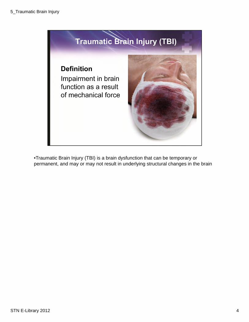
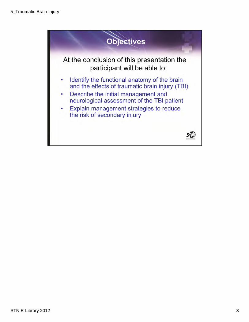
•Traumatic Brain Injury (TBI) is a brain dysfunction that can be temporary or permanent, and may or may not result in underlying structural changes in the brain
4STN E-Library 2012
5_Traumatic Brain Injury
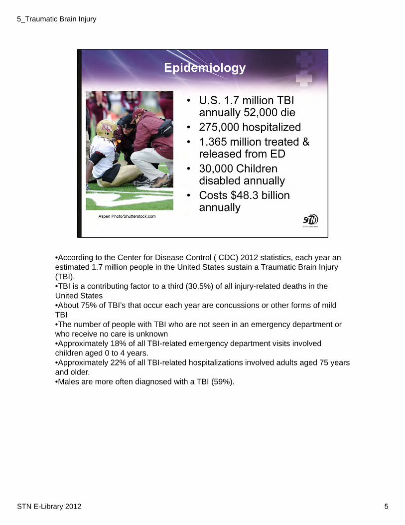
•According to the Center for Disease Control ( CDC) 2012 statistics, each year an estimated 1.7 million people in the United States sustain a Traumatic Brain Injury (TBI).•TBI is a contributing factor to a third (30.5%) of all injury-related deaths in the United States•About 75% of TBI’s that occur each year are concussions or other forms of mild TBI•The number of people with TBI who are not seen in an emergency department or who receive no care is unknown•Approximately 18% of all TBI-related emergency department visits involved children aged 0 to 4 years.•Approximately 22% of all TBI-related hospitalizations involved adults aged 75 years and older.•Males are more often diagnosed with a TBI (59%).
5STN E-Library 2012
5_Traumatic Brain Injury
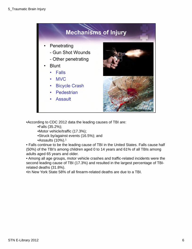
•According to CDC 2012 data the leading causes of TBI are: •Falls (35.2%); •Motor vehicle/traffic (17.3%); •Struck by/against events (16.5%); and •Assaults (10%).1
• Falls continue to be the leading cause of TBI in the United States. Falls cause half (50%) of the TBI’s among children aged 0 to 14 years and 61% of all TBIs among adults aged 65 years and older.• Among all age groups, motor vehicle crashes and traffic-related incidents were the second leading cause of TBI (17.3%) and resulted in the largest percentage of TBI-related deaths (31.8%). •In New York State 58% of all firearm-related deaths are due to a TBI.
6STN E-Library 2012
5_Traumatic Brain Injury
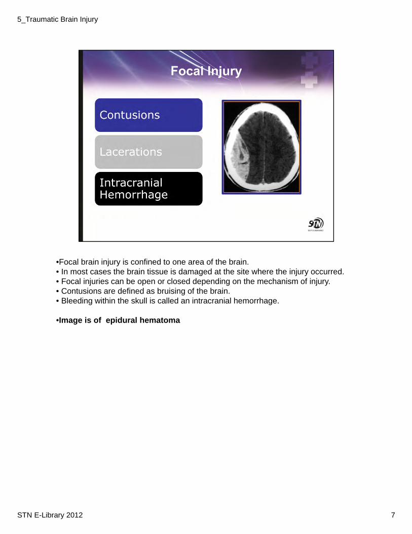
•Focal brain injury is confined to one area of the brain.• In most cases the brain tissue is damaged at the site where the injury occurred.• Focal injuries can be open or closed depending on the mechanism of injury.• Contusions are defined as bruising of the brain. • Bleeding within the skull is called an intracranial hemorrhage.
•Image is of epidural hematoma
7STN E-Library 2012
5_Traumatic Brain Injury
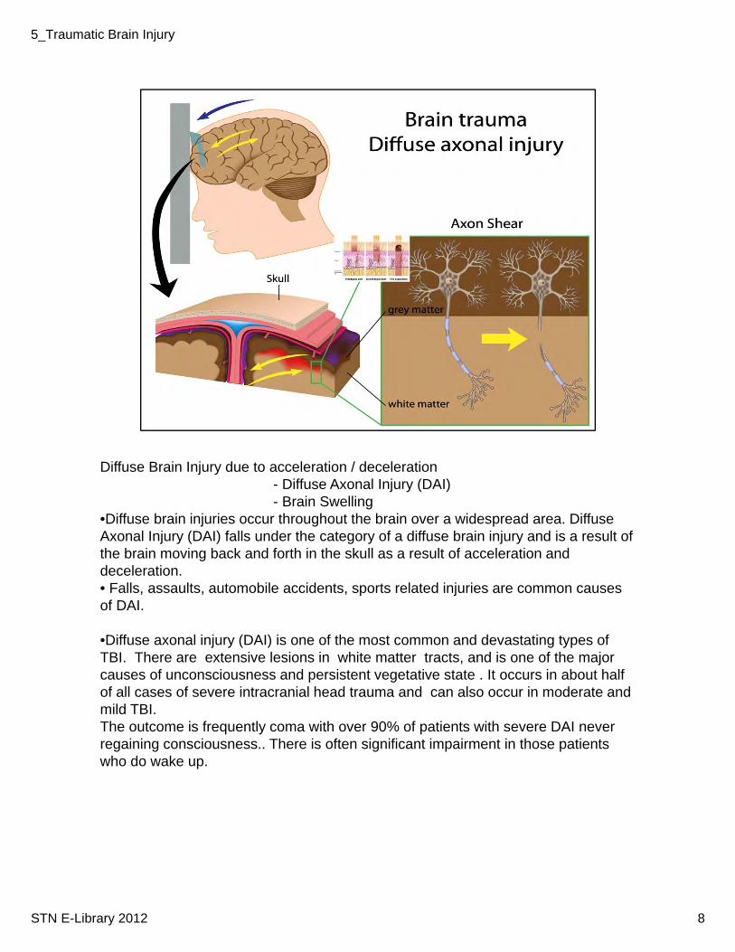
Diffuse Brain Injury due to acceleration / deceleration - Diffuse Axonal Injury (DAI)- Brain Swelling
•Diffuse brain injuries occur throughout the brain over a widespread area. Diffuse Axonal Injury (DAI) falls under the category of a diffuse brain injury and is a result of the brain moving back and forth in the skull as a result of acceleration and deceleration. • Falls, assaults, automobile accidents, sports related injuries are common causes of DAI.
•Diffuse axonal injury (DAI) is one of the most common and devastating types of TBI. There are extensive lesions in white matter tracts, and is one of the major causes of unconsciousness and persistent vegetative state . It occurs in about half of all cases of severe intracranial head trauma and can also occur in moderate and mild TBI.The outcome is frequently coma with over 90% of patients with severe DAI never regaining consciousness.. There is often significant impairment in those patients who do wake up.
8STN E-Library 2012
5_Traumatic Brain Injury
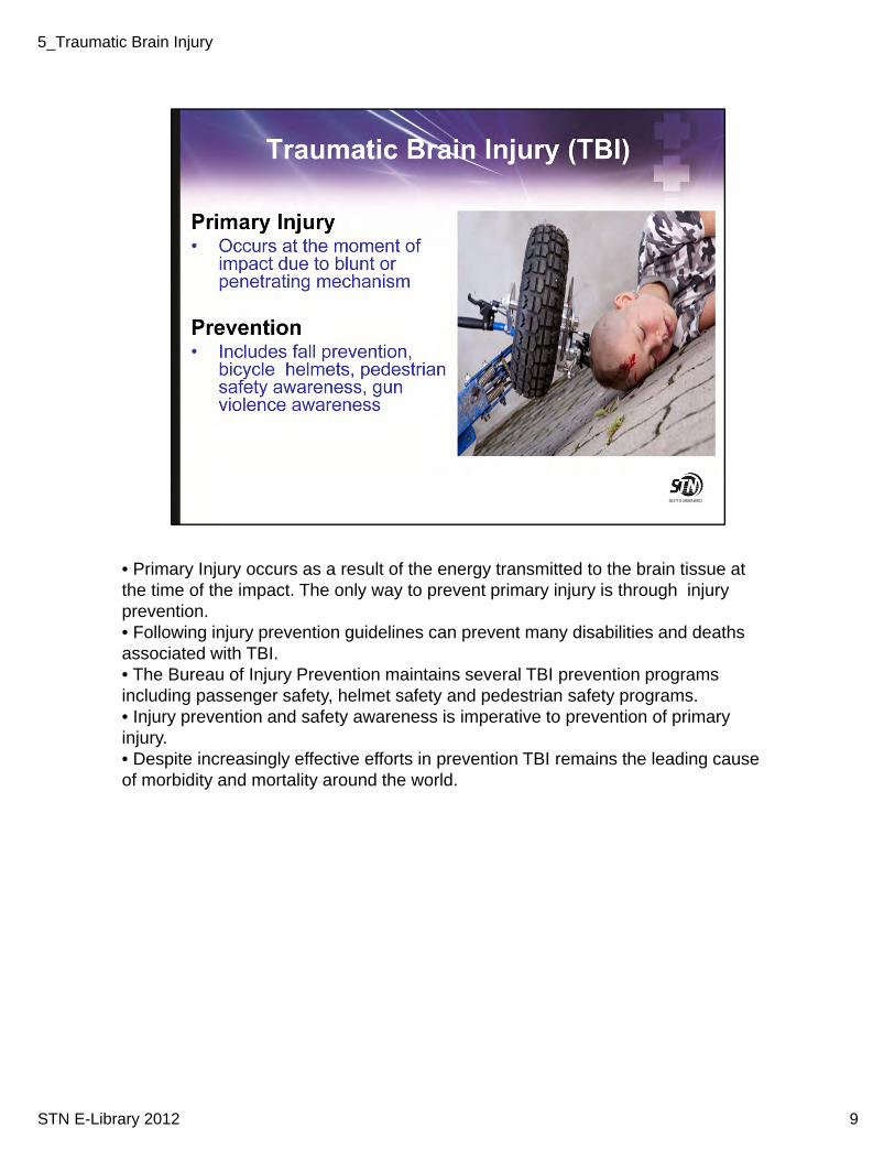
• Primary Injury occurs as a result of the energy transmitted to the brain tissue at the time of the impact. The only way to prevent primary injury is through injury prevention. • Following injury prevention guidelines can prevent many disabilities and deaths associated with TBI.• The Bureau of Injury Prevention maintains several TBI prevention programs including passenger safety, helmet safety and pedestrian safety programs. • Injury prevention and safety awareness is imperative to prevention of primary injury. • Despite increasingly effective efforts in prevention TBI remains the leading cause of morbidity and mortality around the world.
9STN E-Library 2012
5_Traumatic Brain Injury
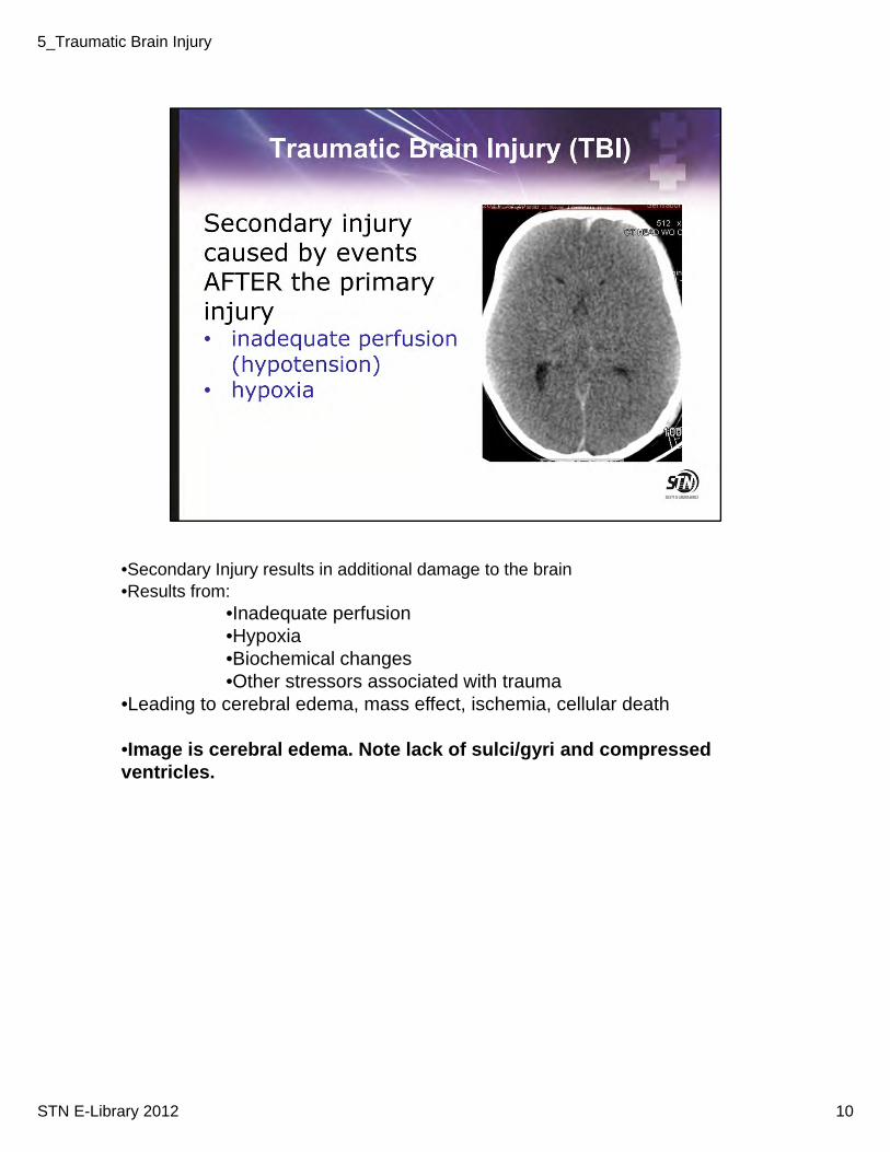
•Secondary Injury results in additional damage to the brain•Results from:
•Inadequate perfusion •Hypoxia•Biochemical changes•Other stressors associated with trauma
•Leading to cerebral edema, mass effect, ischemia, cellular death
•Image is cerebral edema. Note lack of sulci/gyri and compressed ventricles.
10STN E-Library 2012
5_Traumatic Brain Injury
•Key to Improving Survival is PREVENTING Secondary Injury
11STN E-Library 2012
5_Traumatic Brain Injury
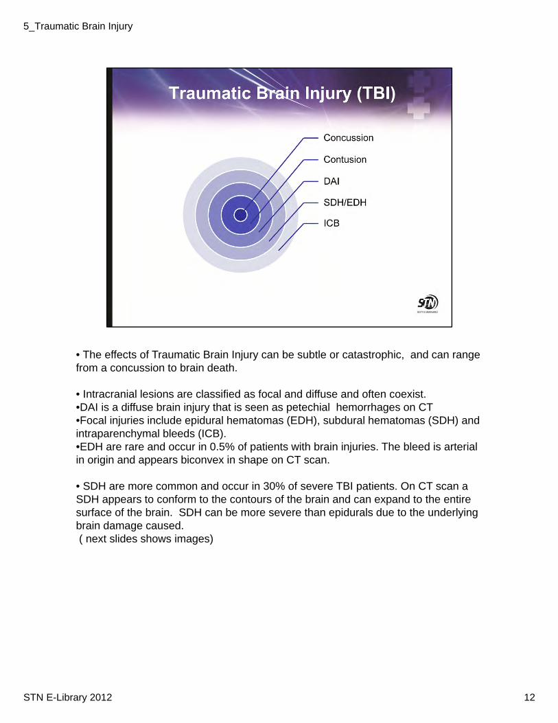
• The effects of Traumatic Brain Injury can be subtle or catastrophic, and can range from a concussion to brain death.
• Intracranial lesions are classified as focal and diffuse and often coexist. •DAI is a diffuse brain injury that is seen as petechial hemorrhages on CT•Focal injuries include epidural hematomas (EDH), subdural hematomas (SDH) and intraparenchymal bleeds (ICB).•EDH are rare and occur in 0.5% of patients with brain injuries. The bleed is arterial in origin and appears biconvex in shape on CT scan.
• SDH are more common and occur in 30% of severe TBI patients. On CT scan a SDH appears to conform to the contours of the brain and can expand to the entire surface of the brain. SDH can be more severe than epidurals due to the underlying brain damage caused.( next slides shows images)
12STN E-Library 2012
5_Traumatic Brain Injury
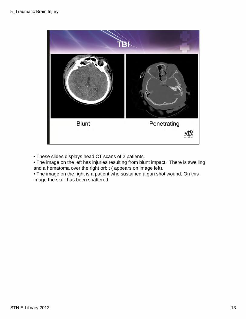
• These slides displays head CT scans of 2 patients. • The image on the left has injuries resulting from blunt impact. There is swelling and a hematoma over the right orbit ( appears on image left). • The image on the right is a patient who sustained a gun shot wound. On this image the skull has been shattered
13STN E-Library 2012
5_Traumatic Brain Injury
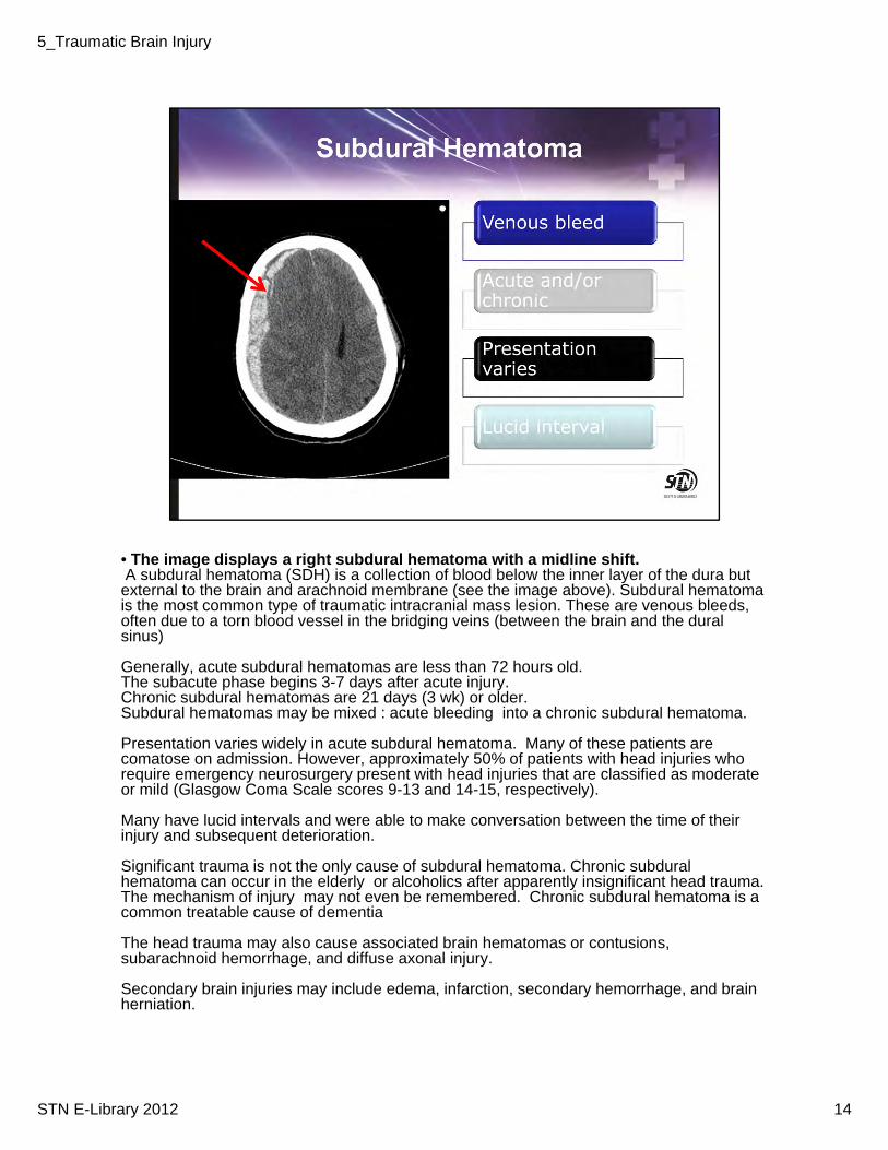
• The image displays a right subdural hematoma with a midline shift. A subdural hematoma (SDH) is a collection of blood below the inner layer of the dura but external to the brain and arachnoid membrane (see the image above). Subdural hematoma is the most common type of traumatic intracranial mass lesion. These are venous bleeds, often due to a torn blood vessel in the bridging veins (between the brain and the dural sinus)
Generally, acute subdural hematomas are less than 72 hours old. The subacute phase begins 3-7 days after acute injury.Chronic subdural hematomas are 21 days (3 wk) or older.Subdural hematomas may be mixed : acute bleeding into a chronic subdural hematoma.
Presentation varies widely in acute subdural hematoma. Many of these patients are comatose on admission. However, approximately 50% of patients with head injuries who require emergency neurosurgery present with head injuries that are classified as moderate or mild (Glasgow Coma Scale scores 9-13 and 14-15, respectively).
Many have lucid intervals and were able to make conversation between the time of their injury and subsequent deterioration.
Significant trauma is not the only cause of subdural hematoma. Chronic subdural hematoma can occur in the elderly or alcoholics after apparently insignificant head trauma. The mechanism of injury may not even be remembered. Chronic subdural hematoma is a common treatable cause of dementia
The head trauma may also cause associated brain hematomas or contusions, subarachnoid hemorrhage, and diffuse axonal injury.
Secondary brain injuries may include edema, infarction, secondary hemorrhage, and brain herniation.
14STN E-Library 2012
5_Traumatic Brain Injury
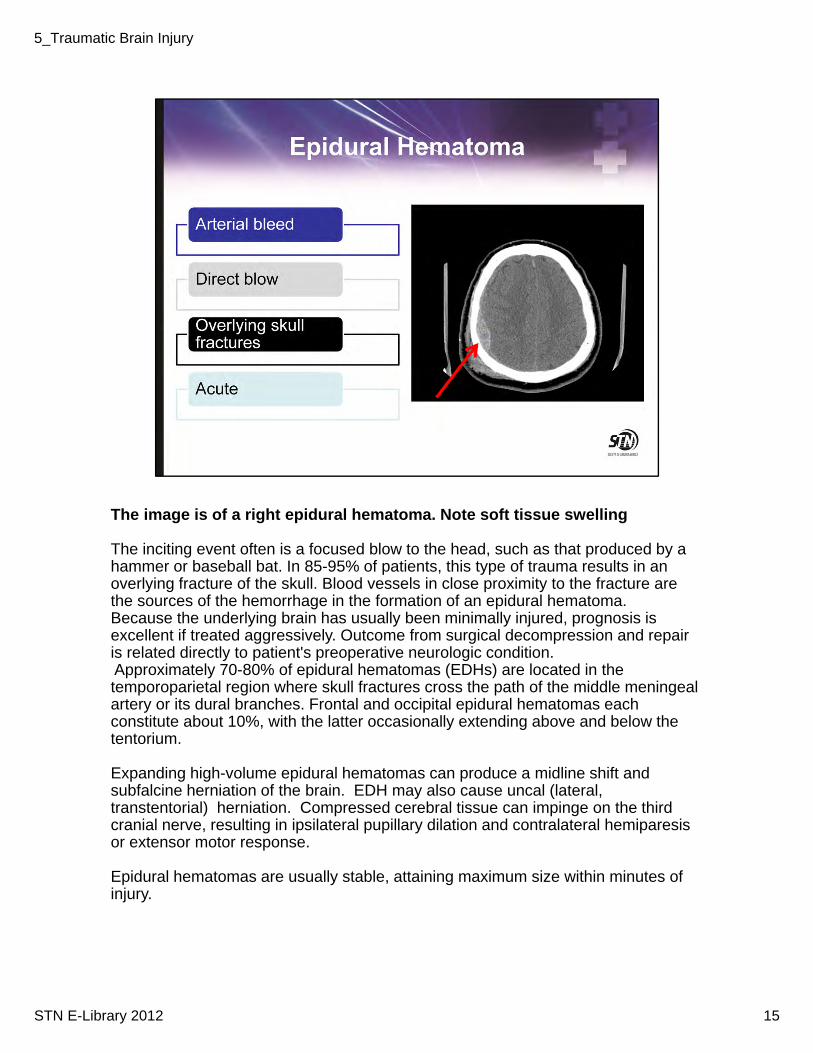
The image is of a right epidural hematoma. Note soft tissue swelling
The inciting event often is a focused blow to the head, such as that produced by a hammer or baseball bat. In 85-95% of patients, this type of trauma results in an overlying fracture of the skull. Blood vessels in close proximity to the fracture are the sources of the hemorrhage in the formation of an epidural hematoma. Because the underlying brain has usually been minimally injured, prognosis is excellent if treated aggressively. Outcome from surgical decompression and repair is related directly to patient's preoperative neurologic condition.Approximately 70-80% of epidural hematomas (EDHs) are located in the temporoparietal region where skull fractures cross the path of the middle meningeal artery or its dural branches. Frontal and occipital epidural hematomas each constitute about 10%, with the latter occasionally extending above and below the tentorium.
Expanding high-volume epidural hematomas can produce a midline shift and subfalcine herniation of the brain. EDH may also cause uncal (lateral, transtentorial) herniation. Compressed cerebral tissue can impinge on the third cranial nerve, resulting in ipsilateral pupillary dilation and contralateral hemiparesis or extensor motor response.
Epidural hematomas are usually stable, attaining maximum size within minutes of injury.
15STN E-Library 2012
5_Traumatic Brain Injury
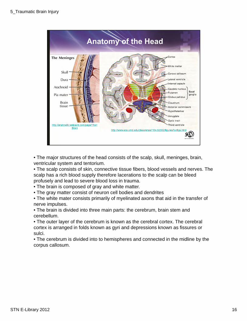
• The major structures of the head consists of the scalp, skull, meninges, brain, ventricular system and tentorium.• The scalp consists of skin, connective tissue fibers, blood vessels and nerves. The scalp has a rich blood supply therefore lacerations to the scalp can be bleed profusely and lead to severe blood loss in trauma.• The brain is composed of gray and white matter. • The gray matter consist of neuron cell bodies and dendrites• The white mater consists primarily of myelinated axons that aid in the transfer of nerve impulses. • The brain is divided into three main parts: the cerebrum, brain stem and cerebellum.• The outer layer of the cerebrum is known as the cerebral cortex. The cerebral cortex is arranged in folds known as gyri and depressions known as fissures or sulci. • The cerebrum is divided into to hemispheres and connected in the midline by the corpus callosum.
16STN E-Library 2012
5_Traumatic Brain Injury
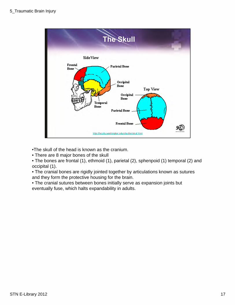
•The skull of the head is known as the cranium. • There are 8 major bones of the skull• The bones are frontal (1), ethmoid (1), parietal (2), sphenpoid (1) temporal (2) and occipital (1).• The cranial bones are rigidly jointed together by articulations known as sutures and they form the protective housing for the brain. • The cranial sutures between bones initially serve as expansion joints but eventually fuse, which halts expandability in adults.
17STN E-Library 2012
5_Traumatic Brain Injury
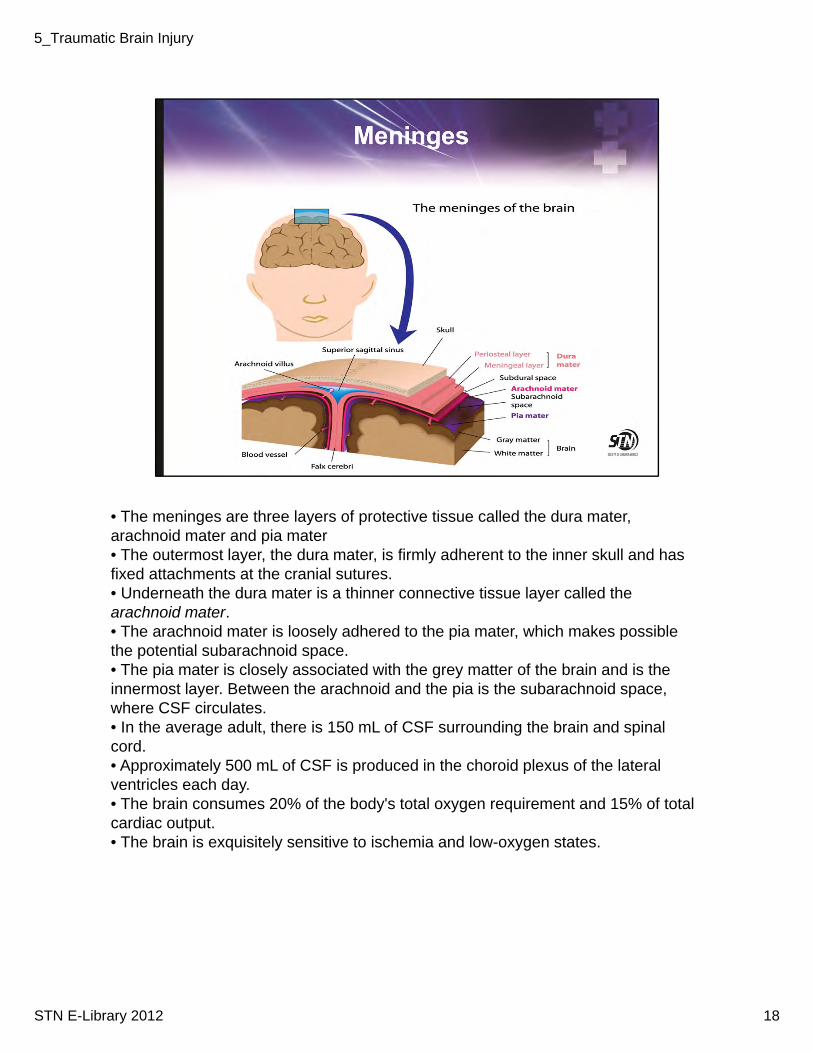
• The meninges are three layers of protective tissue called the dura mater, arachnoid mater and pia mater • The outermost layer, the dura mater, is firmly adherent to the inner skull and has fixed attachments at the cranial sutures.• Underneath the dura mater is a thinner connective tissue layer called thearachnoid mater. • The arachnoid mater is loosely adhered to the pia mater, which makes possible the potential subarachnoid space. • The pia mater is closely associated with the grey matter of the brain and is the innermost layer. Between the arachnoid and the pia is the subarachnoid space, where CSF circulates. • In the average adult, there is 150 mL of CSF surrounding the brain and spinal cord. • Approximately 500 mL of CSF is produced in the choroid plexus of the lateral ventricles each day.• The brain consumes 20% of the body's total oxygen requirement and 15% of total cardiac output. • The brain is exquisitely sensitive to ischemia and low-oxygen states.
18STN E-Library 2012
5_Traumatic Brain Injury
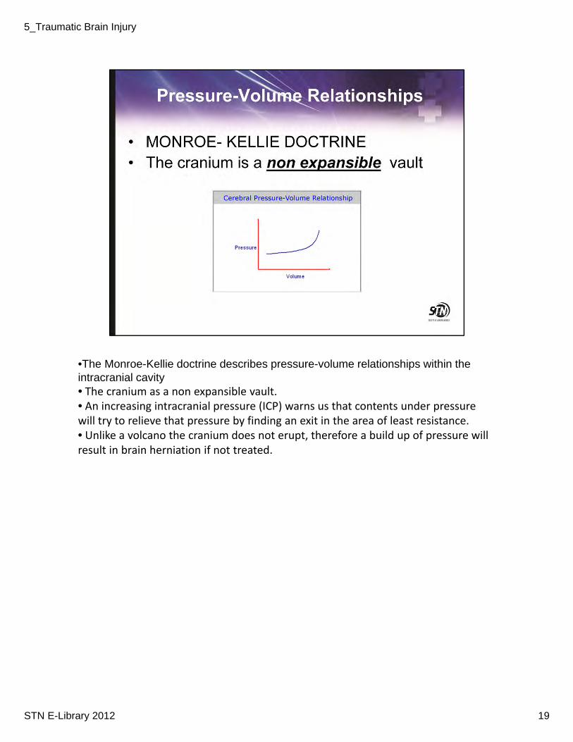
•The Monroe-Kellie doctrine describes pressure-volume relationships within the intracranial cavity• The cranium as a non expansible vault. • An increasing intracranial pressure (ICP) warns us that contents under pressure will try to relieve that pressure by finding an exit in the area of least resistance. • Unlike a volcano the cranium does not erupt, therefore a build up of pressure will result in brain herniation if not treated.
19STN E-Library 2012
5_Traumatic Brain Injury
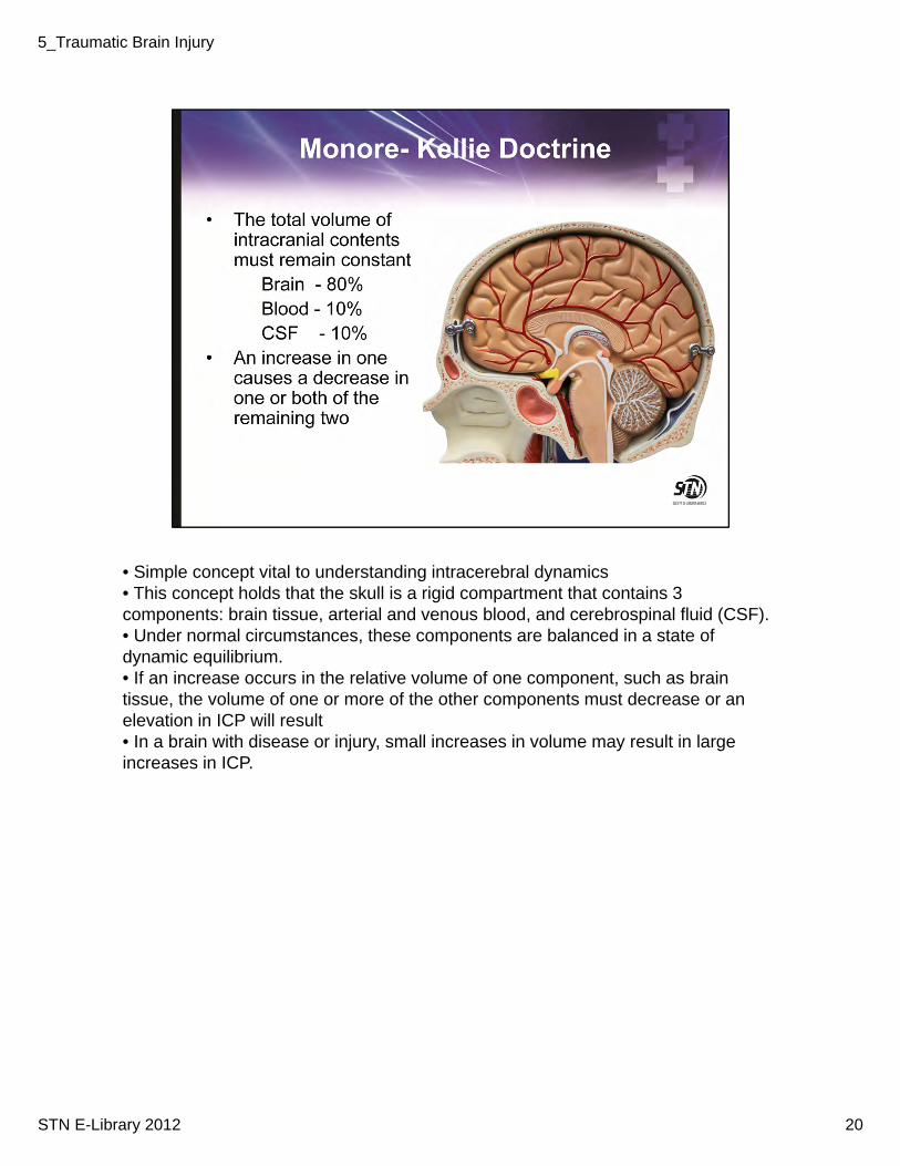
• Simple concept vital to understanding intracerebral dynamics• This concept holds that the skull is a rigid compartment that contains 3 components: brain tissue, arterial and venous blood, and cerebrospinal fluid (CSF). • Under normal circumstances, these components are balanced in a state of dynamic equilibrium. • If an increase occurs in the relative volume of one component, such as brain tissue, the volume of one or more of the other components must decrease or an elevation in ICP will result• In a brain with disease or injury, small increases in volume may result in large increases in ICP.
20STN E-Library 2012
5_Traumatic Brain Injury
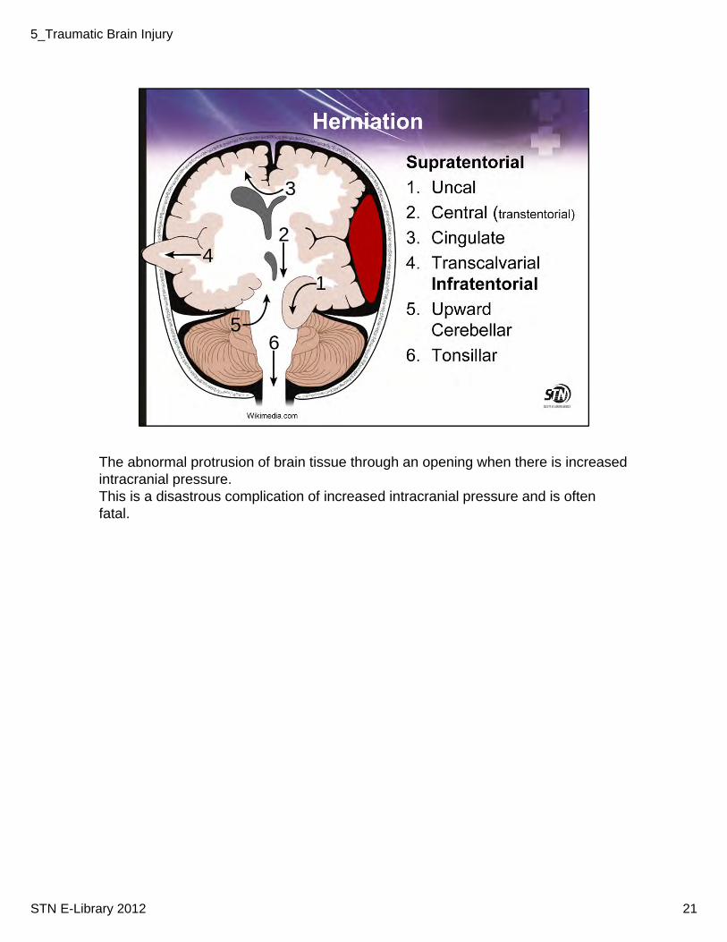
The abnormal protrusion of brain tissue through an opening when there is increased intracranial pressure.This is a disastrous complication of increased intracranial pressure and is often fatal.
21STN E-Library 2012
5_Traumatic Brain Injury
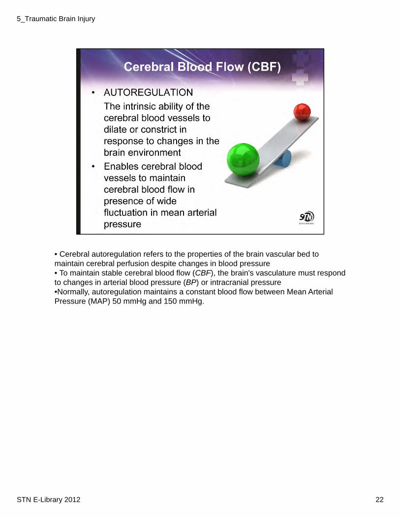
• Cerebral autoregulation refers to the properties of the brain vascular bed to maintain cerebral perfusion despite changes in blood pressure• To maintain stable cerebral blood flow (CBF), the brain's vasculature must respond to changes in arterial blood pressure (BP) or intracranial pressure•Normally, autoregulation maintains a constant blood flow between Mean Arterial Pressure (MAP) 50 mmHg and 150 mmHg.
22STN E-Library 2012
5_Traumatic Brain Injury
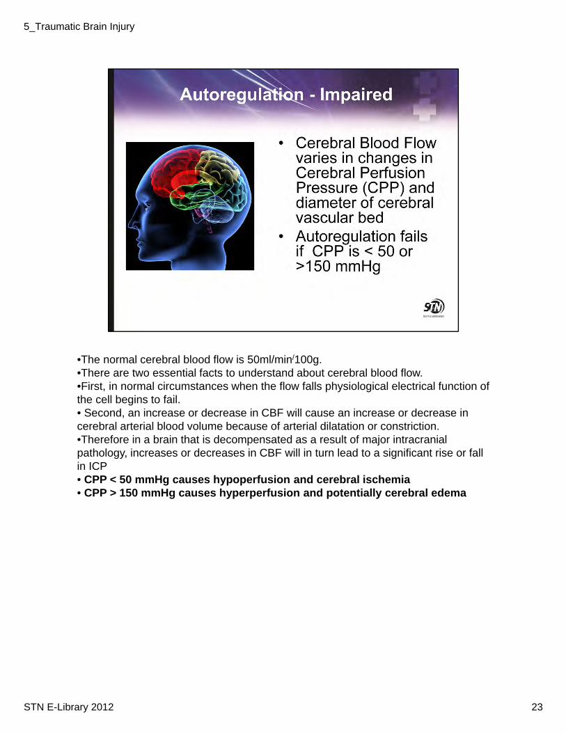
•The normal cerebral blood flow is 50ml/min/100g. •There are two essential facts to understand about cerebral blood flow. •First, in normal circumstances when the flow falls physiological electrical function of the cell begins to fail. • Second, an increase or decrease in CBF will cause an increase or decrease in cerebral arterial blood volume because of arterial dilatation or constriction. •Therefore in a brain that is decompensated as a result of major intracranial pathology, increases or decreases in CBF will in turn lead to a significant rise or fall in ICP• CPP < 50 mmHg causes hypoperfusion and cerebral ischemia• CPP > 150 mmHg causes hyperperfusion and potentially cerebral edema
23STN E-Library 2012
5_Traumatic Brain Injury
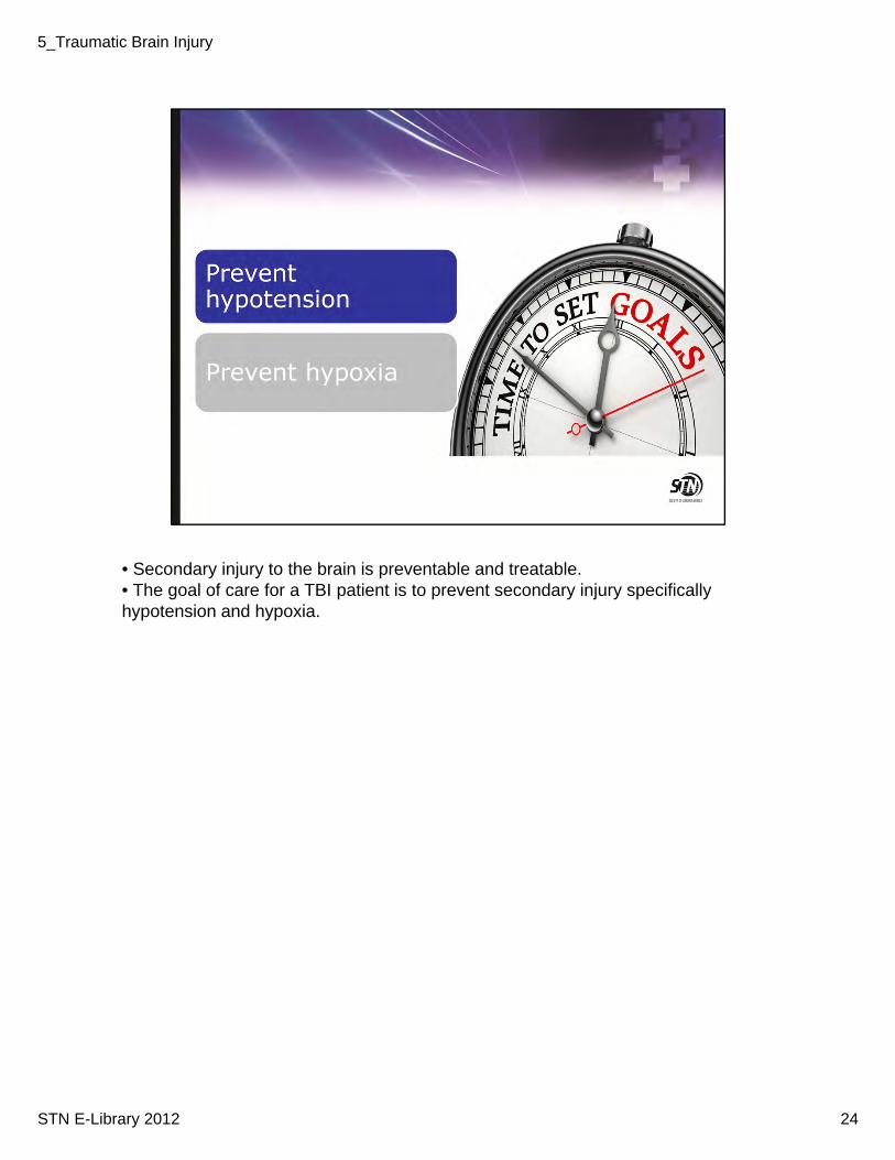
• Secondary injury to the brain is preventable and treatable.• The goal of care for a TBI patient is to prevent secondary injury specifically hypotension and hypoxia.
24STN E-Library 2012
5_Traumatic Brain Injury
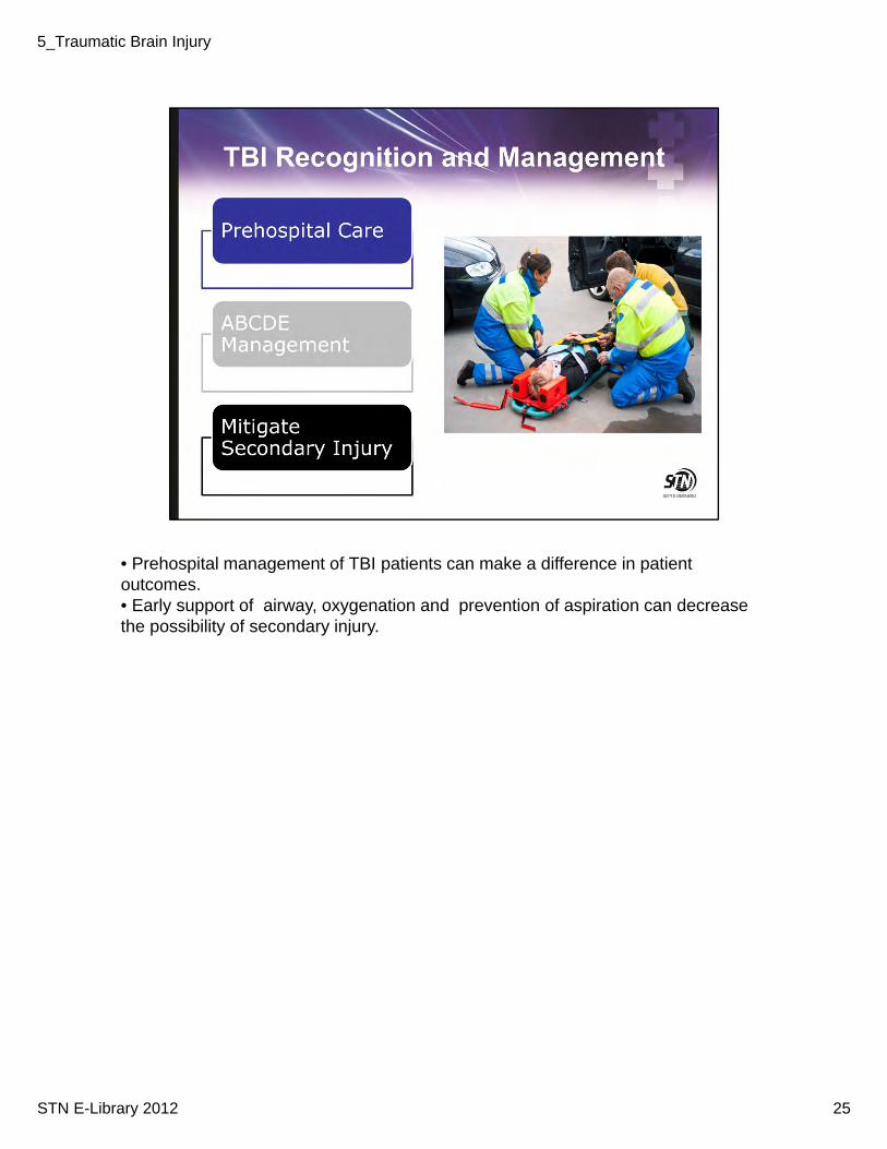
• Prehospital management of TBI patients can make a difference in patient outcomes.• Early support of airway, oxygenation and prevention of aspiration can decrease the possibility of secondary injury.
25STN E-Library 2012
5_Traumatic Brain Injury
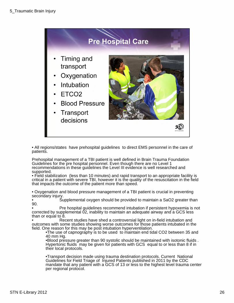
• All regions/states have prehospital guidelines to direct EMS personnel in the care of patients.
Prehospital management of a TBI patient is well defined in Brain Trauma Foundation Guidelines for the pre hospital personnel. Even though there are no Level 1 recommendations in these guidelines the Level III evidence is well researched and supported. • Field stabilization (less than 10 minutes) and rapid transport to an appropriate facility is critical in a patient with severe TBI, however it is the quality of the resuscitation in the field that impacts the outcome of the patient more than speed.
• Oxygenation and blood pressure management of a TBI patient is crucial in preventing secondary injury.• Supplemental oxygen should be provided to maintain a SaO2 greater than 90.• Pre hospital guidelines recommend intubation if persistent hypoxemia is not corrected by supplemental 02, inability to maintain an adequate airway and a GCS less than or equal to 8. • Recent studies have shed a controversial light on in-field intubation and outcomes with some studies showing worse outcomes for those patients intubated in the field. One reason for this may be post intubation hyperventilation.
•The use of capnography is to be used to maintain end tidal CO2 between 35 and 40 mm Hg. •Blood pressure greater than 90 systolic should be maintained with isotonic fluids . Hypertonic fluids may be given for patients with GCS equal to or less than 8 if in their local protocols.
•Transport decision made using trauma destination protocols. Current National Guidelines for Field Triage of Injured Patients published in 2011 by the CDC mandate that any patient with a GCS of 13 or less to the highest level trauma center per regional protocol.
26STN E-Library 2012
5_Traumatic Brain Injury
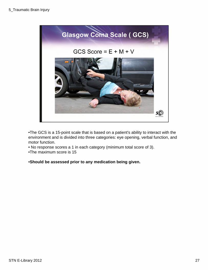
•The GCS is a 15-point scale that is based on a patient's ability to interact with the environment and is divided into three categories: eye opening, verbal function, and motor function. • No response scores a 1 in each category (minimum total score of 3). •The maximum score is 15
•Should be assessed prior to any medication being given.
27STN E-Library 2012
5_Traumatic Brain Injury
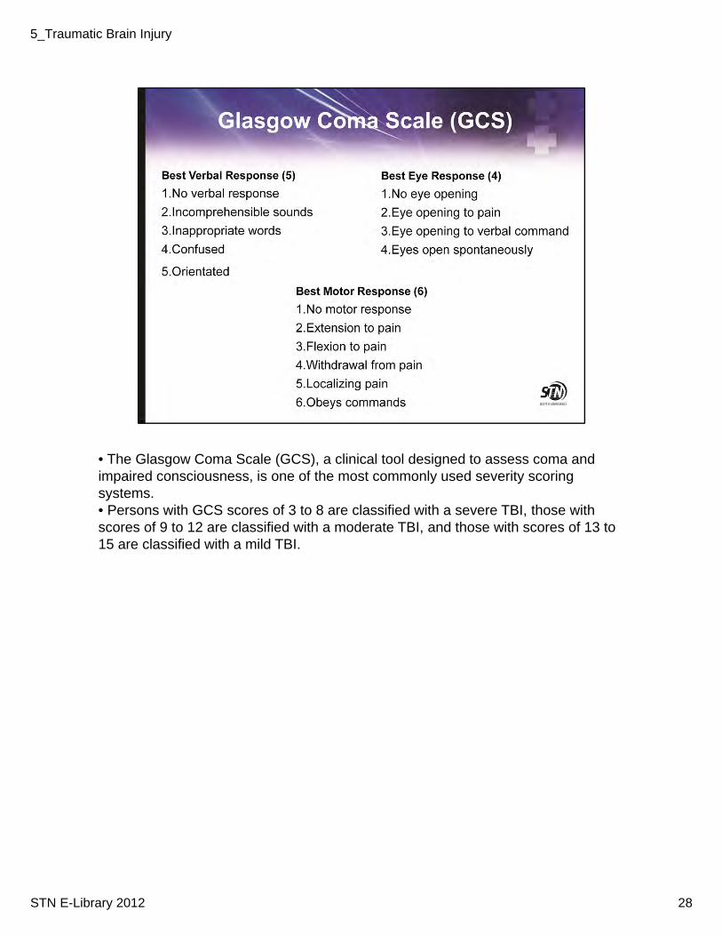
• The Glasgow Coma Scale (GCS), a clinical tool designed to assess coma and impaired consciousness, is one of the most commonly used severity scoring systems.• Persons with GCS scores of 3 to 8 are classified with a severe TBI, those with scores of 9 to 12 are classified with a moderate TBI, and those with scores of 13 to 15 are classified with a mild TBI.
28STN E-Library 2012
5_Traumatic Brain Injury
29STN E-Library 2012
5_Traumatic Brain Injury
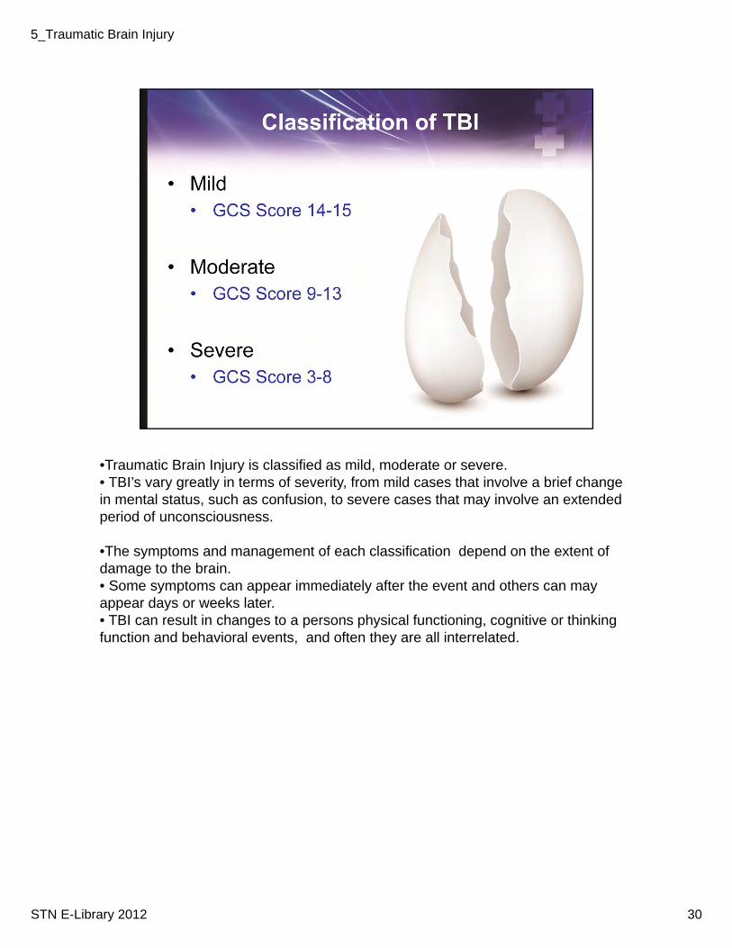
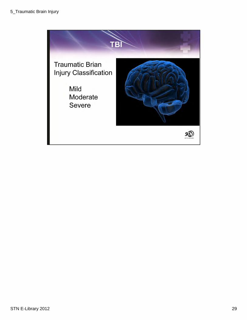
•Traumatic Brain Injury is classified as mild, moderate or severe. • TBI’s vary greatly in terms of severity, from mild cases that involve a brief change in mental status, such as confusion, to severe cases that may involve an extended period of unconsciousness.
•The symptoms and management of each classification depend on the extent of damage to the brain.• Some symptoms can appear immediately after the event and others can may appear days or weeks later. • TBI can result in changes to a persons physical functioning, cognitive or thinking function and behavioral events, and often they are all interrelated.
30STN E-Library 2012
5_Traumatic Brain Injury
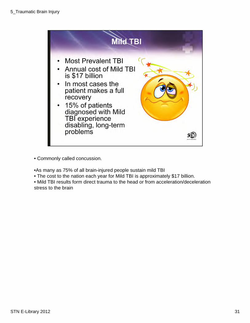
• Commonly called concussion.
•As many as 75% of all brain-injured people sustain mild TBI• The cost to the nation each year for Mild TBI is approximately $17 billion.• Mild TBI results form direct trauma to the head or from acceleration/deceleration stress to the brain
31STN E-Library 2012
5_Traumatic Brain Injury
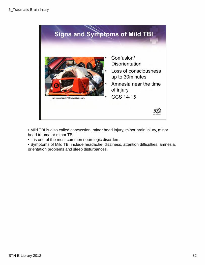
• Mild TBI is also called concussion, minor head injury, minor brain injury, minor head trauma or minor TBI.• It is one of the most common neurologic disorders. • Symptoms of Mild TBI include headache, dizziness, attention difficulties, amnesia, orientation problems and sleep disturbances.
32STN E-Library 2012
5_Traumatic Brain Injury
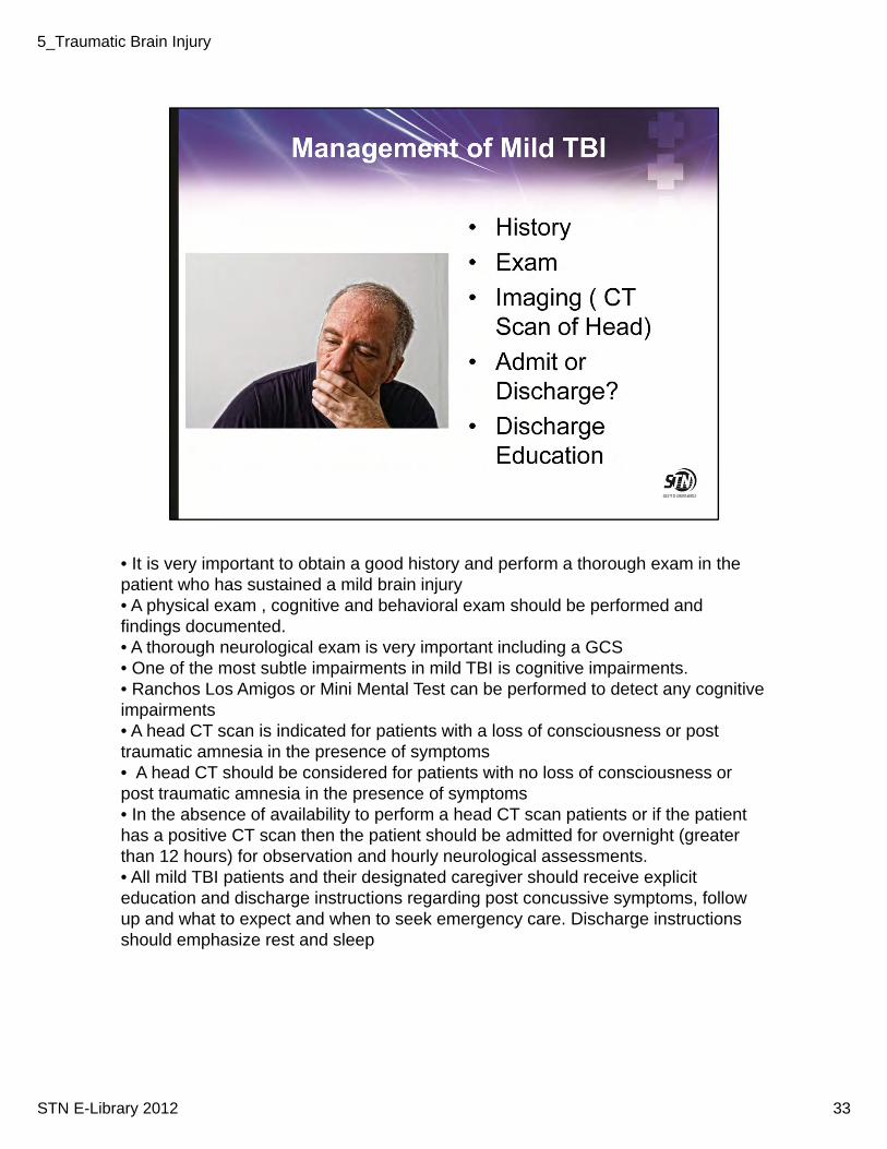
• It is very important to obtain a good history and perform a thorough exam in the patient who has sustained a mild brain injury• A physical exam , cognitive and behavioral exam should be performed and findings documented. • A thorough neurological exam is very important including a GCS• One of the most subtle impairments in mild TBI is cognitive impairments. • Ranchos Los Amigos or Mini Mental Test can be performed to detect any cognitive impairments• A head CT scan is indicated for patients with a loss of consciousness or post traumatic amnesia in the presence of symptoms• A head CT should be considered for patients with no loss of consciousness or post traumatic amnesia in the presence of symptoms• In the absence of availability to perform a head CT scan patients or if the patient has a positive CT scan then the patient should be admitted for overnight (greater than 12 hours) for observation and hourly neurological assessments. • All mild TBI patients and their designated caregiver should receive explicit education and discharge instructions regarding post concussive symptoms, follow up and what to expect and when to seek emergency care. Discharge instructions should emphasize rest and sleep
33STN E-Library 2012
5_Traumatic Brain Injury
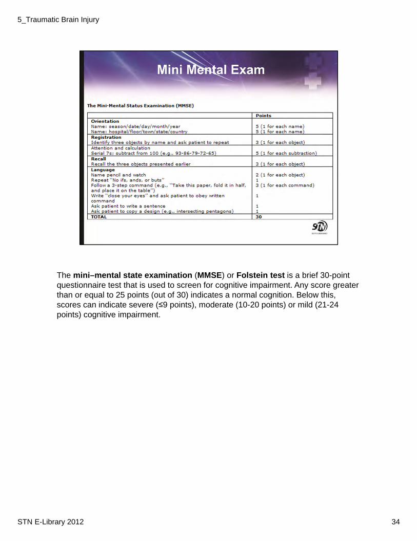
The mini–mental state examination (MMSE) or Folstein test is a brief 30-point questionnaire test that is used to screen for cognitive impairment. Any score greater than or equal to 25 points (out of 30) indicates a normal cognition. Below this, scores can indicate severe (≤9 points), moderate (10-20 points) or mild (21-24 points) cognitive impairment.
34STN E-Library 2012
5_Traumatic Brain Injury
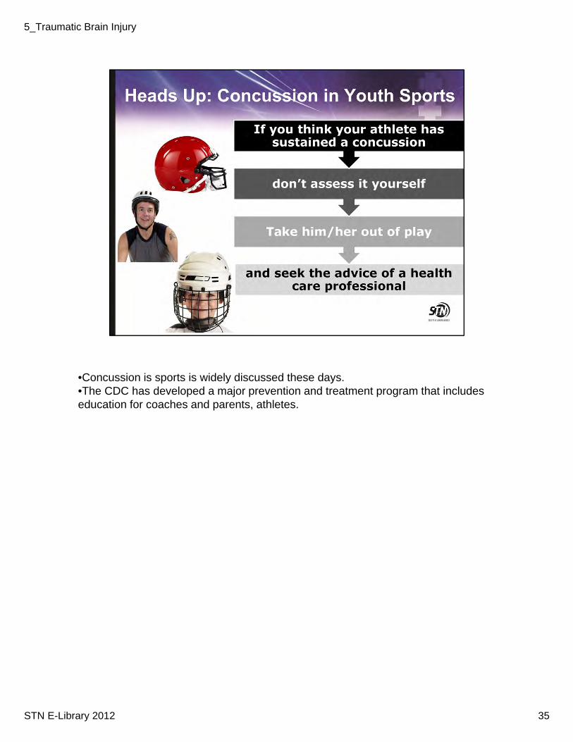
•Concussion is sports is widely discussed these days. •The CDC has developed a major prevention and treatment program that includes education for coaches and parents, athletes.
35STN E-Library 2012
5_Traumatic Brain Injury
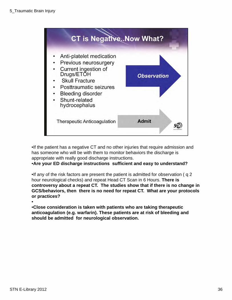
•If the patient has a negative CT and no other injuries that require admission and has someone who will be with them to monitor behaviors the discharge is appropriate with really good discharge instructions. •Are your ED discharge instructions sufficient and easy to understand?
•If any of the risk factors are present the patient is admitted for observation ( q 2 hour neurological checks) and repeat Head CT Scan in 6 Hours. There is controversy about a repeat CT. The studies show that if there is no change in GCS/behaviors, then there is no need for repeat CT. What are your protocols or practices?••Close consideration is taken with patients who are taking therapeutic anticoagulation (e.g. warfarin). These patients are at risk of bleeding and should be admitted for neurological observation.
36STN E-Library 2012
5_Traumatic Brain Injury
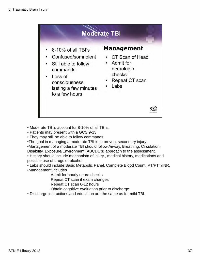
• Moderate TBI’s account for 8-10% of all TBI’s.• Patients may present with a GCS 9-13• They may still be able to follow commands.•The goal in managing a moderate TBI is to prevent secondary injury!•Management of a moderate TBI should follow Airway, Breathing, Circulation, Disability, Exposure/Environment (ABCDE’s) approach to the assessment. • History should include mechanism of injury , medical history, medications and possible use of drugs or alcohol• Labs should include Basic Metabolic Panel, Complete Blood Count, PT/PTT/INR. •Management includes
Admit for hourly neuro checksRepeat CT scan if exam changesRepeat CT scan 6-12 hoursObtain cognitive evaluation prior to discharge
• Discharge instructions and education are the same as for mild TBI.
37STN E-Library 2012
5_Traumatic Brain Injury
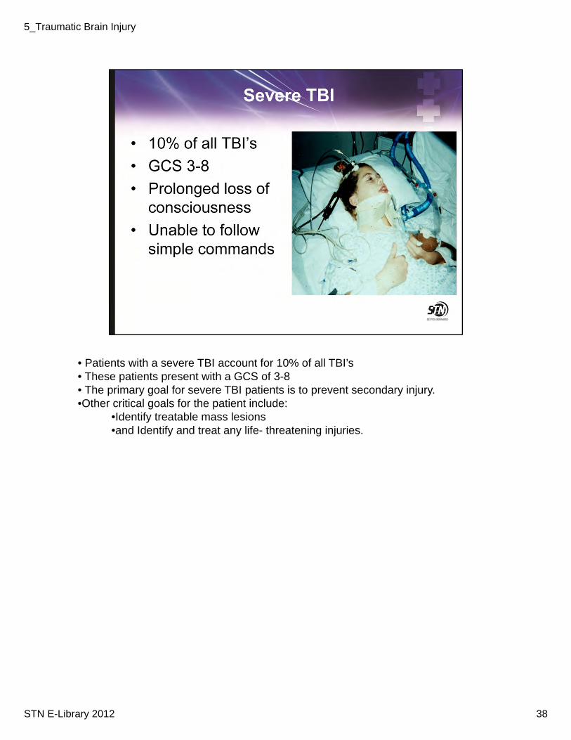
• Patients with a severe TBI account for 10% of all TBI’s• These patients present with a GCS of 3-8• The primary goal for severe TBI patients is to prevent secondary injury. •Other critical goals for the patient include:
•Identify treatable mass lesions •and Identify and treat any life- threatening injuries.
38STN E-Library 2012
5_Traumatic Brain Injury
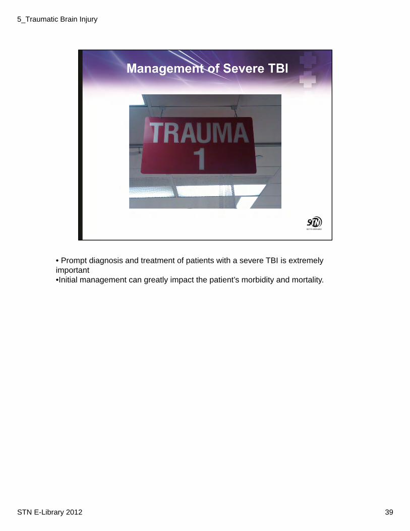
• Prompt diagnosis and treatment of patients with a severe TBI is extremely important•Initial management can greatly impact the patient’s morbidity and mortality.
39STN E-Library 2012
5_Traumatic Brain Injury
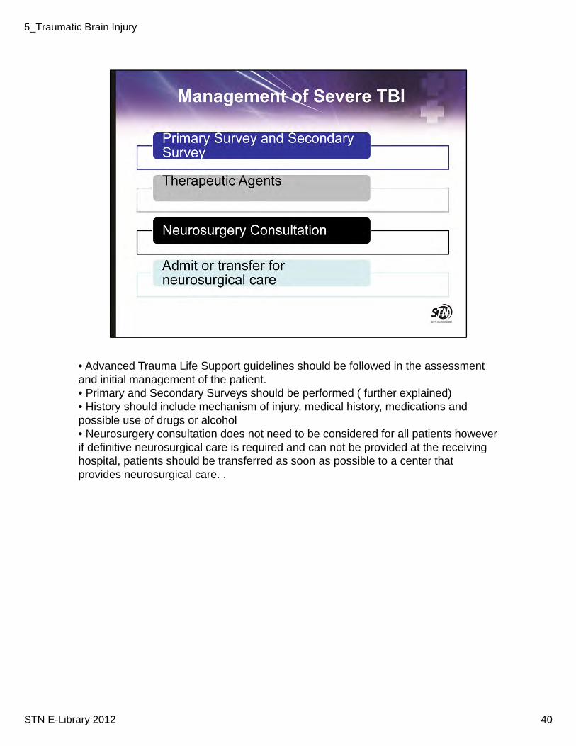
• Advanced Trauma Life Support guidelines should be followed in the assessment and initial management of the patient.• Primary and Secondary Surveys should be performed ( further explained) • History should include mechanism of injury, medical history, medications and possible use of drugs or alcohol• Neurosurgery consultation does not need to be considered for all patients however if definitive neurosurgical care is required and can not be provided at the receiving hospital, patients should be transferred as soon as possible to a center that provides neurosurgical care. .
40STN E-Library 2012
5_Traumatic Brain Injury
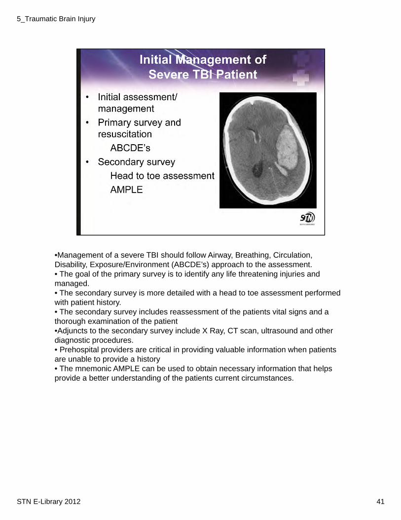
•Management of a severe TBI should follow Airway, Breathing, Circulation, Disability, Exposure/Environment (ABCDE’s) approach to the assessment.• The goal of the primary survey is to identify any life threatening injuries and managed.• The secondary survey is more detailed with a head to toe assessment performed with patient history.• The secondary survey includes reassessment of the patients vital signs and a thorough examination of the patient •Adjuncts to the secondary survey include X Ray, CT scan, ultrasound and other diagnostic procedures. • Prehospital providers are critical in providing valuable information when patients are unable to provide a history• The mnemonic AMPLE can be used to obtain necessary information that helps provide a better understanding of the patients current circumstances.
41STN E-Library 2012
5_Traumatic Brain Injury
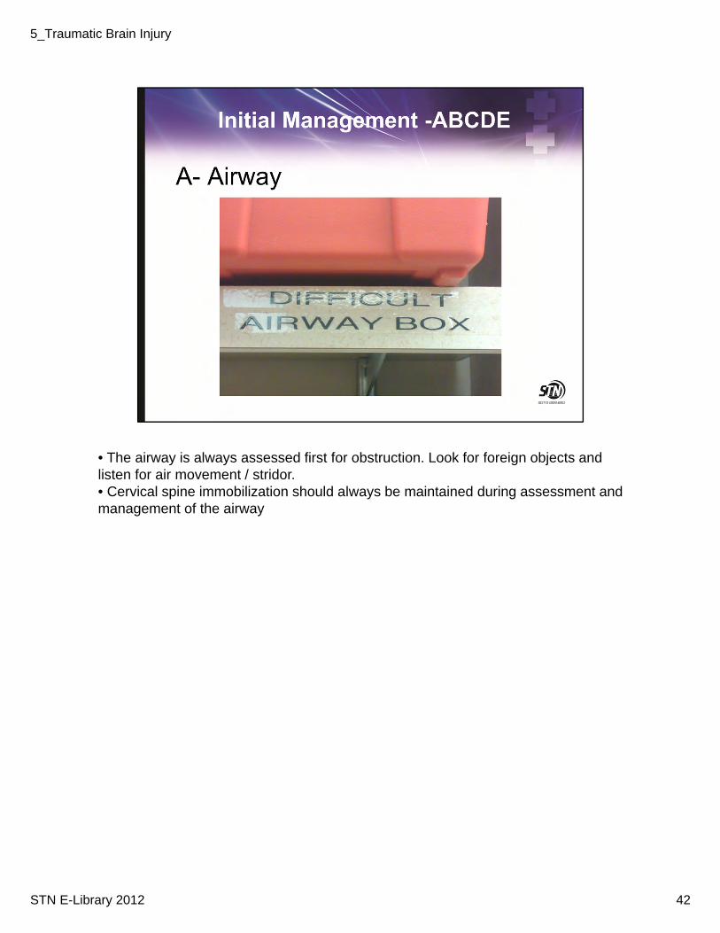
• The airway is always assessed first for obstruction. Look for foreign objects and listen for air movement / stridor. • Cervical spine immobilization should always be maintained during assessment and management of the airway
42STN E-Library 2012
5_Traumatic Brain Injury
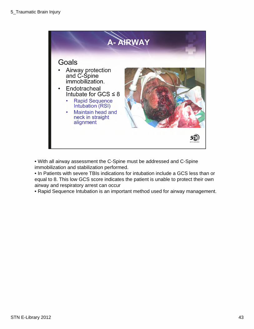
• With all airway assessment the C-Spine must be addressed and C-Spine immobilization and stabilization performed.• In Patients with severe TBIs indications for intubation include a GCS less than or equal to 8. This low GCS score indicates the patient is unable to protect their own airway and respiratory arrest can occur• Rapid Sequence Intubation is an important method used for airway management.
43STN E-Library 2012
5_Traumatic Brain Injury
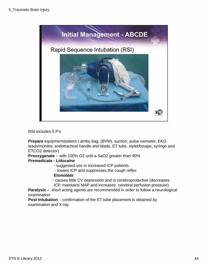
RSI includes 5 P’s
Prepare equipment/patient ( ambu bag, (BVM), suction, pulse oximeter, EKG leads/monitor, endotracheal handle and blade, ET tube, stylet/bougie, syringe and ETCO2 detector)Preoxygenate – with 100% O2 until a SaO2 greater than 90%Premedicate - Lidocaine
- suggested use in increased ICP patients- lowers ICP and suppresses the cough reflex
Etomidate: - causes little CV depression and is cerebroprotective (decreasesICP, maintains MAP and increases cerebral perfusion pressure)
Paralysis – short acting agents are recommended in order to follow a neurological examinationPost Intubation – confirmation of the ET tube placement is obtained by examination and X-ray
44STN E-Library 2012
5_Traumatic Brain Injury
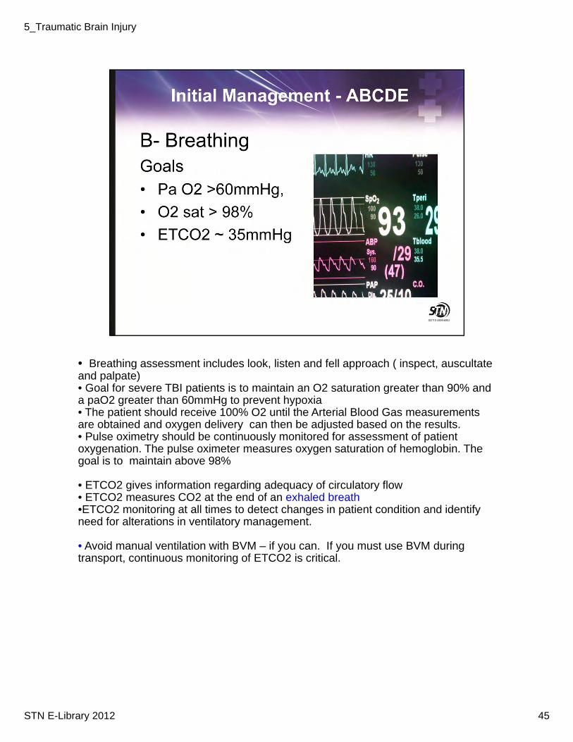
• Breathing assessment includes look, listen and fell approach ( inspect, auscultate and palpate) • Goal for severe TBI patients is to maintain an O2 saturation greater than 90% and a paO2 greater than 60mmHg to prevent hypoxia• The patient should receive 100% O2 until the Arterial Blood Gas measurements are obtained and oxygen delivery can then be adjusted based on the results. • Pulse oximetry should be continuously monitored for assessment of patient oxygenation. The pulse oximeter measures oxygen saturation of hemoglobin. The goal is to maintain above 98%
• ETCO2 gives information regarding adequacy of circulatory flow• ETCO2 measures CO2 at the end of an exhaled breath•ETCO2 monitoring at all times to detect changes in patient condition and identify need for alterations in ventilatory management.
• Avoid manual ventilation with BVM – if you can. If you must use BVM during transport, continuous monitoring of ETCO2 is critical.
45STN E-Library 2012
5_Traumatic Brain Injury
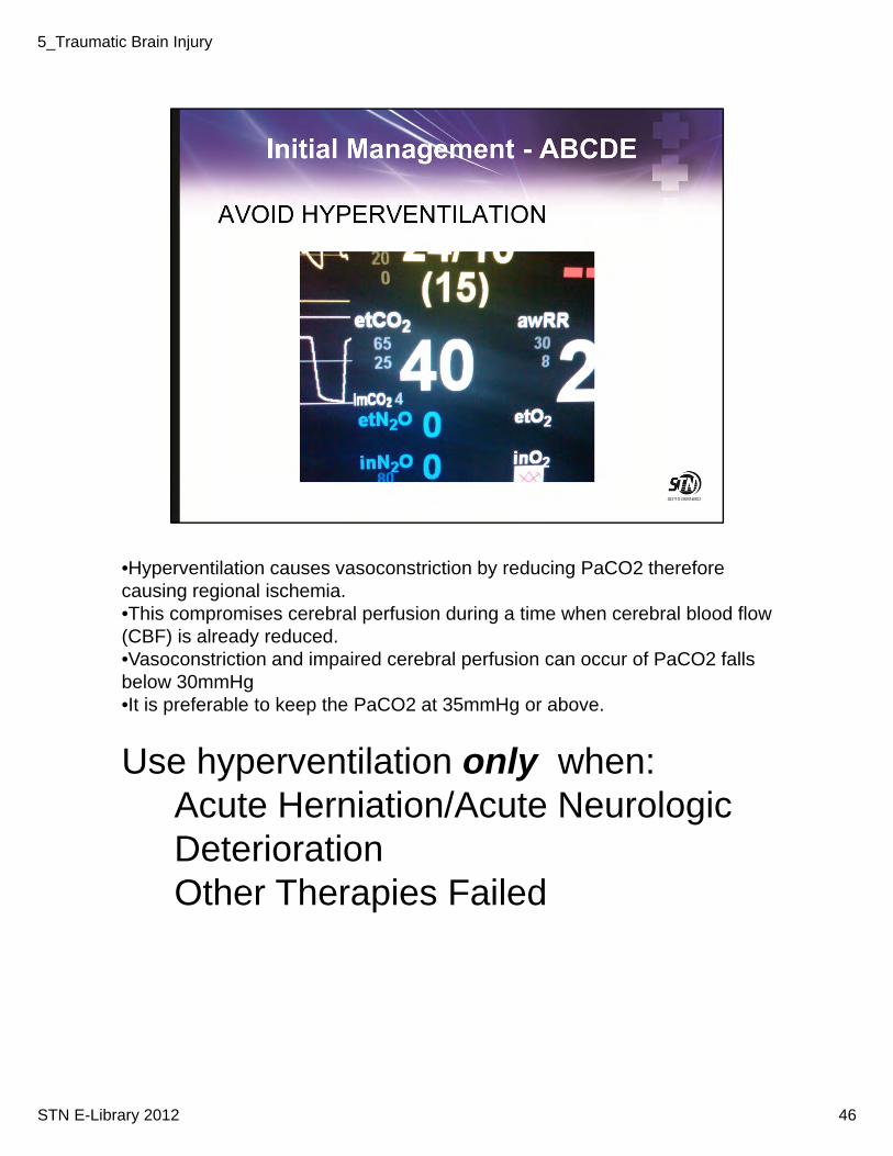
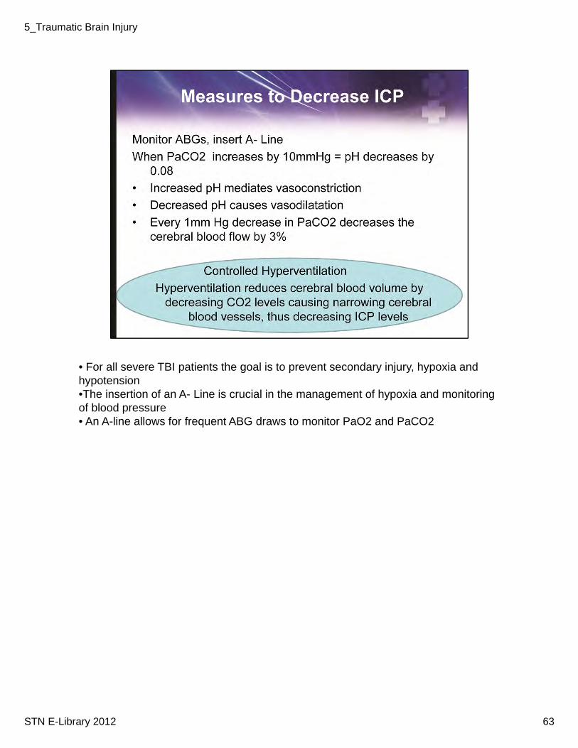
•Hyperventilation causes vasoconstriction by reducing PaCO2 therefore causing regional ischemia. •This compromises cerebral perfusion during a time when cerebral blood flow (CBF) is already reduced.•Vasoconstriction and impaired cerebral perfusion can occur of PaCO2 falls below 30mmHg•It is preferable to keep the PaCO2 at 35mmHg or above.
Use hyperventilation only when:Acute Herniation/Acute Neurologic DeteriorationOther Therapies Failed
46STN E-Library 2012
5_Traumatic Brain Injury
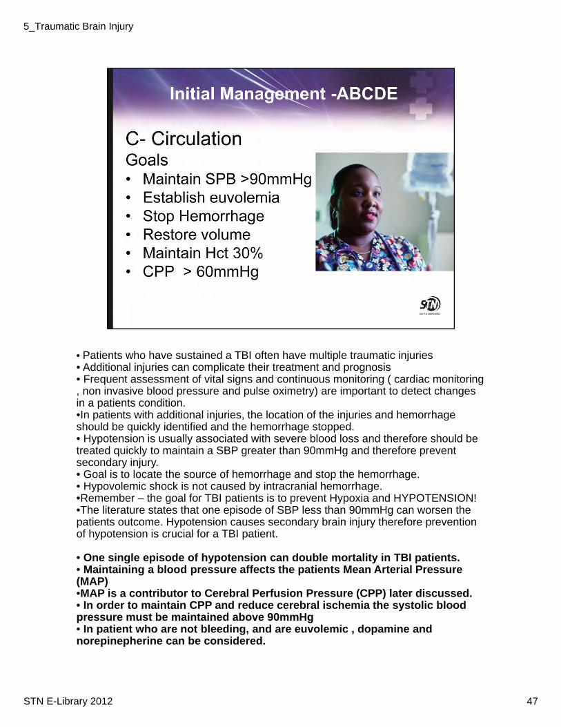
• Patients who have sustained a TBI often have multiple traumatic injuries• Additional injuries can complicate their treatment and prognosis• Frequent assessment of vital signs and continuous monitoring ( cardiac monitoring , non invasive blood pressure and pulse oximetry) are important to detect changes in a patients condition.•In patients with additional injuries, the location of the injuries and hemorrhage should be quickly identified and the hemorrhage stopped. • Hypotension is usually associated with severe blood loss and therefore should be treated quickly to maintain a SBP greater than 90mmHg and therefore prevent secondary injury.• Goal is to locate the source of hemorrhage and stop the hemorrhage.• Hypovolemic shock is not caused by intracranial hemorrhage. •Remember – the goal for TBI patients is to prevent Hypoxia and HYPOTENSION!•The literature states that one episode of SBP less than 90mmHg can worsen the patients outcome. Hypotension causes secondary brain injury therefore prevention of hypotension is crucial for a TBI patient.
• One single episode of hypotension can double mortality in TBI patients.• Maintaining a blood pressure affects the patients Mean Arterial Pressure (MAP)•MAP is a contributor to Cerebral Perfusion Pressure (CPP) later discussed. • In order to maintain CPP and reduce cerebral ischemia the systolic blood pressure must be maintained above 90mmHg• In patient who are not bleeding, and are euvolemic , dopamine and norepinepherine can be considered.
47STN E-Library 2012
5_Traumatic Brain Injury
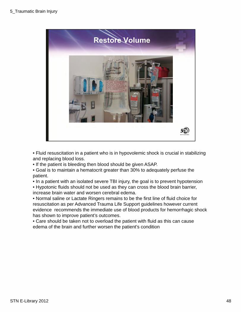
• Fluid resuscitation in a patient who is in hypovolemic shock is crucial in stabilizing and replacing blood loss.• If the patient is bleeding then blood should be given ASAP.• Goal is to maintain a hematocrit greater than 30% to adequately perfuse the patient.• In a patient with an isolated severe TBI injury, the goal is to prevent hypotension• Hypotonic fluids should not be used as they can cross the blood brain barrier, increase brain water and worsen cerebral edema.• Normal saline or Lactate Ringers remains to be the first line of fluid choice for resuscitation as per Advanced Trauma Life Support guidelines however current evidence recommends the immediate use of blood products for hemorrhagic shock has shown to improve patient’s outcomes. • Care should be taken not to overload the patient with fluid as this can cause edema of the brain and further worsen the patient’s condition
48STN E-Library 2012
5_Traumatic Brain Injury
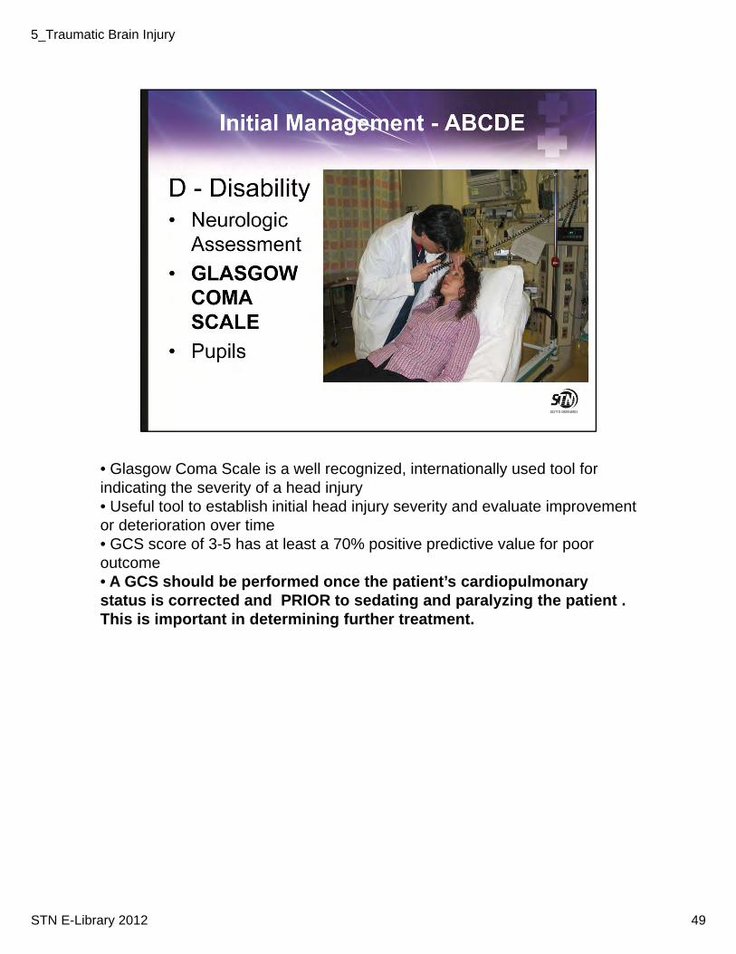
• Glasgow Coma Scale is a well recognized, internationally used tool for indicating the severity of a head injury• Useful tool to establish initial head injury severity and evaluate improvement or deterioration over time• GCS score of 3-5 has at least a 70% positive predictive value for poor outcome• A GCS should be performed once the patient’s cardiopulmonary status is corrected and PRIOR to sedating and paralyzing the patient . This is important in determining further treatment.
49STN E-Library 2012
5_Traumatic Brain Injury
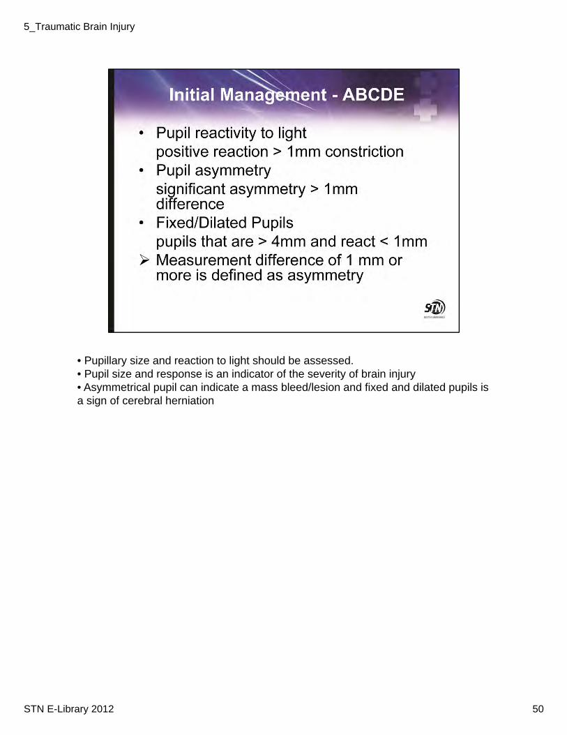
• Pupillary size and reaction to light should be assessed.• Pupil size and response is an indicator of the severity of brain injury• Asymmetrical pupil can indicate a mass bleed/lesion and fixed and dilated pupils is a sign of cerebral herniation
50STN E-Library 2012
5_Traumatic Brain Injury
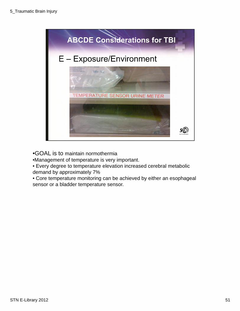
•GOAL is to maintain normothermia•Management of temperature is very important. • Every degree to temperature elevation increased cerebral metabolic demand by approximately 7%• Core temperature monitoring can be achieved by either an esophageal sensor or a bladder temperature sensor.
51STN E-Library 2012
5_Traumatic Brain Injury
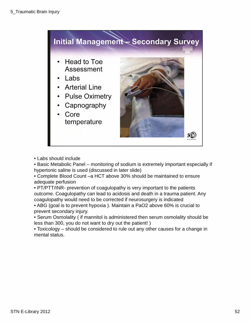
• Labs should include • Basic Metabolic Panel – monitoring of sodium is extremely important especially if hypertonic saline is used (discussed in later slide)• Complete Blood Count –a HCT above 30% should be maintained to ensure adequate perfusion • PT/PTT/INR- prevention of coagulopathy is very important to the patients outcome. Coagulopathy can lead to acidosis and death in a trauma patient. Any coagulopathy would need to be corrected if neurosurgery is indicated• ABG (goal is to prevent hypoxia ). Maintain a PaO2 above 60% is crucial to prevent secondary injury. • Serum Osmolality ( if mannitol is administered then serum osmolality should be less than 300, you do not want to dry out the patient! ) • Toxicology – should be considered to rule out any other causes for a change in mental status.
52STN E-Library 2012
5_Traumatic Brain Injury
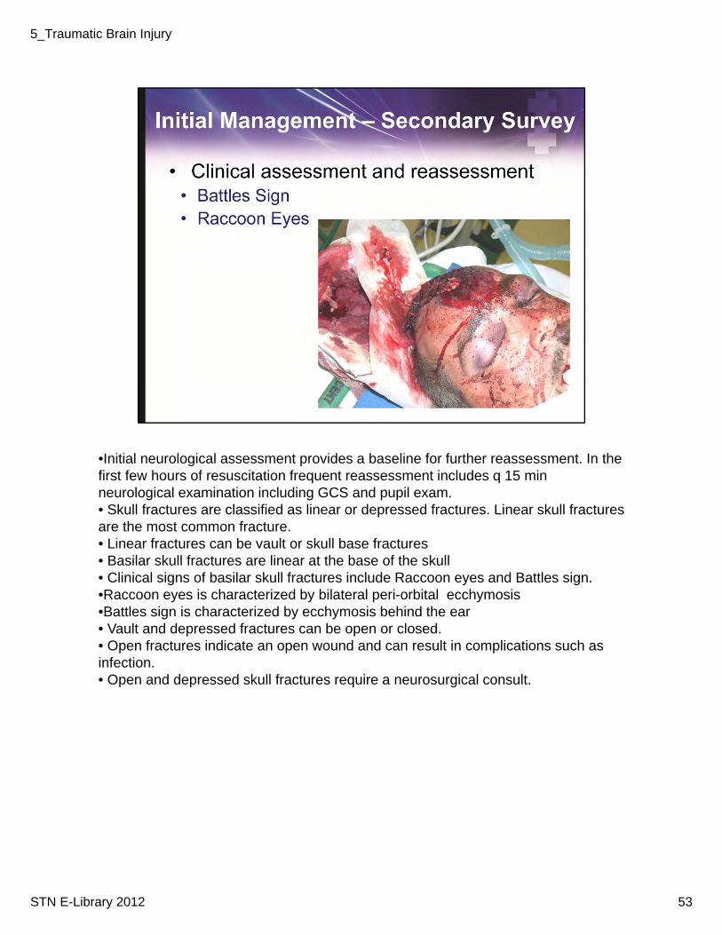
•Initial neurological assessment provides a baseline for further reassessment. In the first few hours of resuscitation frequent reassessment includes q 15 min neurological examination including GCS and pupil exam. • Skull fractures are classified as linear or depressed fractures. Linear skull fractures are the most common fracture. • Linear fractures can be vault or skull base fractures• Basilar skull fractures are linear at the base of the skull• Clinical signs of basilar skull fractures include Raccoon eyes and Battles sign.•Raccoon eyes is characterized by bilateral peri-orbital ecchymosis •Battles sign is characterized by ecchymosis behind the ear• Vault and depressed fractures can be open or closed.• Open fractures indicate an open wound and can result in complications such as infection. • Open and depressed skull fractures require a neurosurgical consult.
53STN E-Library 2012
5_Traumatic Brain Injury
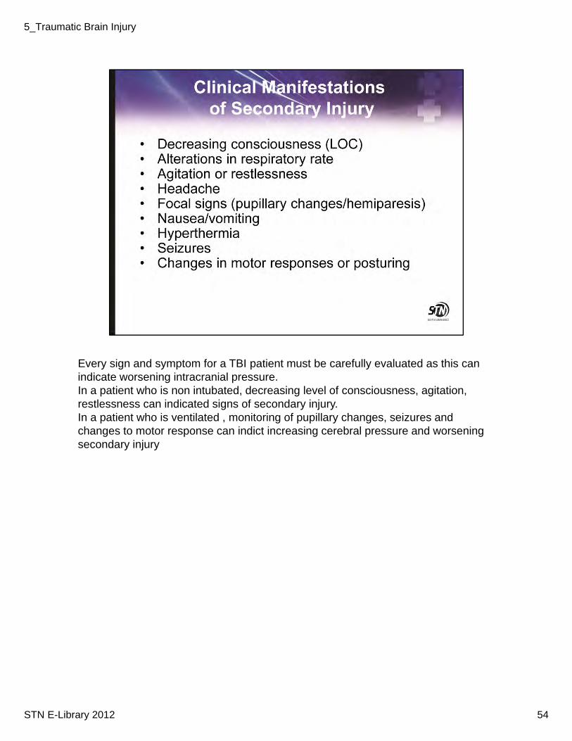
Every sign and symptom for a TBI patient must be carefully evaluated as this can indicate worsening intracranial pressure. In a patient who is non intubated, decreasing level of consciousness, agitation, restlessness can indicated signs of secondary injury.In a patient who is ventilated , monitoring of pupillary changes, seizures and changes to motor response can indict increasing cerebral pressure and worsening secondary injury
54STN E-Library 2012
5_Traumatic Brain Injury
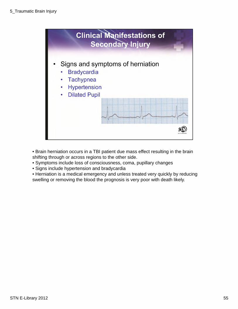
• Brain herniation occurs in a TBI patient due mass effect resulting in the brain shifting through or across regions to the other side.• Symptoms include loss of consciousness, coma, pupillary changes• Signs include hypertension and bradycardia• Herniation is a medical emergency and unless treated very quickly by reducing swelling or removing the blood the prognosis is very poor with death likely.
55STN E-Library 2012
5_Traumatic Brain Injury
• Cushing’s Triad is a late sign of increased ICP and is the last attempt for the brain to compensate during herniation• Cushing’s triad is defined as bradycardia, widening pulse pressure and irregular respirations
56STN E-Library 2012
5_Traumatic Brain Injury
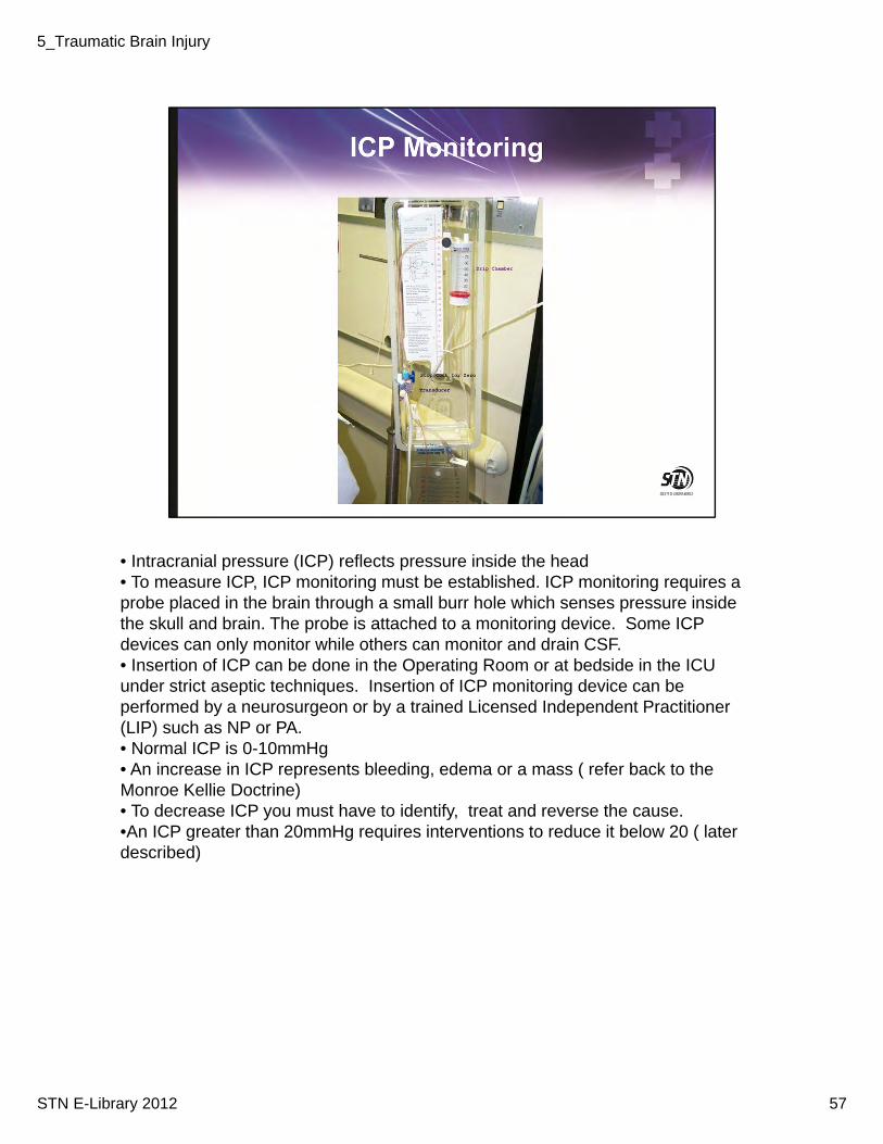
• Intracranial pressure (ICP) reflects pressure inside the head• To measure ICP, ICP monitoring must be established. ICP monitoring requires a probe placed in the brain through a small burr hole which senses pressure inside the skull and brain. The probe is attached to a monitoring device. Some ICP devices can only monitor while others can monitor and drain CSF.• Insertion of ICP can be done in the Operating Room or at bedside in the ICU under strict aseptic techniques. Insertion of ICP monitoring device can be performed by a neurosurgeon or by a trained Licensed Independent Practitioner (LIP) such as NP or PA.• Normal ICP is 0-10mmHg• An increase in ICP represents bleeding, edema or a mass ( refer back to the Monroe Kellie Doctrine)• To decrease ICP you must have to identify, treat and reverse the cause.•An ICP greater than 20mmHg requires interventions to reduce it below 20 ( later described)
57STN E-Library 2012
5_Traumatic Brain Injury
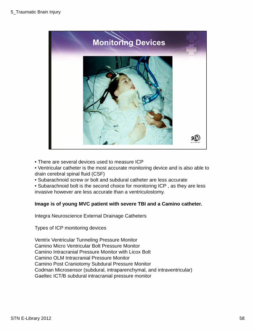
• There are several devices used to measure ICP• Ventricular catheter is the most accurate monitoring device and is also able to drain cerebral spinal fluid (CSF)• Subarachnoid screw or bolt and subdural catheter are less accurate • Subarachnoid bolt is the second choice for monitoring ICP , as they are less invasive however are less accurate than a ventriculostomy.
Image is of young MVC patient with severe TBI and a Camino catheter.
Integra Neuroscience External Drainage Catheters
Types of ICP monitoring devices
Ventrix Ventricular Tunneling Pressure MonitorCamino Micro Ventricular Bolt Pressure MonitorCamino Intracranial Pressure Monitor with Licox BoltCamino OLM Intracranial Pressure MonitorCamino Post Craniotomy Subdural Pressure MonitorCodman Microsensor (subdural, intraparenchymal, and intraventricular)Gaeltec ICT/B subdural intracranial pressure monitor
58STN E-Library 2012
5_Traumatic Brain Injury

• Ventriculostomy is the most frequently used monitoring device•It allows for measuring of the cranial pressure and drainage of cerebral spinal fluid (CSF). CSF drainage helps to reduce ICP• Ventriculostomies are at more risk of infection as they are the most invasive.
59STN E-Library 2012
5_Traumatic Brain Injury
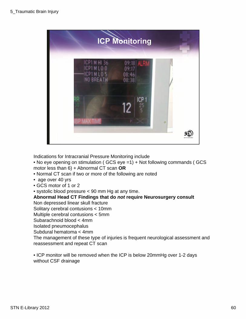
Indications for Intracranial Pressure Monitoring include• No eye opening on stimulation ( GCS eye =1) + Not following commands ( GCS motor less than 6) + Abnormal CT scan OR• Normal CT scan if two or more of the following are noted • age over 40 yrs• GCS motor of 1 or 2• systolic blood pressure < 90 mm Hg at any time.Abnormal Head CT Findings that do not require Neurosurgery consultNon depressed linear skull fracture Solitary cerebral contusions < 10mmMultiple cerebral contusions < 5mmSubarachnoid blood < 4mmIsolated pneumocephalusSubdural hematoma < 4mmThe management of these type of injuries is frequent neurological assessment and reassessment and repeat CT scan
• ICP monitor will be removed when the ICP is below 20mmHg over 1-2 days without CSF drainage
60STN E-Library 2012
5_Traumatic Brain Injury
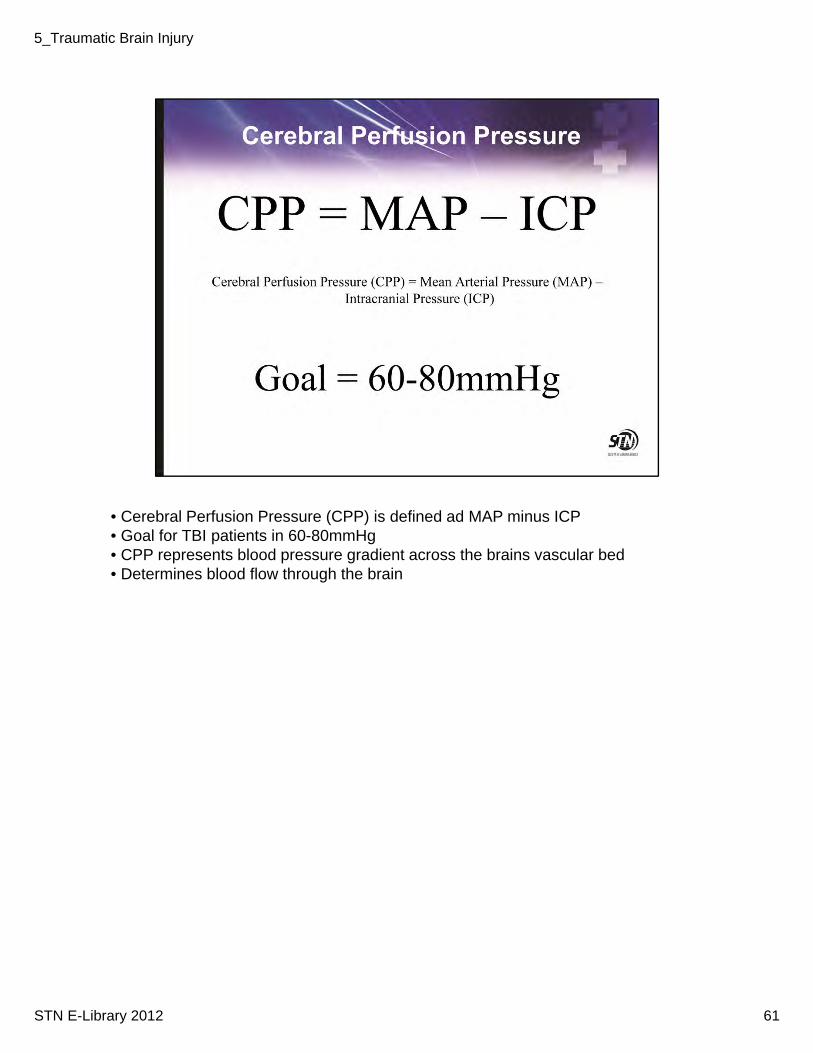
• Cerebral Perfusion Pressure (CPP) is defined ad MAP minus ICP• Goal for TBI patients in 60-80mmHg• CPP represents blood pressure gradient across the brains vascular bed• Determines blood flow through the brain
61STN E-Library 2012
5_Traumatic Brain Injury
•Neurosurgical involvement should be consulted early for evacuation of intracranial mass lesions, penetrating injuries, and depressed skull fractures • The crisis within the trauma systems today is the national shortage of neurosurgeons• The consensus however is that all emergency neurosurgeries should be performed by a neurosurgeon. • If neurosurgical services are not available the patient must be stabilized and considered for early transfer to a level of care that can provide neurosurgical consultation.
62STN E-Library 2012
5_Traumatic Brain Injury
• For all severe TBI patients the goal is to prevent secondary injury, hypoxia and hypotension•The insertion of an A- Line is crucial in the management of hypoxia and monitoring of blood pressure• An A-line allows for frequent ABG draws to monitor PaO2 and PaCO2
63STN E-Library 2012
5_Traumatic Brain Injury
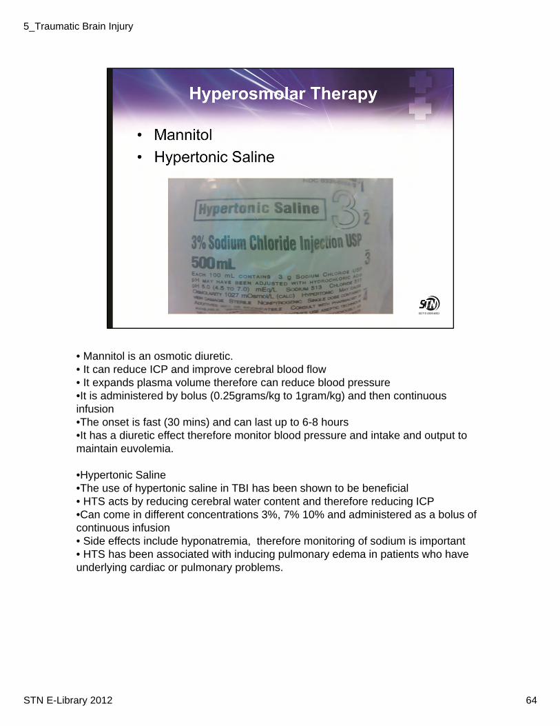
• Mannitol is an osmotic diuretic. • It can reduce ICP and improve cerebral blood flow• It expands plasma volume therefore can reduce blood pressure•It is administered by bolus (0.25grams/kg to 1gram/kg) and then continuous infusion•The onset is fast (30 mins) and can last up to 6-8 hours•It has a diuretic effect therefore monitor blood pressure and intake and output to maintain euvolemia.
•Hypertonic Saline •The use of hypertonic saline in TBI has been shown to be beneficial• HTS acts by reducing cerebral water content and therefore reducing ICP•Can come in different concentrations 3%, 7% 10% and administered as a bolus of continuous infusion• Side effects include hyponatremia, therefore monitoring of sodium is important• HTS has been associated with inducing pulmonary edema in patients who have underlying cardiac or pulmonary problems.
64STN E-Library 2012
5_Traumatic Brain Injury
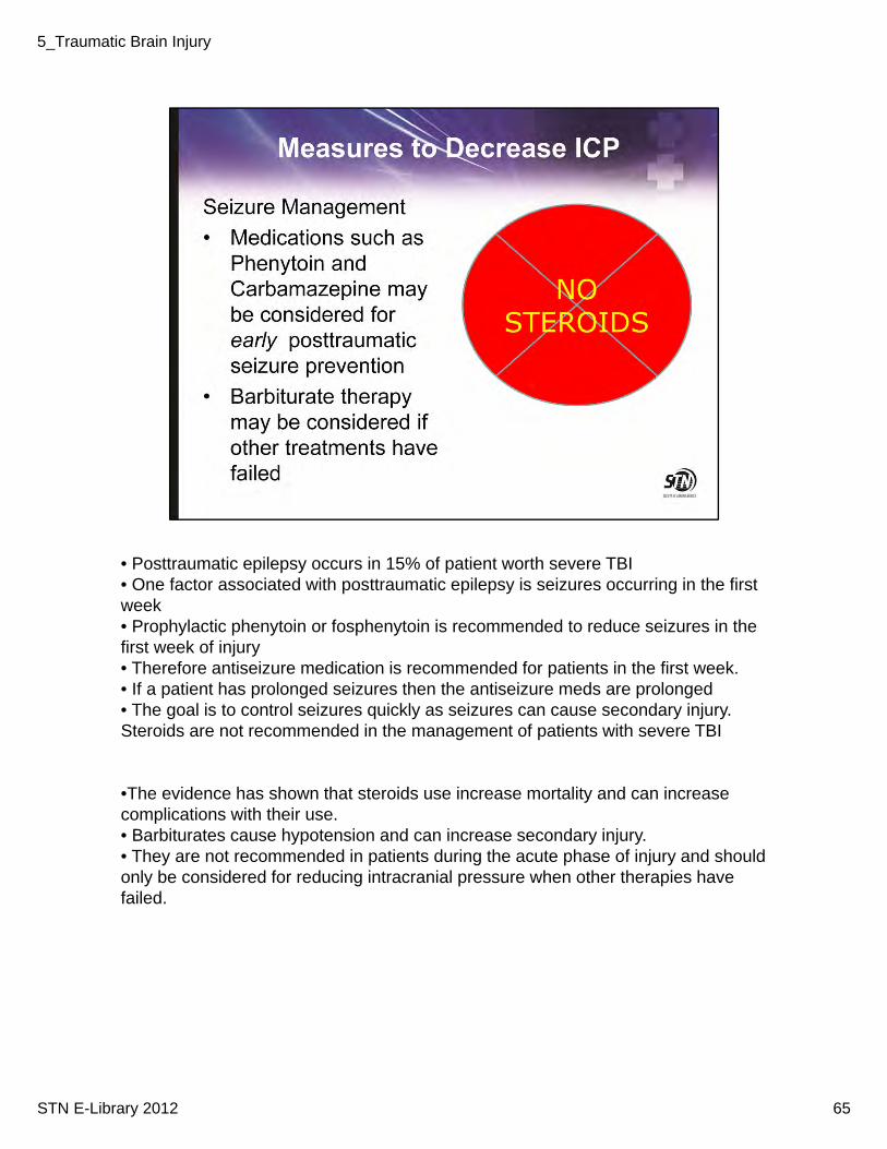
• Posttraumatic epilepsy occurs in 15% of patient worth severe TBI• One factor associated with posttraumatic epilepsy is seizures occurring in the first week• Prophylactic phenytoin or fosphenytoin is recommended to reduce seizures in the first week of injury• Therefore antiseizure medication is recommended for patients in the first week.• If a patient has prolonged seizures then the antiseizure meds are prolonged• The goal is to control seizures quickly as seizures can cause secondary injury. Steroids are not recommended in the management of patients with severe TBI
•The evidence has shown that steroids use increase mortality and can increase complications with their use. • Barbiturates cause hypotension and can increase secondary injury.• They are not recommended in patients during the acute phase of injury and should only be considered for reducing intracranial pressure when other therapies have failed.
65STN E-Library 2012
5_Traumatic Brain Injury
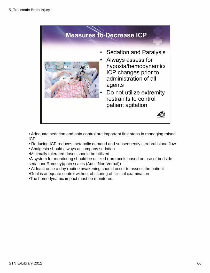
• Adequate sedation and pain control are important first steps in managing raised ICP• Reducing ICP reduces metabolic demand and subsequently cerebral blood flow• Analgesia should always accompany sedation•Minimally tolerated doses should be utilized •A system for monitoring should be utilized ( protocols based on use of bedside sedation( Ramsey)/pain scales (Adult Non Verbal))• At least once a day routine awakening should occur to assess the patient•Goal is adequate control without obscuring of clinical examination•The hemodynamic impact must be monitored.
66STN E-Library 2012
5_Traumatic Brain Injury
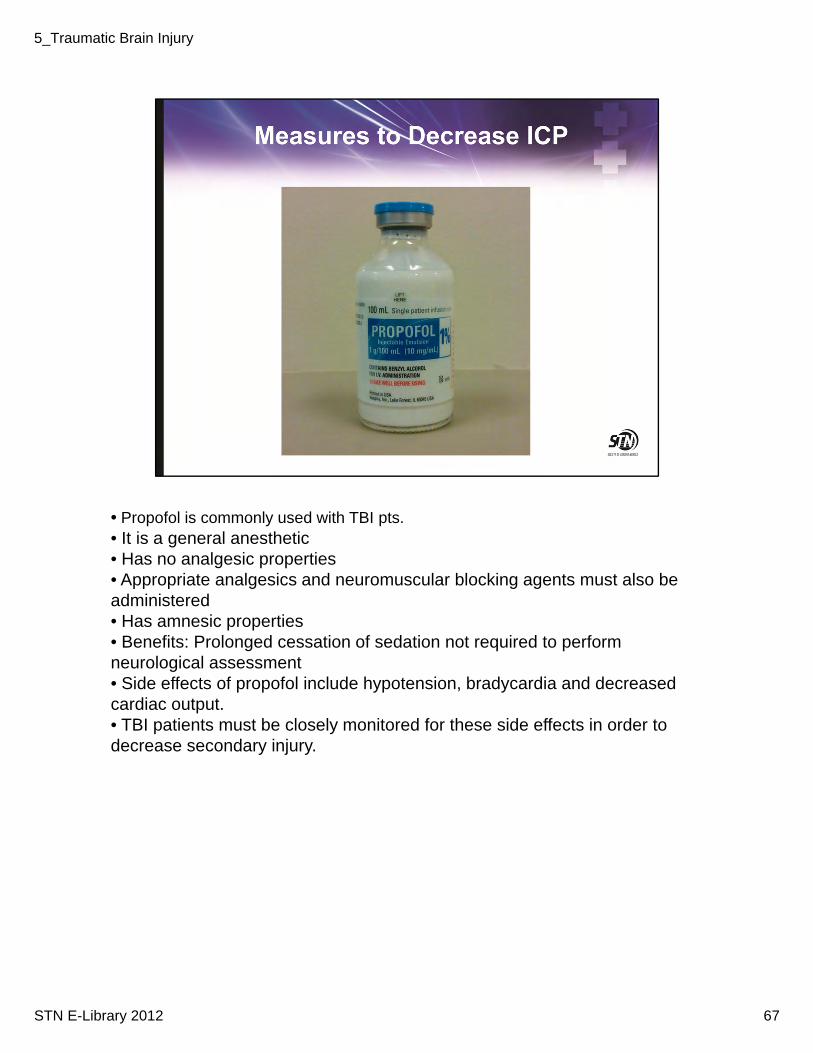
• Propofol is commonly used with TBI pts.• It is a general anesthetic• Has no analgesic properties• Appropriate analgesics and neuromuscular blocking agents must also be administered• Has amnesic properties• Benefits: Prolonged cessation of sedation not required to perform neurological assessment• Side effects of propofol include hypotension, bradycardia and decreased cardiac output. • TBI patients must be closely monitored for these side effects in order to decrease secondary injury.
67STN E-Library 2012
5_Traumatic Brain Injury
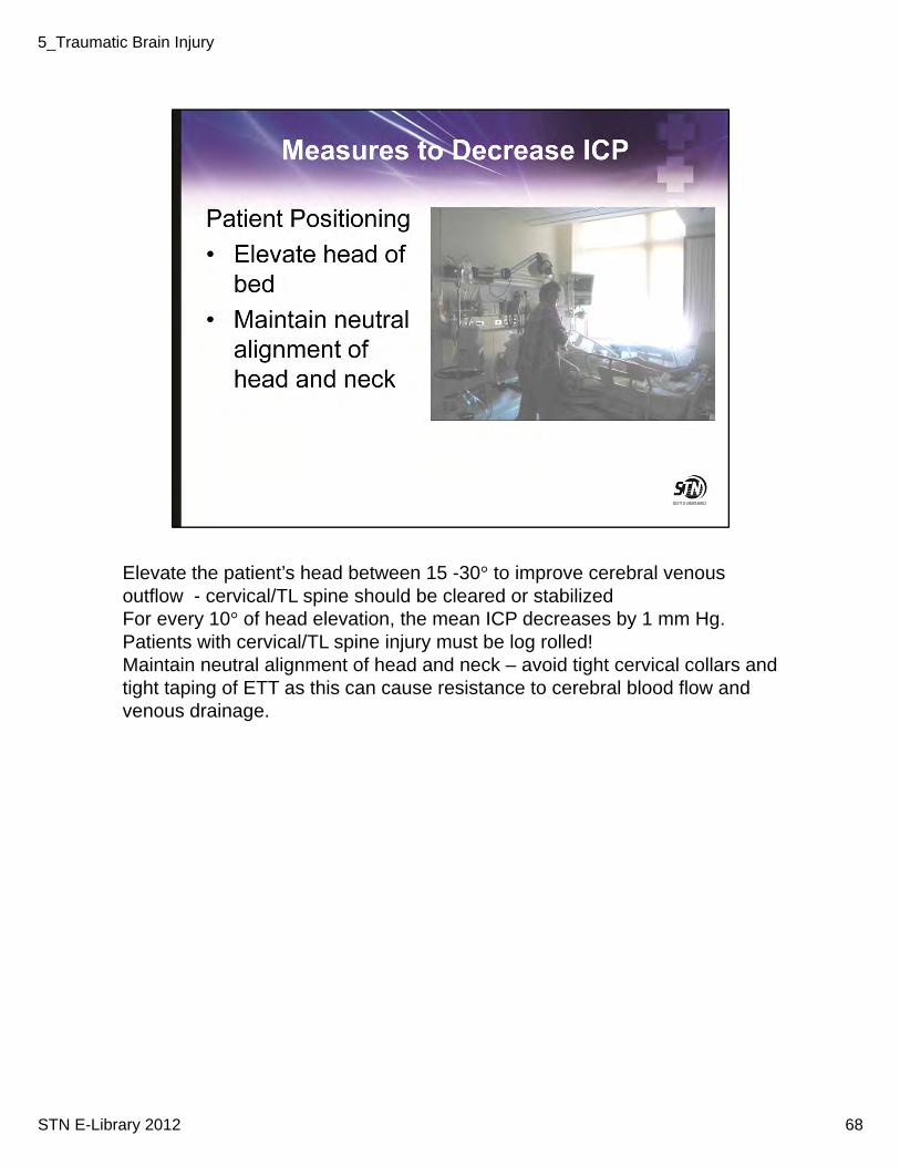
Elevate the patient’s head between 15 -30° to improve cerebral venous outflow - cervical/TL spine should be cleared or stabilizedFor every 10° of head elevation, the mean ICP decreases by 1 mm Hg.Patients with cervical/TL spine injury must be log rolled!Maintain neutral alignment of head and neck – avoid tight cervical collars and tight taping of ETT as this can cause resistance to cerebral blood flow and venous drainage.
68STN E-Library 2012
5_Traumatic Brain Injury
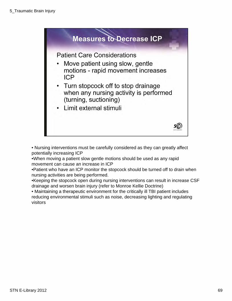
• Nursing interventions must be carefully considered as they can greatly affect potentially increasing ICP•When moving a patient slow gentle motions should be used as any rapid movement can cause an increase in ICP•Patient who have an ICP monitor the stopcock should be turned off to drain when nursing activities are being performed. •Keeping the stopcock open during nursing interventions can result in increase CSF drainage and worsen brain injury (refer to Monroe Kellie Doctrine) • Maintaining a therapeutic environment for the critically ill TBI patient includes reducing environmental stimuli such as noise, decreasing lighting and regulating visitors
69STN E-Library 2012
5_Traumatic Brain Injury
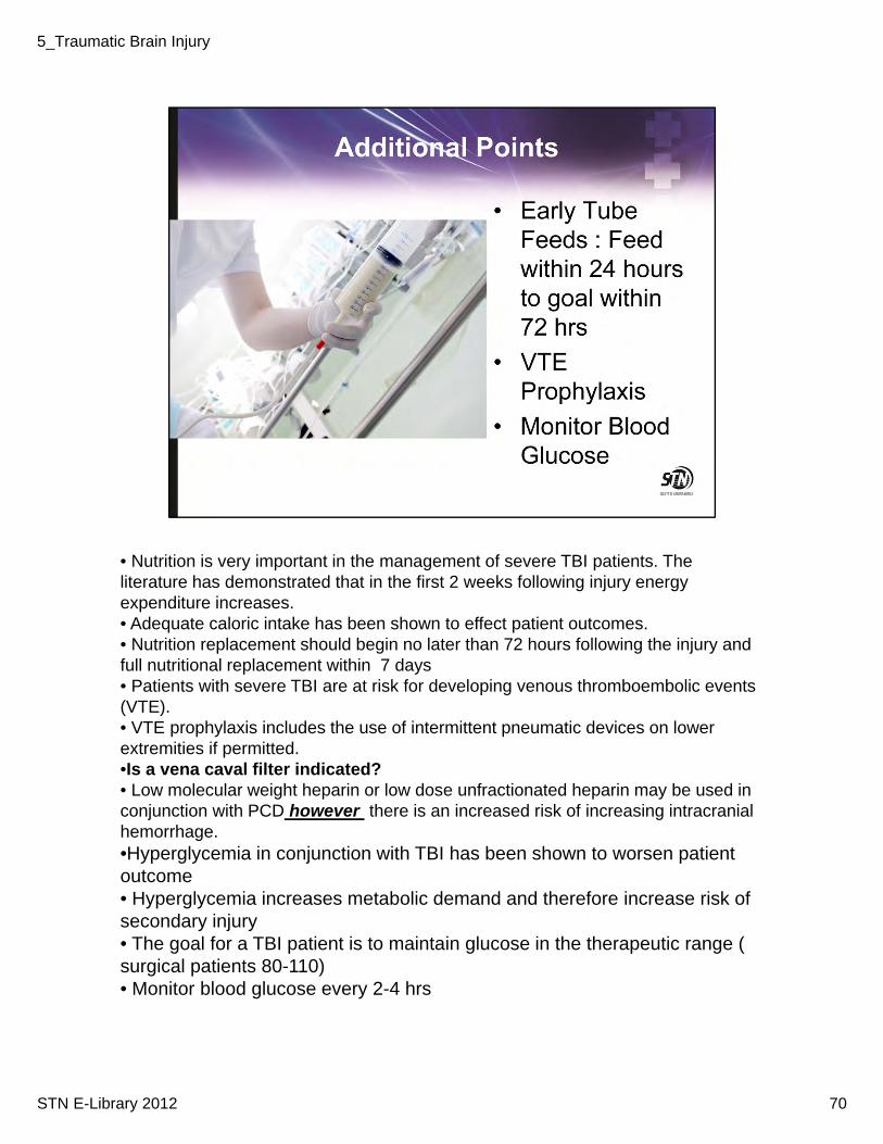
• Nutrition is very important in the management of severe TBI patients. The literature has demonstrated that in the first 2 weeks following injury energy expenditure increases. • Adequate caloric intake has been shown to effect patient outcomes.• Nutrition replacement should begin no later than 72 hours following the injury and full nutritional replacement within 7 days• Patients with severe TBI are at risk for developing venous thromboembolic events (VTE). • VTE prophylaxis includes the use of intermittent pneumatic devices on lower extremities if permitted. •Is a vena caval filter indicated?• Low molecular weight heparin or low dose unfractionated heparin may be used in conjunction with PCD however there is an increased risk of increasing intracranial hemorrhage. •Hyperglycemia in conjunction with TBI has been shown to worsen patient outcome• Hyperglycemia increases metabolic demand and therefore increase risk of secondary injury • The goal for a TBI patient is to maintain glucose in the therapeutic range ( surgical patients 80-110)• Monitor blood glucose every 2-4 hrs
70STN E-Library 2012
5_Traumatic Brain Injury
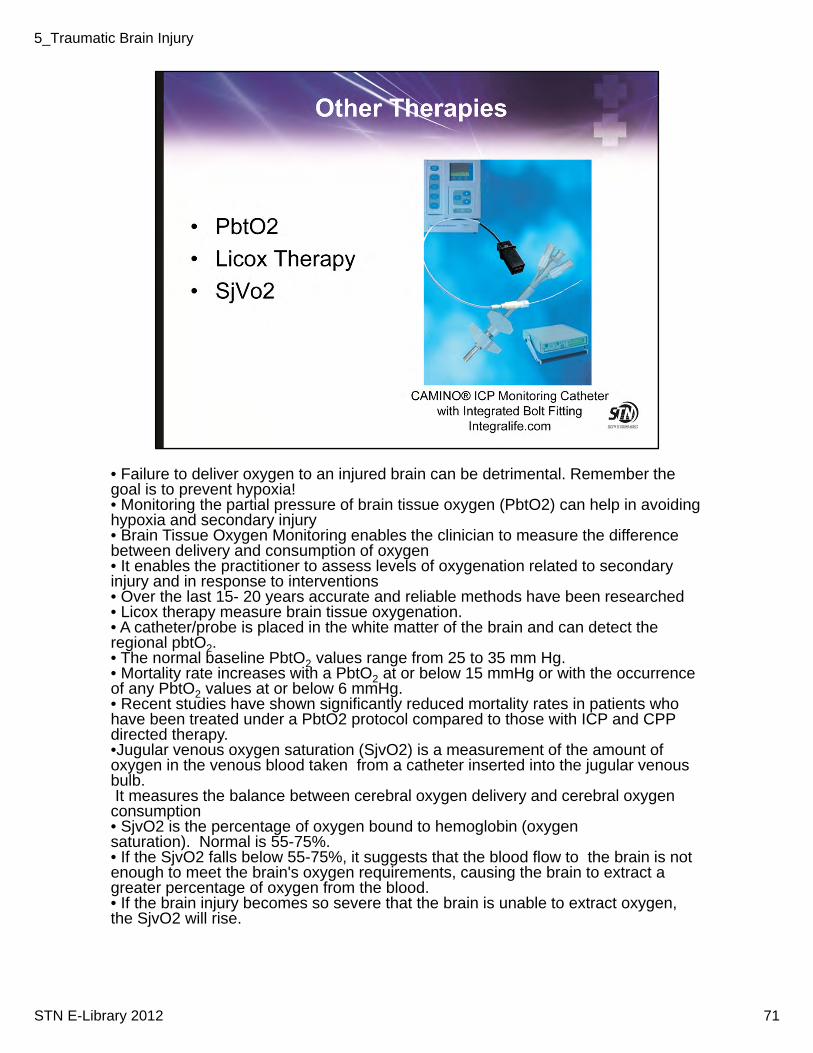
• Failure to deliver oxygen to an injured brain can be detrimental. Remember the goal is to prevent hypoxia!• Monitoring the partial pressure of brain tissue oxygen (PbtO2) can help in avoiding hypoxia and secondary injury• Brain Tissue Oxygen Monitoring enables the clinician to measure the difference between delivery and consumption of oxygen• It enables the practitioner to assess levels of oxygenation related to secondary injury and in response to interventions• Over the last 15- 20 years accurate and reliable methods have been researched• Licox therapy measure brain tissue oxygenation. • A catheter/probe is placed in the white matter of the brain and can detect the regional pbtO2. • The normal baseline PbtO2 values range from 25 to 35 mm Hg. • Mortality rate increases with a PbtO2 at or below 15 mmHg or with the occurrence of any PbtO2 values at or below 6 mmHg.• Recent studies have shown significantly reduced mortality rates in patients who have been treated under a PbtO2 protocol compared to those with ICP and CPP directed therapy. •Jugular venous oxygen saturation (SjvO2) is a measurement of the amount of oxygen in the venous blood taken from a catheter inserted into the jugular venous bulb. It measures the balance between cerebral oxygen delivery and cerebral oxygen
consumption• SjvO2 is the percentage of oxygen bound to hemoglobin (oxygen saturation). Normal is 55-75%.• If the SjvO2 falls below 55-75%, it suggests that the blood flow to the brain is not enough to meet the brain's oxygen requirements, causing the brain to extract a greater percentage of oxygen from the blood.• If the brain injury becomes so severe that the brain is unable to extract oxygen, the SjvO2 will rise.
71STN E-Library 2012
5_Traumatic Brain Injury
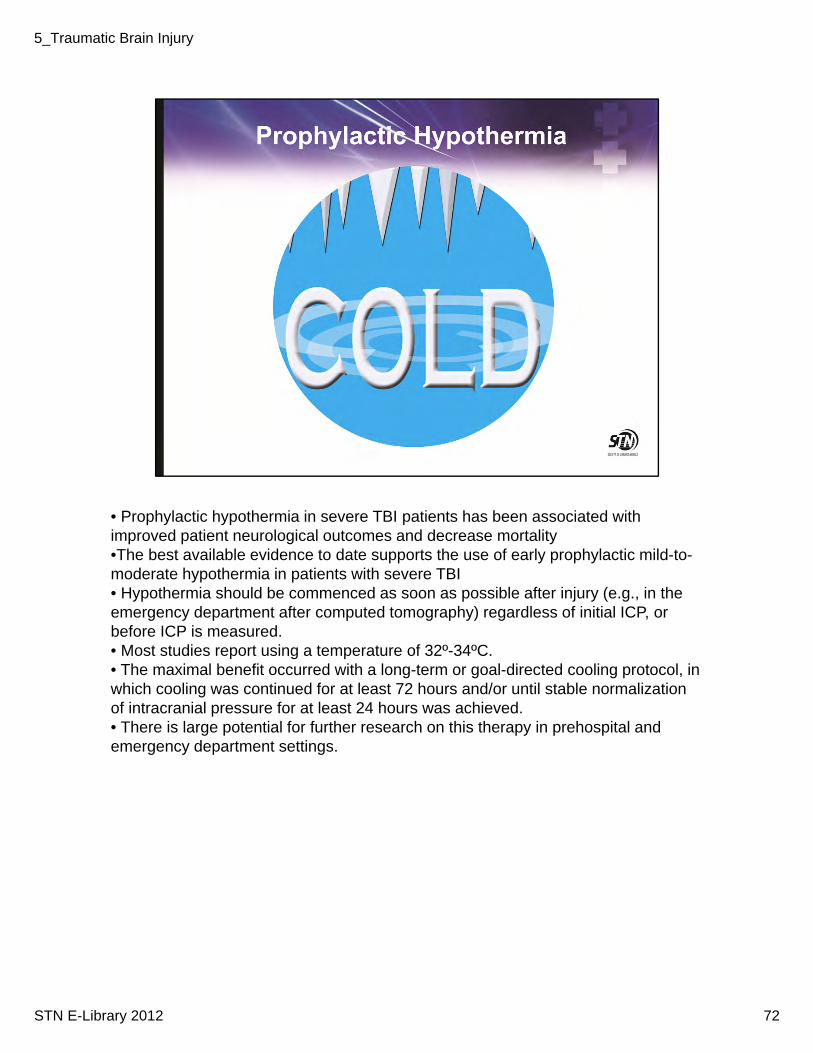
• Prophylactic hypothermia in severe TBI patients has been associated with improved patient neurological outcomes and decrease mortality•The best available evidence to date supports the use of early prophylactic mild-to-moderate hypothermia in patients with severe TBI• Hypothermia should be commenced as soon as possible after injury (e.g., in the emergency department after computed tomography) regardless of initial ICP, or before ICP is measured. • Most studies report using a temperature of 32º-34ºC. • The maximal benefit occurred with a long-term or goal-directed cooling protocol, in which cooling was continued for at least 72 hours and/or until stable normalization of intracranial pressure for at least 24 hours was achieved.• There is large potential for further research on this therapy in prehospital and emergency department settings.
72STN E-Library 2012
5_Traumatic Brain Injury
• The diagnosis of brain death should be considered after all measures have been taken to restore normal physiological parameters.• All medications should be reviewed for potential CNS side effects• Criteria for brain death include
• nonreactive pupils• GCS = 3• absent brainstem reflexes ( no gag reflex, Dolls eyes)• failed apnea test ( no spontaneous ventilations )Confirmation of brain death includes• cerebral angiogram•EEG ( electroencephalography) showing no activityOrgan procurement agencies should be notified according to hospital and Procurement Agency. Review your hospital protocols for Brain Death and Organ Procurement.
73STN E-Library 2012
5_Traumatic Brain Injury
Useful Websites
Brain Trauma Foundationhttp://www.braintrauma.org/
National Institute of Neurological Disorders and Strokehttp://www.ninds.nih.gov/Brain Injury Association of Americahttp://www.biausa.org/Brain Trauma Foundation Learning Portalhttp://www.btflearning.org/go/HomeAmerican Association of Neuroscience Nurseshttp://www.aann.org/
74STN E-Library 2012
5_Traumatic Brain Injury