Embed Size (px)
Citation preview
4-3: Cell Organelles + 4-3: Cell Organelles + FeaturesFeatures
Eukaryotic cells have many membrane Eukaryotic cells have many membrane systemssystems Divide cells into compartments that function Divide cells into compartments that function
together to keep a cell alivetogether to keep a cell alive
Plasma Membrane Plasma Membrane (Factory Doors)(Factory Doors)
Location: surrounds the cell + its partsLocation: surrounds the cell + its parts Functions: Functions:
Controls the ease w/ which substances pass into Controls the ease w/ which substances pass into and out of the cell - - known as and out of the cell - - known as selectively selectively permeablepermeable
Separates internal rxns with external environmentSeparates internal rxns with external environment Allows the cell to excrete wastes and interact with Allows the cell to excrete wastes and interact with
environmentenvironment
Description: made of lipids + proteinsDescription: made of lipids + proteins
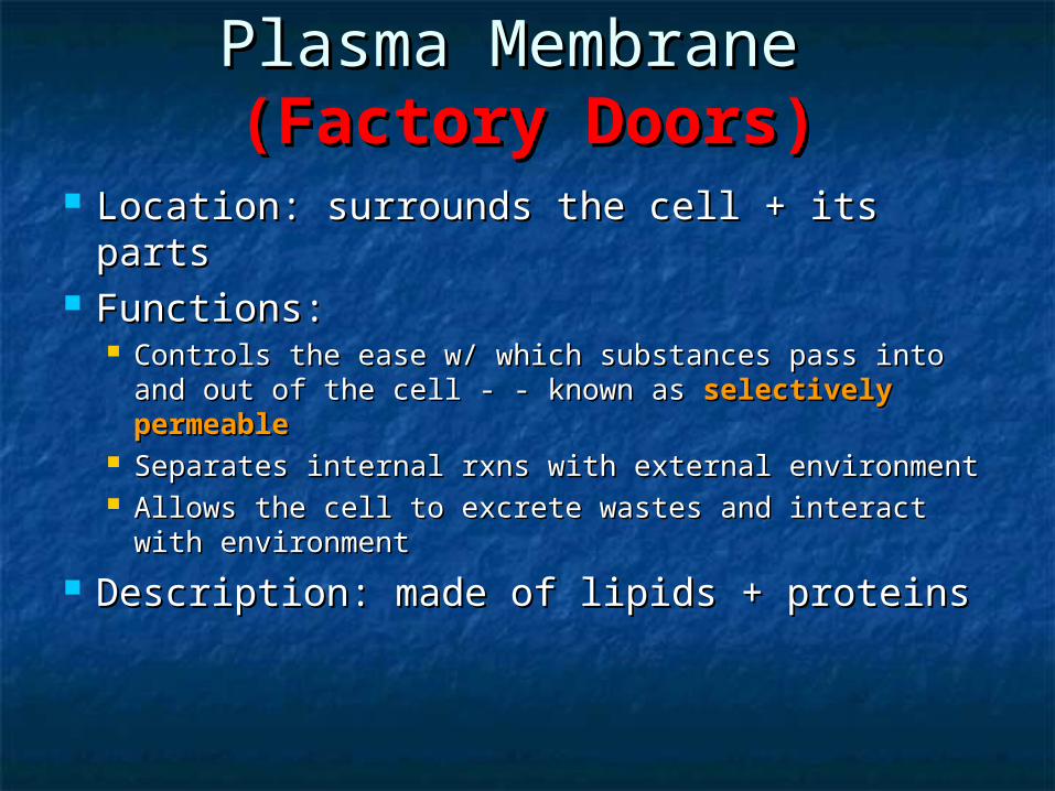
Membrane LipidsMembrane Lipids
Major type – Major type – phospholipidsphospholipids Hydrophilic (head) – Hydrophilic (head) –
Hydrophobic (tail)Hydrophobic (tail) Forms a lipid bilayerForms a lipid bilayer
Heads outward, tails Heads outward, tails inwardinward
Contains sterols Contains sterols between the tailsbetween the tails
Example: CholesterolExample: Cholesterol
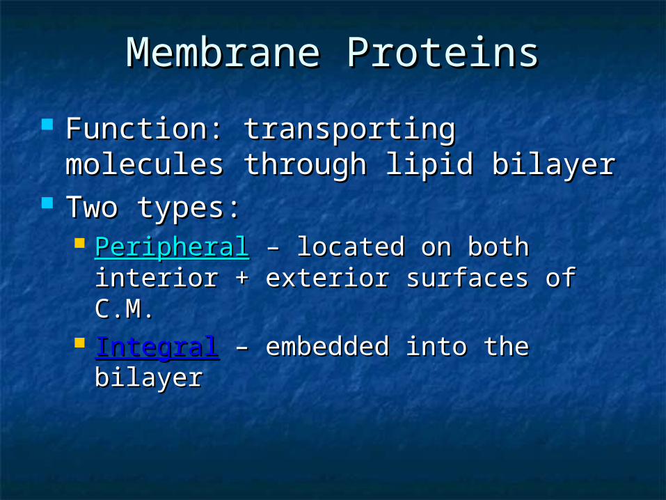
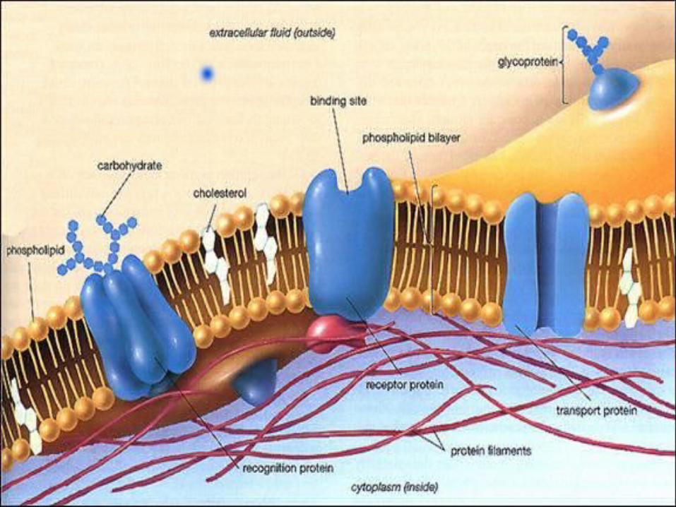
Membrane ProteinsMembrane Proteins
Function: transporting molecules Function: transporting molecules through lipid bilayerthrough lipid bilayer
Two types:Two types: PeripheralPeripheral – located on both interior + – located on both interior +
exterior surfaces of C.M.exterior surfaces of C.M. IntegralIntegral – embedded into the bilayer– embedded into the bilayer
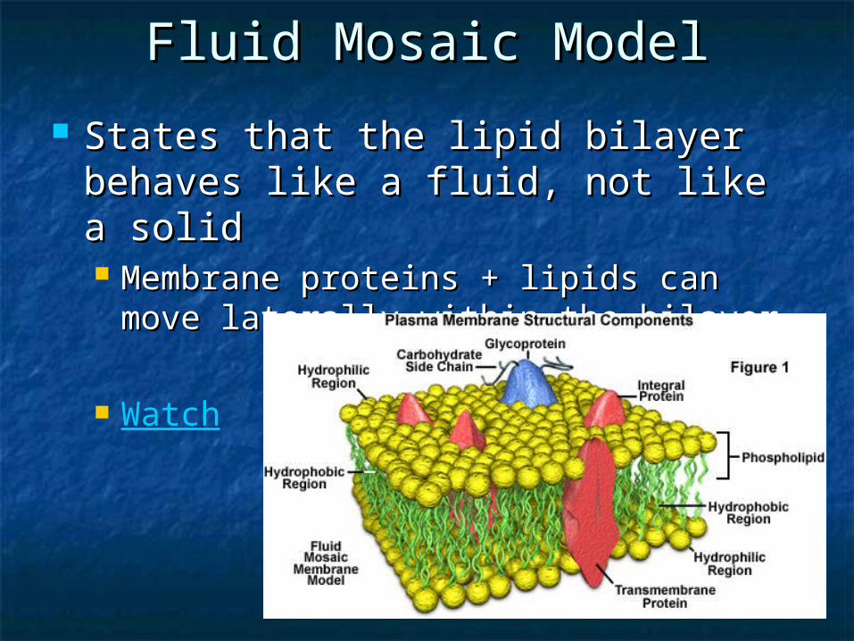
Fluid Mosaic ModelFluid Mosaic Model
States that the lipid bilayer behaves States that the lipid bilayer behaves like a fluid, not like a solidlike a fluid, not like a solid Membrane proteins + lipids can move Membrane proteins + lipids can move
laterally within the bilayerlaterally within the bilayer
Watch
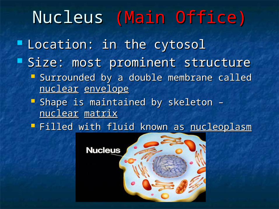
Nucleus Nucleus (Main Office)(Main Office) Location: in the cytosolLocation: in the cytosol Size: most prominent structureSize: most prominent structure
Surrounded by a double membrane called Surrounded by a double membrane called nuclearnuclear envelopeenvelope
Shape is maintained by skeleton – Shape is maintained by skeleton – nuclearnuclear matrixmatrix
Filled with fluid known as Filled with fluid known as nucleoplasmnucleoplasm
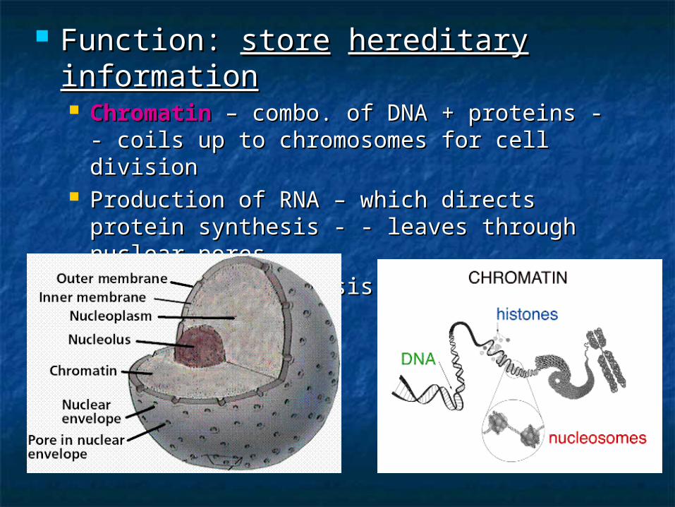
Function: Function: storestore hereditaryhereditary informationinformation ChromatinChromatin – combo. of DNA + proteins - - – combo. of DNA + proteins - -
coils up to chromosomes for cell divisioncoils up to chromosomes for cell division Production of RNA – which directs protein Production of RNA – which directs protein
synthesis - - leaves through nuclear poressynthesis - - leaves through nuclear pores NucleolusNucleolus – synthesis of ribosomes – synthesis of ribosomes
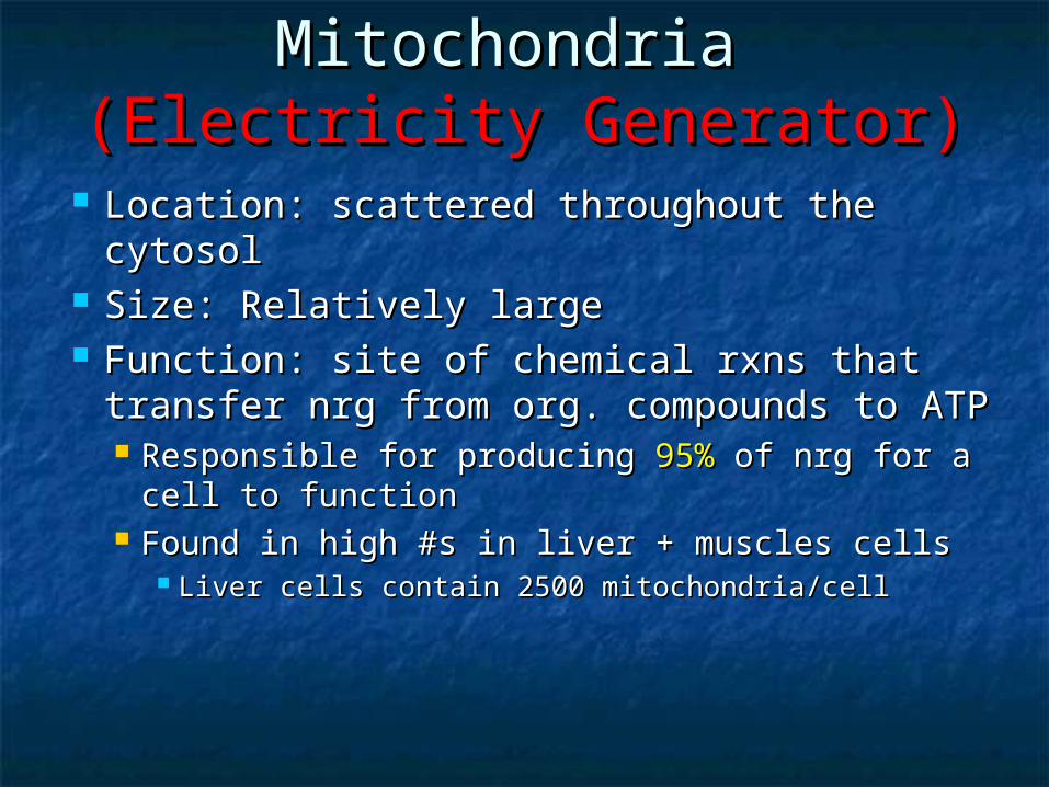
Mitochondria Mitochondria (Electricity Generator)(Electricity Generator)
Location: scattered throughout the Location: scattered throughout the cytosolcytosol
Size: Relatively largeSize: Relatively large Function: site of chemical rxns that Function: site of chemical rxns that
transfer nrg from org. compounds to ATPtransfer nrg from org. compounds to ATP Responsible for producing Responsible for producing 95% 95% of nrg for a of nrg for a
cell to functioncell to function Found in high #s in liver + muscles cellsFound in high #s in liver + muscles cells
Liver cells contain 2500 mitochondria/cellLiver cells contain 2500 mitochondria/cell
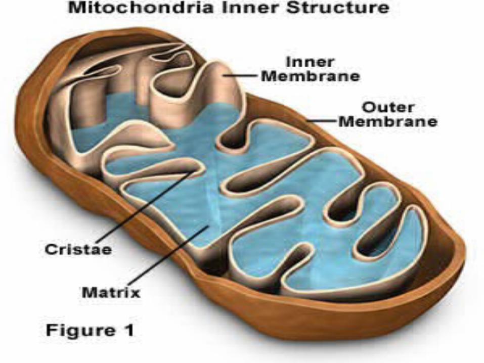
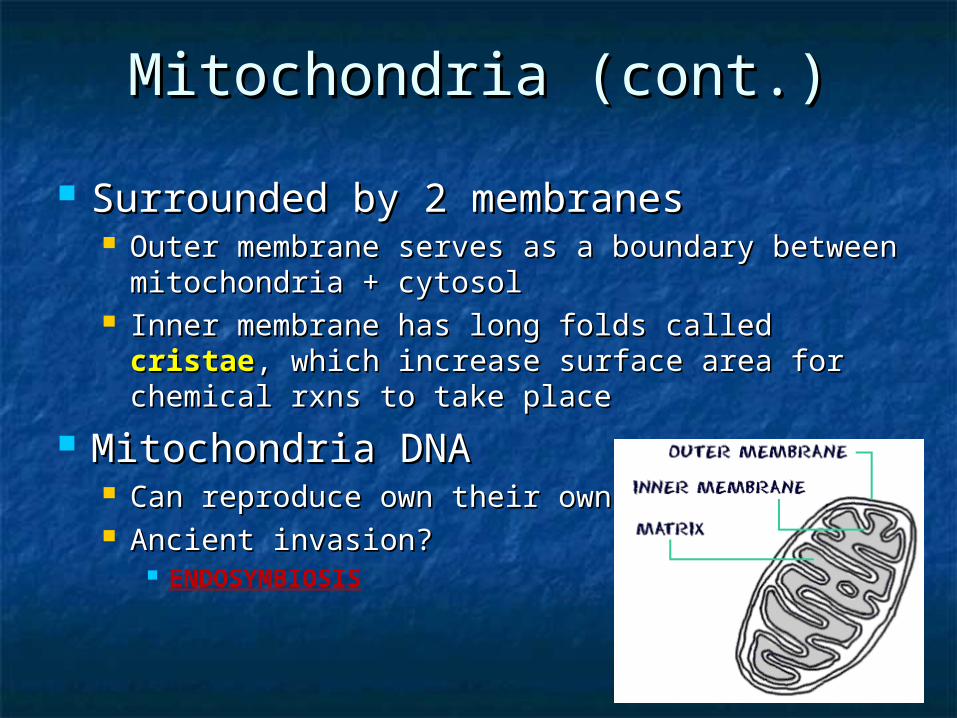
Mitochondria (cont.)Mitochondria (cont.)
Surrounded by 2 membranesSurrounded by 2 membranes Outer membrane serves as a boundary between Outer membrane serves as a boundary between
mitochondria + cytosolmitochondria + cytosol Inner membrane has long folds called Inner membrane has long folds called cristaecristae, ,
which increase surface area for chemical rxns to which increase surface area for chemical rxns to take placetake place
Mitochondria DNAMitochondria DNA Can reproduce own their ownCan reproduce own their own Ancient invasion?Ancient invasion?
ENDOSYMBIOSIS
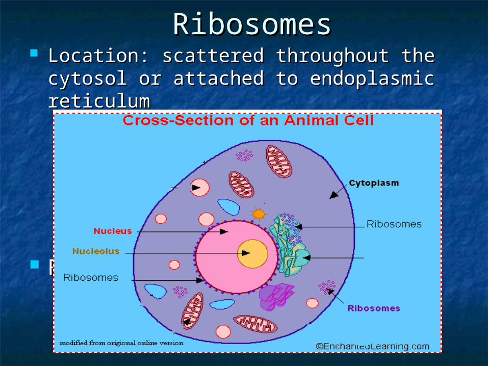
RibosomesRibosomes Location: scattered throughout the cytosol Location: scattered throughout the cytosol
or attached to endoplasmic reticulumor attached to endoplasmic reticulum Made in nucleolus, completed in cytoplasmMade in nucleolus, completed in cytoplasm NOT membrane bound – evolution (prokaryotic NOT membrane bound – evolution (prokaryotic
cells)cells) Size: relatively small + most numerous; Size: relatively small + most numerous;
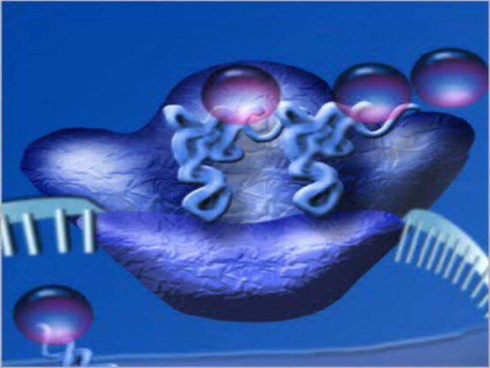
made of protein + RNAmade of protein + RNA Function: Function: Protein synthesisProtein synthesis
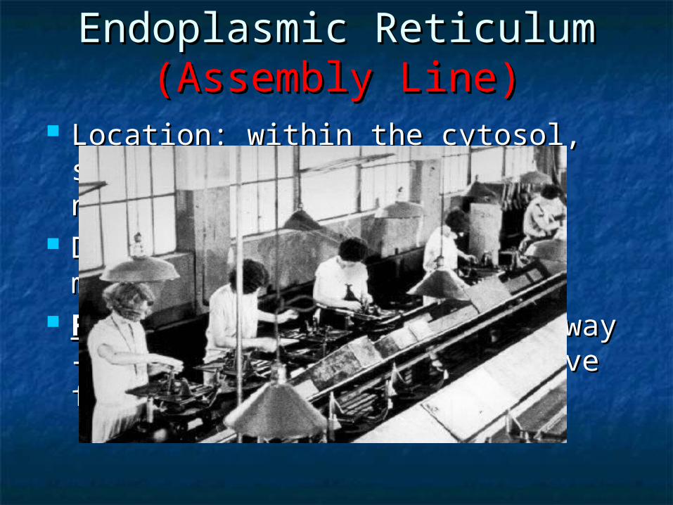
Endoplasmic Reticulum Endoplasmic Reticulum (Assembly Line)(Assembly Line)
Location: within the cytosol, Location: within the cytosol, sometimes attached to the nucleussometimes attached to the nucleus
Description: cisternae – membranous Description: cisternae – membranous tubes + sacstubes + sacs
FunctionFunction: Intracellular highway – a : Intracellular highway – a path for molecules to move from one path for molecules to move from one part to anotherpart to another
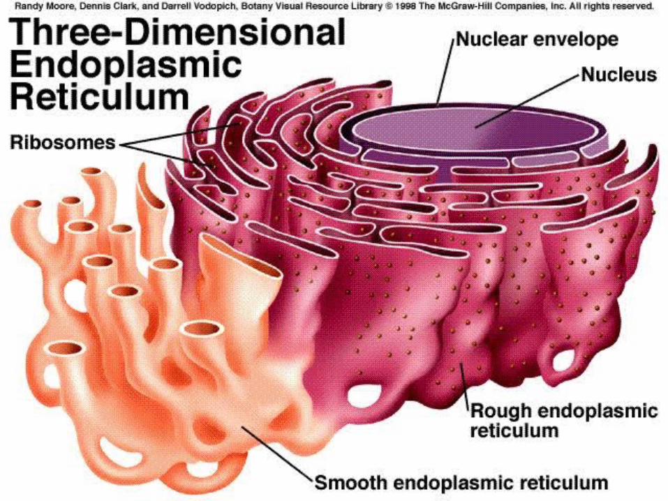
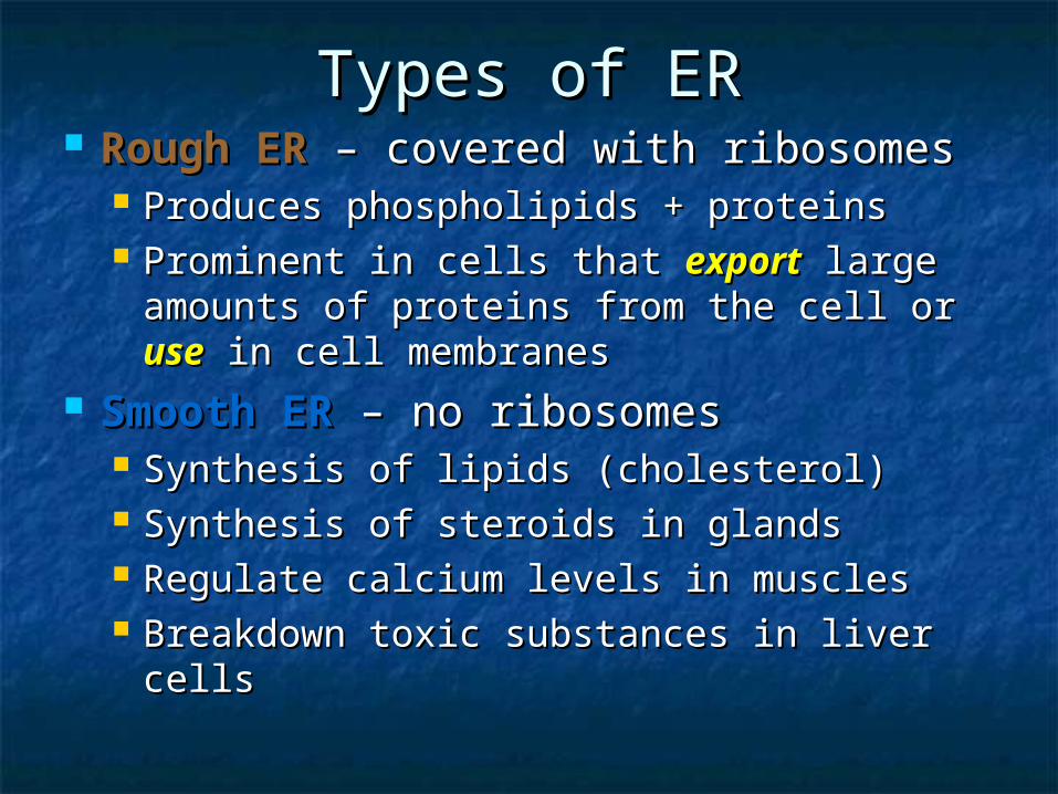
Types of ERTypes of ER Rough ER Rough ER – covered with ribosomes– covered with ribosomes
Produces phospholipids + proteins Produces phospholipids + proteins Prominent in cells that Prominent in cells that exportexport large large
amounts of proteins from the cell or amounts of proteins from the cell or useuse in cell membranesin cell membranes
Smooth ER Smooth ER – no ribosomes – no ribosomes Synthesis of lipids (cholesterol)Synthesis of lipids (cholesterol) Synthesis of steroids in glandsSynthesis of steroids in glands Regulate calcium levels in musclesRegulate calcium levels in muscles Breakdown toxic substances in liver cellsBreakdown toxic substances in liver cells
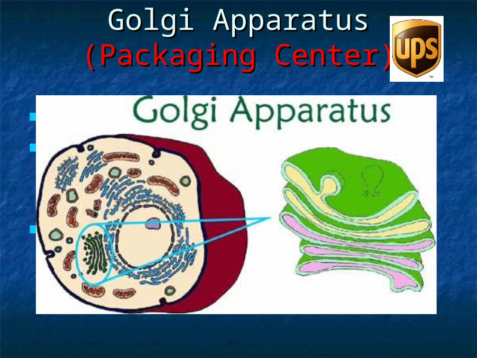
Golgi ApparatusGolgi Apparatus(Packaging Center)(Packaging Center)
Location: within the cytosolLocation: within the cytosol Appearance: system of membranes; Appearance: system of membranes;
series of flattened sacsseries of flattened sacs Function: works with ER to modify Function: works with ER to modify
proteins for transport from cellproteins for transport from cell
VesiclesVesicles Small, spherical sacsSmall, spherical sacs Classified by contentsClassified by contents
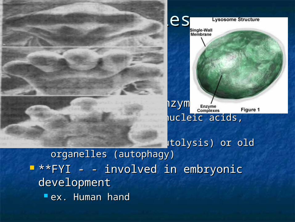
LysosomesLysosomes Made by GolgiMade by Golgi Contain digestive enzymes Contain digestive enzymes
breakdown proteins, nucleic acids, carbs, breakdown proteins, nucleic acids, carbs, fatsfats
Digests old cells (autolysis) or old organelles Digests old cells (autolysis) or old organelles (autophagy)(autophagy)
**FYI - - involved in embryonic **FYI - - involved in embryonic development development
ex. Human handex. Human hand
VesiclesVesicles PeroxisomesPeroxisomes
Abundant in liver + kidney cellsAbundant in liver + kidney cells Neutralize free radicals (Oxygen ions), Neutralize free radicals (Oxygen ions),
detoxify alcohol + other drugsdetoxify alcohol + other drugs Break down fatty acids for Break down fatty acids for nrgnrg source source
GlyoxysomesGlyoxysomes Found in plant seedsFound in plant seeds Help break down fats to supply embryo Help break down fats to supply embryo
with foodwith food
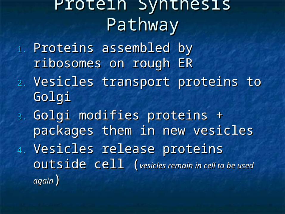
Protein Synthesis PathwayProtein Synthesis Pathway
1.1. Proteins assembled by ribosomes on Proteins assembled by ribosomes on rough ERrough ER
2.2. Vesicles transport proteins to GolgiVesicles transport proteins to Golgi
3.3. Golgi modifies proteins + packages Golgi modifies proteins + packages them in new vesiclesthem in new vesicles
4.4. Vesicles release proteins outside cell Vesicles release proteins outside cell ((vesicles remain in cell to be used againvesicles remain in cell to be used again))
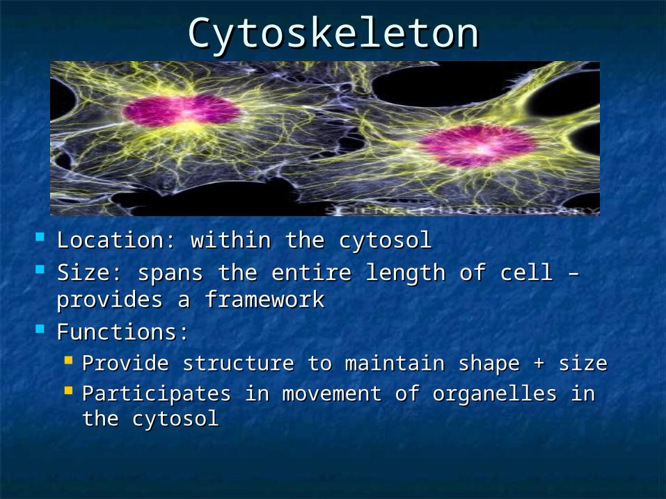
CytoskeletonCytoskeleton
Location: within the cytosolLocation: within the cytosol Size: spans the entire length of cell – provides a Size: spans the entire length of cell – provides a
frameworkframework Functions:Functions:
Provide structure to maintain shape + sizeProvide structure to maintain shape + size Participates in movement of organelles in the Participates in movement of organelles in the
cytosolcytosol
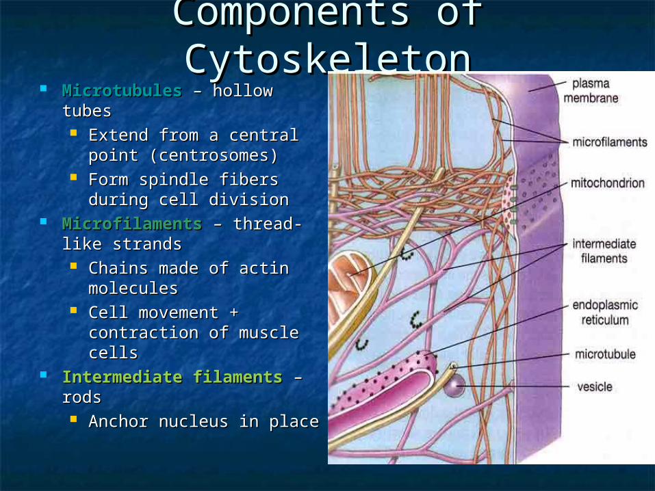
Components of Components of CytoskeletonCytoskeleton
MicrotubulesMicrotubules – hollow – hollow tubestubes Extend from a central Extend from a central
point (centrosomes)point (centrosomes) Form spindle fibers Form spindle fibers
during cell divisionduring cell division MicrofilamentsMicrofilaments – thread- – thread-
like strandslike strands Chains made of actin Chains made of actin
moleculesmolecules Cell movement + Cell movement +
contraction of muscle contraction of muscle cellscells
Intermediate filaments Intermediate filaments – – rodsrods Anchor nucleus in placeAnchor nucleus in place
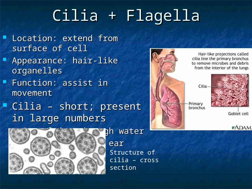
Cilia + FlagellaCilia + Flagella Location: extend from surface Location: extend from surface
of cellof cell Appearance: hair-like organellesAppearance: hair-like organelles Function: assist in movementFunction: assist in movement Cilia – short; present in Cilia – short; present in
large numberslarge numbers Propulsion through waterPropulsion through water Ex. Nose + inner earEx. Nose + inner ear
Structure of cilia – cross section
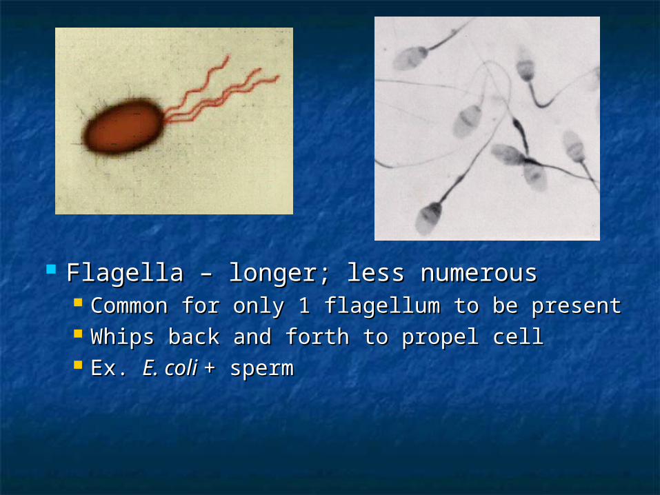
Flagella – longer; less numerousFlagella – longer; less numerous Common for only 1 flagellum to be presentCommon for only 1 flagellum to be present Whips back and forth to propel cellWhips back and forth to propel cell Ex. Ex. E. coli E. coli + sperm+ sperm
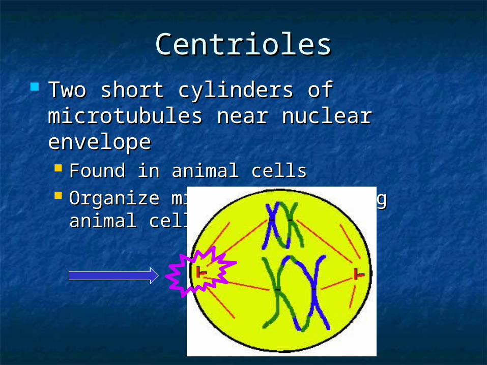
CentriolesCentrioles Two short cylinders of microtubules Two short cylinders of microtubules
near nuclear envelopenear nuclear envelope Found in animal cellsFound in animal cells Organize microtubules during animal Organize microtubules during animal
cell divisioncell division