Embed Size (px)
Citation preview
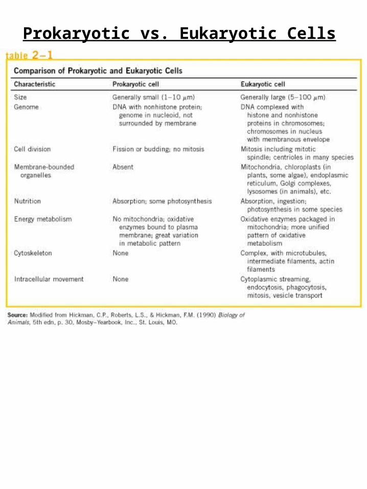
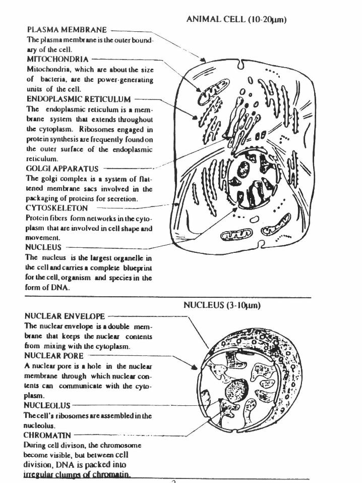
Prokaryotic vs. Eukaryotic Cells
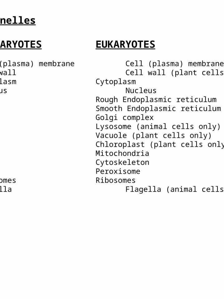
Organelles
PROKARYOTES EUKARYOTES
Cell (plasma) membrane Cell (plasma) membraneCell wall Cell wall (plant cells only)Cytoplasm Cytoplasm Nucleus Nucleus
Rough Endoplasmic reticulumSmooth Endoplasmic reticulum Golgi complexLysosome (animal cells only)Vacuole (plant cells only)Chloroplast (plant cells only)MitochondriaCytoskeletonPeroxisome
Ribosomes RibosomesFlagella Flagella (animal cells only)Pili
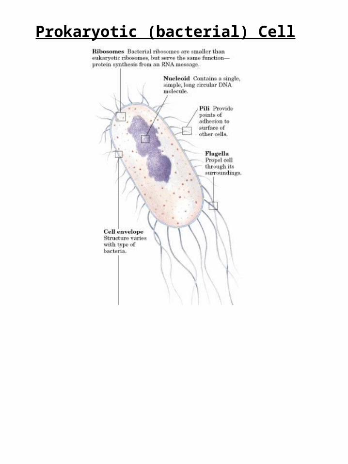
Prokaryotic (bacterial) Cell
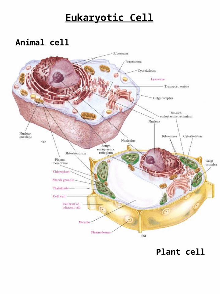
Eukaryotic Cell
Animal cell
Plant cell
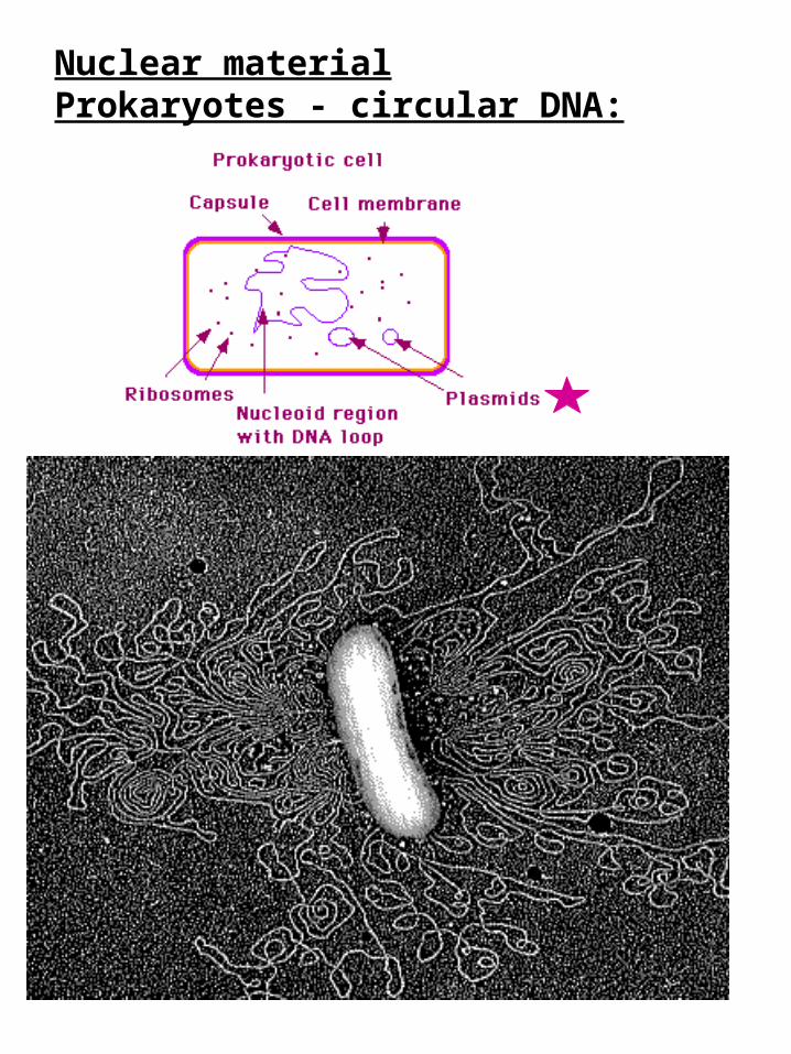
Nuclear materialProkaryotes - circular DNA:
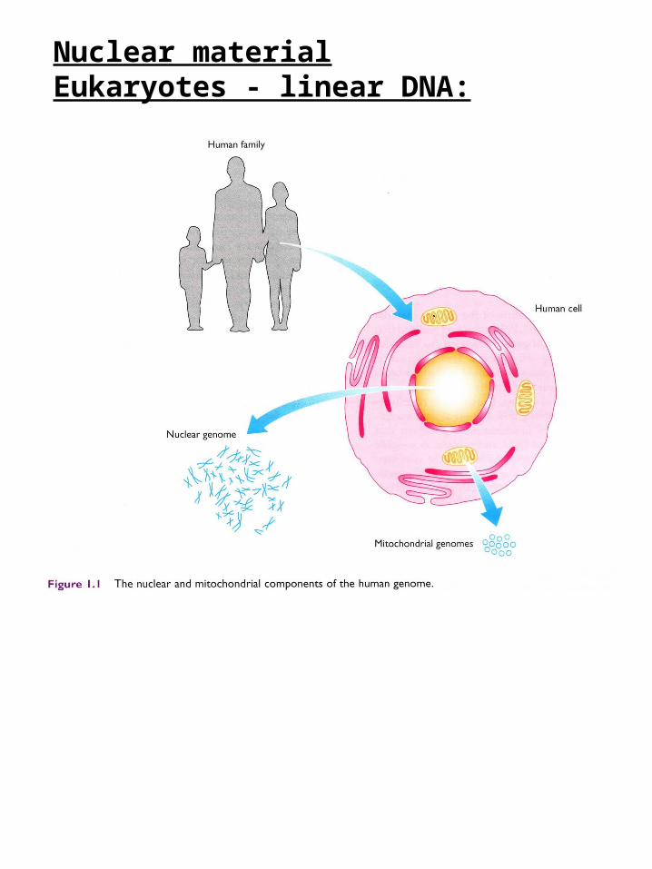
Nuclear materialEukaryotes - linear DNA:
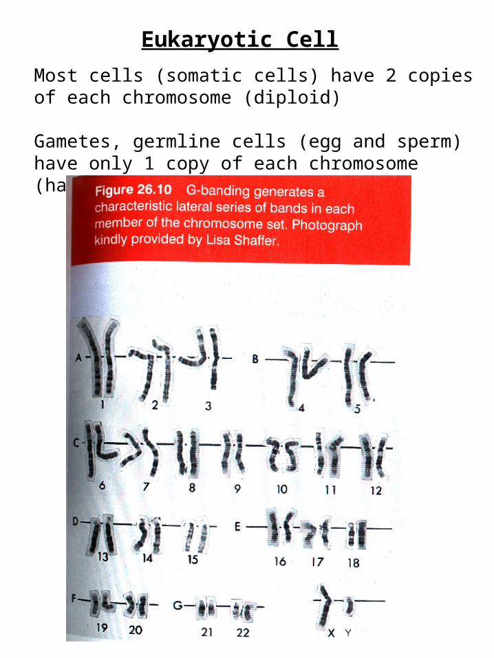
Eukaryotic Cell
Most cells (somatic cells) have 2 copies of each chromosome (diploid)
Gametes, germline cells (egg and sperm) have only 1 copy of each chromosome (haploid)

Eukaryotic CellWe have 2 meters of DNA in all our somatic cellsHOW DOES IT ALL FIT??
Condensation of DNA by proteins!!
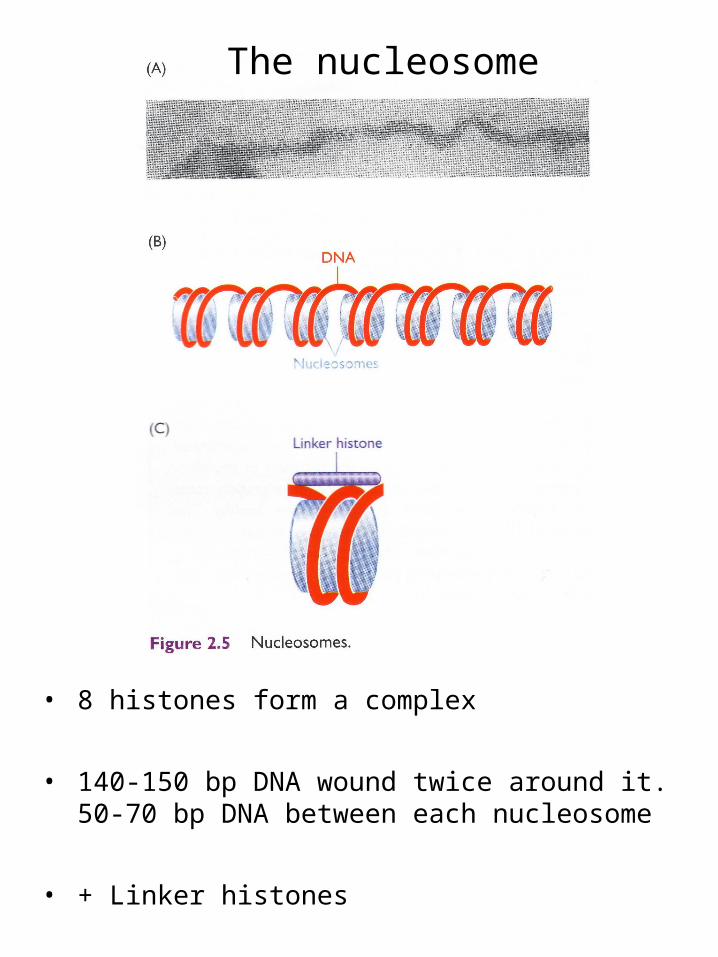
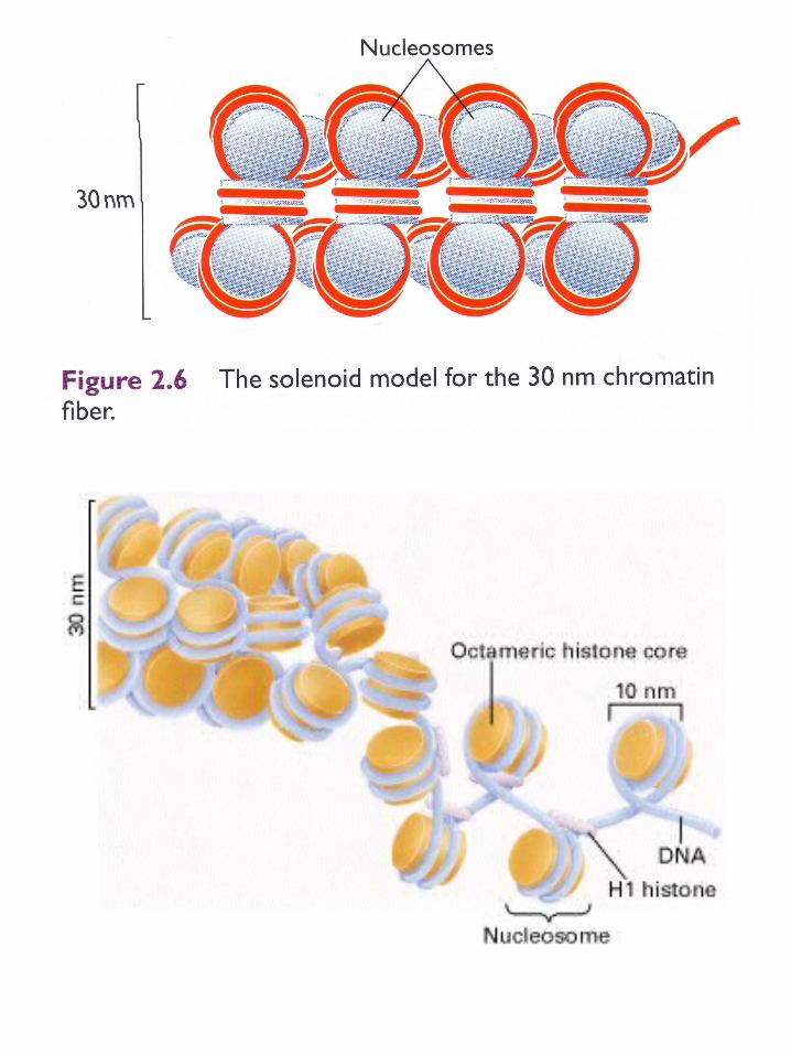
The nucleosome
• 8 histones form a complex
• 140-150 bp DNA wound twice around it. 50-70 bp DNA between each nucleosome
• + Linker histones
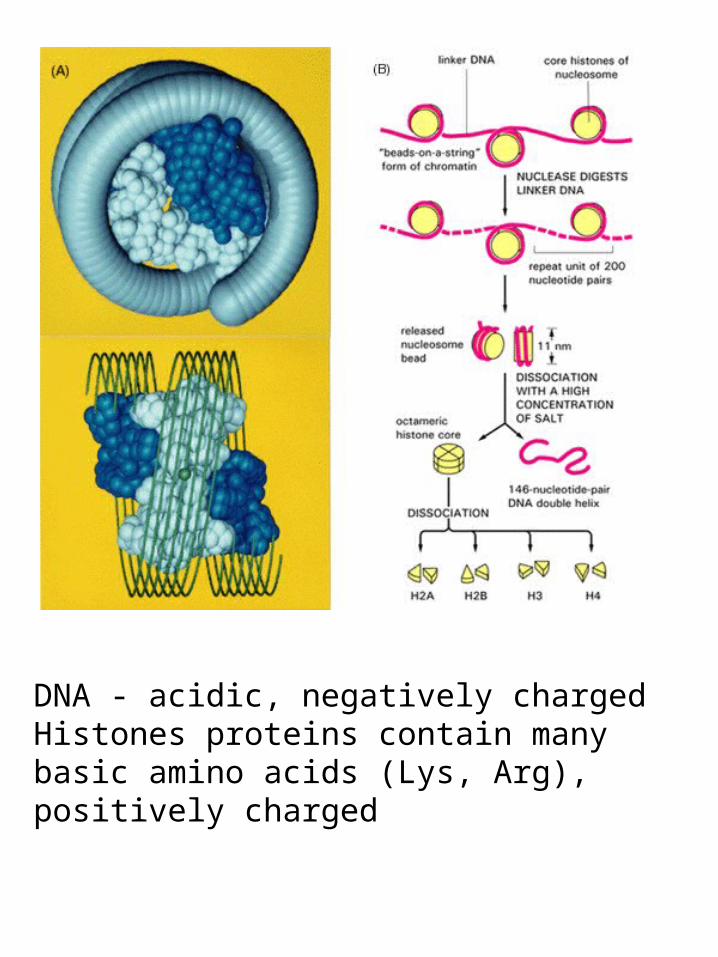
DNA - acidic, negatively chargedHistones proteins contain many basic amino acids (Lys, Arg), positively charged

Beads on a String(in Colorado)
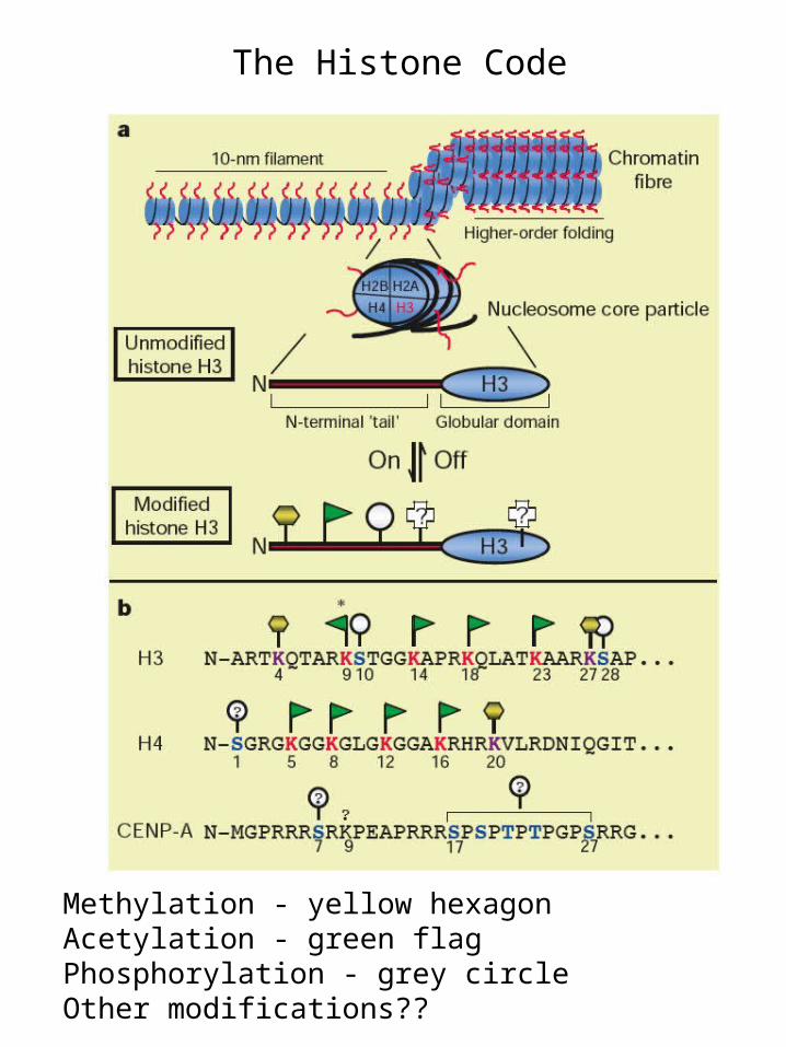
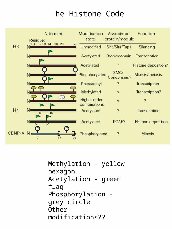
Methylation - yellow hexagonAcetylation - green flagPhosphorylation - grey circle Other modifications??
The Histone Code
Methylation - yellow hexagonAcetylation - green flagPhosphorylation - grey circle Other modifications??
The Histone Code
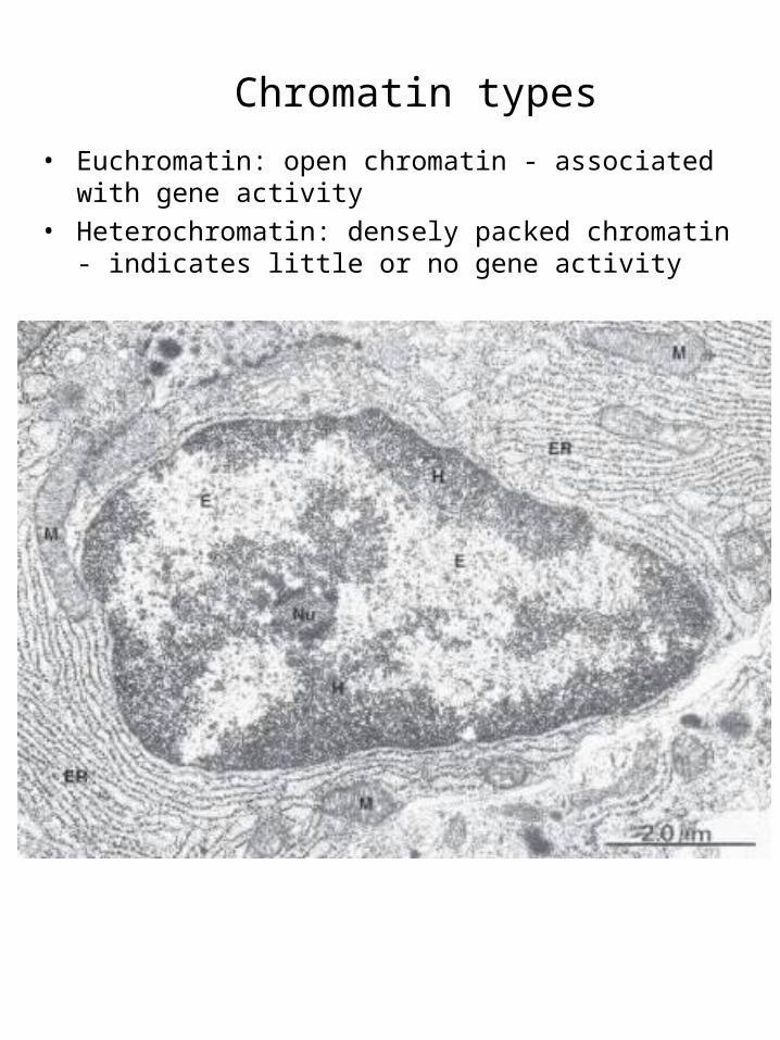
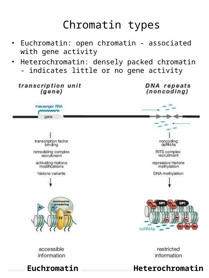
Chromatin types
• Euchromatin: open chromatin - associated with gene activity
• Heterochromatin: densely packed chromatin - indicates little or no gene activity
Chromatin types
• Euchromatin: open chromatin - associated with gene activity
• Heterochromatin: densely packed chromatin - indicates little or no gene activity
Euchromatin Heterochromatin
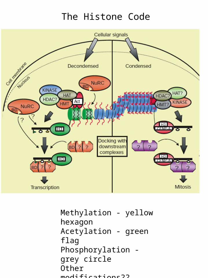
Methylation - yellow hexagonAcetylation - green flagPhosphorylation - grey circle Other modifications??
The Histone Code
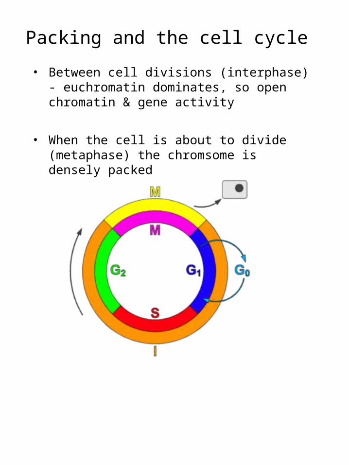
Packing and the cell cycle
• Between cell divisions (interphase) - euchromatin dominates, so open chromatin & gene activity
• When the cell is about to divide (metaphase) the chromsome is densely packed
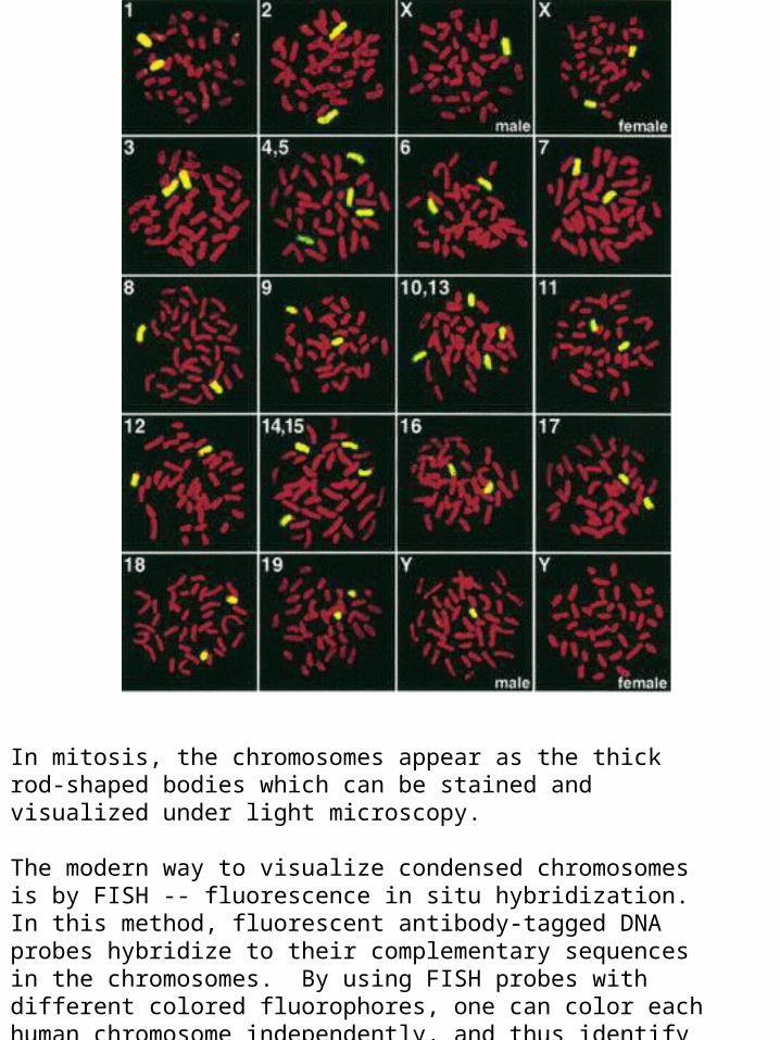
In mitosis, the chromosomes appear as the thick rod-shaped bodies which can be stained and visualized under light microscopy.
The modern way to visualize condensed chromosomes is by FISH -- fluorescence in situ hybridization. In this method, fluorescent antibody-tagged DNA probes hybridize to their complementary sequences in the chromosomes. By using FISH probes with different colored fluorophores, one can color each human chromosome independently, and thus identify all 23 chromosomes. This is called chromosome painting.
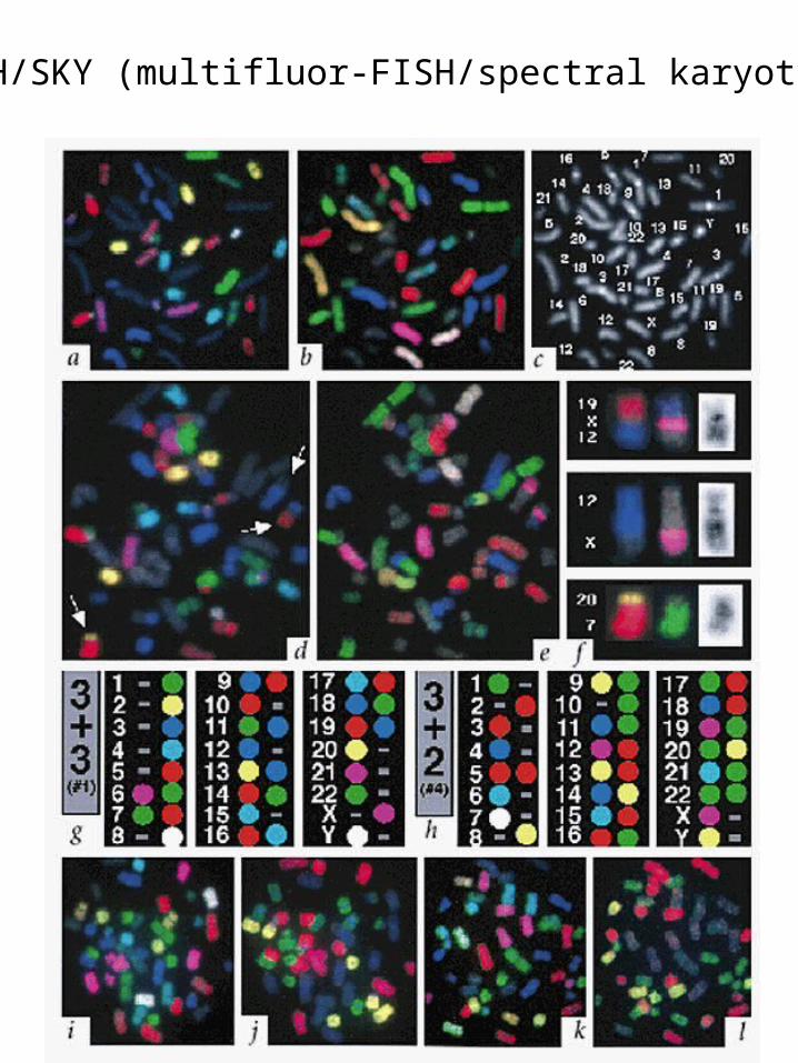
M-FISH/SKY (multifluor-FISH/spectral karyotyping)
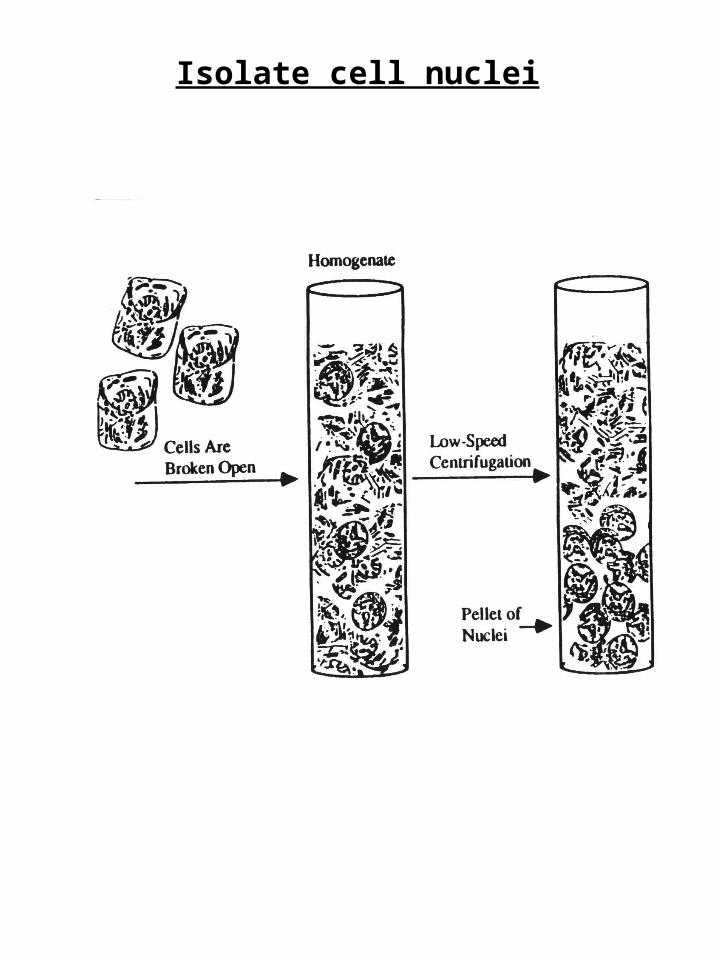
Isolate cell nuclei
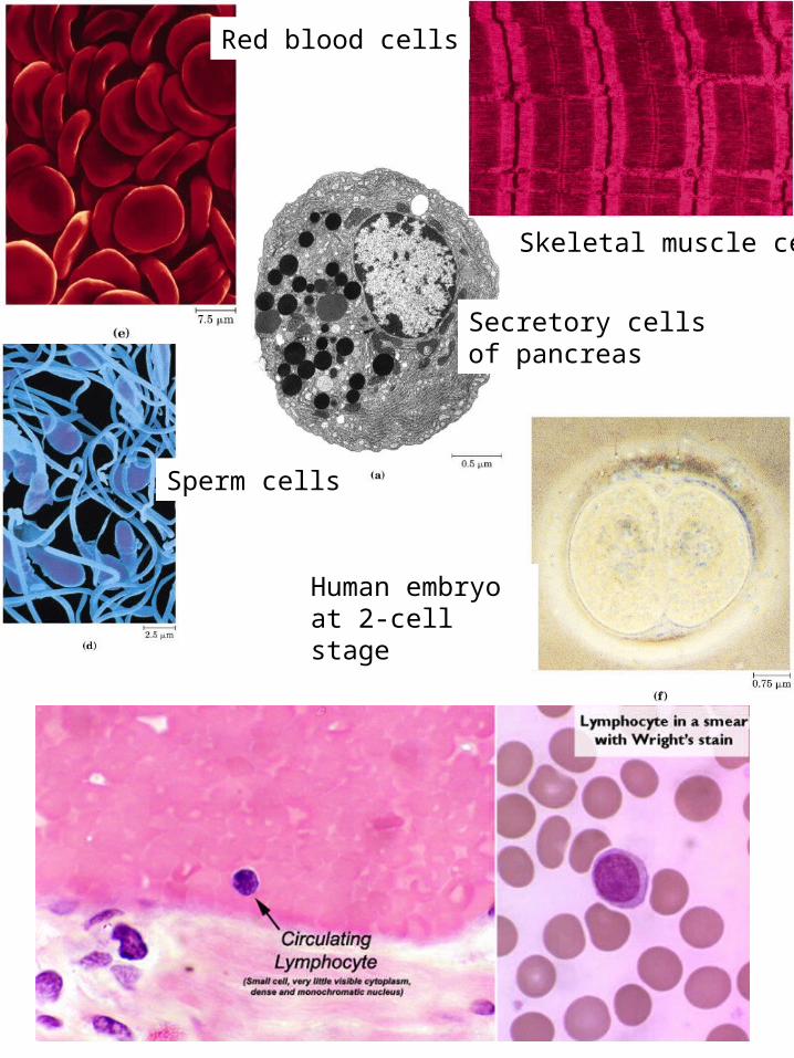
Secretory cells of pancreas
Skeletal muscle cell
Sperm cells
Red blood cells
Human embryo at 2-cell stage
Cell breakageAKA cell disruption, cell disintegration, lysis
Goal - destroy outer cell membrane without destroying organelle membranes
Cells broken open (plasma membrane dissolved) by:
Mechanical Chemical• freeze-thaw • solubilize with detergents• grinding • organic solvents• shearing (homogenizer) • alkali treatment• shearing (french press) • enzymatic digestion
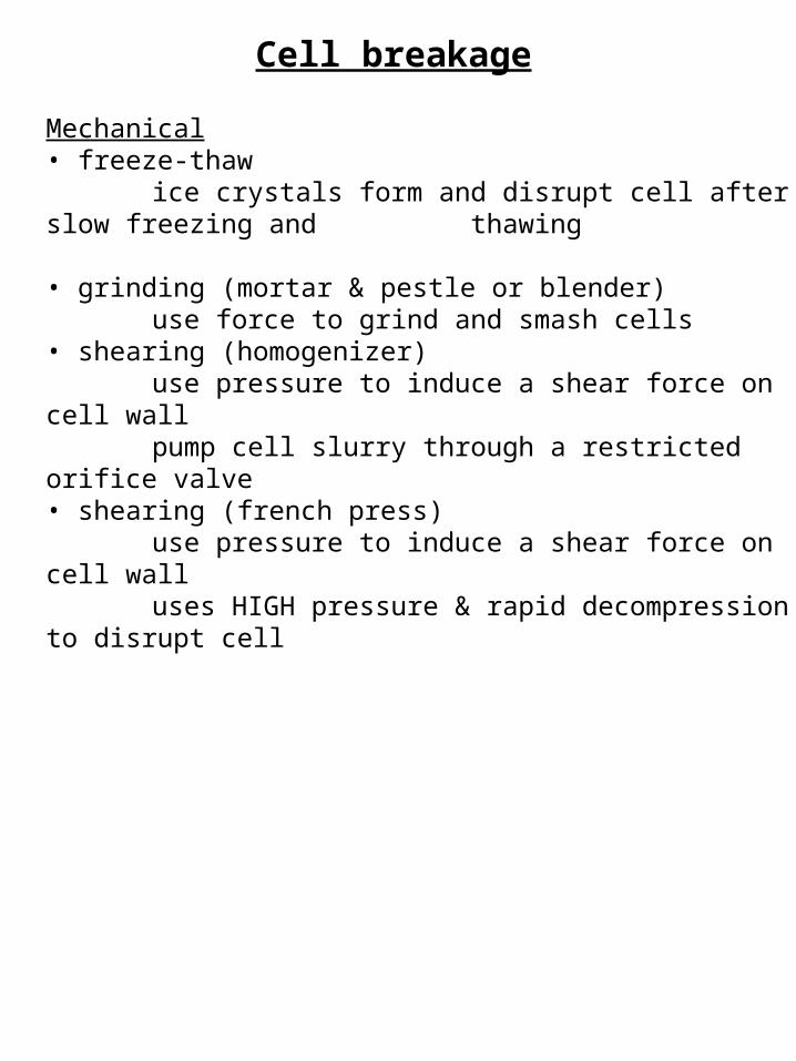
Cell breakage
Mechanical• freeze-thaw
ice crystals form and disrupt cell after slow freezing and thawing
• grinding (mortar & pestle or blender)use force to grind and smash cells
• shearing (homogenizer)use pressure to induce a shear force on cell wallpump cell slurry through a restricted orifice valve
• shearing (french press)use pressure to induce a shear force on cell wall uses HIGH pressure & rapid decompression to disrupt cell
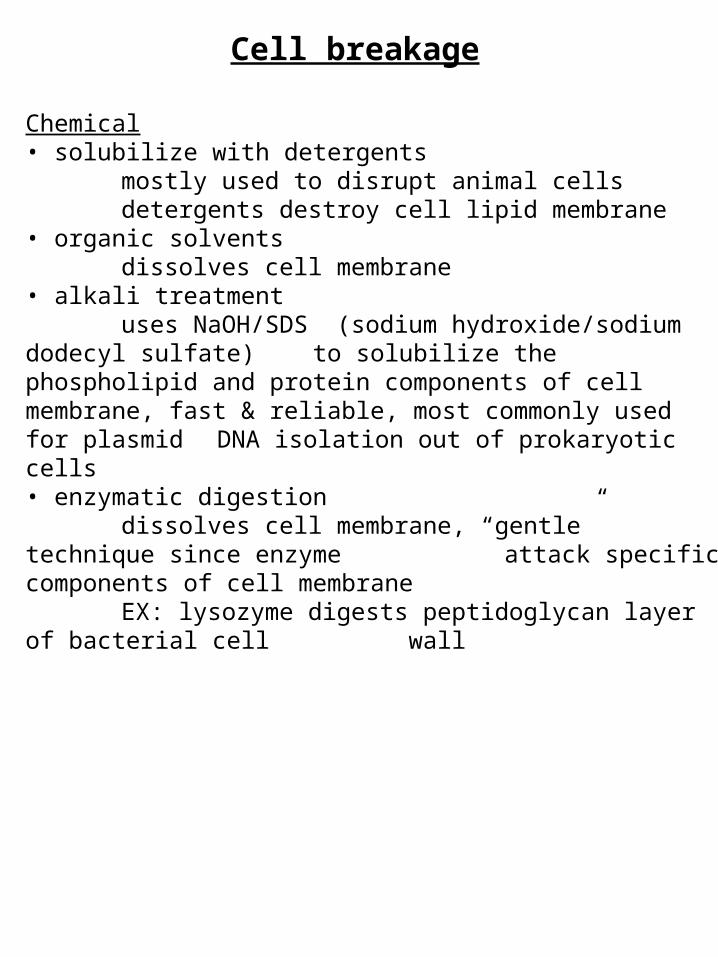
Cell breakage
Chemical• solubilize with detergents
mostly used to disrupt animal cellsdetergents destroy cell lipid membrane
• organic solventsdissolves cell membrane
• alkali treatmentuses NaOH/SDS (sodium hydroxide/sodium dodecyl sulfate) to solubilize the phospholipid and protein components of cell membrane, fast & reliable, most commonly used for plasmid DNA isolation out of prokaryotic cells
• enzymatic digestiondissolves cell membrane, “gentle” technique since enzyme attack specific components of cell membraneEX: lysozyme digests peptidoglycan layer of bacterial cell wall
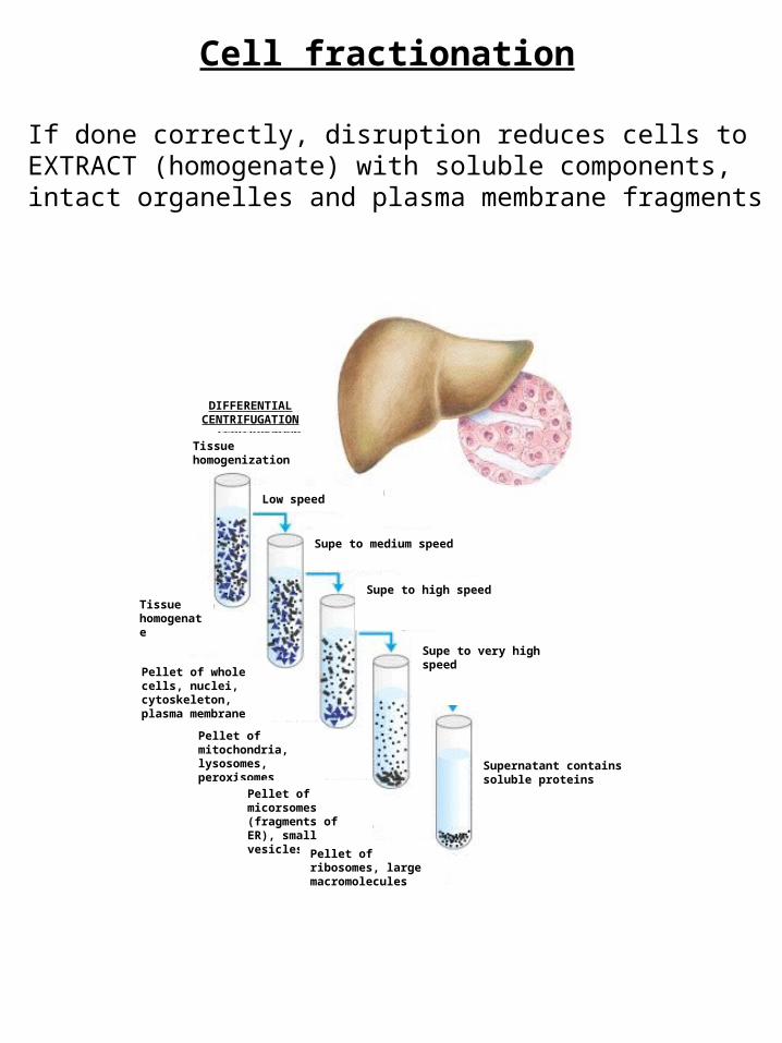
Cell fractionation
If done correctly, disruption reduces cells to EXTRACT (homogenate) with soluble components, intact organelles and plasma membrane fragments
Low speed
Tissue homogenization
DIFFERENTIAL CENTRIFUGATION
Supe to medium speed
Tissue homogenate
Supe to high speed
Pellet of whole cells, nuclei, cytoskeleton, plasma membrane
Pellet of mitochondria, lysosomes, peroxisomes
Pellet of micorsomes (fragments of ER), small vesicles
Pellet of ribosomes, large macromolecules
Supe to very high speed
Supernatant contains soluble proteins
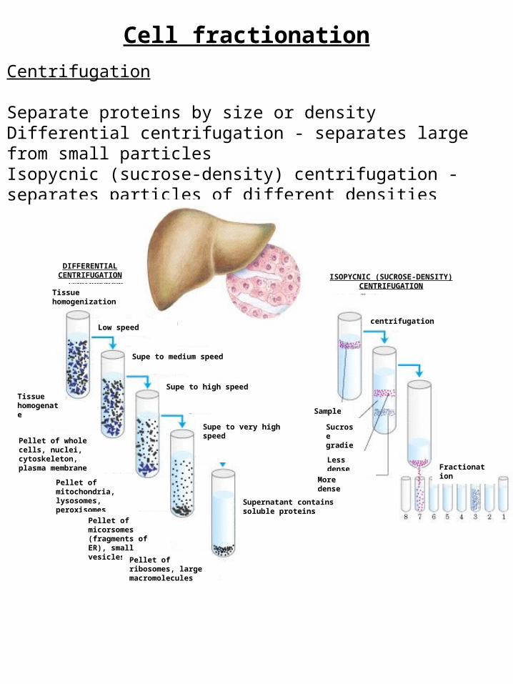
Cell fractionation
Centrifugation
Separate proteins by size or densityDifferential centrifugation - separates large from small particlesIsopycnic (sucrose-density) centrifugation - separates particles of different densities
Low speed
Tissue homogenization
DIFFERENTIAL CENTRIFUGATION
Supe to medium speed
Tissue homogenate
Supe to high speed
Pellet of whole cells, nuclei, cytoskeleton, plasma membrane
Pellet of mitochondria, lysosomes, peroxisomes
Pellet of micorsomes (fragments of ER), small vesicles
Pellet of ribosomes, large macromolecules
Supe to very high speed
Supernatant contains soluble proteins
ISOPYCNIC (SUCROSE-DENSITY)CENTRIFUGATION
centrifugation
Sample
Sucrose gradient
Less dense
More dense
Fractionation
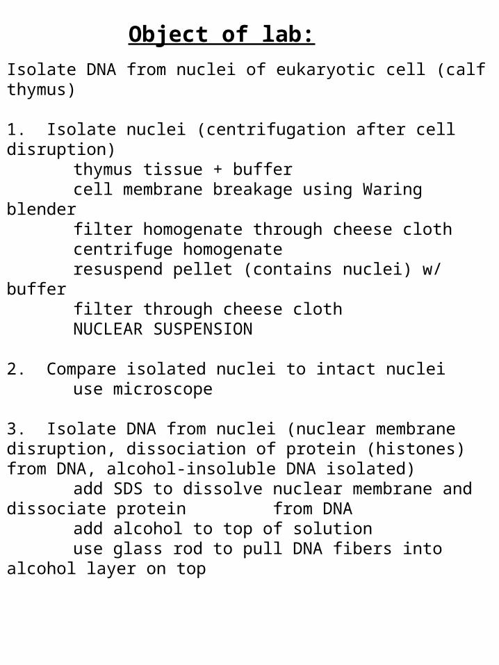
Object of lab:
Isolate DNA from nuclei of eukaryotic cell (calf thymus)
1. Isolate nuclei (centrifugation after cell disruption)thymus tissue + buffercell membrane breakage using Waring blenderfilter homogenate through cheese clothcentrifuge homogenateresuspend pellet (contains nuclei) w/ buffer filter through cheese clothNUCLEAR SUSPENSION
2. Compare isolated nuclei to intact nucleiuse microscope
3. Isolate DNA from nuclei (nuclear membrane disruption, dissociation of protein (histones) from DNA, alcohol-insoluble DNA isolated)
add SDS to dissolve nuclear membrane and dissociate protein from DNA
add alcohol to top of solutionuse glass rod to pull DNA fibers into alcohol layer on top