Embed Size (px)
Citation preview
Biology Module 1 “Cells as the Basis of Life” PhotoMastercopyright © 2005-17 KEEP IT SIMPLE SCIENCEwww.keepitsimplescience.com.au
Page 1 Inspection Copy for school evaluationonly. Copying NOT permitted.
KISS Resources for NSW Syllabuses & Australian Curriculum.
KEEP IT SIMPLE SCIENCEPhotoMaster Format
Biology Module 1
Cells as the Basis of LifeTopic
Outline
2. Cell Structures
1. DifferentTypes of Cells
What is this topic about?To keep it as simple as possible, (K.I.S.S. Principle) this topic covers:
1. DIFFERENT TYPES of CELLSEukaryotic & prokaryotic cells. How we know these things...
light microscopes, electron microscopes, x-ray crystallography, isotopic “tracers”.
2. CELL STRUCTURESMain features of plant & animal cells. Organelles... the nucleus, mitochondria, E.R.,
ribosomes, golgi body, lysosomes, chloroplasts. Structure of membranes.
3. CELL FUNCTIONSa) STUFF GETS IN & OUT
Diffusion & osmosis. Active v. passive transport. Endocytosis & exocytosis. Importance of the SA/Vol. ratio.
b) FOOD & ENERGY for CELLSPhotosynthesis & cellular respiration. What cells need, and need to get rid of.
c) BIOCHEMICAL CONTROL... ENZYMESProperties & importance of enzymes. Effects of temperature & pH on enzyme activity.
Cells as theBasis of Life
keep it simple science®
c) BiochemicalControl... Enzymes
b) Food &Energy for
Cells
a) Stuff GetsIn & Out
Eukaryotic & Prokaryotic
Plant v. animal
Diffusion & osmosis
Photosynthesis
Properties of enzymes
Effects of temperature & pHon enzyme activity
Cellular respiration
What cells need
Active v. passivetransport
Endocytosis& Exocytosis
Importance ofSA/Vol. ratio
Major organelles visible with light& electron microscopes.
Membrane structure
Technologies tounderstand cells
3. CellFunctions
School Inspection only.Copying NOT permitted.
KISS Resources for NSW Syllabuses & Australian Curriculum.
Biology Module 1 “Cells as the Basis of Life” PhotoMastercopyright © 2005-17 KEEP IT SIMPLE SCIENCEwww.keepitsimplescience.com.au
Page 2 Inspection Copy for school evaluationonly. Copying NOT permitted.
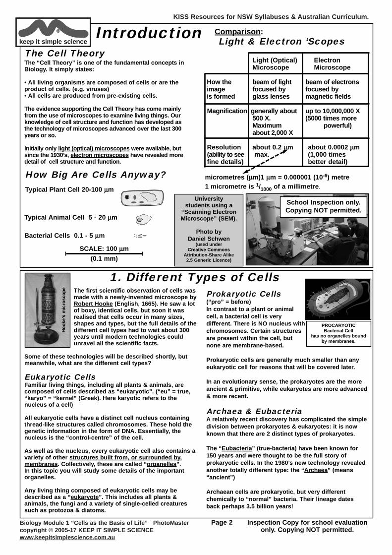
keep it simple science® Introduction Comparison:
Light & Electron ‘Scopes
Light (Optical) ElectronMicroscope Microscope
How the beam of light beam of electronsimage focused by focused by is formed glass lenses magnetic fields
Magnification generally about up to 10,000,000 X500 X. (5000 times moreMaximum powerful)about 2,000 X
Resolution about 0.2 μμm about 0.0002 μμm(ability to see max. (1,000 times fine details) better detail)
micrometres (μμm)1 μμm = 0.000001 (10-6) metre1 micrometre is 1/1000 of a millimetre.
The Cell TheoryThe “Cell Theory” is one of the fundamental concepts inBiology. It simply states:
• All living organisms are composed of cells or are theproduct of cells. (e.g. viruses)• All cells are produced from pre-existing cells.
The evidence supporting the Cell Theory has come mainlyfrom the use of microscopes to examine living things. Ourknowledge of cell structure and function has developed asthe technology of microscopes advanced over the last 300years or so.
Initially only light (optical) microscopes were available, butsince the 1930’s, electron microscopes have revealed moredetail of cell structure and function.
How Big Are Cells Anyway?Typical Plant Cell 20-100 μμm
Bacterial Cells 0.1 - 5 μμm
Typical Animal Cell 5 - 20 μμm
SCALE: 100 μμm(0.1 mm)
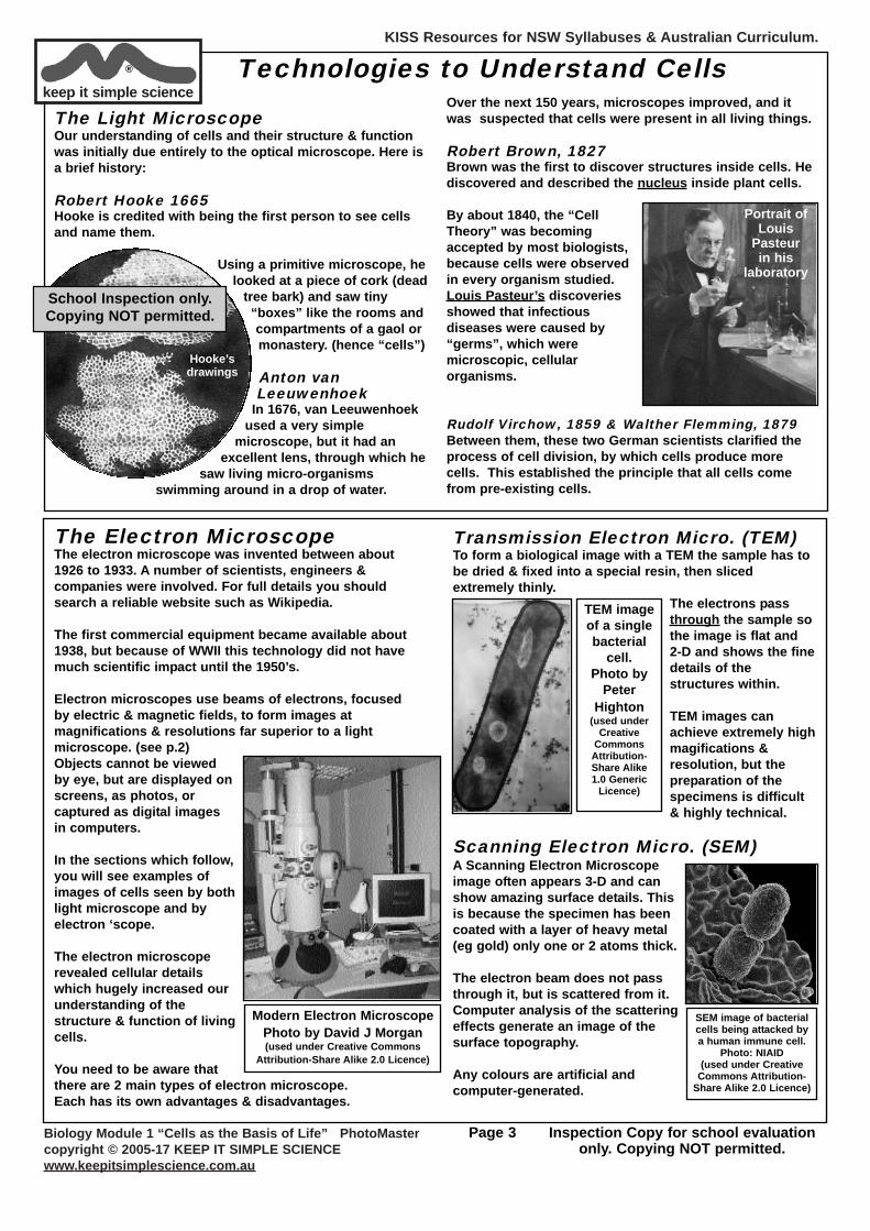
University students using a
“Scanning ElectronMicroscope” (SEM).
Photo byDaniel Schwen
(used under Creative Commons
Attribution-Share Alike2.5 Generic Licence)
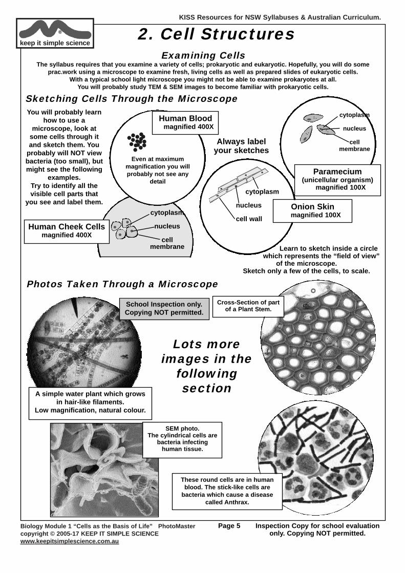
1. Different Types of CellsThe first scientific observation of cells wasmade with a newly-invented microscope byRobert Hooke (English, 1665). He saw a lotof boxy, identical cells, but soon it wasrealised that cells occur in many sizes,shapes and types, but the full details of thedifferent cell types had to wait about 300years until modern technologies couldunravel all the scientific facts.
Some of these technologies will be described shortly, butmeanwhile, what are the different cell types?
Eukaryotic CellsFamiliar living things, including all plants & animals, arecomposed of cells described as “eukaryotic”. (“eu” = true,“karyo” = “kernel” (Greek). Here karyotic refers to thenucleus of a cell)
All eukaryotic cells have a distinct cell nucleus containingthread-like structures called chromosomes. These hold thegenetic information in the form of DNA. Essentially, thenucleus is the “control-centre” of the cell.
As well as the nucleus, every eukaryotic cell also contains avariety of other structures built from, or surrounded by,membranes. Collectively, these are called “organelles”. In this topic you will study some details of the importantorganelles.
Any living thing composed of eukaryotic cells may bedescribed as a “eukaryote”. This includes all plants &animals, the fungi and a variety of single-celled creaturessuch as protozoa & diatoms.
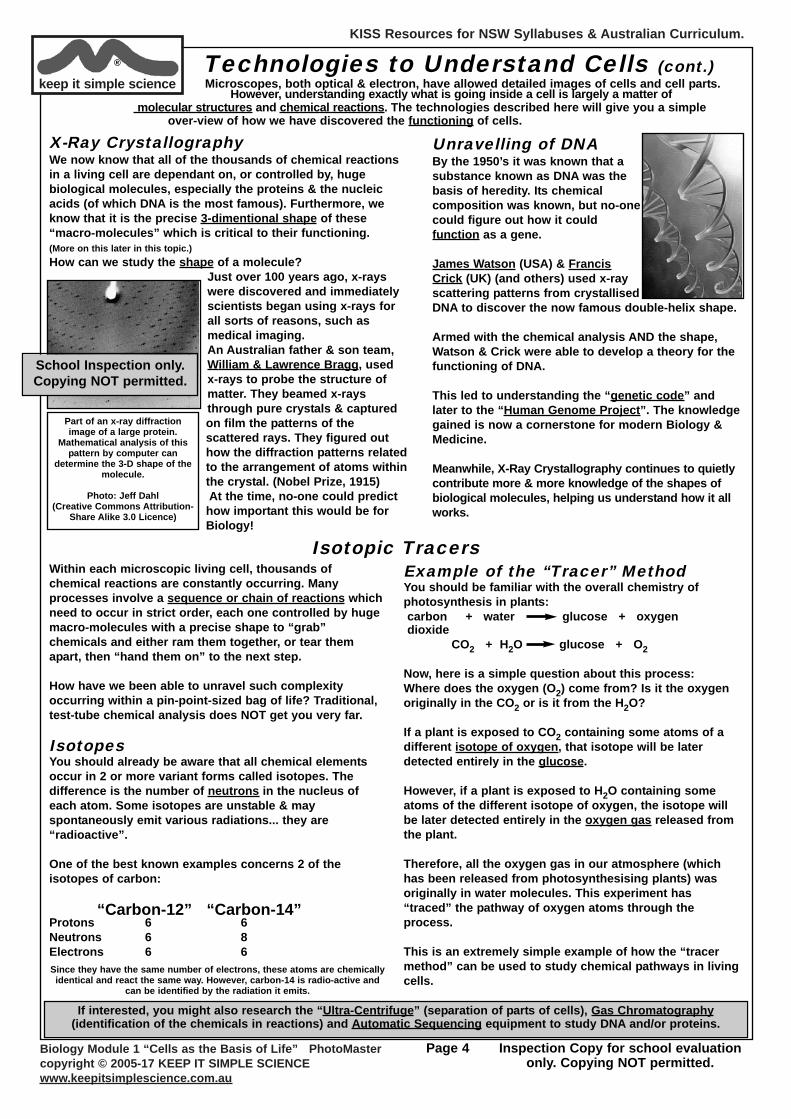
Prokaryotic Cells (“pro” = before)In contrast to a plant or animalcell, a bacterial cell is verydifferent. There is NO nucleus withchromosomes. Certain structuresare present within the cell, butnone are membrane-based.
Prokaryotic cells are generally much smaller than anyeukaryotic cell for reasons that will be covered later.
In an evolutionary sense, the prokaryotes are the moreancient & primitive, while eukaryotes are more advanced& more recent.
Archaea & EubacteriaA relatively recent discovery has complicated the simpledivision between prokaryotes & eukaryotes: it is nowknown that there are 2 distinct types of prokaryotes.
The “Eubacteria” (true-bacteria) have been known for150 years and were thought to be the full story ofprokaryotic cells. In the 1980’s new technology revealedanother totally different type: the “Archaea” (means“ancient”)
Archaean cells are prokaryotic, but very differentchemically to “normal” bacteria. Their lineage datesback perhaps 3.5 billion years!
Hoo
ke’s
mic
rosc
ope
PROCARYOTIC Bacterial Cell
has no organelles boundby membranes.
School Inspection only.Copying NOT permitted.
KISS Resources for NSW Syllabuses & Australian Curriculum.
Biology Module 1 “Cells as the Basis of Life” PhotoMastercopyright © 2005-17 KEEP IT SIMPLE SCIENCEwww.keepitsimplescience.com.au
Page 3 Inspection Copy for school evaluationonly. Copying NOT permitted.
keep it simple science® Technologies to Understand Cells
The Light MicroscopeOur understanding of cells and their structure & functionwas initially due entirely to the optical microscope. Here isa brief history:
Robert Hooke 1665Hooke is credited with being the first person to see cellsand name them.
Using a primitive microscope, helooked at a piece of cork (dead
tree bark) and saw tiny“boxes” like the rooms andcompartments of a gaol ormonastery. (hence “cells”)
Anton vanLeeuwenhoek
In 1676, van Leeuwenhoekused a very simple
microscope, but it had anexcellent lens, through which he
saw living micro-organismsswimming around in a drop of water.
Hooke’sdrawings
Over the next 150 years, microscopes improved, and itwas suspected that cells were present in all living things.
Robert Brown, 1827Brown was the first to discover structures inside cells. Hediscovered and described the nucleus inside plant cells.
By about 1840, the “CellTheory” was becomingaccepted by most biologists,because cells were observedin every organism studied.Louis Pasteur’s discoveriesshowed that infectiousdiseases were caused by“germs”, which weremicroscopic, cellularorganisms.
Rudolf Virchow, 1859 & Walther Flemming, 1879Between them, these two German scientists clarified theprocess of cell division, by which cells produce morecells. This established the principle that all cells comefrom pre-existing cells.
Portrait of Louis
Pasteur in his
laboratory
The Electron MicroscopeThe electron microscope was invented between about1926 to 1933. A number of scientists, engineers &companies were involved. For full details you shouldsearch a reliable website such as Wikipedia.
The first commercial equipment became available about1938, but because of WWII this technology did not havemuch scientific impact until the 1950’s.
Electron microscopes use beams of electrons, focusedby electric & magnetic fields, to form images atmagnifications & resolutions far superior to a lightmicroscope. (see p.2)Objects cannot be viewedby eye, but are displayed onscreens, as photos, orcaptured as digital imagesin computers.
In the sections which follow,you will see examples ofimages of cells seen by bothlight microscope and byelectron ‘scope.
The electron microscoperevealed cellular detailswhich hugely increased ourunderstanding of thestructure & function of livingcells.
You need to be aware thatthere are 2 main types of electron microscope. Each has its own advantages & disadvantages.
Transmission Electron Micro. (TEM)To form a biological image with a TEM the sample has tobe dried & fixed into a special resin, then slicedextremely thinly.
The electrons passthrough the sample sothe image is flat and 2-D and shows the finedetails of thestructures within.
TEM images canachieve extremely highmagifications &resolution, but thepreparation of thespecimens is difficult& highly technical.
Scanning Electron Micro. (SEM)A Scanning Electron Microscopeimage often appears 3-D and canshow amazing surface details. Thisis because the specimen has beencoated with a layer of heavy metal(eg gold) only one or 2 atoms thick.
The electron beam does not passthrough it, but is scattered from it.Computer analysis of the scatteringeffects generate an image of thesurface topography.
Any colours are artificial andcomputer-generated.
Modern Electron MicroscopePhoto by David J Morgan(used under Creative Commons
Attribution-Share Alike 2.0 Licence)
TEM imageof a singlebacterial
cell.Photo by
PeterHighton
(used under Creative
CommonsAttribution-Share Alike1.0 Generic
Licence)
SEM image of bacterialcells being attacked bya human immune cell.
Photo: NIAID(used under Creative
Commons Attribution-Share Alike 2.0 Licence)
School Inspection only.Copying NOT permitted.
KISS Resources for NSW Syllabuses & Australian Curriculum.
Biology Module 1 “Cells as the Basis of Life” PhotoMastercopyright © 2005-17 KEEP IT SIMPLE SCIENCEwww.keepitsimplescience.com.au
Page 4 Inspection Copy for school evaluationonly. Copying NOT permitted.
Technologies to Understand Cells (cont.)Microscopes, both optical & electron, have allowed detailed images of cells and cell parts.
However, understanding exactly what is going inside a cell is largely a matter of molecular structures and chemical reactions. The technologies described here will give you a simple
over-view of how we have discovered the functioning of cells.
X-Ray CrystallographyWe now know that all of the thousands of chemical reactionsin a living cell are dependant on, or controlled by, hugebiological molecules, especially the proteins & the nucleicacids (of which DNA is the most famous). Furthermore, weknow that it is the precise 3-dimentional shape of these“macro-molecules” which is critical to their functioning.(More on this later in this topic.)How can we study the shape of a molecule?
Just over 100 years ago, x-rayswere discovered and immediatelyscientists began using x-rays forall sorts of reasons, such asmedical imaging.An Australian father & son team,William & Lawrence Bragg, used x-rays to probe the structure ofmatter. They beamed x-raysthrough pure crystals & capturedon film the patterns of thescattered rays. They figured outhow the diffraction patterns relatedto the arrangement of atoms withinthe crystal. (Nobel Prize, 1915)At the time, no-one could predicthow important this would be forBiology!
Unravelling of DNABy the 1950’s it was known that asubstance known as DNA was thebasis of heredity. Its chemicalcomposition was known, but no-onecould figure out how it couldfunction as a gene.
James Watson (USA) & FrancisCrick (UK) (and others) used x-rayscattering patterns from crystallisedDNA to discover the now famous double-helix shape.
Armed with the chemical analysis AND the shape,Watson & Crick were able to develop a theory for thefunctioning of DNA.
This led to understanding the “genetic code” andlater to the “Human Genome Project”. The knowledgegained is now a cornerstone for modern Biology &Medicine.
Meanwhile, X-Ray Crystallography continues to quietlycontribute more & more knowledge of the shapes ofbiological molecules, helping us understand how it allworks.
Part of an x-ray diffractionimage of a large protein.
Mathematical analysis of thispattern by computer can
determine the 3-D shape of themolecule.
Photo: Jeff Dahl(Creative Commons Attribution-
Share Alike 3.0 Licence)
keep it simple science®
School Inspection only.Copying NOT permitted.
Within each microscopic living cell, thousands ofchemical reactions are constantly occurring. Manyprocesses involve a sequence or chain of reactions whichneed to occur in strict order, each one controlled by hugemacro-molecules with a precise shape to “grab”chemicals and either ram them together, or tear themapart, then “hand them on” to the next step.
How have we been able to unravel such complexityoccurring within a pin-point-sized bag of life? Traditional,test-tube chemical analysis does NOT get you very far.
Isotopes You should already be aware that all chemical elementsoccur in 2 or more variant forms called isotopes. Thedifference is the number of neutrons in the nucleus ofeach atom. Some isotopes are unstable & mayspontaneously emit various radiations... they are“radioactive”.
One of the best known examples concerns 2 of theisotopes of carbon:
“Carbon-12” “Carbon-14”Protons 6 6Neutrons 6 8Electrons 6 6
Example of the “Tracer” MethodYou should be familiar with the overall chemistry ofphotosynthesis in plants:carbon + water glucose + oxygendioxide
CO2 + H2O glucose + O2
Now, here is a simple question about this process:Where does the oxygen (O2) come from? Is it the oxygenoriginally in the CO2 or is it from the H2O?
If a plant is exposed to CO2 containing some atoms of adifferent isotope of oxygen, that isotope will be laterdetected entirely in the glucose.
However, if a plant is exposed to H2O containing someatoms of the different isotope of oxygen, the isotope willbe later detected entirely in the oxygen gas released fromthe plant.
Therefore, all the oxygen gas in our atmosphere (whichhas been released from photosynthesising plants) wasoriginally in water molecules. This experiment has“traced” the pathway of oxygen atoms through theprocess.
This is an extremely simple example of how the “tracermethod” can be used to study chemical pathways in livingcells.
Isotopic Tracers
Since they have the same number of electrons, these atoms are chemicallyidentical and react the same way. However, carbon-14 is radio-active and
can be identified by the radiation it emits.
If interested, you might also research the “Ultra-Centrifuge” (separation of parts of cells), Gas Chromatography(identification of the chemicals in reactions) and Automatic Sequencing equipment to study DNA and/or proteins.
KISS Resources for NSW Syllabuses & Australian Curriculum.
Biology Module 1 “Cells as the Basis of Life” PhotoMastercopyright © 2005-17 KEEP IT SIMPLE SCIENCEwww.keepitsimplescience.com.au
Page 5 Inspection Copy for school evaluationonly. Copying NOT permitted.
keep it simple science®
Examining CellsThe syllabus requires that you examine a variety of cells; prokaryotic and eukaryotic. Hopefully, you will do some
prac.work using a microscope to examine fresh, living cells as well as prepared slides of eukaryotic cells. With a typical school light microscope you might not be able to examine prokaryotes at all.
You will probably study TEM & SEM images to become familiar with prokaryotic cells.
You will probably learnhow to use a
microscope, look atsome cells through itand sketch them. You
probably will NOT viewbacteria (too small), butmight see the following
examples.Try to identify all thevisible cell parts that
you see and label them.
cytoplasm
nucleus
cellmembrane
Paramecium (unicellular organism)
magnified 100X
cytoplasm
nucleus
cellmembrane
Human Cheek Cellsmagnified 400X
Human Bloodmagnified 400X
Learn to sketch inside a circlewhich represents the “field of view”
of the microscope. Sketch only a few of the cells, to scale.
Always label your sketches
Even at maximummagnification you willprobably not see any
detail
Sketching Cells Through the Microscope
Cross-Section of partof a Plant Stem.
SEM photo. The cylindrical cells are
bacteria infecting human tissue.
Lots moreimages in the
followingsection
2. Cell Structures
Photos Taken Through a Microscope
cytoplasm
nucleus
cell wallOnion Skinmagnified 100X
A simple water plant which growsin hair-like filaments.
Low magnification, natural colour.
These round cells are in humanblood. The stick-like cells are
bacteria which cause a diseasecalled Anthrax.
School Inspection only.Copying NOT permitted.
KISS Resources for NSW Syllabuses & Australian Curriculum.
Biology Module 1 “Cells as the Basis of Life” PhotoMastercopyright © 2005-17 KEEP IT SIMPLE SCIENCEwww.keepitsimplescience.com.au
Page 6 Inspection Copy for school evaluationonly. Copying NOT permitted.
keep it simple science®
School Inspection only.Copying NOT permitted.
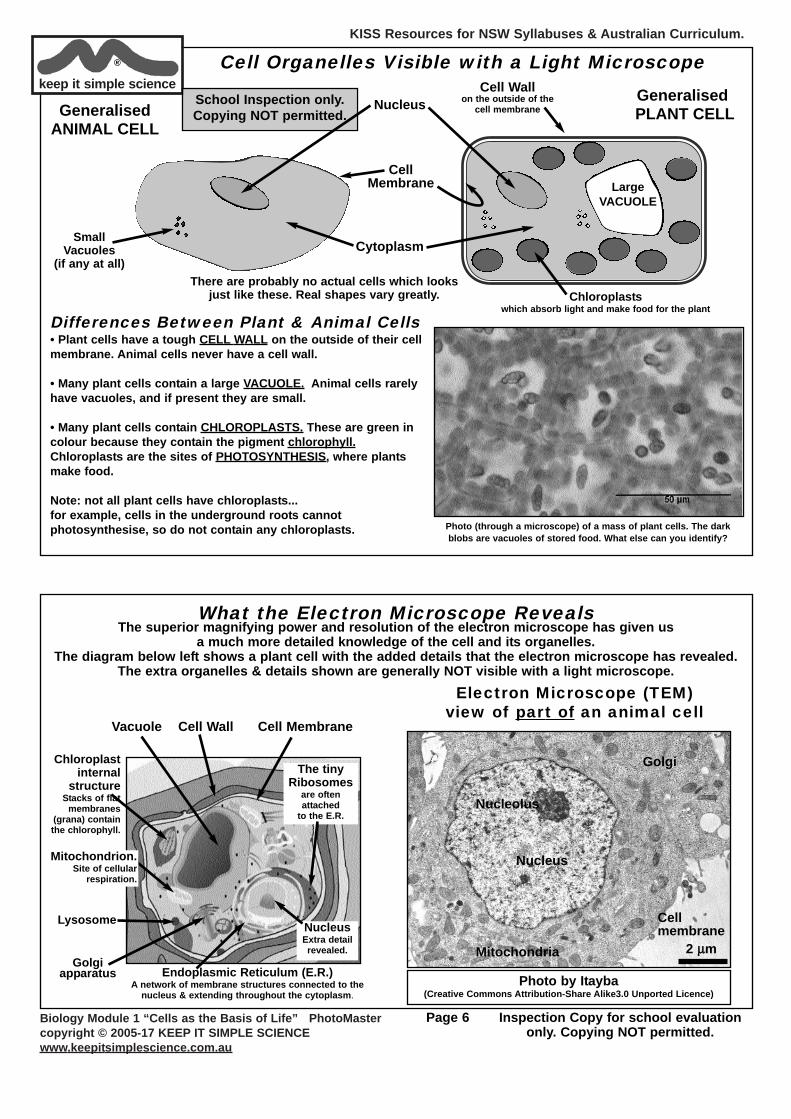
Cell Organelles Visible with a Light Microscope
Generalised ANIMAL CELL
SmallVacuoles
(if any at all)
GeneralisedPLANT CELL
There are probably no actual cells which looksjust like these. Real shapes vary greatly.
CellMembrane
Nucleus
Cytoplasm
Cell Wall on the outside of the
cell membrane
Chloroplasts which absorb light and make food for the plant
LargeVACUOLE
Differences Between Plant & Animal Cells• Plant cells have a tough CELL WALL on the outside of their cellmembrane. Animal cells never have a cell wall.
• Many plant cells contain a large VACUOLE. Animal cells rarelyhave vacuoles, and if present they are small.
• Many plant cells contain CHLOROPLASTS. These are green incolour because they contain the pigment chlorophyll.Chloroplasts are the sites of PHOTOSYNTHESIS, where plantsmake food.
Note: not all plant cells have chloroplasts... for example, cells in the underground roots cannotphotosynthesise, so do not contain any chloroplasts. Photo (through a microscope) of a mass of plant cells. The dark
blobs are vacuoles of stored food. What else can you identify?
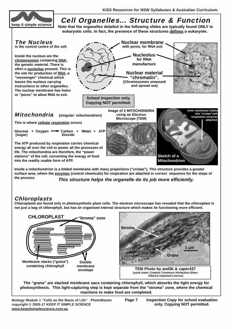
What the Electron Microscope RevealsThe superior magnifying power and resolution of the electron microscope has given us
a much more detailed knowledge of the cell and its organelles. The diagram below left shows a plant cell with the added details that the electron microscope has revealed.
The extra organelles & details shown are generally NOT visible with a light microscope.
Chloroplast internal
structureStacks of flat
membranes(grana) containthe chlorophyll.
Lysosome
Golgiapparatus
Vacuole Cell Wall Cell Membrane
NucleusExtra detailrevealed.
The tiny Ribosomes
are oftenattached
to the E.R.
Endoplasmic Reticulum (E.R.)A network of membrane structures connected to the
nucleus & extending throughout the cytoplasm.
Nucleus
Nucleolus
Cellmembrane
Golgi
Mitochondria
Electron Microscope (TEM)view of part of an animal cell
2 μμm
Photo by Itayba(Creative Commons Attribution-Share Alike3.0 Unported Licence)
Mitochondrion.Site of cellular
respiration.
KISS Resources for NSW Syllabuses & Australian Curriculum.
Biology Module 1 “Cells as the Basis of Life” PhotoMastercopyright © 2005-17 KEEP IT SIMPLE SCIENCEwww.keepitsimplescience.com.au
Page 7 Inspection Copy for school evaluationonly. Copying NOT permitted.
Cell Organelles... Structure & FunctionNote that the organelles detailed in the following slides are typically found ONLY in
eukaryotic cells. In fact, the presence of these structures defines a eukaryote.
Nuclear material“chromatin”.
(Chromosomes unwoundand spread out)
The Nucleus is the control centre of the cell.
Inside the nucleus are thechromosomes containing DNA,the genetic material. There isoften a nucleolus present. This isthe site for production of RNA, a“messenger” chemical whichleaves the nucleus carryinginstructions to other organelles.The nuclear membrane has holesor “pores” to allow RNA to exit.
Mitochondria (singular: mitochondrion)
This is where cellular respiration occurs
Glucose + Oxygen Carbon + Water + ATP(sugar) Dioxide
The ATP produced by respiration carries chemicalenergy all over the cell to power all the processes oflife. The mitochondria are therefore, the “powerstations” of the cell, converting the energy of foodinto the readily usable form of ATP.
Inside a mitochondrion is a folded membrane with many projections (“cristae”). This structure provides a greatersurface area, where the enzymes (control chemicals) for respiration are attached in correct sequence for the steps ofthe process.
Inner membrane foldedinto “cristae”with
respiration enzymesattached.
Sketch of aMitochondrion
Image of 2 MITOCHONDRIAusing an ElectronMicroscope (TEM)
Nuclear membranewith pores, for RNA exit
Nucleolusfor RNA
manufacture
keep it simple science®
School Inspection only.Copying NOT permitted.
This structure helps the organelle do its job more efficiently.
Chloroplasts Chloroplasts are found only in photosynthetic plant cells. The electron microscope has revealed that the chloroplast isnot just a bag of chlorophyll, but has an organised internal structure which makes its functioning more efficient.
CHLOROPLAST
Doublemembraneenvelope
Membrane stacks (“grana”) containing chlorophyll
“Stroma” zone
The “grana” are stacked membrane sacs containing chlorophyll, which absorbs the light energy forphotosynthesis. This light-capturing step is kept separate from the “stroma” zone, where the chemical
reactions to make food are completed.
TEM Photo by and3k & caper437(used under Creative Commons Attribution-Share
Alike3.0 Unported Licence)
1 μμm
Grana
Stroma

KISS Resources for NSW Syllabuses & Australian Curriculum.
Biology Module 1 “Cells as the Basis of Life” PhotoMastercopyright © 2005-17 KEEP IT SIMPLE SCIENCEwww.keepitsimplescience.com.au
Page 8 Inspection Copy for school evaluationonly. Copying NOT permitted.
keep it simple science® School Inspection only.
Copying NOT permitted.
Endoplasmic Reticulum (E.R.)E.R. is a network of membranes which form channels and compartments throughout the cytoplasm ofthe cell. Its function can be compared to the internal walls of an office building which divide the buildinginto “rooms” where different operations can be kept separate so that each does not interfere with others.
The E.R. structure provides channels forchemicals and “messengers” to travelaccurately to the correct locations, andfor chemical production to occur inisolation from other operations. Thisstructure helps make cells functionmore efficient.
Often found attached to the E.R. are the tiny Ribosomes. Theseare the sites of production of proteins, the main structural andfunctional chemicals of living cells. RNA “messengers” from thenucleus attach to a ribosome to make the specific proteins thatthe cell needs.
Nucleus
Mitochondrion
E.R.membranescoated withribosomes
More Cell Organelles
ENDOPLASMICRETICULUM
RIBOSOMESattached to membranes
Membranes
Membranes enclosechannels and “rooms”
The Golgi Apparatus is a semi-circular arrangement of membranes which are concerned with packagingchemicals into small membrane sacs (“vesicles”) for storage or secretion.
One type of “vesicle” produced by a Golgi Body is the Lysosome. These membrane sacs contain digestive enzymeswhich can destroy any foreign proteins which enter the cell. Lysosome enzymes also rapidly digest the contents of acell which has died, so that your body can clean up the remains and replace the dead cell.
GOLGI BODY Curvedmembrane sacs
Vesicles pinch-off forstorage or secretion
Lysosomes formthis way
The “membrane-bound” organelles help thecell’s various functions to be carried out withgreater efficiency.
Having these membrane-based organelles isthe defining characteristic of the“Eukaryotic” group of organisms, whichincludes all plants & animals.
Prokaryotic cells (such as bacteria) do havelots of tiny structures inside, but do NOThave any membrane-type organelles, andcan only operate efficiently by being verysmall.
Except for the tiny ribosomes, all the cellorganelles are built from, and surrounded by,membranes.
The membranes provide:-
• the infrastructure of the cell.• channels for chemicals to move through.• packaging for chemicals which need to bestored.• points of attachment for chemicals (enzymes”).• control over what moves in or out of eachorganelle, and in or out of the entire cell.
The Importance of Membranes
KISS Resources for NSW Syllabuses & Australian Curriculum.
Biology Module 1 “Cells as the Basis of Life” PhotoMastercopyright © 2005-17 KEEP IT SIMPLE SCIENCEwww.keepitsimplescience.com.au
Page 9 Inspection Copy for school evaluationonly. Copying NOT permitted.
keep it simple science®
School Inspection only.Copying NOT permitted.
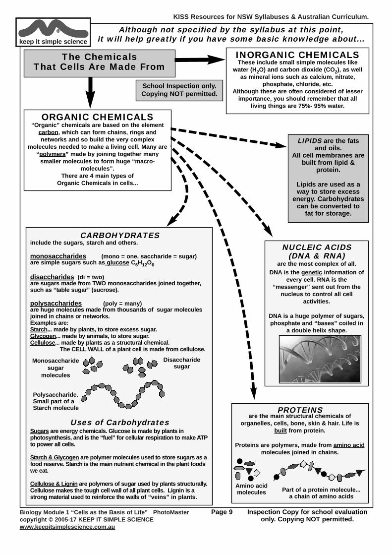
INORGANIC CHEMICALSThese include small simple molecules like
water (H2O) and carbon dioxide (CO2), as wellas mineral ions such as calcium, nitrate,
phosphate, chloride, etc.Although these are often considered of lesser
importance, you should remember that allliving things are 75%- 95% water.
The Chemicals That Cells Are Made From
Although not specified by the syllabus at this point, it will help greatly if you have some basic knowledge about...
CARBOHYDRATES include the sugars, starch and others.
monosaccharides (mono = one, saccharide = sugar)are simple sugars such as glucose C6H12O6
disaccharides (di = two)are sugars made from TWO monosaccharides joined together,such as “table sugar” (sucrose).
polysaccharides (poly = many)are huge molecules made from thousands of sugar moleculesjoined in chains or networks.Examples are:Starch... made by plants, to store excess sugar.Glycogen... made by animals, to store sugar.Cellulose... made by plants as a structural chemical.
The CELL WALL of a plant cell is made from cellulose.
Uses of CarbohydratesSugars are energy chemicals. Glucose is made by plants inphotosynthesis, and is the “fuel” for cellular respiration to make ATPto power all cells.
Starch & Glycogen are polymer molecules used to store sugars as afood reserve. Starch is the main nutrient chemical in the plant foodswe eat.
Cellulose & Lignin are polymers of sugar used by plants structurally.Cellulose makes the tough cell wall of all plant cells. Lignin is astrong material used to reinforce the walls of “veins” in plants.
Polysaccharide.Small part of aStarch molecule
Monosaccharidesugar
molecules
Disaccharidesugar
PROTEINSare the main structural chemicals of
organelles, cells, bone, skin & hair. Life isbuilt from protein.
Proteins are polymers, made from amino acidmolecules joined in chains.
Amino acidmolecules Part of a protein molecule...
a chain of amino acids
LIPIDS are the fatsand oils.
All cell membranes arebuilt from lipid &
protein.
Lipids are used as away to store excess
energy. Carbohydratescan be converted to
fat for storage.
NUCLEIC ACIDS(DNA & RNA)
are the most complex of all. DNA is the genetic information of
every cell. RNA is the“messenger” sent out from the
nucleus to control all cellactivities.
DNA is a huge polymer of sugars,phosphate and “bases” coiled in
a double helix shape.
ORGANIC CHEMICALS“Organic” chemicals are based on the element
carbon, which can form chains, rings andnetworks and so build the very complex
molecules needed to make a living cell. Many are“polymers” made by joining together many
smaller molecules to form huge “macro-molecules”.
There are 4 main types of Organic Chemicals in cells...
KISS Resources for NSW Syllabuses & Australian Curriculum.
Biology Module 1 “Cells as the Basis of Life” PhotoMastercopyright © 2005-17 KEEP IT SIMPLE SCIENCEwww.keepitsimplescience.com.au
Page 10 Inspection Copy for school evaluationonly. Copying NOT permitted.
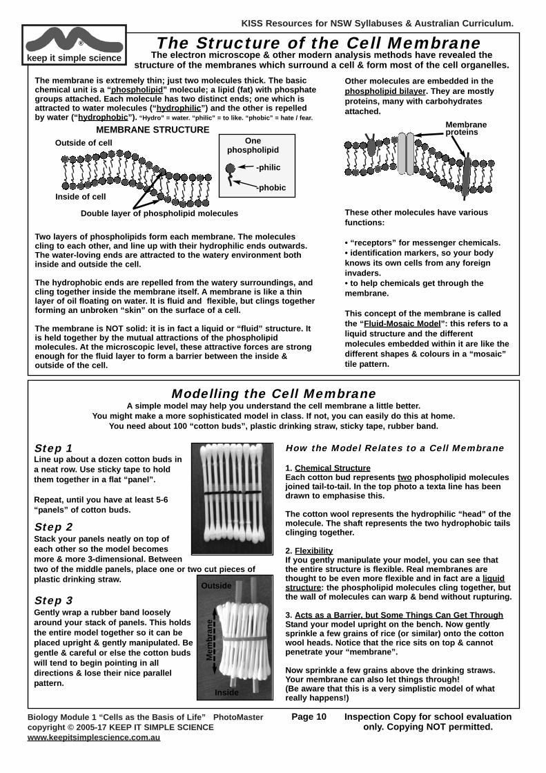
keep it simple science® The Structure of the Cell Membrane
The electron microscope & other modern analysis methods have revealed thestructure of the membranes which surround a cell & form most of the cell organelles.
The membrane is extremely thin; just two molecules thick. The basicchemical unit is a “phospholipid” molecule; a lipid (fat) with phosphategroups attached. Each molecule has two distinct ends; one which isattracted to water molecules (“hydrophilic”) and the other is repelledby water (“hydrophobic”). “Hydro” = water. “philic” = to like. “phobic” = hate / fear.
Two layers of phospholipids form each membrane. The moleculescling to each other, and line up with their hydrophilic ends outwards.The water-loving ends are attracted to the watery environment bothinside and outside the cell.
The hydrophobic ends are repelled from the watery surroundings, andcling together inside the membrane itself. A membrane is like a thinlayer of oil floating on water. It is fluid and flexible, but clings togetherforming an unbroken “skin” on the surface of a cell.
The membrane is NOT solid: it is in fact a liquid or “fluid” structure. Itis held together by the mutual attractions of the phospholipidmolecules. At the microscopic level, these attractive forces are strongenough for the fluid layer to form a barrier between the inside &outside of the cell.
Other molecules are embedded in thephospholipid bilayer. They are mostlyproteins, many with carbohydratesattached.
These other molecules have variousfunctions:
• “receptors” for messenger chemicals.• identification markers, so your bodyknows its own cells from any foreigninvaders.• to help chemicals get through themembrane.
This concept of the membrane is calledthe “Fluid-Mosaic Model”: this refers to aliquid structure and the differentmolecules embedded within it are like thedifferent shapes & colours in a “mosaic”tile pattern.
Membraneproteins
Onephospholipid
-philic
-phobic
MEMBRANE STRUCTUREOutside of cell
Inside of cell
Double layer of phospholipid molecules
Modelling the Cell MembraneA simple model may help you understand the cell membrane a little better.
You might make a more sophisticated model in class. If not, you can easily do this at home.You need about 100 “cotton buds”, plastic drinking straw, sticky tape, rubber band.
Step 1Line up about a dozen cotton buds ina neat row. Use sticky tape to holdthem together in a flat “panel”.
Repeat, until you have at least 5-6“panels” of cotton buds.
How the Model Relates to a Cell Membrane
1. Chemical StructureEach cotton bud represents two phospholipid moleculesjoined tail-to-tail. In the top photo a texta line has beendrawn to emphasise this.
The cotton wool represents the hydrophilic “head” of themolecule. The shaft represents the two hydrophobic tailsclinging together.
2. FlexibilityIf you gently manipulate your model, you can see thatthe entire structure is flexible. Real membranes arethought to be even more flexible and in fact are a liquidstructure: the phospholipid molecules cling together, butthe wall of molecules can warp & bend without rupturing.
3. Acts as a Barrier, but Some Things Can Get ThroughStand your model upright on the bench. Now gentlysprinkle a few grains of rice (or similar) onto the cottonwool heads. Notice that the rice sits on top & cannotpenetrate your “membrane”.
Now sprinkle a few grains above the drinking straws.Your membrane can also let things through!(Be aware that this is a very simplistic model of whatreally happens!)
Step 2Stack your panels neatly on top ofeach other so the model becomesmore & more 3-dimensional. Betweentwo of the middle panels, place one or two cut pieces ofplastic drinking straw.
Step 3Gently wrap a rubber band looselyaround your stack of panels. This holdsthe entire model together so it can beplaced upright & gently manipulated. Begentle & careful or else the cotton budswill tend to begin pointing in alldirections & lose their nice parallelpattern.
Outside
Inside
Mem
bran
e
KISS Resources for NSW Syllabuses & Australian Curriculum.
Biology Module 1 “Cells as the Basis of Life” PhotoMastercopyright © 2005-17 KEEP IT SIMPLE SCIENCEwww.keepitsimplescience.com.au
Page 11 Inspection Copy for school evaluationonly. Copying NOT permitted.
3. Cell Functionsa) How Stuff Gets In & Out
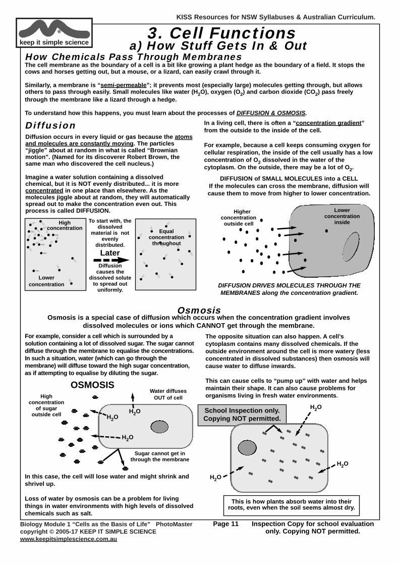
How Chemicals Pass Through MembranesThe cell membrane as the boundary of a cell is a bit like growing a plant hedge as the boundary of a field. It stops thecows and horses getting out, but a mouse, or a lizard, can easily crawl through it.
Similarly, a membrane is “semi-permeable”; it prevents most (especially large) molecules getting through, but allowsothers to pass through easily. Small molecules like water (H2O), oxygen (O2) and carbon dioxide (CO2) pass freelythrough the membrane like a lizard through a hedge.
To understand how this happens, you must learn about the processes of DIFFUSION & OSMOSIS.
DiffusionDiffusion occurs in every liquid or gas because the atomsand molecules are constantly moving. The particles“jiggle” about at random in what is called “Brownianmotion”. (Named for its discoverer Robert Brown, thesame man who discovered the cell nucleus.)
Imagine a water solution containing a dissolvedchemical, but it is NOT evenly distributed... it is moreconcentrated in one place than elsewhere. As themolecules jiggle about at random, they will automaticallyspread out to make the concentration even out. Thisprocess is called DIFFUSION.
To start with, thedissolved
material is notevenly
distributed.
Diffusioncauses the
dissolved soluteto spread out
uniformly.
Highconcentration
Lowerconcentration
Equalconcentration
throughout
Later
In a living cell, there is often a “concentration gradient”from the outside to the inside of the cell.
For example, because a cell keeps consuming oxygen forcellular respiration, the inside of the cell usually has a lowconcentration of O2 dissolved in the water of thecytoplasm. On the outside, there may be a lot of O2.
DIFFUSION DRIVES MOLECULES THROUGH THEMEMBRANES along the concentration gradient.
DIFFUSION of SMALL MOLECULES into a CELLIf the molecules can cross the membrane, diffusion willcause them to move from higher to lower concentration.
Higherconcentration
outside cell
Lowerconcentration
inside
keep it simple science®
School Inspection only.Copying NOT permitted.
OsmosisOsmosis is a special case of diffusion which occurs when the concentration gradient involves
dissolved molecules or ions which CANNOT get through the membrane.For example, consider a cell which is surrounded by asolution containing a lot of dissolved sugar. The sugar cannotdiffuse through the membrane to equalise the concentrations.In such a situation, water (which can go through themembrane) will diffuse toward the high sugar concentration,as if attempting to equalise by diluting the sugar.
In this case, the cell will lose water and might shrink andshrivel up.
Loss of water by osmosis can be a problem for livingthings in water environments with high levels of dissolvedchemicals such as salt.
Sugar cannot get inthrough the membrane
OSMOSISWater diffuses
OUT of cell
H2O
H2OH2O
Highconcentration
of sugaroutside cell
The opposite situation can also happen. A cell’scytoplasm contains many dissolved chemicals. If theoutside environment around the cell is more watery (lessconcentrated in dissolved substances) then osmosis willcause water to diffuse inwards.
This can cause cells to “pump up” with water and helpsmaintain their shape. It can also cause problems fororganisms living in fresh water environments.
This is how plants absorb water into theirroots, even when the soil seems almost dry.
H2O
H2O
H2O

KISS Resources for NSW Syllabuses & Australian Curriculum.
Biology Module 1 “Cells as the Basis of Life” PhotoMastercopyright © 2005-17 KEEP IT SIMPLE SCIENCEwww.keepitsimplescience.com.au
Page 12 Inspection Copy for school evaluationonly. Copying NOT permitted.
keep it simple science® Observing Diffusion & Osmosis
DiffusionYou might do one of these activities yourself, or seeit demonstrated.
OsmosisYour teacher may have a more sophisticatedexperiment for you to do. If not, this is a simpleactivity you could do in class or even at home.
Cut 2 pieces of fresh celery from the same stick.Trim them to be exactly the same length.
If possible, pat dry with a tissue, then weigh eachto the nearest 0.1g and record.
Place each into a small beaker of water or saltsolution, as shown and leave overnight.
Fluids (liquids and gases) seem to be able to mixthemselves together automatically... “Diffusion”.
The explanation is in the Moving-Particle Model ofmatter. In liquids and gases, the particles are
moving around. If 2 different gases or liquids areside-by-side, then the moving particles will
automatically mix. Any dissolved molecules will spread out evenly.
Is diffusion faster in liquid or gas?What effect would temperature have?
Next day, pat each piece dry with a tissue and re-weigh. One piece of celery may have lost a smallamount of mass.
Compare their lengths. They might not be exactly the same any more.
Bend or cut each piece and note the texture and“crispness”. One will be hard and crisp, the othersofter and “rubbery”.
Try to explain these results on the basis ofmovement of water (NOT salt!!) in/out of the livingcelery cells.
Gas Jar of air
glassseparator
When the separator is
removed, thetwo gases mix
themselvestogether.Gas Jar of
brown gas
The food colourspreads out
through the waterby itself.
Without anystirring, it auto-
mixes through thewater.
one dropof foodcolourdye
Water
Identical pieces ofcelery soaked indifferent liquids
for 24 hours.
One loses waterdue to osmosis.
PureWater
conc.Saltsoln.
Schematicdiagram of a cell
membrane,according to
the “FluidMosaic Model”.
Image byLady of Hats
(Mariana Ruiz)
School Inspection only.Copying NOT permitted.
KISS Resources for NSW Syllabuses & Australian Curriculum.
Biology Module 1 “Cells as the Basis of Life” PhotoMastercopyright © 2005-17 KEEP IT SIMPLE SCIENCEwww.keepitsimplescience.com.au
Page 13 Inspection Copy for school evaluationonly. Copying NOT permitted.
keep it simple science® Other Ways Substances Get Through Membranes
Passive & Active TransportDiffusion and Osmosis are vitally important formany chemicals (especially water) to get in and outof cells. Diffusion and osmosis happenautomatically and without the cell having to useany energy. We say these are “passive transport”processes.
What about all the other important chemicals whichcannot get through the membrane? Many proteins,carbohydrates and other molecules regularly moveinto or out of cells. How do they get in or out?
Cells have other ways to deliberately movesubstances across the membrane apart fromdiffusion and osmosis. The membrane containsspecial protein channels & mechanisms which can“carry” chemicals through the membrane.
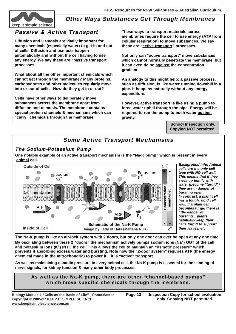
Some Active Transport MechanismsThe Sodium-Potassium PumpOne notable example of an active transport mechanism is the “Na-K pump” which is present in everyanimal cell.
Background info: Animalcells are the only celltype with NO cell wall.This means that if theyswell up tightly withwater (become “turgid”)they are in danger ofbursting open. In contrast, a plant cellhas a tough, rigid cellwall. If a plant cellbecomes turgid there islittle danger ofbursting... plantshabitually keep theircells turgid to supporttheir leaves, etc.
The Na-K pump is like an air-lock system with 2 doors, but only one door can ever be open at any one time.By oscillating between these 2 “doors” the mechanism actively pumps sodium ions (Na+) OUT of the celland potassium ions (K+) INTO the cell. This allows the cell to maintain an “osmotic pressure” whichprevents it absorbing excess water and bursting. Note how the “2-door system” requires ATP (the energychemical made in the mitrochondria) to power it... it is “active” transport.As well as maintaining osmotic pressure in every animal cell, the Na-K pump is essential for the sending ofnerve signals, for kidney function & many other body processes.
Outside of Cell
Inside of Cell
Con
cent
ratio
nm
aint
aine
d
Schematic of the Na-K PumpImage by Lady of Hats (Mariana Ruiz)
As well as the Na-K pump, there are other “channel-based pumps”which move specific chemicals through the membrane.
These ways to transport materials acrossmembranes require the cell to use energy (ATP fromcellular respiration) to move substances. We saythese are “active transport” processes.
Not only can “active transport” move substanceswhich cannot normally penetrate the membrane, butit can even do so against the concentrationgradient.
An analogy to this might help: a passive process,such as diffusion, is like water running downhill in apipe. It happens naturally without any energyexpenditure.
However, active transport is like using a pump toforce water uphill through the pipe. Energy will berequired to run the pump to push water againstgravity.
School Inspection only.Copying NOT permitted.
KISS Resources for NSW Syllabuses & Australian Curriculum.
Biology Module 1 “Cells as the Basis of Life” PhotoMastercopyright © 2005-17 KEEP IT SIMPLE SCIENCEwww.keepitsimplescience.com.au
Page 14 Inspection Copy for school evaluationonly. Copying NOT permitted.
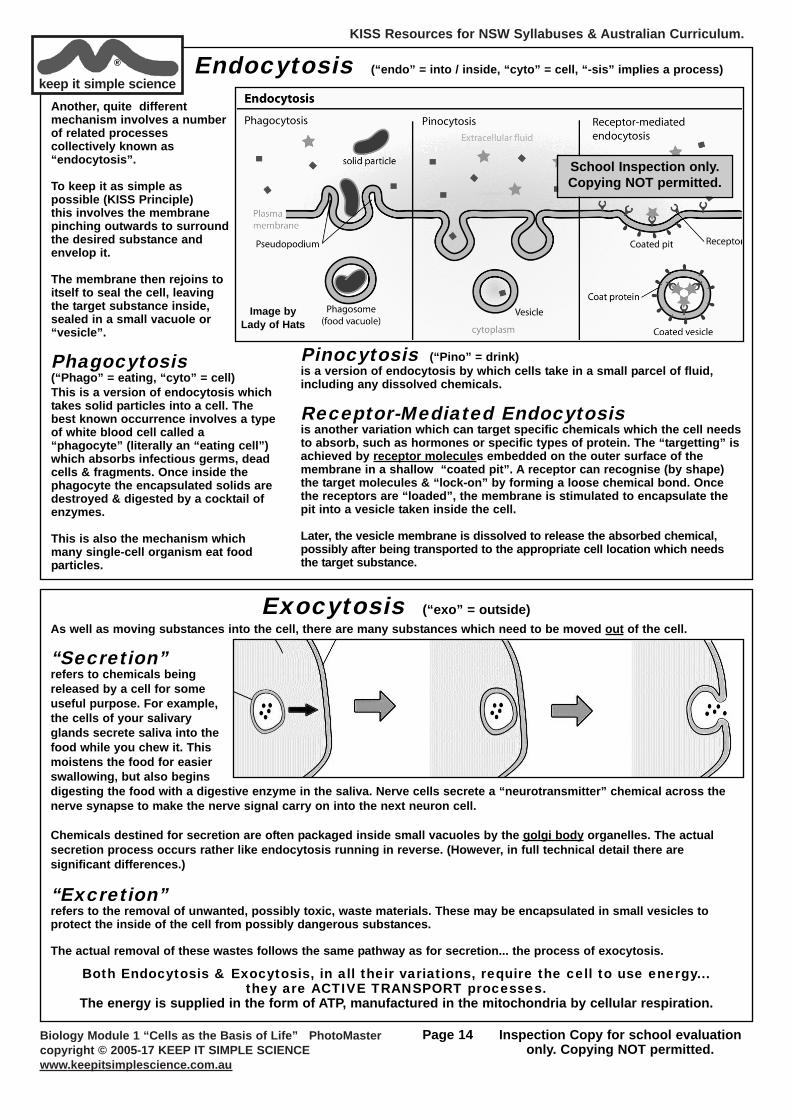
keep it simple science® Endocytosis (“endo” = into / inside, “cyto” = cell, “-sis” implies a process)
Another, quite differentmechanism involves a numberof related processescollectively known as“endocytosis”.
To keep it as simple aspossible (KISS Principle)this involves the membranepinching outwards to surroundthe desired substance andenvelop it.
The membrane then rejoins toitself to seal the cell, leavingthe target substance inside,sealed in a small vacuole or“vesicle”.
Phagocytosis(“Phago” = eating, “cyto” = cell)This is a version of endocytosis whichtakes solid particles into a cell. Thebest known occurrence involves a typeof white blood cell called a“phagocyte” (literally an “eating cell”)which absorbs infectious germs, deadcells & fragments. Once inside thephagocyte the encapsulated solids aredestroyed & digested by a cocktail ofenzymes.
This is also the mechanism whichmany single-cell organism eat foodparticles.
Image by Lady of Hats
Pinocytosis (“Pino” = drink)is a version of endocytosis by which cells take in a small parcel of fluid,including any dissolved chemicals.
Receptor-Mediated Endocytosisis another variation which can target specific chemicals which the cell needsto absorb, such as hormones or specific types of protein. The “targetting” isachieved by receptor molecules embedded on the outer surface of themembrane in a shallow “coated pit”. A receptor can recognise (by shape)the target molecules & “lock-on” by forming a loose chemical bond. Oncethe receptors are “loaded”, the membrane is stimulated to encapsulate thepit into a vesicle taken inside the cell.
Later, the vesicle membrane is dissolved to release the absorbed chemical,possibly after being transported to the appropriate cell location which needsthe target substance.
Exocytosis (“exo” = outside)As well as moving substances into the cell, there are many substances which need to be moved out of the cell.
“Secretion”refers to chemicals beingreleased by a cell for someuseful purpose. For example,the cells of your salivaryglands secrete saliva into thefood while you chew it. Thismoistens the food for easierswallowing, but also beginsdigesting the food with a digestive enzyme in the saliva. Nerve cells secrete a “neurotransmitter” chemical across thenerve synapse to make the nerve signal carry on into the next neuron cell.
Chemicals destined for secretion are often packaged inside small vacuoles by the golgi body organelles. The actualsecretion process occurs rather like endocytosis running in reverse. (However, in full technical detail there aresignificant differences.)
“Excretion”refers to the removal of unwanted, possibly toxic, waste materials. These may be encapsulated in small vesicles toprotect the inside of the cell from possibly dangerous substances.
The actual removal of these wastes follows the same pathway as for secretion... the process of exocytosis.
Both Endocytosis & Exocytosis, in all their variations, require the cell to use energy...they are ACTIVE TRANSPORT processes.
The energy is supplied in the form of ATP, manufactured in the mitochondria by cellular respiration.
School Inspection only.Copying NOT permitted.
KISS Resources for NSW Syllabuses & Australian Curriculum.
Biology Module 1 “Cells as the Basis of Life” PhotoMastercopyright © 2005-17 KEEP IT SIMPLE SCIENCEwww.keepitsimplescience.com.au
Page 15 Inspection Copy for school evaluationonly. Copying NOT permitted.
keep it simple science®
Length ofone side= 1 unit
Length ofone side= 2 units
Length ofone side= 3 units
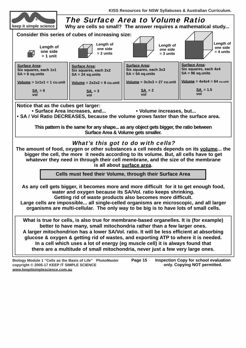
The Surface Area to Volume RatioWhy are cells so small? The answer requires a mathematical study...
Length ofone side= 4 units
Consider this series of cubes of increasing size:
Surface Area:Six squares, each 1x1SA = 6 sq.units
Volume = 1x1x1 = 1 cu.unit
SA = 6vol
Surface Area:Six squares, each 2x2SA = 24 sq.units
Volume = 2x2x2 = 8 cu.unit
SA = 3vol
Surface Area:Six squares, each 3x3SA = 54 sq.units
Volume = 3x3x3 = 27 cu.unit
SA = 2vol
Surface Area:Six squares, each 4x4SA = 96 sq.units
Volume = 4x4x4 = 64 cu.unit
SA = 1.5vol
What’s this got to do with cells?The amount of food, oxygen or other substances a cell needs depends on its volume... the
bigger the cell, the more it needs according to its volume. But, all cells have to getwhatever they need in through their cell membrane, and the size of the membrane
is all about surface area.
As any cell gets bigger, it becomes more and more difficult for it to get enough food,water and oxygen because its SA/Vol. ratio keeps shrinking. Getting rid of waste products also becomes more difficult.
Large cells are impossible... all single-celled organisms are microscopic, and all largerorganisms are multi-cellular. The only way to be big is to have lots of small cells.
Notice that as the cubes get larger:• Surface Area increases, and... • Volume increases, but...
• SA / Vol Ratio DECREASES, because the volume grows faster than the surface area.
This pattern is the same for any shape... as any object gets bigger, the ratio between Surface Area & Volume gets smaller.
Cells must feed their Volume, through their Surface Area
What is true for cells, is also true for membrane-based organelles. It is (for example)better to have many, small mitochondria rather than a few larger ones.
A larger mitochondrion has a lower SA/Vol. ratio. It will be less efficient at absorbingglucose & oxygen & getting rid of wastes, and exporting ATP to where it is needed.
In a cell which uses a lot of energy (eg muscle cell) it is always found that there are a multitude of small mitochondria, never just a few very large ones.
KISS Resources for NSW Syllabuses & Australian Curriculum.
Biology Module 1 “Cells as the Basis of Life” PhotoMastercopyright © 2005-17 KEEP IT SIMPLE SCIENCEwww.keepitsimplescience.com.au
Page 16 Inspection Copy for school evaluationonly. Copying NOT permitted.
keep it simple science®
School Inspection only.Copying NOT permitted.
Why are Prokaryotes so Small?Now we will try to answer a question which arose near the verybeginning of this topic.
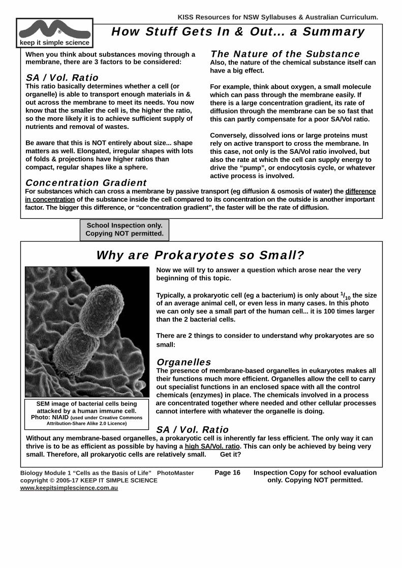
Typically, a prokaryotic cell (eg a bacterium) is only about 1/10 the sizeof an average animal cell, or even less in many cases. In this photowe can only see a small part of the human cell... it is 100 times largerthan the 2 bacterial cells.
There are 2 things to consider to understand why prokaryotes are sosmall:
OrganellesThe presence of membrane-based organelles in eukaryotes makes alltheir functions much more efficient. Organelles allow the cell to carryout specialist functions in an enclosed space with all the controlchemicals (enzymes) in place. The chemicals involved in a processare concentrated together where needed and other cellular processescannot interfere with whatever the organelle is doing.
SA / Vol. RatioWithout any membrane-based organelles, a prokaryotic cell is inherently far less efficient. The only way it canthrive is to be as efficient as possible by having a high SA/Vol. ratio. This can only be achieved by being verysmall. Therefore, all prokaryotic cells are relatively small. Get it?
SEM image of bacterial cells beingattacked by a human immune cell.
Photo: NIAID (used under Creative Commons Attribution-Share Alike 2.0 Licence)
How Stuff Gets In & Out... a SummaryWhen you think about substances moving through amembrane, there are 3 factors to be considered:
SA / Vol. RatioThis ratio basically determines whether a cell (ororganelle) is able to transport enough materials in &out across the membrane to meet its needs. You nowknow that the smaller the cell is, the higher the ratio,so the more likely it is to achieve sufficient supply ofnutrients and removal of wastes.
Be aware that this is NOT entirely about size... shapematters as well. Elongated, irregular shapes with lotsof folds & projections have higher ratios thancompact, regular shapes like a sphere.
Concentration GradientFor substances which can cross a membrane by passive transport (eg diffusion & osmosis of water) the differencein concentration of the substance inside the cell compared to its concentration on the outside is another importantfactor. The bigger this difference, or “concentration gradient”, the faster will be the rate of diffusion.
The Nature of the SubstanceAlso, the nature of the chemical substance itself canhave a big effect.
For example, think about oxygen, a small moleculewhich can pass through the membrane easily. Ifthere is a large concentration gradient, its rate ofdiffusion through the membrane can be so fast thatthis can partly compensate for a poor SA/Vol ratio.
Conversely, dissolved ions or large proteins mustrely on active transport to cross the membrane. Inthis case, not only is the SA/Vol ratio involved, butalso the rate at which the cell can supply energy todrive the “pump”, or endocytosis cycle, or whateveractive process is involved.
KISS Resources for NSW Syllabuses & Australian Curriculum.
Biology Module 1 “Cells as the Basis of Life” PhotoMastercopyright © 2005-17 KEEP IT SIMPLE SCIENCEwww.keepitsimplescience.com.au
Page 17 Inspection Copy for school evaluationonly. Copying NOT permitted.
keep it simple science® 3. Cell Functions
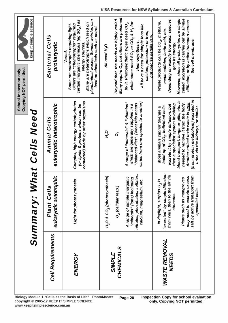
b) Food & Energy for CellsAutotrophs & Heterotrophs (auto = “self”, hetero = “other, not self”, troph = “to eat, feeding”)An autotroph is an organism that makes its own food from simple inorganic chemicals, plus an energy source. All plantsare autotrophic, making their own food by photosynthesis. Any organism that cannot make its own food must be aheterotroph. All animals are heterotrophic, and so are the fungi and most bacteria. A heterotrophic animal eats plants orother animals which have eaten plants, and so on according to the food chain involved.
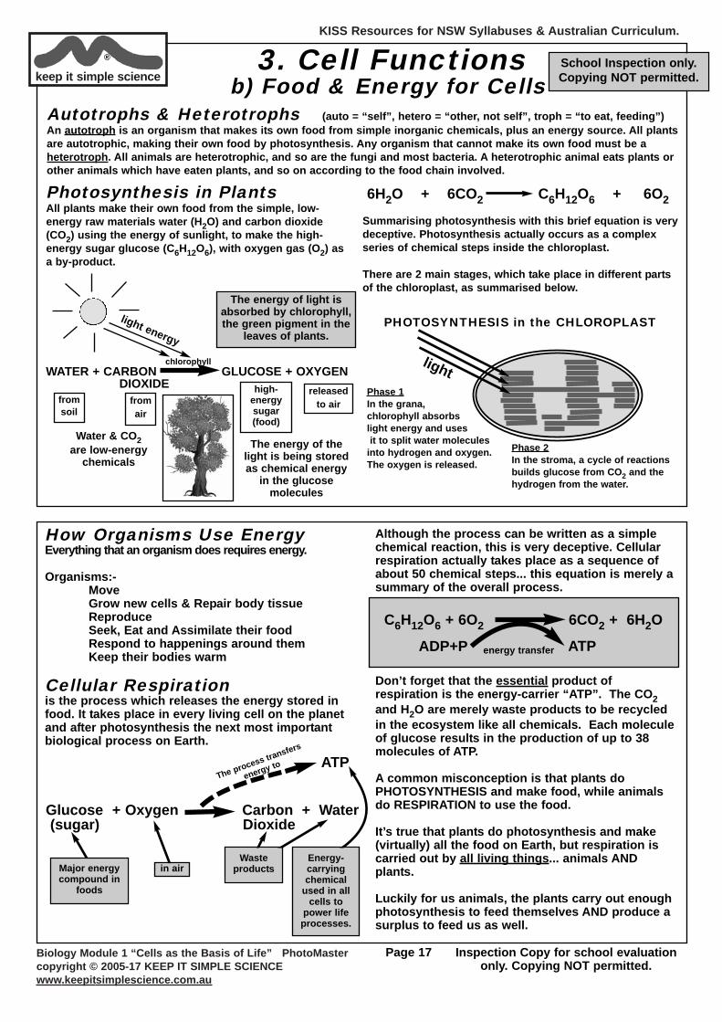
Photosynthesis in PlantsAll plants make their own food from the simple, low-energy raw materials water (H2O) and carbon dioxide(CO2) using the energy of sunlight, to make the high-energy sugar glucose (C6H12O6), with oxygen gas (O2) asa by-product.
lightPhase 1In the grana,chlorophyll absorbslight energy and usesit to split water molecules
into hydrogen and oxygen. The oxygen is released.
PHOTOSYNTHESIS in the CHLOROPLAST
Phase 2In the stroma, a cycle of reactionsbuilds glucose from CO2 and thehydrogen from the water.
Summarising photosynthesis with this brief equation is verydeceptive. Photosynthesis actually occurs as a complexseries of chemical steps inside the chloroplast.
There are 2 main stages, which take place in different partsof the chloroplast, as summarised below.
WATER + CARBON GLUCOSE + OXYGENDIOXIDE
chlorophyll
light energy
The energy of light isabsorbed by chlorophyll,the green pigment in the
leaves of plants.
high-energysugar(food)
fromsoil
fromair
releasedto air
Water & CO2are low-energy
chemicals
6H2O + 6CO2 C6H12O6 + 6O2
The energy of thelight is being storedas chemical energy
in the glucosemolecules
How Organisms Use EnergyEverything that an organism does requires energy.
Organisms:-MoveGrow new cells & Repair body tissueReproduceSeek, Eat and Assimilate their foodRespond to happenings around themKeep their bodies warm
Cellular Respirationis the process which releases the energy stored infood. It takes place in every living cell on the planetand after photosynthesis the next most importantbiological process on Earth.
Although the process can be written as a simplechemical reaction, this is very deceptive. Cellularrespiration actually takes place as a sequence ofabout 50 chemical steps... this equation is merely asummary of the overall process.
Don’t forget that the essential product ofrespiration is the energy-carrier “ATP”. The CO2and H2O are merely waste products to be recycledin the ecosystem like all chemicals. Each moleculeof glucose results in the production of up to 38molecules of ATP.
A common misconception is that plants doPHOTOSYNTHESIS and make food, while animalsdo RESPIRATION to use the food.
It’s true that plants do photosynthesis and make(virtually) all the food on Earth, but respiration iscarried out by all living things... animals ANDplants.
Luckily for us animals, the plants carry out enoughphotosynthesis to feed themselves AND produce asurplus to feed us as well.
C6H12O6 + 6O2 6CO2 + 6H2O
energy transfer ATPADP+P
Glucose + Oxygen Carbon + Water (sugar) Dioxide
ATPThe process transfers
energy to
Major energycompound in
foods
in airWaste
productsEnergy-carryingchemical
used in allcells to
power lifeprocesses.
School Inspection only.Copying NOT permitted.
KISS Resources for NSW Syllabuses & Australian Curriculum.
Biology Module 1 “Cells as the Basis of Life” PhotoMastercopyright © 2005-17 KEEP IT SIMPLE SCIENCEwww.keepitsimplescience.com.au
Page 18 Inspection Copy for school evaluationonly. Copying NOT permitted.
keep it simple science®
School Inspection only.Copying NOT permitted.
What Happens to Glucose in a Plant?If photosynthesis only makes glucose, where do all the other biological chemicals
in a plant come from?Glucose is a monosaccharide sugar, a member of thecarbohydrate group. It is easy for a plant to convertglucose into other types of carbohydrate.
GLUCOSEmolecules
joined in pairs
joined in 1000’s
(polymerisation)
Other sugars, such as sucrose
CELLULOSEfor building new
cell walls STARCHfor storage of food
In fact, plants convert glucose to STARCH so rapidlythat the cells in a plant leaf become packed with
starch grains when it is photosynthesising.
Glucose can also be converted chemically into lipids... fatsand oils, since they contain exactly the same chemicalelements (carbon, hydrogen & oxygen only - CHO).
GLUCOSE LIPIDS (oils)
Making proteins and nucleic acids is more difficult, sincethese contain additional chemical elements, especiallynitrogen, phosphorus and sulfur.
This is where the “minerals” such as nitrate, phosphate andsulfate come in. Soil minerals are often called “plantnutrients”, and a gardener may say he/she is “feeding” theplants when applying fertiliser, but these minerals are NOTfood.
They are the essential ingredients needed so plants can makeproteins and DNA etc, from the real food... glucose.
GLUCOSE Polymerisation
Aminoacids PROTEIN
Amino acids
chemicalconversion
Soil minerals:nitrate, sulfate, etc
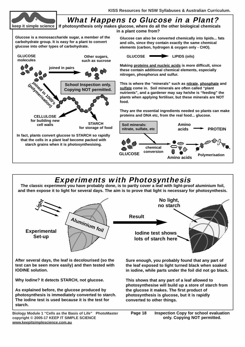
Experiments with PhotosynthesisThe classic experiment you have probably done, is to partly cover a leaf with light-proof aluminium foil,
and then expose it to light for several days. The aim is to prove that light is necessary for photosynthesis.
Ligh
t
Iodine test showslots of starch here
No light,no starch
After several days, the leaf is decolourised (so thetest can be seen more easily) and then tested withIODINE solution.
Why Iodine? It detects STARCH, not glucose.
As explained before, the glucose produced byphotosynthesis is immediately converted to starch.The iodine test is used because it is the test forstarch.
Sure enough, you probably found that any part ofthe leaf exposed to light turned black when soakedin iodine, while parts under the foil did not go black.
This shows that any part of a leaf allowed tophotosynthesise will build up a store of starch fromthe glucose it makes. The first product ofphotosynthesis is glucose, but it is rapidlyconverted to other things.
ExperimentalSet-up
ResultAluminium foil
KISS Resources for NSW Syllabuses & Australian Curriculum.
Biology Module 1 “Cells as the Basis of Life” PhotoMastercopyright © 2005-17 KEEP IT SIMPLE SCIENCEwww.keepitsimplescience.com.au
Page 19 Inspection Copy for school evaluationonly. Copying NOT permitted.
keep it simple science®
A Note About ATPATP stands for “adenosine tri-phosphate”. The molecule can be represented by this simplediagram:
The bond holding the3rd phosphate group
contains a lot ofchemical energy.
P P PAdenosine 3 phosphate groups
High-energy bond
ATP will readily transfer the 3rd phosphate group toother chemicals (with help from an enzyme). Whenthis occurs, energy is transferred which can forceother reactions to go.
The molecule now has only 2 phosphate groups, soit is called “ADP”.
P P PADP=adenosine di-phosphate
Energy transfer when P-group is detached
As you have learned previously, in all ecosystemsthere is a constant input and flow of energy via thefood chains, while the chemicals such as H2O, O2,and CO2 simply get re-cycled over and over.
Most Important Process on EarthPhotosynthesis makes virtually all the food on Earth,for all living things. It also makes all the oxygen in the
atmosphere for us animals to breathe.
For these two reasons, photosynthesis has to beconsidered the most important biological
process on the planet.
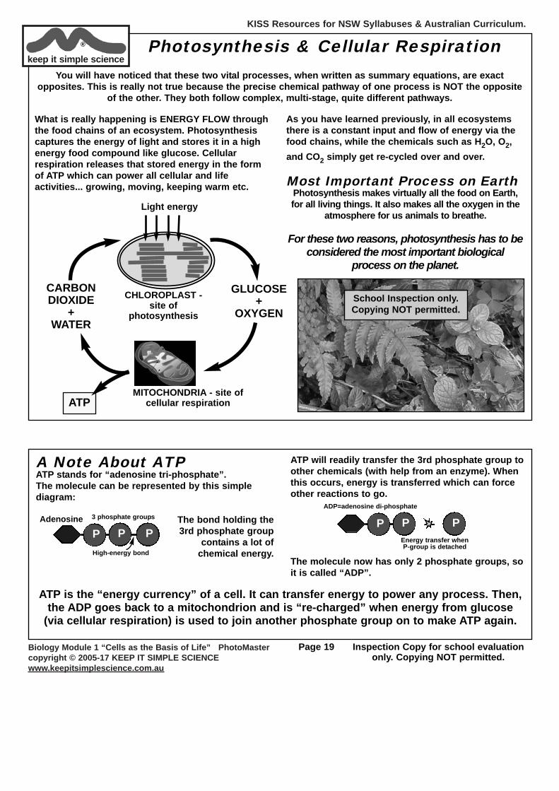
Photosynthesis & Cellular Respiration
What is really happening is ENERGY FLOW throughthe food chains of an ecosystem. Photosynthesiscaptures the energy of light and stores it in a highenergy food compound like glucose. Cellularrespiration releases that stored energy in the formof ATP which can power all cellular and lifeactivities... growing, moving, keeping warm etc.
Light energy
MITOCHONDRIA - site ofcellular respiration
GLUCOSE+
OXYGEN
ATP
CHLOROPLAST -site of
photosynthesis
CARBONDIOXIDE
+WATER
You will have noticed that these two vital processes, when written as summary equations, are exactopposites. This is really not true because the precise chemical pathway of one process is NOT the opposite
of the other. They both follow complex, multi-stage, quite different pathways.
ATP is the “energy currency” of a cell. It can transfer energy to power any process. Then,the ADP goes back to a mitochondrion and is “re-charged” when energy from glucose
(via cellular respiration) is used to join another phosphate group on to make ATP again.
School Inspection only.Copying NOT permitted.
KISS Resources for NSW Syllabuses & Australian Curriculum.
Biology Module 1 “Cells as the Basis of Life” PhotoMastercopyright © 2005-17 KEEP IT SIMPLE SCIENCEwww.keepitsimplescience.com.au
Page 20 Inspection Copy for school evaluationonly. Copying NOT permitted.
keep
itsi
mpl
e sc
ienc
e®
Scho
ol In
spec
tion
only
.C
opyi
ng N
OT
perm
itted
.
Sum
mar
y: W
hat
Cel
ls N
eed
Pla
nt C
ells
euka
ryot
ic a
utot
roph
ic
Ligh
t for
pho
tosy
nthe
sis
Cel
l Req
uire
men
ts
ENER
GY
SIM
PLE
CH
EMIC
ALS
WA
STE
REM
OVA
LN
EED
S
Ani
mal
Cel
lseu
kary
otic
het
erot
roph
ic
Com
plex
, hig
h en
ergy
car
bohy
drat
es(o
r lip
ids
& p
rote
ins
whi
ch c
an b
eco
nver
ted)
mad
e by
oth
er o
rgan
ism
s
Bac
teri
al C
ells
prok
aryo
tic
Varie
d.
Som
e ar
e au
totr
ophs
req
uirin
g lig
ht.
Oth
ers
are
“che
mot
roph
s” r
equi
ring
cert
ain
inor
gani
c ch
emic
als
(eg
SO2
) as
ener
gy s
ourc
es.
Man
y ar
e he
tero
trop
hs w
hich
feed
on
plan
t/ani
mal
was
tes.
Som
es w
eird
os c
anfe
ed o
n ch
emic
als
such
as
petr
ol.
H2O
& C
O2
(pho
tosy
nthe
sis)
O2
(cel
lula
r re
sp.)
Ara
nge
of s
impl
e in
orga
nic
“min
eral
s” (i
ons)
incl
udin
gni
trat
es, p
hosp
hate
s, s
ulfa
tes,
calc
ium
, mag
nesi
um, e
tc.
In d
aylig
ht, s
urpl
us O
2is
“exc
rete
d” b
y si
mpl
e di
ffusi
onfr
om c
ells
, the
n to
the
air
via
stom
ates
.
Plan
ts s
uch
as m
angr
oves
may
nee
d to
exc
rete
exc
ess
salt
by a
ctiv
e tr
ansp
ort f
rom
spec
ialis
t cel
ls.
Mos
t ani
mal
s ca
nnot
tole
rate
a
build
-up
of C
O2.
Indi
vidu
al c
ells
excr
ete
it by
sim
ple
diffu
sion
, but
then
a s
peci
alis
t sys
tem
invo
lvin
gbl
ood
tran
spor
t, lu
ngs
or g
ills,
etc
. is
need
ed to
rem
ove
it fr
om th
e bo
dy.
Ano
ther
crit
ical
toxi
c w
aste
is u
rea
(from
pro
tein
met
abol
ism
) exc
rete
d in
urin
e vi
a th
e ki
dney
s, o
r si
mila
r.
Was
te p
rodu
cts
can
be C
O2,
met
hane
,m
etal
sul
fides
, lac
tic a
cid,
etc
. de
pend
ing
on e
xact
ly h
ow e
ach
spec
ies
gets
its
ener
gy.
How
ever
, sin
ce a
ll pr
okar
yote
s ar
e si
ngle
-ce
lled,
exc
retio
n is
car
ried
out b
y si
mpl
edi
ffusi
on, o
r by
act
ive
tran
spor
t acr
oss
the
cell
mem
bran
e.
All
need
H2O
Bey
ond
that
, the
nee
ds a
re h
ighl
y va
ried.
Man
y re
quire
O2,
but
oth
ers
are
pois
oned
by it
. Pho
tosy
nthe
tic ty
pes
need
CO
2,w
hile
som
e ne
ed S
O2
or C
O2
& H
2fo
rch
emos
ynth
esis
.A
ll ha
ve a
nee
d fo
r si
mpl
e io
ns li
keca
lciu
m, p
otas
sium
or
iron,
but p
reci
se d
etai
ls v
ary.
H2O O2
Ara
nge
of “
min
eral
s” &
“vi
tam
ins”
whi
ch a
re g
ener
ally
sup
plie
d in
a“b
alan
ced
diet
”. (W
hat t
his
mea
nsva
ries
from
one
spe
cies
to a
noth
er)
KISS Resources for NSW Syllabuses & Australian Curriculum.
Biology Module 1 “Cells as the Basis of Life” PhotoMastercopyright © 2005-17 KEEP IT SIMPLE SCIENCEwww.keepitsimplescience.com.au
Page 21 Inspection Copy for school evaluationonly. Copying NOT permitted.
keep it simple science®
School Inspection only.Copying NOT permitted.
Metabolism is ChemistryEverything that happens inside a living cell is really amatter of chemistry... “metabolism”. For example...
• For your body to grow, cells must divide and add moremembranes, cytoplasm and organelles. This involves thechemical construction of new DNA molecules, newphospholipids for membranes and so on.
• All these chemical reactions require energy. Energy isdelivered by the ATP molecule, itself the product of aseries of chemical reactions in the mitochondria... cellular respiration.
All of these reactions are “metabolism”: the sum total ofall the thousands of chemical reactions going onconstantly in all the billions of cells in your body.
EnzymesEvery reaction requires a catalyst... a chemical whichspeeds the reaction up and makes it happen, withoutbeing changed in the process. In living cells there is acatalyst for every different reaction.
Biological catalysts are called enzymes. • Enzymes are protein molecules.• Each has a particular 3-dimensional shape, which fits its“substrate” perfectly.• Enzymes are highly “specific”. This means that eachenzyme will only catalyse one particular reaction; no other.• Enzymes only work effectively in a relatively narrowrange of temperature and pH (acidity).
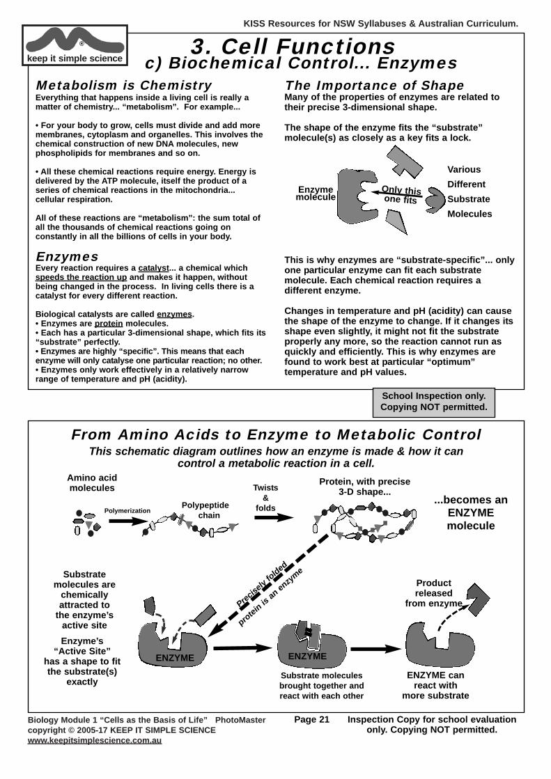
The Importance of ShapeMany of the properties of enzymes are related totheir precise 3-dimensional shape.
The shape of the enzyme fits the “substrate”molecule(s) as closely as a key fits a lock.
This is why enzymes are “substrate-specific”... onlyone particular enzyme can fit each substratemolecule. Each chemical reaction requires adifferent enzyme.
Changes in temperature and pH (acidity) can causethe shape of the enzyme to change. If it changes itsshape even slightly, it might not fit the substrateproperly any more, so the reaction cannot run asquickly and efficiently. This is why enzymes arefound to work best at particular “optimum”temperature and pH values.
Various DifferentSubstrateMolecules
Only thisone fits
Enzymemolecule
3. Cell Functionsc) Biochemical Control... Enzymes
PolymerizationPolypeptide
chain
Productreleased
from enzyme
Substrate molecules are
chemicallyattracted to
the enzyme’sactive site
Protein, with precise3-D shape...
Substrate moleculesbrought together andreact with each other
Amino acidmolecules Twists
&folds
...becomes anENZYMEmolecule
Enzyme’s “Active Site”
has a shape to fitthe substrate(s)
exactly
ENZYME ENZYME
ENZYME canreact with
more substrate
From Amino Acids to Enzyme to Metabolic Control
Precisely folded
protein is an enzyme
This schematic diagram outlines how an enzyme is made & how it can control a metabolic reaction in a cell.
KISS Resources for NSW Syllabuses & Australian Curriculum.
Biology Module 1 “Cells as the Basis of Life” PhotoMastercopyright © 2005-17 KEEP IT SIMPLE SCIENCEwww.keepitsimplescience.com.au
Page 22 Inspection Copy for school evaluationonly. Copying NOT permitted.
keep it simple science®
Temperature
1/tim
e ta
ken
for
reac
tion
(rat
e)
You may have measured the rate of a chemical reaction being catalysedby an enzyme, such as:• the rate of milk clotting by junket tablets.
• the rate of digestion of some starch by amylase.
• the rate of decomposition of hydrogen peroxide by “catalase” enzyme.
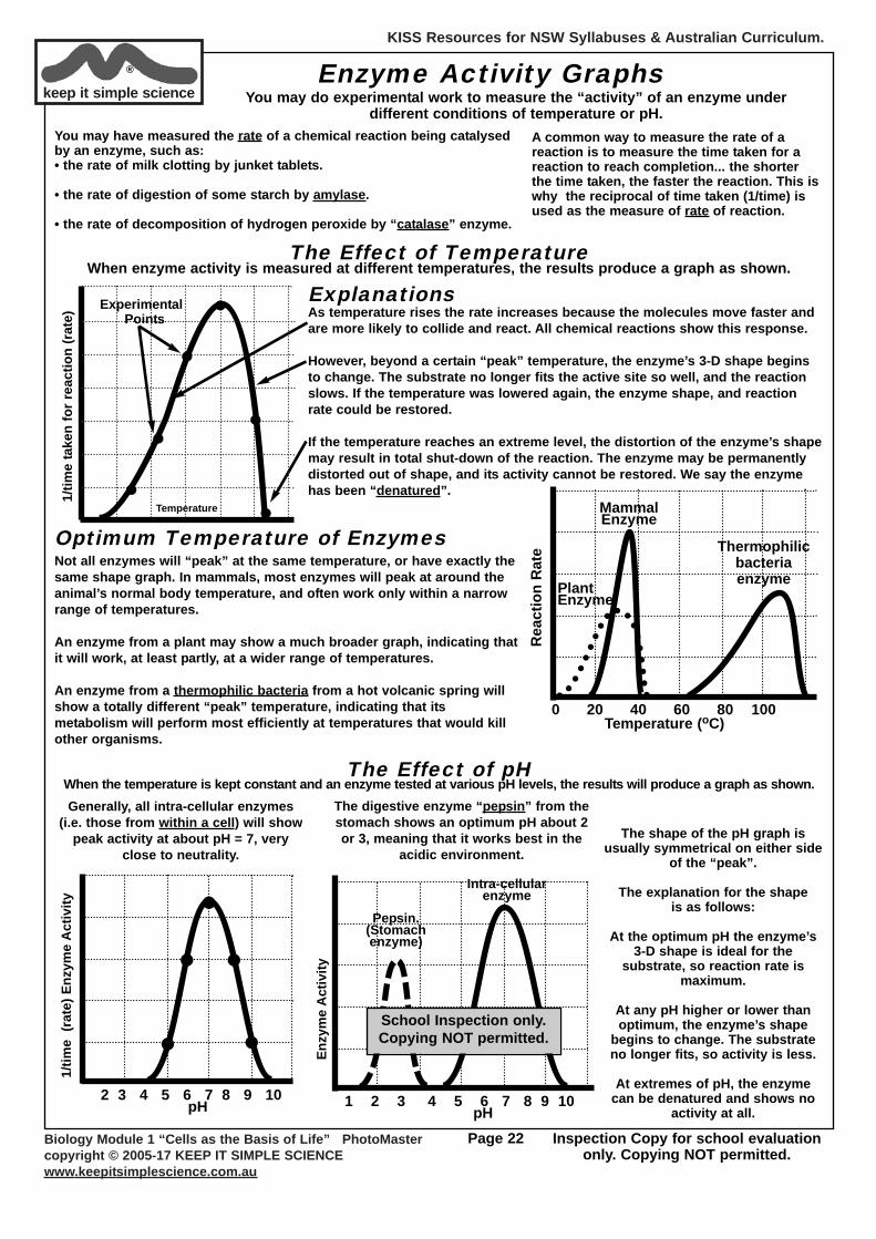
Enzyme Activity GraphsYou may do experimental work to measure the “activity” of an enzyme under
different conditions of temperature or pH.A common way to measure the rate of areaction is to measure the time taken for areaction to reach completion... the shorterthe time taken, the faster the reaction. This iswhy the reciprocal of time taken (1/time) isused as the measure of rate of reaction.
The Effect of TemperatureWhen enzyme activity is measured at different temperatures, the results produce a graph as shown.
ExplanationsAs temperature rises the rate increases because the molecules move faster andare more likely to collide and react. All chemical reactions show this response.
However, beyond a certain “peak” temperature, the enzyme’s 3-D shape beginsto change. The substrate no longer fits the active site so well, and the reactionslows. If the temperature was lowered again, the enzyme shape, and reactionrate could be restored.
If the temperature reaches an extreme level, the distortion of the enzyme’s shapemay result in total shut-down of the reaction. The enzyme may be permanentlydistorted out of shape, and its activity cannot be restored. We say the enzymehas been “denatured”.
ExperimentalPoints
Not all enzymes will “peak” at the same temperature, or have exactly thesame shape graph. In mammals, most enzymes will peak at around theanimal’s normal body temperature, and often work only within a narrowrange of temperatures.
An enzyme from a plant may show a much broader graph, indicating thatit will work, at least partly, at a wider range of temperatures.
An enzyme from a thermophilic bacteria from a hot volcanic spring willshow a totally different “peak” temperature, indicating that itsmetabolism will perform most efficiently at temperatures that would killother organisms.
0 20 40 60 80 100Temperature (oC)
Rea
ctio
n R
ate
MammalEnzyme
PlantEnzyme
Thermophilicbacteriaenzyme
Optimum Temperature of Enzymes
The shape of the pH graph is usually symmetrical on either side
of the “peak”.
The explanation for the shape is as follows:
At the optimum pH the enzyme’s3-D shape is ideal for the
substrate, so reaction rate ismaximum.
At any pH higher or lower thanoptimum, the enzyme’s shape
begins to change. The substrateno longer fits, so activity is less.
At extremes of pH, the enzymecan be denatured and shows no
activity at all.2 3 4 5 6 7 8 9 10
pH
1/tim
e (r
ate)
Enz
yme
Act
ivity
Enzy
me
Act
ivity
1 2 3 4 5 6 7 8 9 10 pH
Intra-cellular enzyme
Pepsin.(Stomachenzyme)
The Effect of pHWhen the temperature is kept constant and an enzyme tested at various pH levels, the results will produce a graph as shown.Generally, all intra-cellular enzymes
(i.e. those from within a cell) will showpeak activity at about pH = 7, very
close to neutrality.
The digestive enzyme “pepsin” from thestomach shows an optimum pH about 2or 3, meaning that it works best in the
acidic environment.
School Inspection only.Copying NOT permitted.

KISS Resources for NSW Syllabuses & Australian Curriculum.
Biology Module 1 “Cells as the Basis of Life” PhotoMastercopyright © 2005-17 KEEP IT SIMPLE SCIENCEwww.keepitsimplescience.com.au
Page 23 Inspection Copy for school evaluationonly. Copying NOT permitted.
A Note About the pH Scale (in case you’ve forgotten)The acidity or alkalinity of any solution is measured on a numerical scale known as “pH”.
On the pH scale, anything which is neutral(neither acid nor alkaline) has a pH = 7.
The inside environment of a cell, and most parts of an organism’s body, is always very close to pH 7... i.e. neutral.An exception is in the stomach where conditions are strongly acidic. (approx. pH 2)
76 8543 119 10
Neutralincreasingacidity
increasingalkalinity
The “Bottom Line” for a Cell to ThriveNow that you know some basics about enzymes, we can end this topic by
discussing just why animals (and people) can die in a “heat wave” (or a blizzard), why pouring vinegar on weeds kills them and why it is so important to get rid of the CO2 your cells are constantly
producing while making their vital ATP.
Temperature Impacts on CellsTo stay alive, a human’s body temperature must be close to 37oC. If it varies by more than about 4oC eitherside of this, it is life-threatening! Now you can figure out why.
Impacts of Changing pHLikewise, the pH of your cellular & body fluids (eg blood) is also critical for your survival.
Same reason... if the pH goes up or down by just 0.5 of a pH unit some critical enzyme molecules willchange their 3-D shape & might not fit their substrate properly. This could slow down, or stop, some vitalbiochemical pathway.
This is why it is (for example) very important to get rid of the CO2 you constantly produce in your hundredsof billions of cells busily making ATP by cellular respiration. The problem with CO2 is not that it is“poisonous” in some vague, mysterious way. Specifically, its danger is pH change! When CO2 dissolves inyour blood beyond certain concentrations, it increases acidity. This can quickly lead to “acidosis” in yourbody fluids which can kill you (by malfunction of vital enzymes) within minutes.
Temperature 32 37 42En
zym
e A
ctiv
ity
Somewhere in your cells there are critical chemical pathwayscontrolled by enzymes which have very narrow activity curves, asshown by this graph.
These pathways might be in one of the many steps in cellularrespiration in all your mitochondria. Maybe it’s an enzymeinvolved with exocytosis of a neuro-transmitter which passesnerve signals from one nerve cell in your brain to another.Whatever it is, it is vital to your survival.
Now look at the graph:If your body temperature drops to 32o, or goes above, say, 40o,this enzyme will STOP FUNCTIONING. This could stop ATPproduction, or stop nerve signals in your brain. Either one couldstop your heart!
Body temperature & pH are critical to survival because vital enzymes can only performefficiently in a narrow range of temperature and/or pH.
keep it simple science®
School Inspection only.Copying NOT permitted.