Embed Size (px)
Citation preview
Dr. Chris Doumen
Week 112401 : Anatomy/Physiology
NeuroTransmitters andReceptors
Neurotransmitter are molecular triggers.They require binding to a specificreceptor. Which in turn sets in motion asequence of events that translates to acellular event. The type of receptordetermines the specificity of cellularactions.
Somatic versus Autonomic
In the somatic nervous system, theactual motor neuron starts in the CNSand the singular axon connects with askeletal muscle at the neuromuscularjunction. In this synapse, theneurotransmitter is released andconnects with a receptor on the motorend plate ( muscle membrane area ofthe synapse). From previous discussion, itis known that the information “hand-off” must be an excitatory signal. Thus,the membrane area of the skeletalmuscle must experience EPSP’s.
This signal hand-off is accomplished byAcetylCholine and the receptor is achemically stimulated Na+ channel, theopening of which results in adepolarization.
In the autonomic system, things are alittle more complicated since now we aredealing with two synapses.
• First synapse is in the auronomicganglion
• Second synapse is at the targetorgan
The neurotransmitters in the autonomicsystem are quite simple since there areonly two :
• Acetylcholine• NorEpinephrine
Neurons that release Acetylcholine arecalled CHOLINERGIC neurons (fibers)
Neurons that release NorEpinephrine ( orepinephrine) are called ADRENERGICfibers.
Similarly, receptors that bindAcetylcholine are referred to asCHOLINERGIC receptors and those thatbind the catecholamines ( Norepinephrineand epinephrine) are called ADENERGICreceptors.
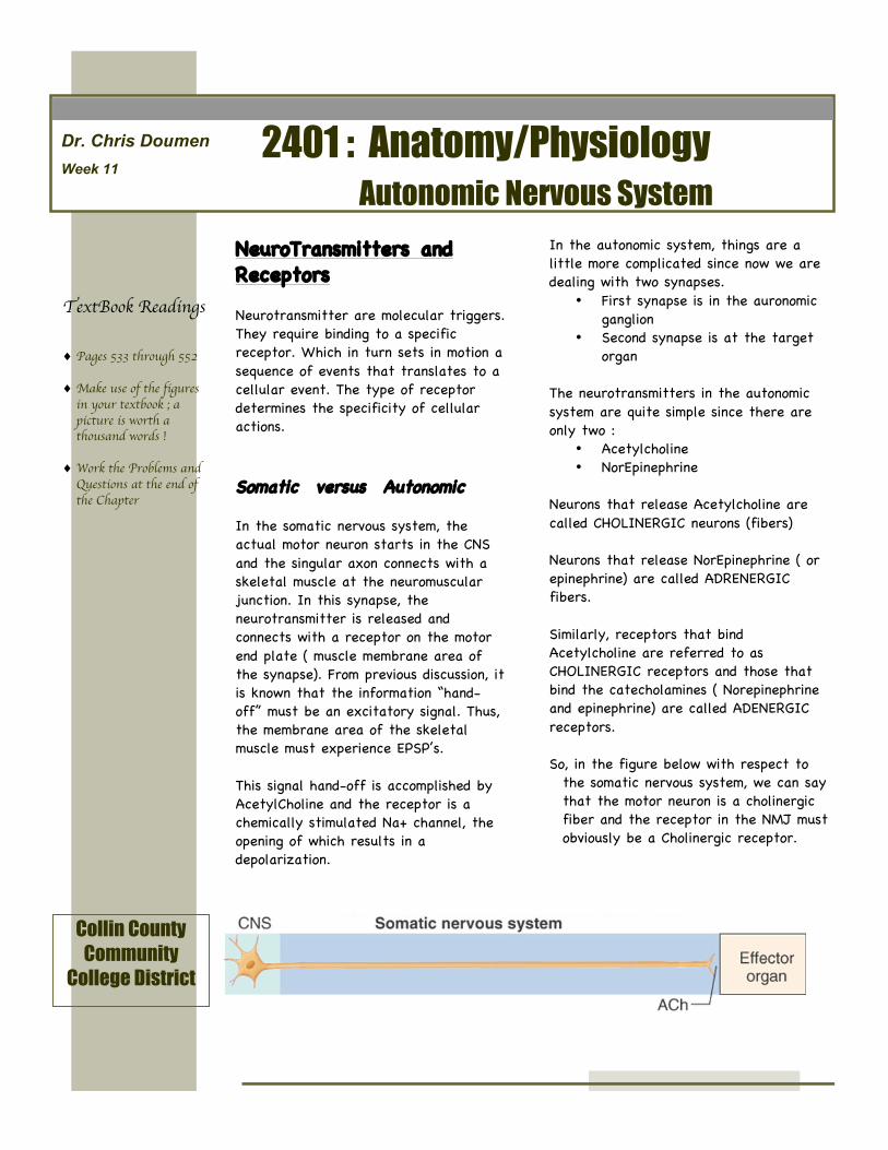
So, in the figure below with respect tothe somatic nervous system, we can saythat the motor neuron is a cholinergicfiber and the receptor in the NMJ mustobviously be a Cholinergic receptor.
Autonomic Nervous System
Collin CountyCommunity
College District
TextBook Readings
♦ Pages 533 through 552
♦ Make use of the figuresin your textbook ; apicture is worth athousand words !
♦ Work the Problems andQuestions at the end ofthe Chapter
Cholinergic Receptors
There are two kinds of cholinergic receptors which functionally the same because they bindAcetylcholine but are pharmacologically different because they bind other agonist moleculesdifferently. They are in addition functionally different because the waythe receptor sets inmotion cellular events.
The two cholinergic receptors are• Nicotinic Receptors• Muscarinic Receptors
Cholinergic Nicotinic Receptors
• They are members of a superfamily of ligand-gated membrane channels that mediatefast signal transmission at synapses.
• They bind nicotine (tabacco product) as an agonistmolecule
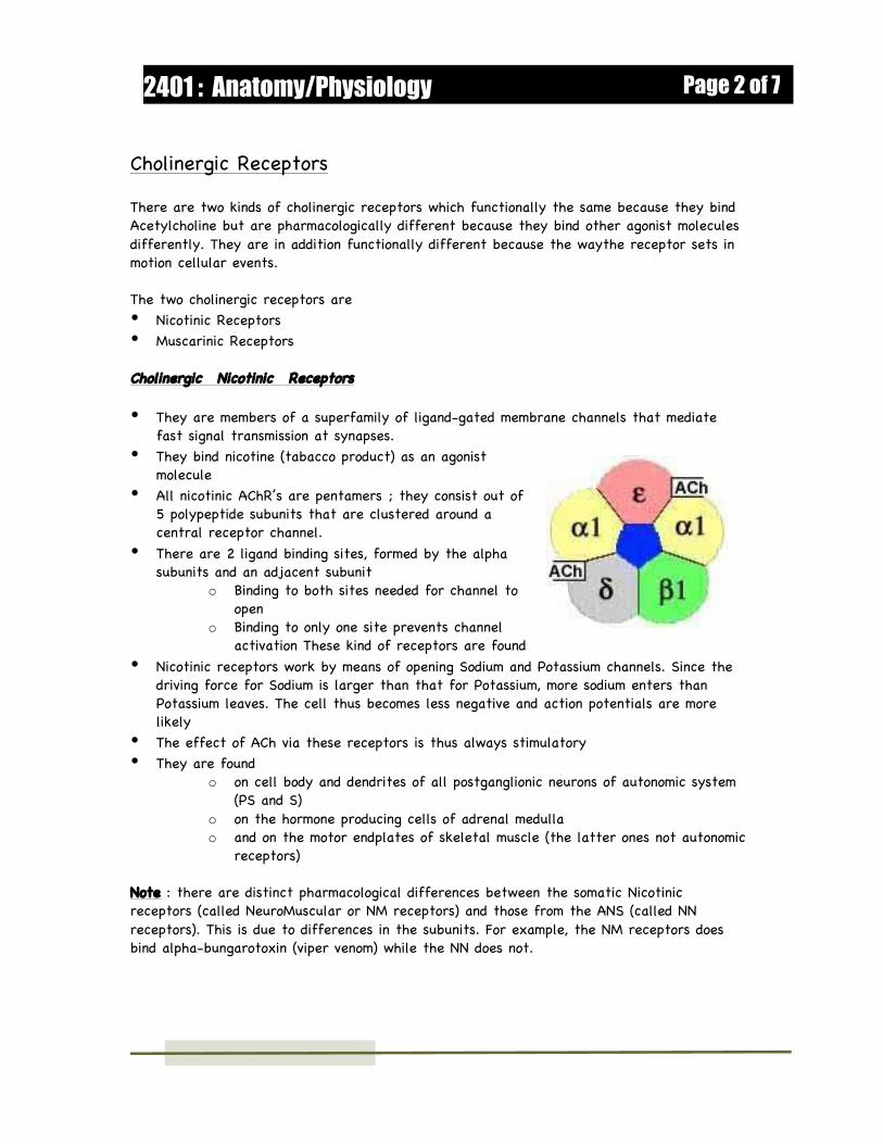
• All nicotinic AChR’s are pentamers ; they consist out of5 polypeptide subunits that are clustered around acentral receptor channel.
• There are 2 ligand binding sites, formed by the alphasubunits and an adjacent subunit
o Binding to both sites needed for channel toopen
o Binding to only one site prevents channelactivation These kind of receptors are found
• Nicotinic receptors work by means of opening Sodium and Potassium channels. Since thedriving force for Sodium is larger than that for Potassium, more sodium enters thanPotassium leaves. The cell thus becomes less negative and action potentials are morelikely
• The effect of ACh via these receptors is thus always stimulatory• They are found
o on cell body and dendrites of all postganglionic neurons of autonomic system(PS and S)
o on the hormone producing cells of adrenal medullao and on the motor endplates of skeletal muscle (the latter ones not autonomic
receptors)
Note : there are distinct pharmacological differences between the somatic Nicotinicreceptors (called NeuroMuscular or NM receptors) and those from the ANS (called NNreceptors). This is due to differences in the subunits. For example, the NM receptors doesbind alpha-bungarotoxin (viper venom) while the NN does not.
Page 2 of 72401 : Anatomy/Physiology
Cholinergic Muscarinic Receptors
• Bind muscarine ( mushroom poison)• Found on all effectors cells stimulated by postganglionic cholinergic fibers• Muscarinic Receptors operate as G-protein coupled receptors and mediate their response
by activating a variety of intra-cellular pathways.• There are two importanrt system
• c-AMP (adenylate cyclase) system• PIP2 (Phosholipase C ) system
• The c-AMP system has been described previously• The PIP2 system is almost identical to the Adenylate cyclase mechanism.
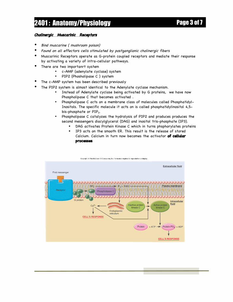
• Instead of Adenylate cyclase being activated by G proteins, we have nowPhospholipase C that becomes activated .
• Phospholipase C acts on a membrane class of molecules called Phosphatidyl-Inositols. The specific molecule it acts on is called phosphatidylinositol 4,5-bis-phosphate or PIP2.
• Phospholipase C catalyzes the hydrolysis of PIP2 and produces produces thesecond messengers diacylglycerol (DAG) and inositol tris-phosphate (IP3).
DAG activates Protein Kinase C which in turns phsphorylates proteins IP3 acts on the smooth ER. This result is the release of stored
Calcium. Calcium in turn now becomes the activator of cellularprocesses
• • • • • • • • • • • • • •
Page 3 of 72401 : Anatomy/Physiology
There are 5 subtypes of Muscarinic receptors: M1 , M2, M3, M4, M5. They all seem to workby means of G-proteins . The Effect can be stimulatory or inhibitory, depending on the kindof receptor in the target organ.
• M1 receptors for example act via Phospholipase C and tend to decrease theconductancefor K + ions. This thus results in a local depolarization.
• The M2 receptors (located in the heart) work by inhibiting Adenylate cyclase. Thereduced levels of cAMP results in an increase in K+ conductance, resulting in localhyperpolarization
• M3 receptors are found in the smnooth muscles and glands of the gut and activatePhoshoplipase C (mediating contraction)
• M4 and M5 receptors are found in the CNS and work by inhibiting Adenyulate cyclaseand activating Phospholipase respectively.
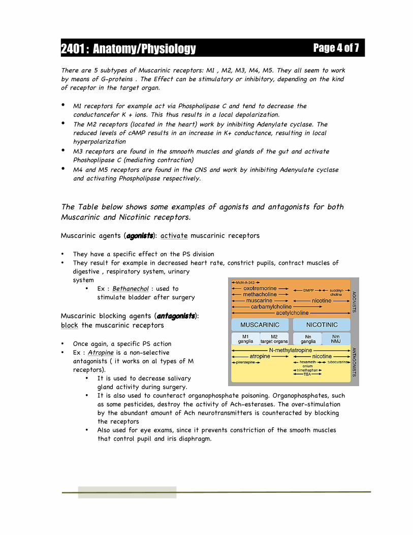
The Table below shows some examples of agonists and antagonists for bothMuscarinic and Nicotinic receptors.
Muscarinic agents (agonists): activate muscarinic receptors
• They have a specific effect on the PS division• They result for example in decreased heart rate, constrict pupils, contract muscles of
digestive , respiratory system, urinarysystem
• Ex : Bethanechol : used tostimulate bladder after surgery
Muscarinic blocking agents (antagonists):block the muscarinic receptors
• Once again, a specific PS action• Ex : Atropine is a non-selective
antagonists ( it works on al types of Mreceptors).
• It is used to decrease salivarygland activity during surgery.
• It is also used to counteract organophosphate poisoning. Organophosphates, suchas some pesticides, destroy the activity of Ach-esterases. The over-stimulationby the abundant amount of Ach neurotransmitters is counteracted by blockingthe receptors
• Also used for eye exams, since it prevents constriction of the smooth musclesthat control pupil and iris diaphragm.
Page 4 of 72401 : Anatomy/Physiology
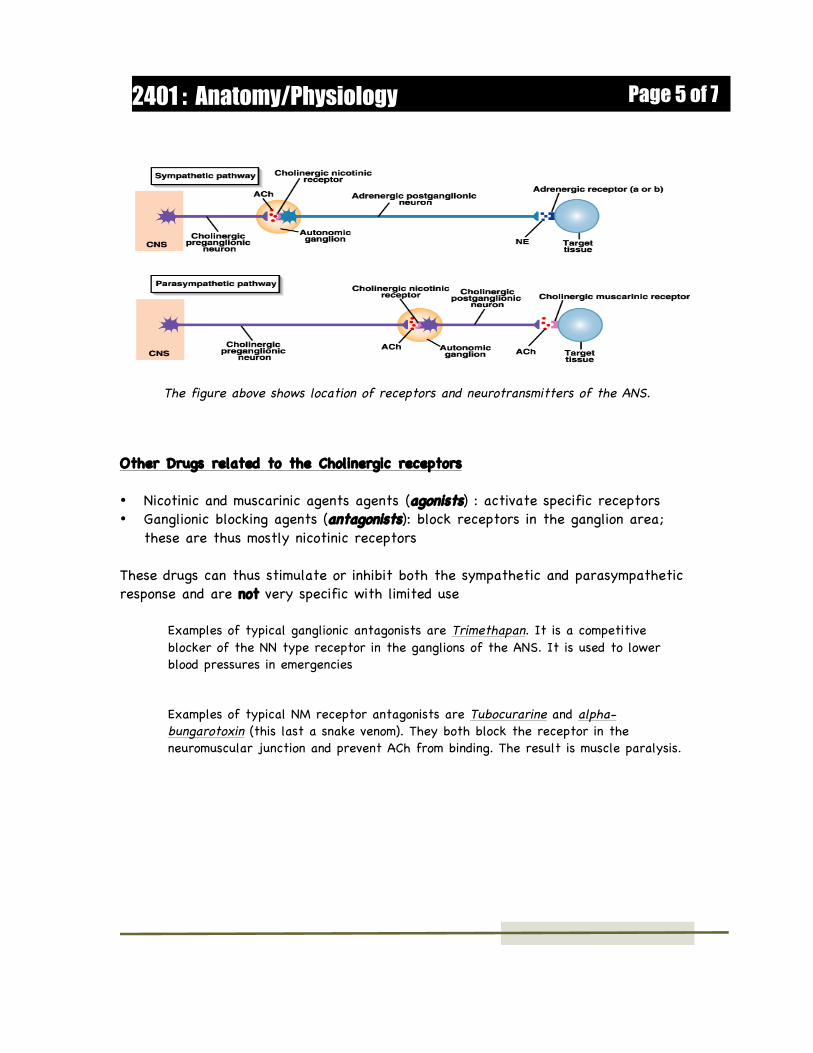
The figure above shows location of receptors and neurotransmitters of the ANS.
Other Drugs related to the Cholinergic receptors • Nicotinic and muscarinic agents agents (agonists) : activate specific receptors• Ganglionic blocking agents (antagonists): block receptors in the ganglion area;
these are thus mostly nicotinic receptors These drugs can thus stimulate or inhibit both the sympathetic and parasympatheticresponse and are not very specific with limited use
Examples of typical ganglionic antagonists are Trimethapan. It is a competitiveblocker of the NN type receptor in the ganglions of the ANS. It is used to lowerblood pressures in emergencies
Examples of typical NM receptor antagonists are Tubocurarine and alpha-bungarotoxin (this last a snake venom). They both block the receptor in theneuromuscular junction and prevent ACh from binding. The result is muscle paralysis.
Page 5 of 72401 : Anatomy/Physiology
Adrenergic Receptors (bind NE or epineprhrine) There are 2 major classes • Alpha receptors :
• When activated, usually produce excitatory responses of smooth muscle• There are subtypes : α1 and α2• A1 mostly found in smooth muscle of peripheral nervous system such as blood
vessels, digestive system• Example : activating these receptors will result contraction in smooth muscle of
blood vessels
• Beta receptors• When activated, usually produce inhibitory responses of smooth muscle (exception
however is the heart where the beta receptors are stimulatory)• There are 3 subtypes :
• Beta 1 : located mostly in heart tissue and the effect is increase contractility• Beta 2 : located mostly in coronaries of the heart, arterioles of peripheral
system and bronchi of the lungs; the effect is relaxation• Beta 3 : located mostly in adipose tissue and the effect is lipolysis
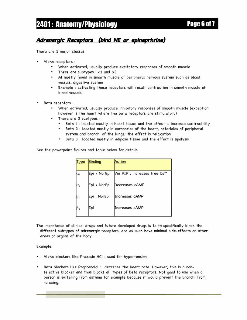
See the powerpoint figures and table below for details.
Type Binding Action
α1 Epi > NorEpi Via PIP , increases free Ca++
α2 Epi > NorEpi Decreases cAMP
β1 Epi , NorEpi Increases cAMP
β2 Epi Increases cAMP
The importance of clinical drugs and future developed drugs is to to specifically block thedifferent subtypes of adrenergic receptors, and as such have minimal side-effects on otherareas or organs of the body.
Example: • Alpha blockers like Prazosin HCl : used for hypertension
• Beta blockers like Propranolol : decrease the heart rate. However, this is a non-selective blocker and thus blocks all types of beta receptors. Not good to use when aperson is suffering from asthma for example because it would prevent the bronchi fromrelaxing.
Page 6 of 72401 : Anatomy/Physiology
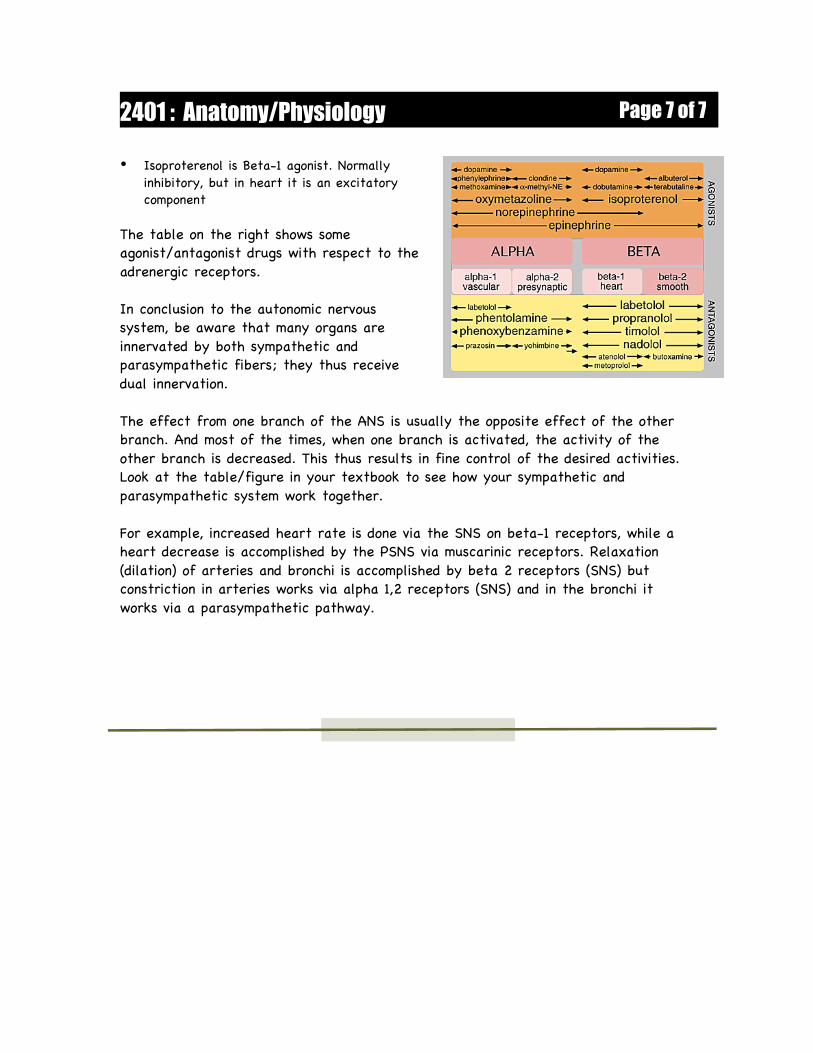
• Isoproterenol is Beta-1 agonist. Normallyinhibitory, but in heart it is an excitatorycomponent
The table on the right shows someagonist/antagonist drugs with respect to theadrenergic receptors.
In conclusion to the autonomic nervoussystem, be aware that many organs areinnervated by both sympathetic andparasympathetic fibers; they thus receivedual innervation.
The effect from one branch of the ANS is usually the opposite effect of the otherbranch. And most of the times, when one branch is activated, the activity of theother branch is decreased. This thus results in fine control of the desired activities.Look at the table/figure in your textbook to see how your sympathetic andparasympathetic system work together.
For example, increased heart rate is done via the SNS on beta-1 receptors, while aheart decrease is accomplished by the PSNS via muscarinic receptors. Relaxation(dilation) of arteries and bronchi is accomplished by beta 2 receptors (SNS) butconstriction in arteries works via alpha 1,2 receptors (SNS) and in the bronchi itworks via a parasympathetic pathway.
Page 7 of 72401 : Anatomy/Physiology