Embed Size (px)
Citation preview
1
1
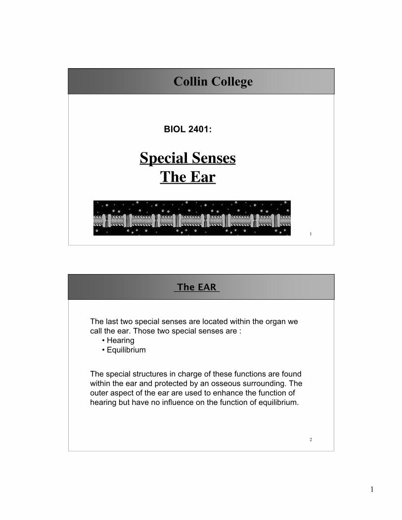
Collin College
BIOL 2401:
Special SensesThe Ear
2
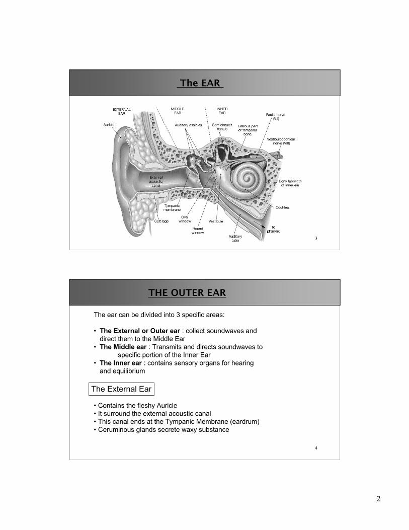
The EAR
The last two special senses are located within the organ wecall the ear. Those two special senses are :
• Hearing• Equilibrium
The special structures in charge of these functions are foundwithin the ear and protected by an osseous surrounding. Theouter aspect of the ear are used to enhance the function ofhearing but have no influence on the function of equilibrium.
2
3
The EAR
4
THE OUTER EAR
The ear can be divided into 3 specific areas:
• The External or Outer ear : collect soundwaves anddirect them to the Middle Ear
• The Middle ear : Transmits and directs soundwaves tospecific portion of the Inner Ear
• The Inner ear : contains sensory organs for hearingand equilibrium
The External Ear
• Contains the fleshy Auricle• It surround the external acoustic canal• This canal ends at the Tympanic Membrane (eardrum)• Ceruminous glands secrete waxy substance

3
5
THE MIDDLE EAR
The MIDDLE Ear
• Contains 3 auditory ossicles that connect thetympanic membrane with the inner ear
• Malleus or Hammer : connects at threepoints with the eardrum
• Incus or Anvil : connect Malleus to Stapes• Stapes or Stirrup: is connected to the oval
window of the inner ear
• Communicates with the nasopharynx via the pharyngotympanic tubeor Eustachian Tube
6
THE MIDDLE EAR
The MIDDLE Ear
• Damage to the tympanic membrane and ossiclesis partially prevented by two muscles
• Tensor tympani : stiffens the eardum bypulling on the malleus
• Stapedius muscle: prevents excessivemovement of stapes on the oval window
• Sound waves result in a vibrating eardrum. These vibrations arepassed on via these ossicles and focused onto the oval window
• Since the tympanic membrane is 22x larger than the oval window,considerable amplification occurs

4
7
THE INNER EAR
The INNER Ear
• Contains the receptors for hearing andequilibrium
• These receptors lie within fluid filled tubesreferred to as the membraneous labyrinth :the fluid is called the endolymph
• The endolymph is a-typical in that it has ahigh potassium and low sodiumconcentration.
• The bony labyrinth surrounds and protectsthis membraneous labyrinth. Betweenbony and membraneous labyrinth flows afluid called the perilymph.
Bony labyrinth
Membraneous labyrinth
perilymph
endolymph
8
THE INNER EAR
The Bony labyrinth isdivided into
• Cochlea• Vestibule• Semicircular canals
5
9
THE INNER EAR
The INNER Ear
• The Vestibule consist out of saccule and utricle : they containreceptors that are important for sensation of gravity and linearacceleration
• Receptors in The semicircular canals sense rotation of the head
• Together, Vestibule and semicircular canals are called thevestibular complex
• The cochlea is a spiral-shaped bony chamber that houses thecochlear duct and the receptors for hearing.
• The sensory receptors of the inner ear are called hair-cells
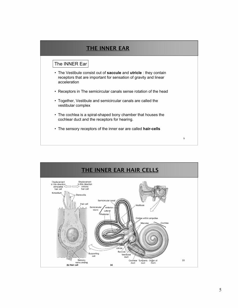
10
THE INNER EAR HAIR CELLS

6
11
THE INNER EAR HAIR CELLS
• Hair cells have a tall kinocilium and many stereocilia (decreasing inlength)
• They are all connected to each other by links• Movement of stereocilia towards the kinocilium, pulls ion channels open
(K+ channels)
12
THE INNER EAR
Movement of stereocilia towards the kinocilium thus creates a change inmembrane potential and increases the number of action potentials in theafferent nerve (the vestibular or cochlear nerve depending on where weare).

7
13
The Sense of HEARING
They pick up the sound vibrations which have beentransmitted from Tympanic membrane via the middle earossicles to the oval window and transformed into waves inthe perilymph.
• The organs for hearing are located within the Cochlearduct, within the cochlea
• The cochlear duct, filled with endolymph, itself issandwiched between the vestibular duct and the tympanicduct (filled with perilynph)
• The receptor Hair cells of the cochlear duct are organizedwithin the Spiral Organ of Corti
14
The Sense of HEARING
(filled with endolymph)

8
15
The Sense of HEARING
• Even though the chochlea isspiral-shaped, it is actually atube.
• We can visualize theorganization of the cochlear ductbetter if we imagine it beingrolled out
Oval window withS. vestibuli
Cochlear duct or Scala Media
• This wraps around at the endinto the Scala Tympani, whichends at the Round window
Round window withS. tympani
• Oval window connects to theScala Vestibuli.
• In the middle is the Scala media,which contains the hair cells andspiral organ of Corti.
16
The Sense of HEARING
• Basilar membrane separatesS. Media from S. Tympani
Oval window withS. vestibuli
Cochlear duct or Scala Media
Round window withS. tympani
Basilar membrane
Vestibular membrane
• The organ of Corti sits on thebasilar membrane within theScala Media
• Vestibular membraneseparates S.Vestibuli fromS. media
• S. vestibuli and S. tympani formone contineous duct, filled withperilymph.
• S. media is filled with endolymph.• Endolymph is rich in K+ and similar to intracellular fluid.

9
17
The Sense of HEARING
18
The Sense of HEARING
• The organ of Corti with the hair cellsis the actual organ of hearing.
• They respond to the pressurewaves generated in the endolymphby the action of the tympanicmembrane on the perilymph.
10
19
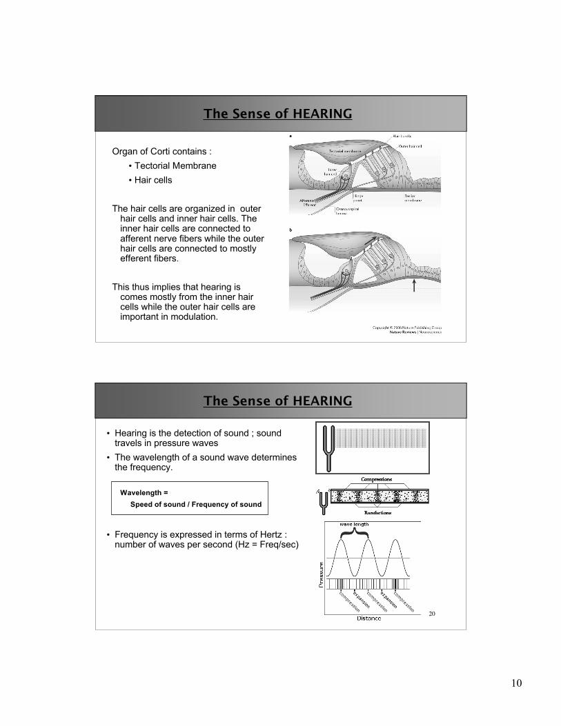
The Sense of HEARING
Organ of Corti contains :• Tectorial Membrane• Hair cells
The hair cells are organized in outerhair cells and inner hair cells. Theinner hair cells are connected toafferent nerve fibers while the outerhair cells are connected to mostlyefferent fibers.
This thus implies that hearing iscomes mostly from the inner haircells while the outer hair cells areimportant in modulation.
20
The Sense of HEARING
• Hearing is the detection of sound ; soundtravels in pressure waves
• The wavelength of a sound wave determinesthe frequency.
Wavelength = Speed of sound / Frequency of sound
• Frequency is expressed in terms of Hertz :number of waves per second (Hz = Freq/sec)

11
21
The Sense of HEARING
• The pitch of a sound relates to the Freq.• High pitch = high frequency, short
wavelength• Low pitch = low frequency, high
wavelength
Examples :For sound in air ( 1235 km/hr)
(= 1235000 m/3600 sec) or (= 343 m/sec)
Wavelength = speed/Freq
Frequency Wavelength(Hertz, or cycles / second) (meters) 100 3.43 1000 0.343 10,000 0.0343
22
The Sense of HEARING
The wave's amplitude is the change inpressure as the sound wave passesby.
The amplitude of a wave is related tothe amount of energy it carries.
• A high amplitude wave carries a largeamount of energy (louder)
• A low amplitude wave carries a smallamount of energy (quieter)
As the amplitude of the sound waveincreases, the intensity of the soundincreases.
Relative sound intensities are oftengiven in units named decibels (dB).
12
23
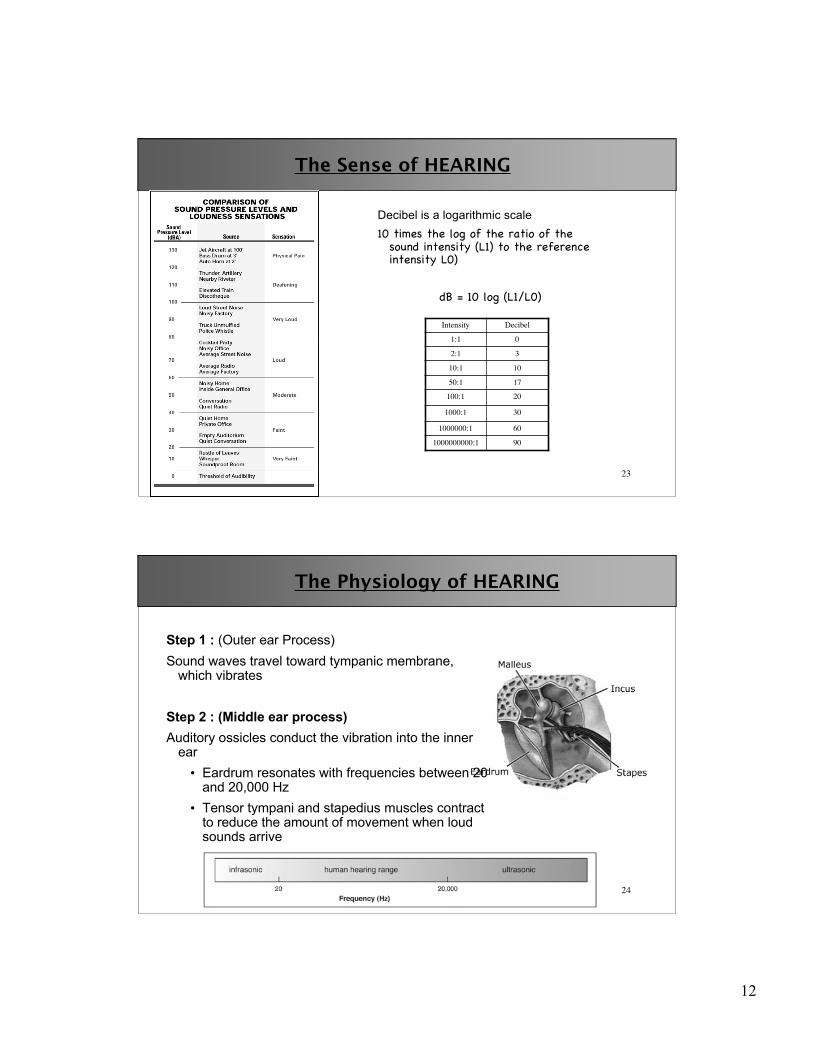
The Sense of HEARING
Decibel is a logarithmic scale10 times the log of the ratio of the
sound intensity (L1) to the referenceintensity L0)
dB = 10 log (L1/L0)
901000000000:1
601000000:1
301000:1
20100:1
1750:1
1010:1
32:1
01:1
DecibelIntensity
24
The Physiology of HEARING
Step 1 : (Outer ear Process)Sound waves travel toward tympanic membrane,
which vibrates
Step 2 : (Middle ear process)Auditory ossicles conduct the vibration into the inner
ear• Eardrum resonates with frequencies between 20
and 20,000 Hz• Tensor tympani and stapedius muscles contract
to reduce the amount of movement when loudsounds arrive

13
25
The Physiology of HEARING
• The middle ear acts as an impedance-matching device.• Sound waves travel much easier through air (low impedance) than
water (high impedance).
• If sound waves were directed at the oval window (water) almost all ofthe acoustic energy would be reflected back to the middle ear (air) andonly 1% would enter the cochlea.
• This would be a terribly inefficient mechanism.
• To increase the efficiency of the system, the middle ear acts totransform the acoustic energy to mechanical energy which thenstimulates the cochlear fluid.
26
The Physiology of HEARING
• The middle ear also acts to increase the acoustic energy reaching thecochlea by essentially two mechanical phenomenon.
• First, the area of the tympanic membrane is much greater than thatof the stapes footplate (oval window) causing the force applied atthe footplate per square area to be greater than the tympanicmembrane.
• Second, the ossicles act as a lever increasing once again the forceapplied at the stapes footplate. Overall, the increase in soundenergy reaching the cochlea is approximately 22 times.

14
27
The Physiology of HEARING
Step 3 : Inner Ear process
• The cochlea consists of a fluid filled bony canal within which lies the cochlearduct containing the sensory epithelium.
• Energy enters the cochlea via the stapes bone at the oval window and isdissipated through a second opening (which is covered by a membrane) theround window.
• Vibrations of the stapes footplate cause the perilymph to form a wave. Thiswave travels the length of the cochlea. It takes approximately 5 msec to travelthe length of the cochlea.
28
The Physiology of HEARING
Step 4 : Basilar membrane distortion
• As it passes the basilar membrane ofthe cochlear duct,the fluid wave causesthe basilar membrane to move in awave-like fashion (i.e. up and down).The wave form travels the length of thecochlea and is dissipated at the roundwindow
• Due to changes in the mechanical properties of the basilar membrane, theamplitude of vibration changes as one travels along the basilar membrane.Low frequency stimuli cause the greatest vibration of basilar membrane at itsapex, high frequency stimuli at its base.

15
29
The Physiology of HEARING
30
The Physiology of HEARING
16
31
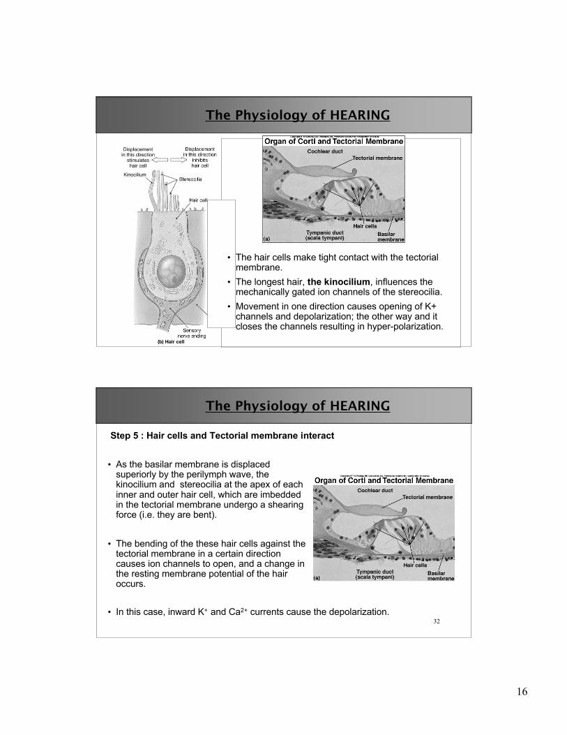
The Physiology of HEARING
• The hair cells make tight contact with the tectorialmembrane.
• The longest hair, the kinocilium, influences themechanically gated ion channels of the stereocilia.
• Movement in one direction causes opening of K+channels and depolarization; the other way and itcloses the channels resulting in hyper-polarization.
32
The Physiology of HEARING
• As the basilar membrane is displacedsuperiorly by the perilymph wave, thekinocilium and stereocilia at the apex of eachinner and outer hair cell, which are imbeddedin the tectorial membrane undergo a shearingforce (i.e. they are bent).
• The bending of the these hair cells against thetectorial membrane in a certain directioncauses ion channels to open, and a change inthe resting membrane potential of the hairoccurs.
Step 5 : Hair cells and Tectorial membrane interact
• In this case, inward K+ and Ca2+ currents cause the depolarization.

17
33
The Physiology of HEARING
Step 5 : Hair cells and Tectorial membrane interact
• The depolarization results in the opening of calcium channels, which resultsin the release of neurotransmitters ( glutamate) at the base of the hair cells.
34
The Physiology of HEARING
Step 6 :
• The hair cell synapse with a bipolar neuron (see next image)
• This causesdepolarization of thebipolar neuron nervefiber, which transmitsthis neural impulsetowards the auditorycenters of the brain.
18
35
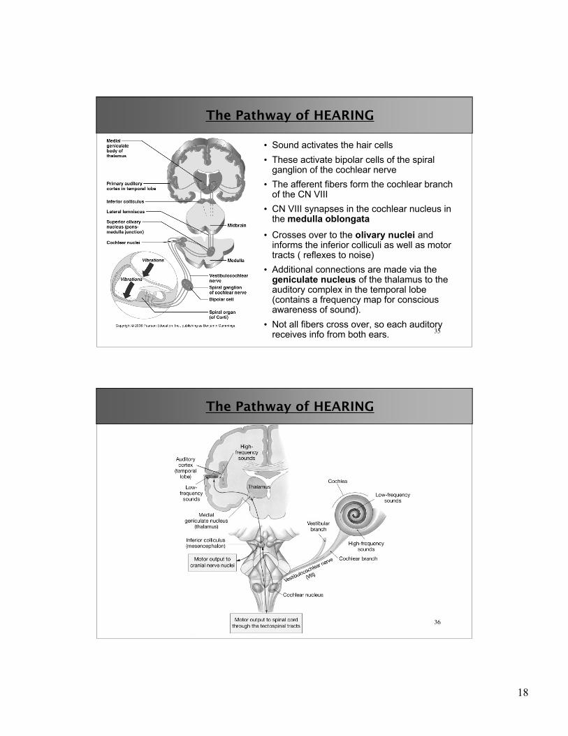
• Crosses over to the olivary nuclei andinforms the inferior colliculi as well as motortracts ( reflexes to noise)
• Additional connections are made via thegeniculate nucleus of the thalamus to theauditory complex in the temporal lobe(contains a frequency map for consciousawareness of sound).
• Not all fibers cross over, so each auditoryreceives info from both ears.
The Pathway of HEARING
• Sound activates the hair cells• These activate bipolar cells of the spiral
ganglion of the cochlear nerve• The afferent fibers form the cochlear branch
of the CN VIII• CN VIII synapses in the cochlear nucleus in
the medulla oblongata
36
The Pathway of HEARING
19
37
HEARING Imbalances
Deafness• Conduction deafness : Due to sound conduction problems
• Wax, perforated eardrum• Otosclerosis (hardening of the ear ossicles)
• Sensorineural deafness : damage to neural structures• Damage to hair cells (broken cilia)• Damage to cochlear nerve or auditory cortex.
Meniere’s Syndrome• Labyrinth disorder and affects all 3 parts of inner ear• Person has repeated attacks of vertigo, nausea, vomiting and balance
problems.• Tinnitus ( ringing sounds in the ear) is common as well.
38
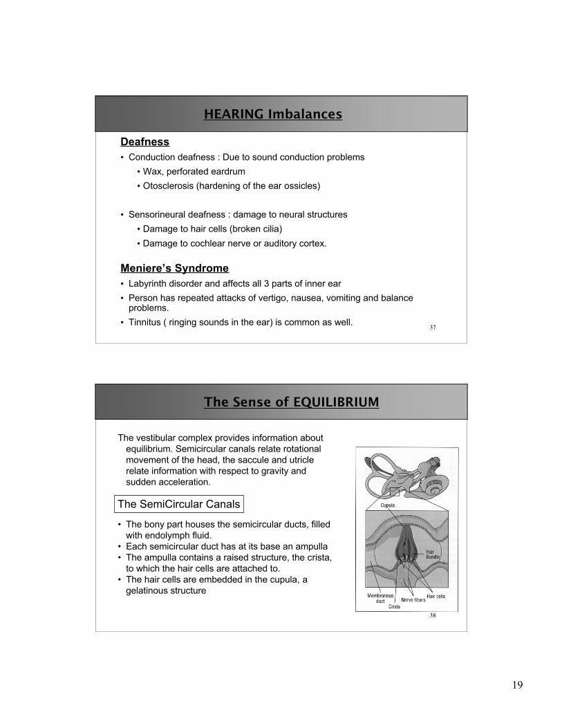
The Sense of EQUILIBRIUM
The vestibular complex provides information aboutequilibrium. Semicircular canals relate rotationalmovement of the head, the saccule and utriclerelate information with respect to gravity andsudden acceleration.
The SemiCircular Canals
• The bony part houses the semicircular ducts, filledwith endolymph fluid.
• Each semicircular duct has at its base an ampulla• The ampulla contains a raised structure, the crista,
to which the hair cells are attached to.• The hair cells are embedded in the cupula, a
gelatinous structure
20
39
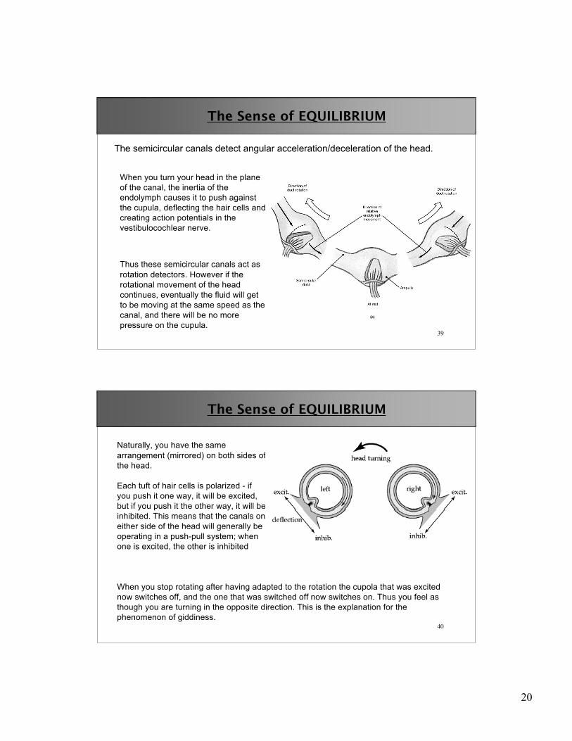
The Sense of EQUILIBRIUM
The semicircular canals detect angular acceleration/deceleration of the head.
When you turn your head in the planeof the canal, the inertia of theendolymph causes it to push againstthe cupula, deflecting the hair cells andcreating action potentials in thevestibulocochlear nerve.
Thus these semicircular canals act asrotation detectors. However if therotational movement of the headcontinues, eventually the fluid will getto be moving at the same speed as thecanal, and there will be no morepressure on the cupula.
40
The Sense of EQUILIBRIUM
Naturally, you have the samearrangement (mirrored) on both sides ofthe head.
Each tuft of hair cells is polarized - ifyou push it one way, it will be excited,but if you push it the other way, it will beinhibited. This means that the canals oneither side of the head will generally beoperating in a push-pull system; whenone is excited, the other is inhibited
When you stop rotating after having adapted to the rotation the cupola that was excitednow switches off, and the one that was switched off now switches on. Thus you feel asthough you are turning in the opposite direction. This is the explanation for thephenomenon of giddiness.
21
41
The Sense of EQUILIBRIUM
• It is important that both sides agree as to what the head is doing. If thereis disagreement, if both sides push at once, then you will feel debilitatingvertigo and nausea.
• This is the reason that infections of the endolymph or damage to the innerear can cause vertigo.
• However, if one vestibular nerve is cut, the brain will gradually get used toonly monitoring one side - this can actually be a treatment for intractablevertigo.
• Drugs such as Dramamine tend to depress the activity of the vestibularnuclei.
42
The Sense of EQUILIBRIUM
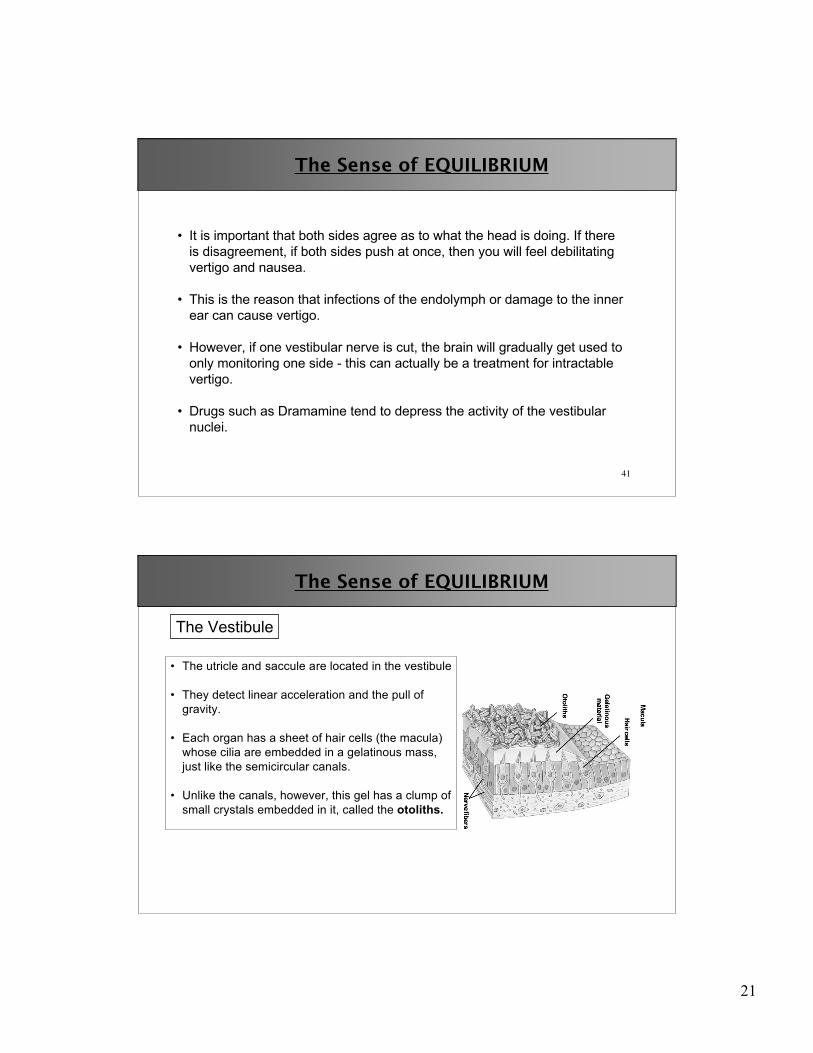
• The utricle and saccule are located in the vestibule
• They detect linear acceleration and the pull ofgravity.
• Each organ has a sheet of hair cells (the macula)whose cilia are embedded in a gelatinous mass,just like the semicircular canals.
• Unlike the canals, however, this gel has a clump ofsmall crystals embedded in it, called the otoliths.
The Vestibule

22
43
The Sense of EQUILIBRIUM
The otoliths provide the inertia, sothat when you move to one side,the otolith-gel mass drags on thehair cells.
Once you are moving at a constantspeed, such as in a car, theotoliths come to equilibrium andyou no longer perceive the motion.
The Vestibule
44
The Sense of EQUILIBRIUM
The hair cells in the utricle and saccule arepolarized, but they are arrayed indifferent directions so that a single sheetof hair cells can detect motion forwardand back, side to side. Each macula cantherefore cover two dimensions ofmovement.
In addition to that :
1. The utricle lies horizontally in the ear, and detects motion in the horizontalplane.
2. The saccule is oriented vertically, so detects motion in the sagittal plane(up and down, forward and back).
23
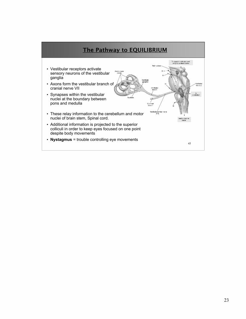
45
The Pathway to EQUILIBRIUM
• Vestibular receptors activatesensory neurons of the vestibularganglia
• Axons form the vestibular branch ofcranial nerve VII
• Synapses within the vestibularnuclei at the boundary betweenpons and medulla
• These relay information to the cerebellum and motornuclei of brain stem, Spinal cord.
• Additional information is projected to the superiorcolliculi in order to keep eyes focused on one pointdespite body movements
• Nystagmus = trouble controlling eye movements


![Collin College Course Syllabus FALL 2013-COLLIN …iws2.collin.edu/mrosenfield/17951.201410[1].pdf · Collin College Course Syllabus FALL 2013-COLLIN COLLEGE ... Determine and use](https://img.pdfslide.us/doc/110x75/5ae7a6e17f8b9aee078e774d/collin-college-course-syllabus-fall-2013-collin-iws2-1pdfcollin-college-course.jpg)