Embed Size (px)
Citation preview
OpenStax-CNX module: m43150 1
2.3.1 Blood circulatory system*
Daniel Williamson
This work is produced by OpenStax-CNX and licensed under the
Creative Commons Attribution License 3.0�
1 Useful links
Cardiovascular system: http://www.biologyinmotion.com/cardio/index.html 1
Khan: Red blood cells http://www.khanacademy.org/video/red-blood-cells?playlist=Biology 2
Khan: Circulatory system and the heart http://www.khanacademy.org/video/circulatory-system-and-the-heart?playlist=Biology 3
YouTube video: Circulatory system http://www.youtube.com/watch?v=D3ZDJgFDdk0 4
Blood �ow animation http://health.howstu�works.com/human-body/systems/circulatory/adam-200078.htm5
Blood Flow animation http://www.sumanasinc.com/webcontent/animations/content/human_heart.html6
The heart and circulation (interactive activity): http://www.kett6.net/adulteducation/heartanimations.html7
Circulation animation: http://www.bbc.co.uk/schools/gcsebitesize/pe/appliedanatomy/0_anatomy_circulatorysys_rev1.shtml 8
2 Overview
All living cells require nutrients and oxygen to survive. Cells produce metabolic waste, which must be re-moved and excreted. The circulatory system is responsible from providing nutrients and removing metabolicwaste.
Unicellular organisms have a simple system to allow for this and it is by di�usion where substancesmove from a high concentration to a low concentration.
Most invertebrates like a grasshopper have an open circulatory system , where blood (haemolymph)bathes the body organs.
By comparison, mammals have a closed circulatory system since blood is contained within bloodvessels.
*Version 1.1: Feb 17, 2012 6:47 am -0600�http://creativecommons.org/licenses/by/3.0/1http://www.biologyinmotion.com/cardio/index.html2http://www.khanacademy.org/video/red-blood-cells?playlist=Biology3http://www.khanacademy.org/video/circulatory-system-and-the-heart?playlist=Biology4http://www.youtube.com/watch?v=D3ZDJgFDdk05http://health.howstu�works.com/human-body/systems/circulatory/adam-200078.htm6http://www.sumanasinc.com/webcontent/animations/content/human_heart.html7http://www.kett6.net/adulteducation/heartanimations.html8http://www.bbc.co.uk/schools/gcsebitesize/pe/appliedanatomy/0_anatomy_circulatorysys_rev1.shtml
http://cnx.org/content/m43150/1.1/
OpenStax-CNX module: m43150 2
3 Pulmonary and Systemic circulatory systems
Open circulatory systemblood is pumped into a heamocoel (an open space or cavity) that surrounds to organs. Muscle movement
also helps to pump then blood. Blood di�uses back the heart. Blood movement is sluggish. There is nodi�erence between the blood and the interstitial �uid. Interstitial �uid is the �uid that surrounds the cells.Blood is not contained within capillaries.
Closed circulatory systemblood is pumped from the heart through arteries and returns to the heart via veins. Blood never leaves
the vascular system (arteries, veins and capillaries). Nutrients, water and metabolic waste di�uses out ofthe vascular system and into the interstitial �uid. Interstitial �uid and blood are separated, by the vascularsystem. Interstitial �uid returns to circulation through the lymphatic system.
3.1 The Human Circulatory System
All mammals have a closed blood circulatory system - blood always �ows inside blood vessels.A double circulatory system = blood passes through the heart twice:
1. Pulmonary circulation: the blood is pumped from the heart to the lungs to oxygenate the bloodand then back to the heart.
2. Systemic circulation (to all the systems): the blood is pumped from the heart to all parts ofthe body and back to the heart again.
3. Coronary circulation : is a circulatory system that supplies the heart muscle with the blood itrequired in order to function.
Very simple simulation of blood �ow through the systemic and pulmonary circulatory systems. The illus-tration shows each of these circulatory systems to be separate loops leaving from one side of the heart andreturning to the other.
http 9 :// 10 www 11 . 12 biologyinmotion 13 . 14 com 15 / 16 cardio 17 / 18 index 19 .20 html 21
Figure: Simpli�ed Diagrammatic sketch of the entire circulatory system. Blood �ows to every inch ofthe body, even to the tips of the �ngers and toes. Lungs provide oxygen to the blood. The digestive systemsupplies nutrients. The kidneys �lter the blood.
9http://www.biologyinmotion.com/cardio/index.html10http://www.biologyinmotion.com/cardio/index.html11http://www.biologyinmotion.com/cardio/index.html12http://www.biologyinmotion.com/cardio/index.html13http://www.biologyinmotion.com/cardio/index.html14http://www.biologyinmotion.com/cardio/index.html15http://www.biologyinmotion.com/cardio/index.html16http://www.biologyinmotion.com/cardio/index.html17http://www.biologyinmotion.com/cardio/index.html18http://www.biologyinmotion.com/cardio/index.html19http://www.biologyinmotion.com/cardio/index.html20http://www.biologyinmotion.com/cardio/index.html21http://www.biologyinmotion.com/cardio/index.html
http://cnx.org/content/m43150/1.1/
OpenStax-CNX module: m43150 3
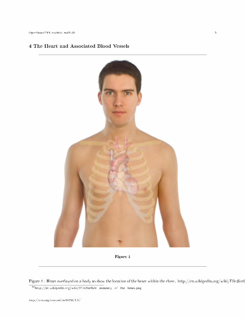
4 The Heart and Associated Blood Vessels
Figure 1
Figure 1 : Heart overlayed on a body so show the location of the heart within the chest. http://en.wikipedia.org/wiki/File:Surface_anatomy_of_the_heart.png22
22http://en.wikipedia.org/wiki/File:Surface_anatomy_of_the_heart.png
http://cnx.org/content/m43150/1.1/
OpenStax-CNX module: m43150 4
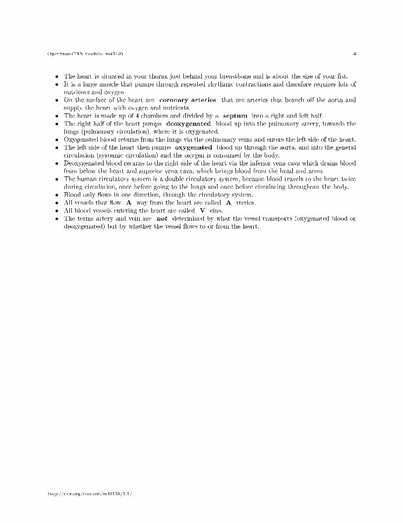
• The heart is situated in your thorax just behind your breastbone and is about the size of your �st.• It is a large muscle that pumps through repeated rhythmic contractions and therefore requires lots of
nutrients and oxygen.• On the surface of the heart are coronary arteries that are arteries that branch o� the aorta and
supply the heart with oxygen and nutrients.• The heart is made up of 4 chambers and divided by a septum into a right and left half.• The right half of the heart pumps deoxygenated blood up into the pulmonary artery, towards the
lungs (pulmonary circulation), where it is oxygenated.• Oxygenated blood returns from the lungs via the pulmonary veins and enters the left side of the heart.• The left side of the heart then pumps oxygenated blood up through the aorta, and into the general
circulation (systemic circulation) and the oxygen is consumed by the body.• Deoxygenated blood returns to the right side of the heart via the inferior vena cava which drains blood
from below the heart and superior vena cava, which brings blood from the head and arms.• The human circulatory system is a double circulatory system, because blood travels to the heart twice
during circulation, once before going to the lungs and once before circulating throughout the body.• Blood only �ows in one direction, through the circulatory system.• All vessels that �ow A way from the heart are called A rteries.• All blood vessels entering the heart are called V eins.• The terms artery and vein are not determined by what the vessel transports (oxygenated blood or
deoxygenated) but by whether the vessel �ows to or from the heart.
http://cnx.org/content/m43150/1.1/
OpenStax-CNX module: m43150 5
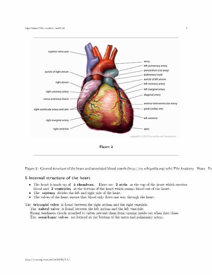
Figure 2
Figure 2 : General structure of the heart and associated blood vessels (http://en.wikipedia.org/wiki/File:Anatomy_Heart_English_Tiesworks.jpg)
5 internal structure of the heart
• The heart is made up of 4 chambers. There are 2 atria at the top of the heart which receivesblood and 2 ventricles at the bottom of the heart which pumps blood out of the heart.
• The septum divides the left and right side of the heart.• The valves of the heart ensure that blood only �ows one way through the heart.
The tricuspid valve is found between the right atrium and the right ventricle.The mitral valve is found between the left atrium and the left ventricle.Strong tendinous chords attached to valves prevent them from turning inside out when they close.The semi-lunar valves are located at the bottom of the aorta and pulmonary artery.
http://cnx.org/content/m43150/1.1/
OpenStax-CNX module: m43150 6
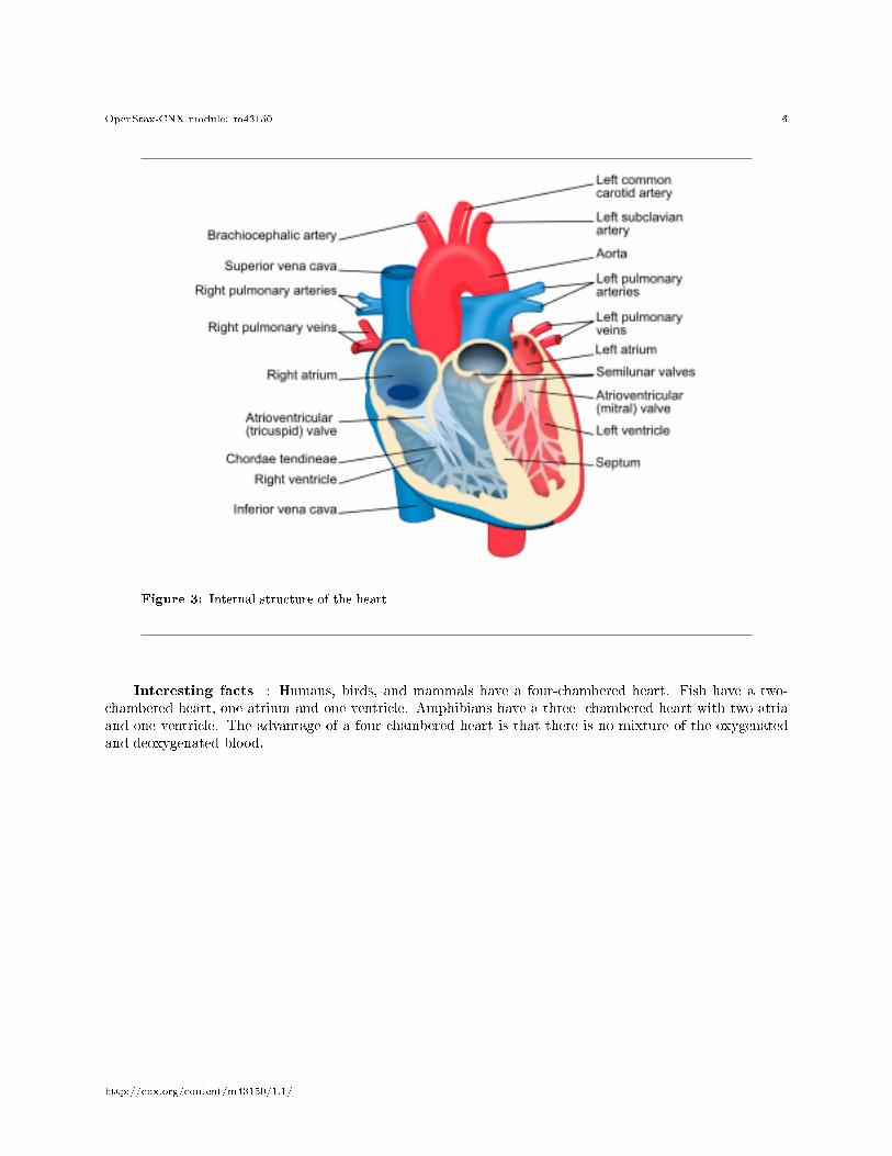
Figure 3: Internal structure of the heart
Interesting facts : Humans, birds, and mammals have a four-chambered heart. Fish have a two-chambered heart, one atrium and one ventricle. Amphibians have a three- chambered heart with two atriaand one ventricle. The advantage of a four chambered heart is that there is no mixture of the oxygenatedand deoxygenated blood.
http://cnx.org/content/m43150/1.1/
OpenStax-CNX module: m43150 7
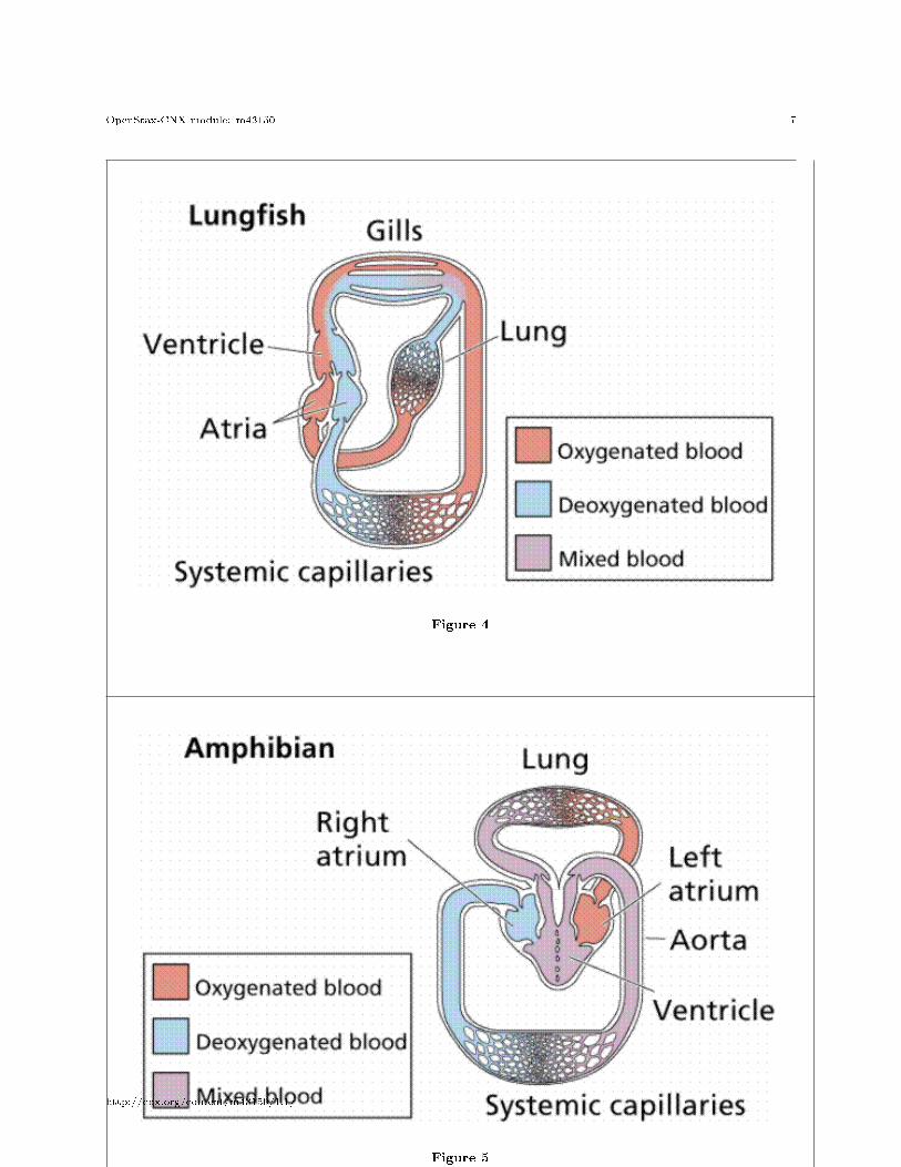
Figure 4
Figure 5
continued on next page
http://cnx.org/content/m43150/1.1/
OpenStax-CNX module: m43150 8
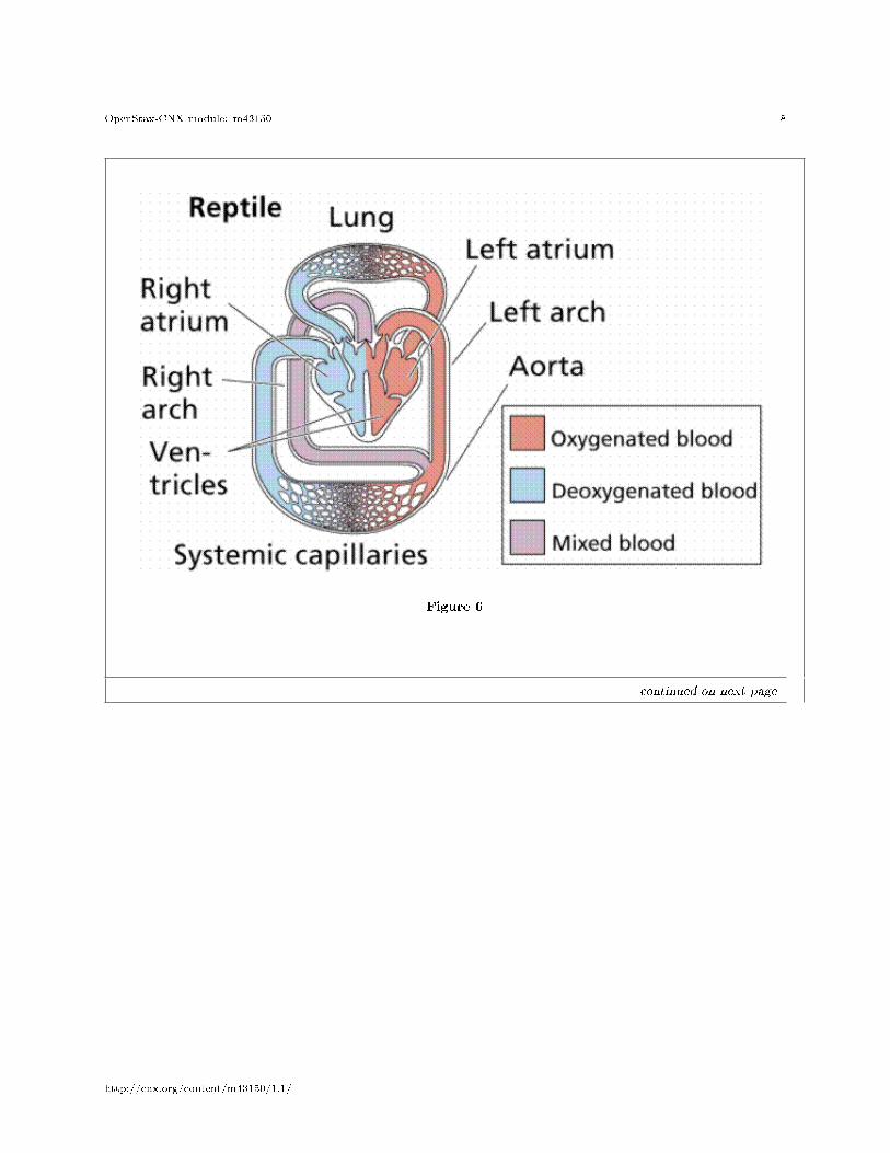
Figure 6
continued on next page
http://cnx.org/content/m43150/1.1/
OpenStax-CNX module: m43150 9
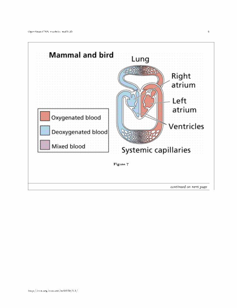
Figure 7
continued on next page
http://cnx.org/content/m43150/1.1/
OpenStax-CNX module: m43150 10
Figure 4. The relationship of the heart and circulatory system to major visceral organs.
Table 1
The circulatory song http://www.youtube.com/watch?v=q0s-1MC1hcE&NR=1 23
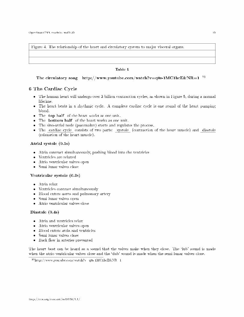
6 The Cardiac Cycle
• The human heart will undergo over 3 billion contraction cycles, as shown in Figure 5, during a normallifetime.
• The heart beats in a rhythmic cycle. A complete cardiac cycle is one round of the heart pumpingblood.
• The top half of the heart works as one unit.• The bottom half of the heart works as one unit.• The sino-atrial node (pacemaker) starts and regulates the process.• The cardiac cycle consists of two parts: systole (contraction of the heart muscle) and diastole
(relaxation of the heart muscle).
Atrial systole (0.1s)
• Atria contract simultaneously, pushing blood into the ventricles• Ventricles are relaxed• Atrio ventricular valves open• Semi lunar valves close
Ventricular systole (0.3s)
• Atria relax• Ventricles contract simultaneously• Blood enters aorta and pulmonary artery• Semi lunar valves open• Atrio ventricular valves close
Diastole (0.4s)
• Atria and ventricles relax• Atrio ventricular valves open• Blood enters atria and ventricles• Semi lunar valves close• Back �ow in arteries prevented
The heart beat can be heard as a sound that the valves make when they close. The `lub' sound is madewhen the atrio ventricular valves close and the `dub' sound is made when the semi lunar valves close.
23http://www.youtube.com/watch?v=q0s-1MC1hcE&NR=1
http://cnx.org/content/m43150/1.1/
OpenStax-CNX module: m43150 11
Figure 8
Figure 5from mindset � (please check permission from this, found it in Biology 6th edition Campbell and Reece)Cardiac Cycle: �ow of blood through the heartExcellent simple video illustrating the heart cycle.http://www.youtube.com/watch?v=D3ZDJgFDdk0 24
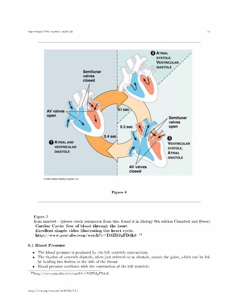
6.1 Blood Pressure
• The blood pressure is produced by the left ventricle contractions.• The rhythm of ventricle diastole, often just referred to as diastole, causes the pulse, which can be felt
by holding two �nders to the side of the throat.• Blood pressure oscillates with the contraction of the left ventricle.
24http://www.youtube.com/watch?v=D3ZDJgFDdk0
http://cnx.org/content/m43150/1.1/
OpenStax-CNX module: m43150 12
Ideal blood pressure for an adult is:Systolic pressure: 120 mm HGDiastolic blood pressure: 80 mm HGA usual rule is that systolic pressure should be 100 plus your age but never more than 140 and diastolic
pressure should not be over 90.
Figure 9
Table 2
Figure 6 The cardiac cycle. Image from Purves et al., Life: The Science of Biology , 4th Edition,by Sinauer Associates ( www.sinauer.com 25 ) and WH Freeman ( www.whfreeman.com 26 ),(please getpermission)
Normal Heart Soundshttp://upload.wikimedia.org/wikipedia/commons/7/72/HROgg.ogg 27
7
8 Lung and pulmonary system
Khan Academy video on the pulmonary system. Overview on breathing.
25http://www.sinauer.com/26http://www.whfreeman.com/27http://upload.wikimedia.org/wikipedia/commons/7/72/HROgg.ogg
http://cnx.org/content/m43150/1.1/
OpenStax-CNX module: m43150 13
http://www.khanacademy.org/video/the-lungs-and-pulmonary-system?playlist=Biology 28
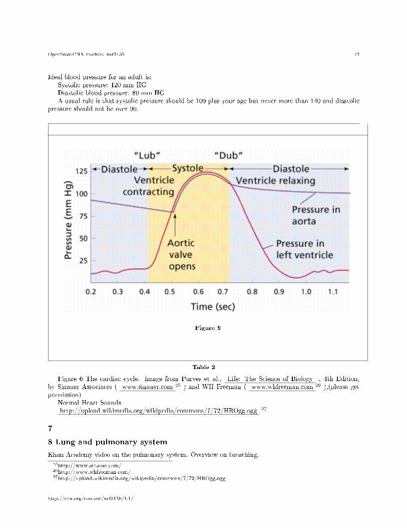
• The lungs serve as the air-blood interface.• Blood from the lungs is pumped into the pulmonary arteries.• From the pulmonary arteries the vascular system branches into smaller and smaller vessels until the
blood is �owing through thin pulmonary capillaries.• These capillaries surround the alveoli in the lungs.• At this point there are only two layers of cells separating the blood from the air.• Carbon dioxide in deoxygenated blood di�used out of the blood.• Oxygen in the lungs di�use in to the blood oxygenating it Oxygen is absorbed.• Oxygenated blood then returns to the heart vial the pulmonary veins.
Figure : Details arteries and veins connecting the heart to the lungs Red blood has been oxygenated, blueblood is deoxygenated. . (Wikipedia - http://en.wikipedia.org/wiki/File:Illu_pulmonary_circuit.jpg)
Figure 10
28http://www.khanacademy.org/video/the-lungs-and-pulmonary-system?playlist=Biology
http://cnx.org/content/m43150/1.1/
OpenStax-CNX module: m43150 14
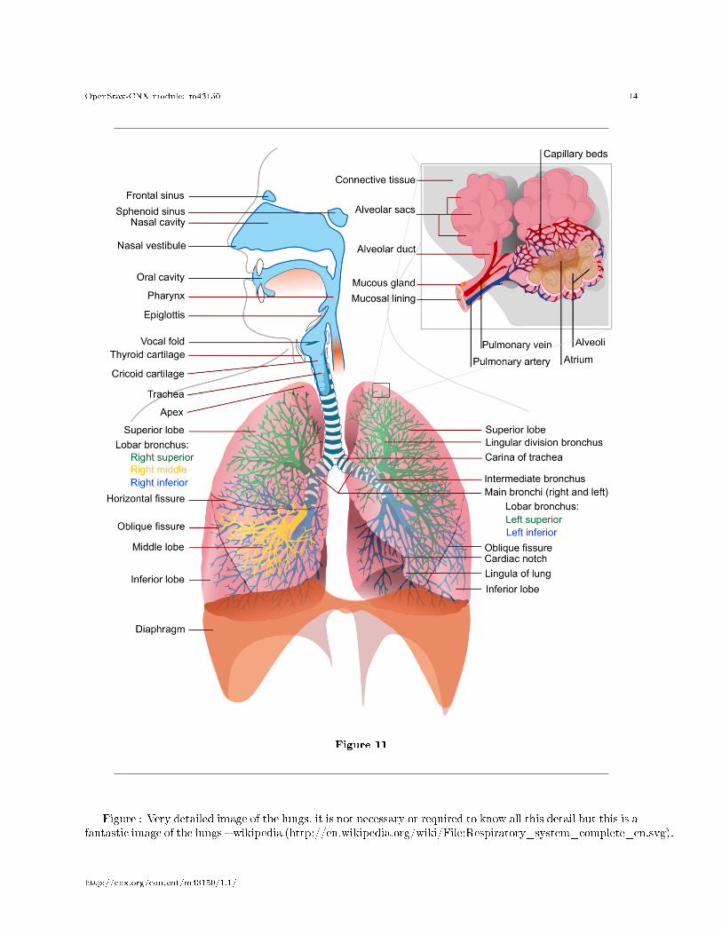
Figure 11
Figure : Very detailed image of the lungs, it is not necessary or required to know all this detail but this is afantastic image of the lungs � wikipedia (http://en.wikipedia.org/wiki/File:Respiratory_system_complete_en.svg).
http://cnx.org/content/m43150/1.1/
OpenStax-CNX module: m43150 15
9 Major organs and systemic system: associated major blood vessels the brain,
small intestines, liver, kidney.
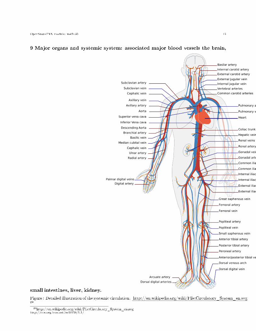
Figure : Detailed illustration of the systemic circulation. http://en.wikipedia.org/wiki/File:Circulatory_System_en.svg29
29http://en.wikipedia.org/wiki/File:Circulatory_System_en.svghttp://cnx.org/content/m43150/1.1/
OpenStax-CNX module: m43150 16
• All the organs of the body are supplied by blood.• Each has an artery supplying the organ with blood from the heart, and veins returning blood to the
heart.• Arteries and veins have been named according to the organ which they supply blood to.
The circulatory system forms a closed system.
• Nutrients enter the circulatory system from the digestive system.• These nutrients �rst move to the liver via the hepatic portal vein, the liver then controls the nutrient
composition of the blood.• Blood passes from the liver to the heart through the hepatic vein.• Nutrients are then circulated throughout the body.• Cells consume the nutrients in the blood and produce metabolic waste. T• his metabolic waste is circulated in the blood, if it remains in the blood the blood would eventually
become toxic.• The kidneys are supplied with blood via the renal arteries and they remove metabolic waste from the
blood, passing it to urine.• The Brain is supplied with blood via the carotid arteries and the vertebral arteries. The blood is
drained via the jugular veins. The brain is supplied with 15% of the total amount of blood pumpedby the heart.
10 Mechanisms for controlling cardiac cycle and heart rate (pulse)
• The cardiac cycle is controlled by nerve �bers extending from nodes of nerve bundles through the heartmuscle.
• An electrical signal is triggered in the node.• The electrical signal then spreads through the �bers and causes the heart muscle to contract.
10.1 There are two nodes:
1. The sinoatrial node (SA), which initiates the heart cycle. Electrical signals spread from the SAacross the atria causing it to contact.
2. The electrical signal also reaches the Atrioventricular node (AV) . Here the signal pauses, beforespreading through the ventricles causing them to contract.
• The SA is able to initiate the electrical signal without any stimulation for the nervous system, but itcan be controlled by the nervous system.
• The brain does not need to tell the heart to beat; it is able to beat on its own.
• The brain can make the heart rate increase, when for instance you are scared or are running.• Hormones are also able to increase the heart rate.
Simple simulation of how electrical activity spreads over the heart.Link : http://en.wikipedia.org/wiki/File:ECG_Principle_fast.gif 30
Measuring pulse rate: http://www.nlm.nih.gov/medlineplus/ency/article/003399.htm 31
30http://en.wikipedia.org/wiki/File:ECG_Principle_fast.gif31http://www.nlm.nih.gov/medlineplus/ency/article/003399.htm
http://cnx.org/content/m43150/1.1/
OpenStax-CNX module: m43150 17
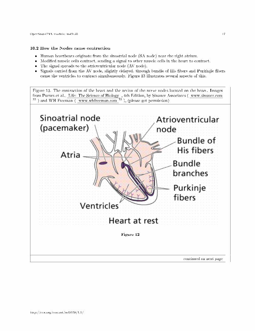
10.2 How the Nodes cause contraction
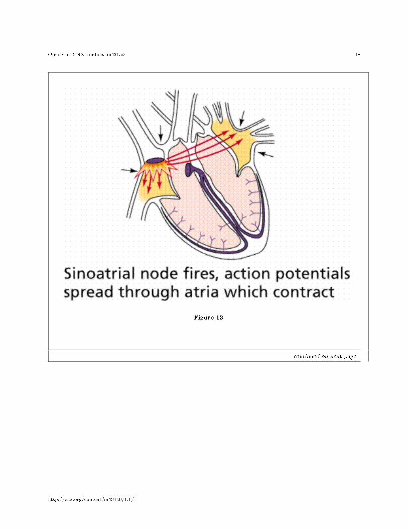
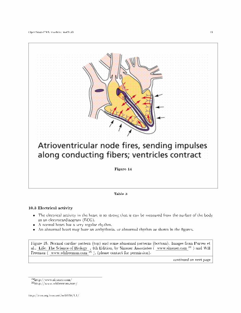
• Human heartbeats originate from the sinoatrial node (SA node) near the right atrium.• Modi�ed muscle cells contract, sending a signal to other muscle cells in the heart to contract.• The signal spreads to the atrioventricular node (AV node).• Signals carried from the AV node, slightly delayed, through bundle of His �bers and Purkinjie �bers
cause the ventricles to contract simultaneously. Figure 13 illustrates several aspects of this.
Figure 13. The contraction of the heart and the action of the nerve nodes located on the heart. Imagesfrom Purves et al., Life: The Science of Biology , 4th Edition, by Sinauer Associates ( www.sinauer.com32 ) and WH Freeman ( www.whfreeman.com 33 ), (please get permission)
Figure 12
continued on next page
http://cnx.org/content/m43150/1.1/
OpenStax-CNX module: m43150 18
Figure 13
continued on next page
http://cnx.org/content/m43150/1.1/
OpenStax-CNX module: m43150 19
Figure 14
Table 3
10.3 Electrical activity
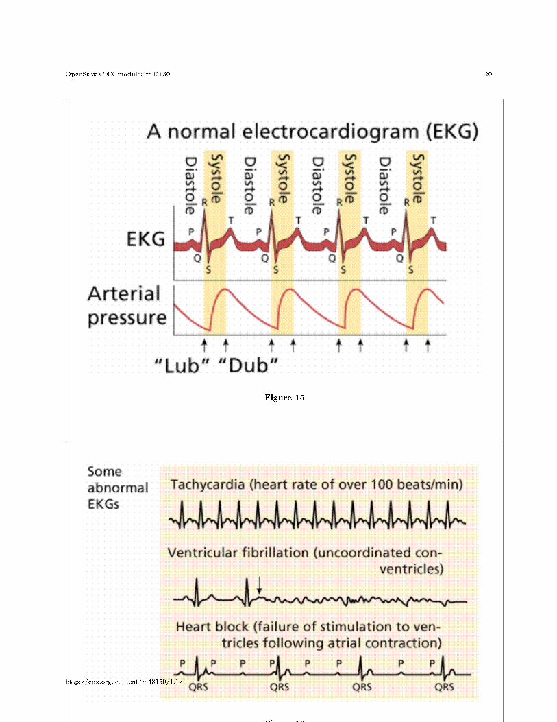
• The electrical activity in the heart is so strong that is can be measured from the surface of the bodyas an electrocardiaogram (ECG).
• A normal heart has a very regular rhythm.• An abnormal heart may have an arrhythmia, or abnormal rhythm as shown in the �gures.
Figure 15. Normal cardiac pattern (top) and some abnormal patterns (bottom). Images from Purves etal., Life: The Science of Biology , 4th Edition, by Sinauer Associates ( www.sinauer.com 34 ) and WHFreeman ( www.whfreeman.com 35 ), (please contact for permission).
continued on next page
32http://www.sinauer.com/33http://www.whfreeman.com/
http://cnx.org/content/m43150/1.1/
OpenStax-CNX module: m43150 20
Figure 15
Figure 16
http://cnx.org/content/m43150/1.1/
OpenStax-CNX module: m43150 21
Table 4
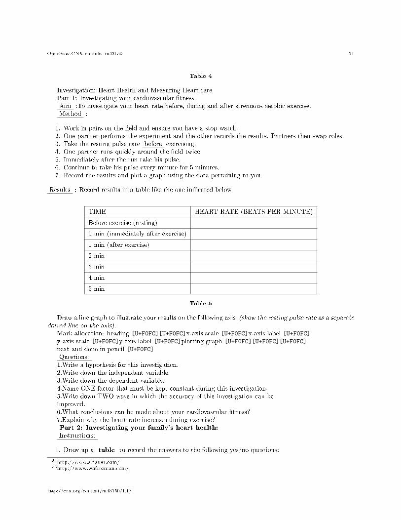
Investigation: Heart Health and Measuring Heart ratePart 1: Investigating your cardiovascular �tnessAim :To investigate your heart rate before, during and after strenuous aerobic exercise.Method :
1. Work in pairs on the �eld and ensure you have a stop watch.2. One partner performs the experiment and the other records the results. Partners then swap roles.3. Take the resting pulse rate before exercising.4. One partner runs quickly around the �eld twice.5. Immediately after the run take his pulse.6. Continue to take his pulse every minute for 5 minutes.7. Record the results and plot a graph using the data pertaining to you.
Results : Record results in a table like the one indicated below
TIME HEART RATE (BEATS PER MINUTE)
Before exercise (resting)
0 min (immediately after exercise)
1 min (after exercise)
2 min
3 min
4 min
5 min
Table 5
Draw a line graph to illustrate your results on the following axis (show the resting pulse rate as a separatedotted line on the axis).
Mark allocation: heading [U+F0FC][U+F0FC]x-axis scale [U+F0FC]x-axis label [U+F0FC]y-axis scale [U+F0FC]y-axis label [U+F0FC]plotting graph [U+F0FC][U+F0FC][U+F0FC]
neat and done in pencil [U+F0FC]Questions:1.Write a hypothesis for this investigation.2.Write down the independent variable.3.Write down the dependent variable.4.Name ONE factor that must be kept constant during this investigation.5.Write down TWO ways in which the accuracy of this investigation can beimproved.6.What conclusions can be made about your cardiovascular �tness?7.Explain why the heart rate increases during exercise?Part 2: Investigating your family's heart health:Instructions:
1. Draw up a table to record the answers to the following yes/no questions:
34http://www.sinauer.com/35http://www.whfreeman.com/
http://cnx.org/content/m43150/1.1/
OpenStax-CNX module: m43150 22
i. Do you smoke?ii. Are you overweight?iii. Do you exercise regularly?iv. Do you follow a healthy diet (low fat, low salt)v. Do you have your blood pressure checked regularly?vi. Do you have a family history of heart and circulatory disease?
1. Survey two adult male family member (father, grandfather or uncle) and two adult female familymembers (mother, grandmother or aunt). Include the adults' �rst name, gender, age and relationshipto you.
3.Record the results in your table. Also indicated the score they obtained:i. yes=0; no=5ii. yes=0; no=5iii. yes=5; no=0iv. yes=5; no=0v. yes=5; no=0vii. yes=0; no=54.Analyse the results by comparing the total score with the following descriptors:30 marks- you take very good care of your heart. Well done!25 marks- you take good care of your heart. Keep it up!20 marks- you take reasonably good care of your heart but need to workon a few aspects where you scored 0.15 marks- you need to take better care of your heart.0-10 marks- you do not look after your heart at all. It's time to make achange to a healthier lifestyle.Assessment Rubric
• Results
0- not done1- poorly presented.2- average presentation of results,but missing some detail.3- aver-age presentation of results, in-cluding all salient features andinformation.4- good presentationof results, but missing somedetail.5- good presentation of re-sults, including all salient fea-tures and information.
5
Table 6
Rich media:Khan Academy
http://cnx.org/content/m43150/1.1/
OpenStax-CNX module: m43150 23
http 36 :// 37 www 38 . 39 khanacademy 40 . 41 org 42 / 43 video 44 / 45 circulatory 46 -47 system 48 - 49 and 50 - 51 the 52 - 53 heart 54 ? 55 playlist 56 = 57 Biology 58
Cardiac Magnetic Resonance imaging of Beating heart: Large magnets are used to create images of theheart inside the body, without the need for surgery.
http://upload.wikimedia.org/wikipedia/commons/7/73/Four_chamber_cardiovascular_m agnetic_resonance_imaging.gif59
View from the tophttp://commons.wikimedia.org/wiki/File:Beating_Heart_axial.gif 60
View from the sidehttp://commons.wikimedia.org/wiki/File:Cardiac_mri_ani_sagittal_bionerd.gif 61
11 Blood Vessels
11.1 Structure and functioning of arteries, veins, capillaries and valves
11.1.1 Arteries
• Arteries carry blood from away from the heart. The pressure created by the pumping heart forcesblood down the arteries.
• Arteries have three layers.
1. Outside layer � connective tissue2. Middle layer � smooth muscle, allows contraction of the arteries to regulate blood �ow and pressure3. Inside layer � single layer of tightly connected simple squamous endothelial cells
• The large arteries close to the heart branch into smaller arterioles (smaller arteries) and eventuallybranch into capillaries.
36http://www.khanacademy.org/video/circulatory-%20system-and-the-heart?playlist=Biology37http://www.khanacademy.org/video/circulatory-%20system-and-the-heart?playlist=Biology38http://www.khanacademy.org/video/circulatory-%20system-and-the-heart?playlist=Biology39http://www.khanacademy.org/video/circulatory-%20system-and-the-heart?playlist=Biology40http://www.khanacademy.org/video/circulatory-%20system-and-the-heart?playlist=Biology41http://www.khanacademy.org/video/circulatory-%20system-and-the-heart?playlist=Biology42http://www.khanacademy.org/video/circulatory-%20system-and-the-heart?playlist=Biology43http://www.khanacademy.org/video/circulatory-%20system-and-the-heart?playlist=Biology44http://www.khanacademy.org/video/circulatory-%20system-and-the-heart?playlist=Biology45http://www.khanacademy.org/video/circulatory-%20system-and-the-heart?playlist=Biology46http://www.khanacademy.org/video/circulatory-%20system-and-the-heart?playlist=Biology47http://www.khanacademy.org/video/circulatory-%20system-and-the-heart?playlist=Biology48http://www.khanacademy.org/video/circulatory-%20system-and-the-heart?playlist=Biology49http://www.khanacademy.org/video/circulatory-%20system-and-the-heart?playlist=Biology50http://www.khanacademy.org/video/circulatory-%20system-and-the-heart?playlist=Biology51http://www.khanacademy.org/video/circulatory-%20system-and-the-heart?playlist=Biology52http://www.khanacademy.org/video/circulatory-%20system-and-the-heart?playlist=Biology53http://www.khanacademy.org/video/circulatory-%20system-and-the-heart?playlist=Biology54http://www.khanacademy.org/video/circulatory-%20system-and-the-heart?playlist=Biology55http://www.khanacademy.org/video/circulatory-%20system-and-the-heart?playlist=Biology56http://www.khanacademy.org/video/circulatory-%20system-and-the-heart?playlist=Biology57http://www.khanacademy.org/video/circulatory-%20system-and-the-heart?playlist=Biology58http://www.khanacademy.org/video/circulatory-%20system-and-the-heart?playlist=Biology59http://upload.wikimedia.org/wikipedia/commons/7/73/Four_chamber_cardiovascular_magnetic_resonance_imaging.gif60http://commons.wikimedia.org/wiki/File:Beating_Heart_axial.gif61http://commons.wikimedia.org/wiki/File:Cardiac_mri_ani_sagittal_bionerd.gif
http://cnx.org/content/m43150/1.1/
OpenStax-CNX module: m43150 24
11.1.1.1 Capillaries
• Capillaries are little more than a single layer or endothelial cells.• Capillaries form intricate networks throughout the tissues.• They allow water, nutrients and gasses to di�use out of the blood and waste materials to di�use into
the blood.• This exchange occurs between the blood and the interstitial �uid.• The interstitial �uid is the �uid surrounding the cells.• The blood never comes into contact with the cells.• The blood and interstitial �uid exchange material, and the interstitial �uid then exchanges material
with the cells.
11.1.1.2 Veins
• The intricate networks formed by the capillaries eventually converge to form venules, (small veins)• The venules then converge to form veins which return the blood to the heart.• Veins only consist of two layers.
1. The outer layer is made up of connective tissue2. The inner layer is made up of endothelial cells.
11.1.1.3 Valves
• Once the blood has passed through the capillaries very little blood pressure remains to return bloodto the heart.
• Instead of pressure from the heart veins use a series of valves to force blood to return to the heart.• Contraction of the muscles squeezes the veins, pushing the blood through them.• The valves cause the blood to �ow in only one direction, back to the heart.
http://cnx.org/content/m43150/1.1/

OpenStax-CNX module: m43150 25
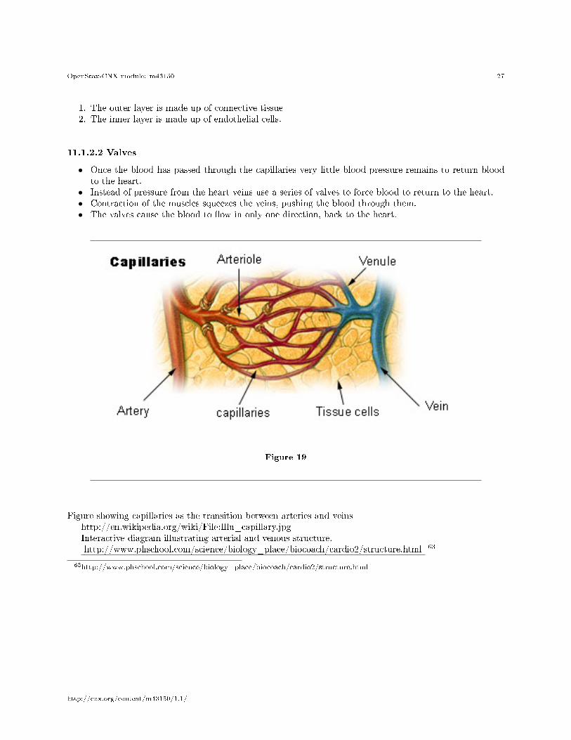
Figure 17
Figure showing capillaries as the transition between arteries and veinshttp://en.wikipedia.org/wiki/File:Illu_capillary.jpgInteractive diagram illustrating arterial and venous structure.http://www.phschool.com/science/biology_place/biocoach/cardio2/structure.html 62
62http://www.phschool.com/science/biology_place/biocoach/cardio2/structure.html
http://cnx.org/content/m43150/1.1/
OpenStax-CNX module: m43150 26
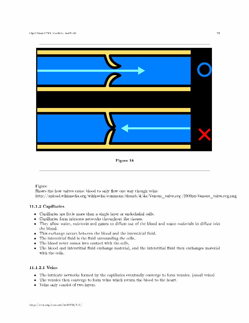
Figure 18
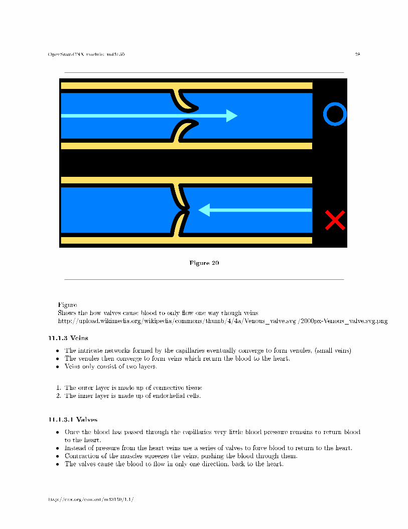
FigureShows the how valves cause blood to only �ow one way though veinshttp://upload.wikimedia.org/wikipedia/commons/thumb/4/4a/Venous_valve.svg /2000px-Venous_valve.svg.png
11.1.2 Capillaries
• Capillaries are little more than a single layer or endothelial cells.• Capillaries form intricate networks throughout the tissues.• They allow water, nutrients and gasses to di�use out of the blood and waste materials to di�use into
the blood.• This exchange occurs between the blood and the interstitial �uid.• The interstitial �uid is the �uid surrounding the cells.• The blood never comes into contact with the cells.• The blood and interstitial �uid exchange material, and the interstitial �uid then exchanges material
with the cells.
11.1.2.1 Veins
• The intricate networks formed by the capillaries eventually converge to form venules, (small veins)• The venules then converge to form veins which return the blood to the heart.• Veins only consist of two layers.
http://cnx.org/content/m43150/1.1/
OpenStax-CNX module: m43150 27
1. The outer layer is made up of connective tissue2. The inner layer is made up of endothelial cells.
11.1.2.2 Valves
• Once the blood has passed through the capillaries very little blood pressure remains to return bloodto the heart.
• Instead of pressure from the heart veins use a series of valves to force blood to return to the heart.• Contraction of the muscles squeezes the veins, pushing the blood through them.• The valves cause the blood to �ow in only one direction, back to the heart.
Figure 19
Figure showing capillaries as the transition between arteries and veinshttp://en.wikipedia.org/wiki/File:Illu_capillary.jpgInteractive diagram illustrating arterial and venous structure.http://www.phschool.com/science/biology_place/biocoach/cardio2/structure.html 63
63http://www.phschool.com/science/biology_place/biocoach/cardio2/structure.html
http://cnx.org/content/m43150/1.1/
OpenStax-CNX module: m43150 28
Figure 20
FigureShows the how valves cause blood to only �ow one way though veinshttp://upload.wikimedia.org/wikipedia/commons/thumb/4/4a/Venous_valve.svg /2000px-Venous_valve.svg.png
11.1.3 Veins
• The intricate networks formed by the capillaries eventually converge to form venules, (small veins)• The venules then converge to form veins which return the blood to the heart.• Veins only consist of two layers.
1. The outer layer is made up of connective tissue2. The inner layer is made up of endothelial cells.
11.1.3.1 Valves
• Once the blood has passed through the capillaries very little blood pressure remains to return bloodto the heart.
• Instead of pressure from the heart veins use a series of valves to force blood to return to the heart.• Contraction of the muscles squeezes the veins, pushing the blood through them.• The valves cause the blood to �ow in only one direction, back to the heart.
http://cnx.org/content/m43150/1.1/

OpenStax-CNX module: m43150 29
Figure 21
Figure showing capillaries as the transition between arteries and veinshttp://en.wikipedia.org/wiki/File:Illu_capillary.jpgInteractive diagram illustrating arterial and venous structure.http://www.phschool.com/science/biology_place/biocoach/cardio2/structure.html 64
64http://www.phschool.com/science/biology_place/biocoach/cardio2/structure.html
http://cnx.org/content/m43150/1.1/
OpenStax-CNX module: m43150 30
Figure 22
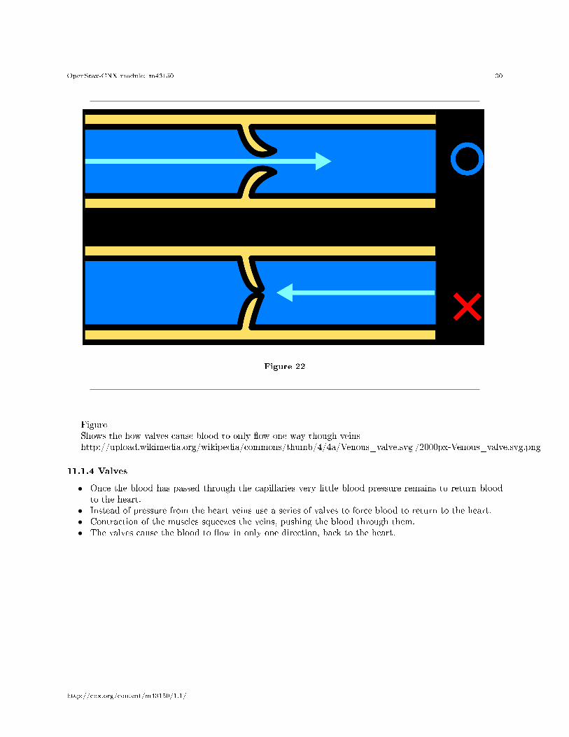
FigureShows the how valves cause blood to only �ow one way though veinshttp://upload.wikimedia.org/wikipedia/commons/thumb/4/4a/Venous_valve.svg /2000px-Venous_valve.svg.png
11.1.4 Valves
• Once the blood has passed through the capillaries very little blood pressure remains to return bloodto the heart.
• Instead of pressure from the heart veins use a series of valves to force blood to return to the heart.• Contraction of the muscles squeezes the veins, pushing the blood through them.• The valves cause the blood to �ow in only one direction, back to the heart.
http://cnx.org/content/m43150/1.1/
OpenStax-CNX module: m43150 31
Figure 23
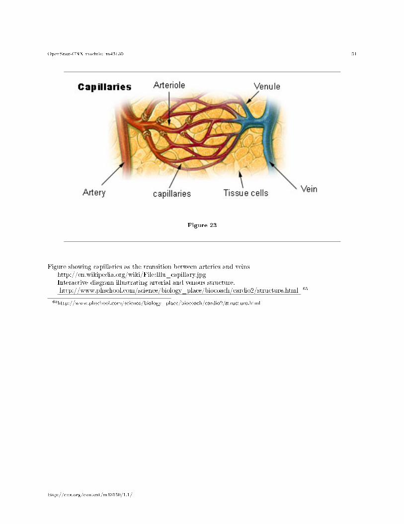
Figure showing capillaries as the transition between arteries and veinshttp://en.wikipedia.org/wiki/File:Illu_capillary.jpgInteractive diagram illustrating arterial and venous structure.http://www.phschool.com/science/biology_place/biocoach/cardio2/structure.html 65
65http://www.phschool.com/science/biology_place/biocoach/cardio2/structure.html
http://cnx.org/content/m43150/1.1/
OpenStax-CNX module: m43150 32
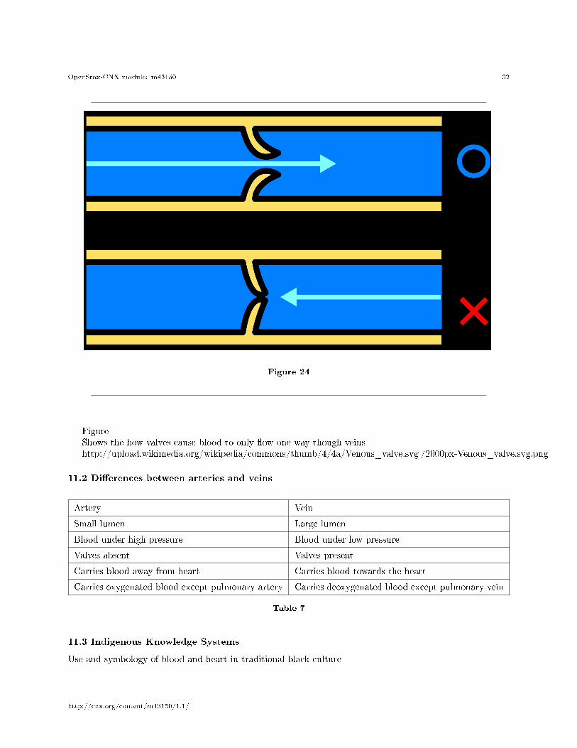
Figure 24
FigureShows the how valves cause blood to only �ow one way though veinshttp://upload.wikimedia.org/wikipedia/commons/thumb/4/4a/Venous_valve.svg /2000px-Venous_valve.svg.png
11.2 Di�erences between arteries and veins
Artery Vein
Small lumen Large lumen
Blood under high pressure Blood under low pressure
Valves absent Valves present
Carries blood away from heart Carries blood towards the heart
Carries oxygenated blood except pulmonary artery Carries deoxygenated blood except pulmonary vein
Table 7
11.3 Indigenous Knowledge Systems
Use and symbology of blood and heart in traditional black culture
http://cnx.org/content/m43150/1.1/
OpenStax-CNX module: m43150 33
12 Fun facts about your heart
1. The average adult heart beats:
• 72 times a minute• 100,000 times a day• 3,600,000 times a year• A billion times during a lifetime.
1. Each day your heart creates enough energy to drive a truck for 32 kilometres.2. Your left lung is smaller than your right one to make room in your chest cavity for your heart.3. Clench your �st - the size of your �st is more or less the size of your heart.4. Laughing is good exercise for your heart. Whenever you laugh, the blood �ow in your heart is increased,
keeping your heart healthy.
Investigation: Practical investigation of sheep's heartVideo: Doing a dissectionhttp://www.hometrainingtools.com/images/videos/Dissection_Video/dissection_�vpl ayer.html?TB_iframe=true&height=390&width=405
66
Equipment:
Figure 25
• 1 sheep heart• Cutting board• Scalpel• textbook
• Cotton• water• funnel• scissors
Table 8
1. EXTERNAL(a)How would you describe the general shape of the heart?(b)Note the grooves on the surface of the heart. In which direction do they run.What do you observe in these grooves.(c)Identify the atria and ventricles. How do they di�er from each other inappearance. What di�erence do you notice between the atria and ventricles.2. If the venae cavae are su�ciently long, insert a funnel into the superior vena cava and tie o�
the inferior vena cava with a piece of cotton . When water is added through the superior vena caveinto the right atrium:
(a)What happens to the wall of the right ventricle?
66http://www.hometrainingtools.com/images/videos/Dissection_Video/dissection_�vplayer.html?TB_iframe=true&height=390&width=405
http://cnx.org/content/m43150/1.1/
OpenStax-CNX module: m43150 34
(b)Press the right ventricle. What do you observe?(c)Release the pressure. What happens?(d)Now press the left ventricle a few times. What do you notice?(e)Now attach funnel to one of the pulmonary veins and tie o� the others(if possible). Pour water down the funnel and press the left ventricle.What do you observe?(f)Release the pressure and press the right ventricle. What do you observe?Remove the funnel and tubes.3. Cut the superior vena cava from the atrium and cut open the wall of the atrium. Dothe same
with the pulmonary vein and left atrium.(a)Describe the appearance of the inner atrial surface.(b)Determine the position of the pulmonary artery and the aorta by inserting aglass rod through these vessel into the chambers of the heart.Name the artery that leaves the right ventricle.Name the artery that leaves the left ventricle.4. Make an incision in the right side of the left ventricle from the oblique groove to the a
pex of the heart.(a)What do you observe between the left atrium and left ventricle?(b)How many �aps do you see?(c)What is the function of these �aps?5. Similarly, make an incision in the left wall of the right ventricle from the oblique groove.(a)How many �aps do you see between the atrium and the ventricle?(b)What do these �aps collectively form?6. Compare the muscular walls of the:(a) atria and the ventricles(b) left and right ventricles7. What do you observe between the two halves of the heart.8. Examine the tendinous cords .(a)Where are their points of attachment?(b)What is their function9. If the pulmonary artery and aorta are long enough, do this question. Using a funnel, pour water
into the pulmonary artery and the aorta.(a)What do you notice?(b)What do you see at the base of these arteries?10. Cut the aorta and pulmonary arteries open longitudinally and examine the valves.(a)How many parts are there to each of these valves?(b)Compare the walls of the aorta and the pulmonary artery and suggest areason for any di�erence you many �nd.
http://cnx.org/content/m43150/1.1/