Embed Size (px)
Citation preview
Circulatory System
Consists of
• Heart
• Blood Vessels
• Blood
Function
• Transports oxygen and nutrients to the cells
• Transports carbon dioxide and metabolic waste away from the cells
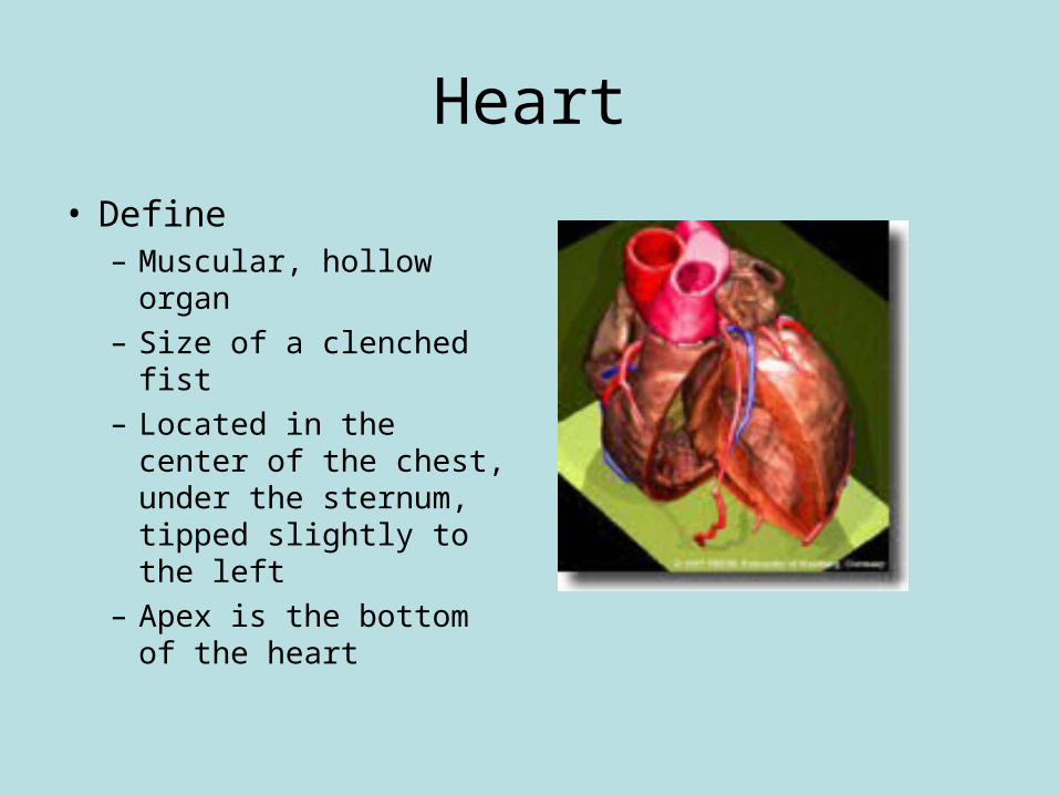
Heart
• Define– Muscular, hollow
organ– Size of a clenched fist– Located in the center
of the chest, under the sternum, tipped slightly to the left
– Apex is the bottom of the heart
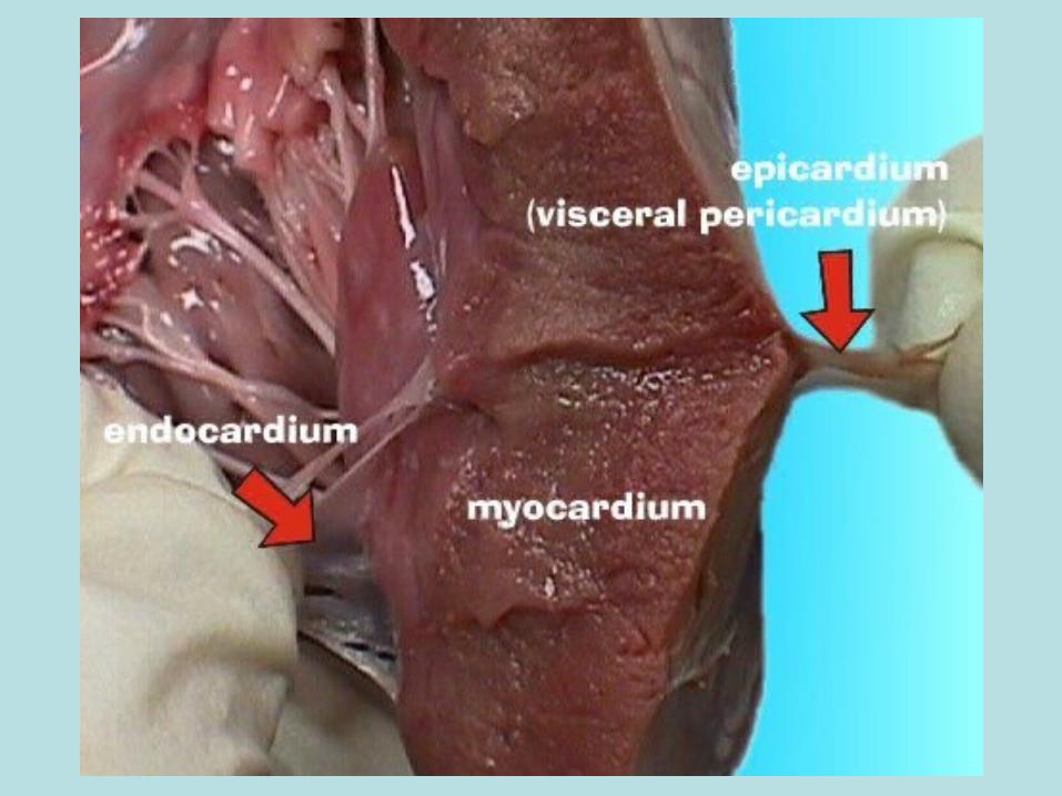
Layers of the Heart
• Endocardium– Epithelial tissue– Lines heart
• Myocardium– Middle layer– Cardiac muscle tissue
• Pericardium– Epithelial tissue– Covers outside of heart
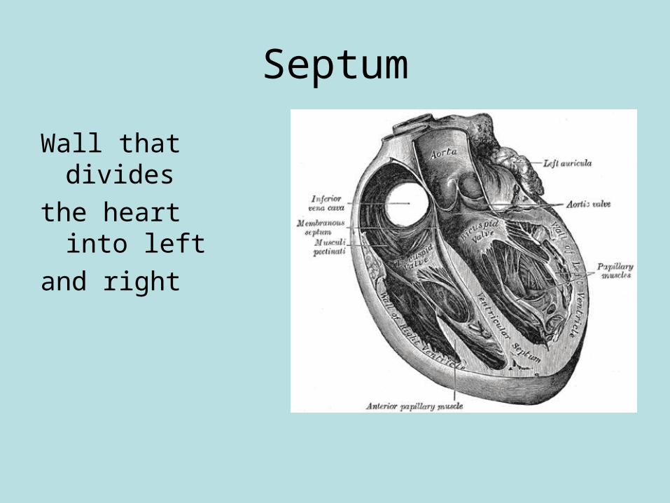
Septum
Wall that divides
the heart into left
and right
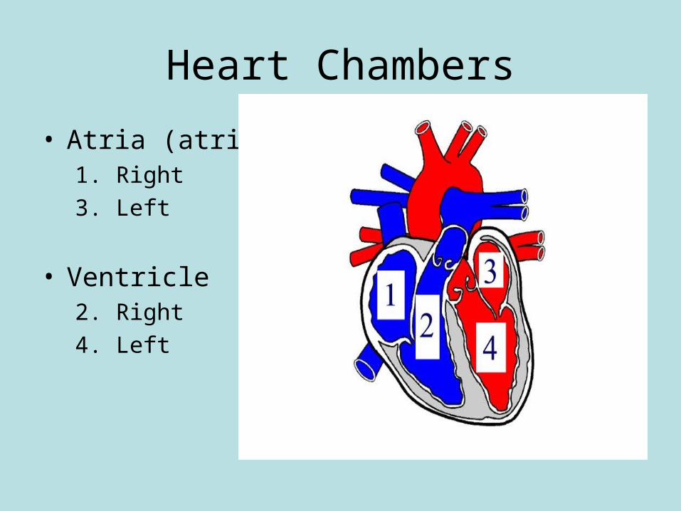
Heart Chambers
• Atria (atrium)1. Right
3. Left
• Ventricle2. Right
4. Left
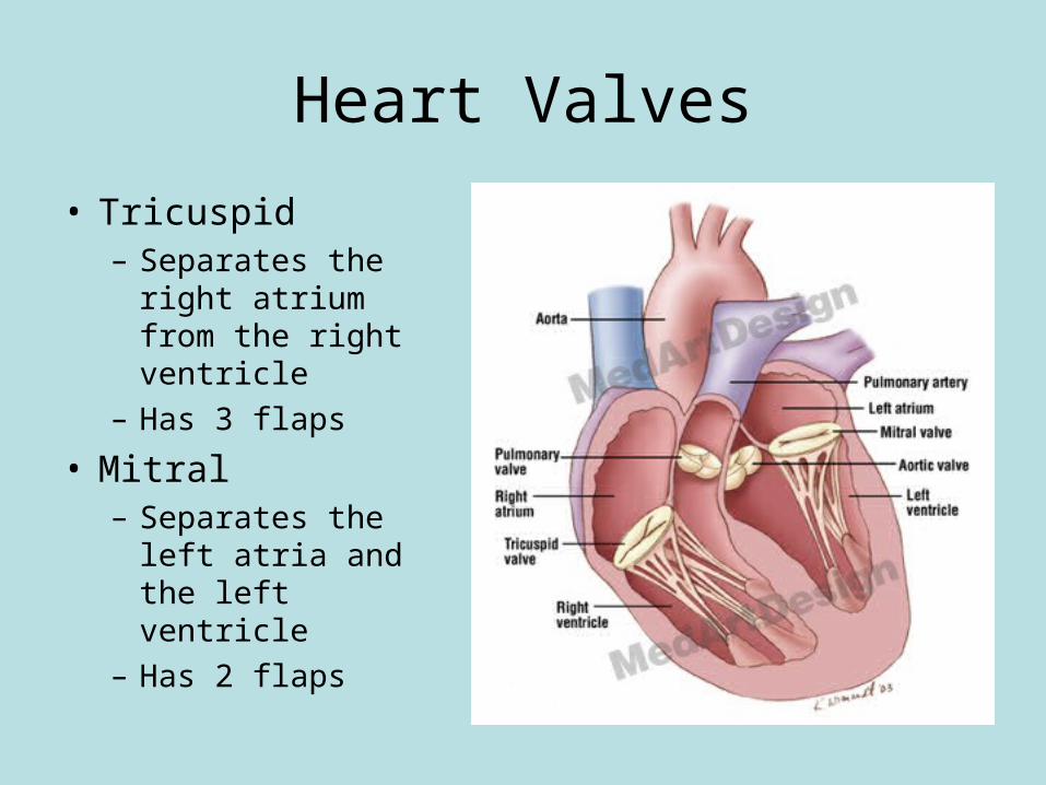
Heart Valves
• Tricuspid– Separates the right
atrium from the right ventricle
– Has 3 flaps
• Mitral– Separates the left
atria and the left ventricle
– Has 2 flaps
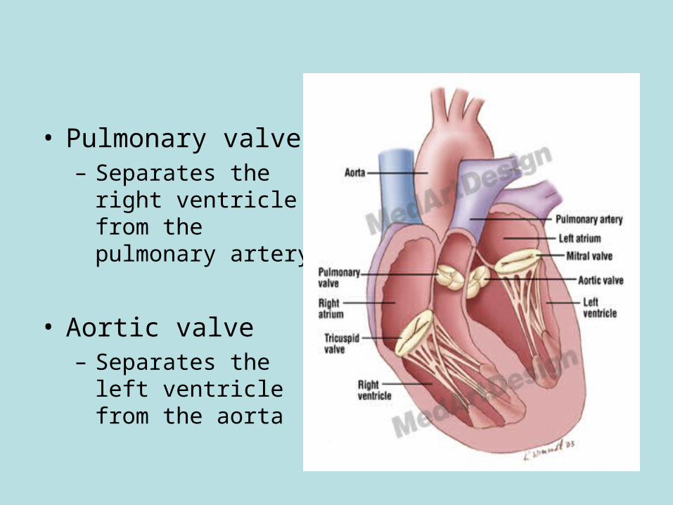
• Pulmonary valve– Separates the right
ventricle from the pulmonary artery
• Aortic valve– Separates the left
ventricle from the aorta
Cardiac Cycle
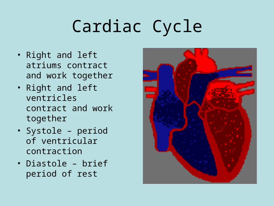
• Right and left atriums contract and work together
• Right and left ventricles contract and work together
• Systole – period of ventricular contraction
• Diastole – brief period of rest
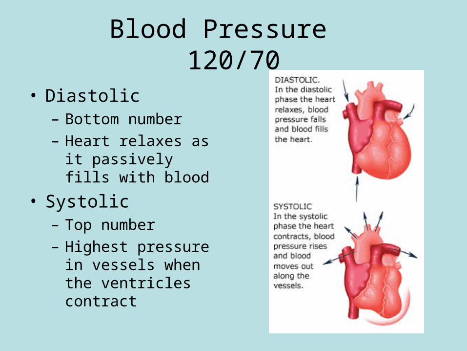
Blood Pressure 120/70
• Diastolic– Bottom number– Heart relaxes as it
passively fills with blood
• Systolic– Top number– Highest pressure in
vessels when the ventricles contract
Pattern of Circulation • Superior and inferior vena cava• Right atrium• Tricuspid valve• Right ventricle• Pulmonary valve• Pulmonary artery• Lungs• Pulmonary veins• Left atrium• Mitral valve• Left ventricle• Aortic valve• Aorta• Arteries• Arteriole• Capillaries• Venules• Veins• Superior and inferior vena cava
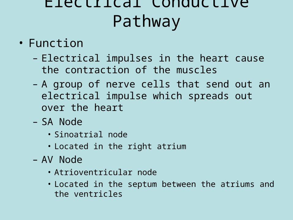
Electrical Conductive Pathway
• Function– Electrical impulses in the heart cause the contraction
of the muscles– A group of nerve cells that send out an electrical
impulse which spreads out over the heart– SA Node
• Sinoatrial node• Located in the right atrium
– AV Node• Atrioventricular node• Located in the septum between the atriums and the
ventricles
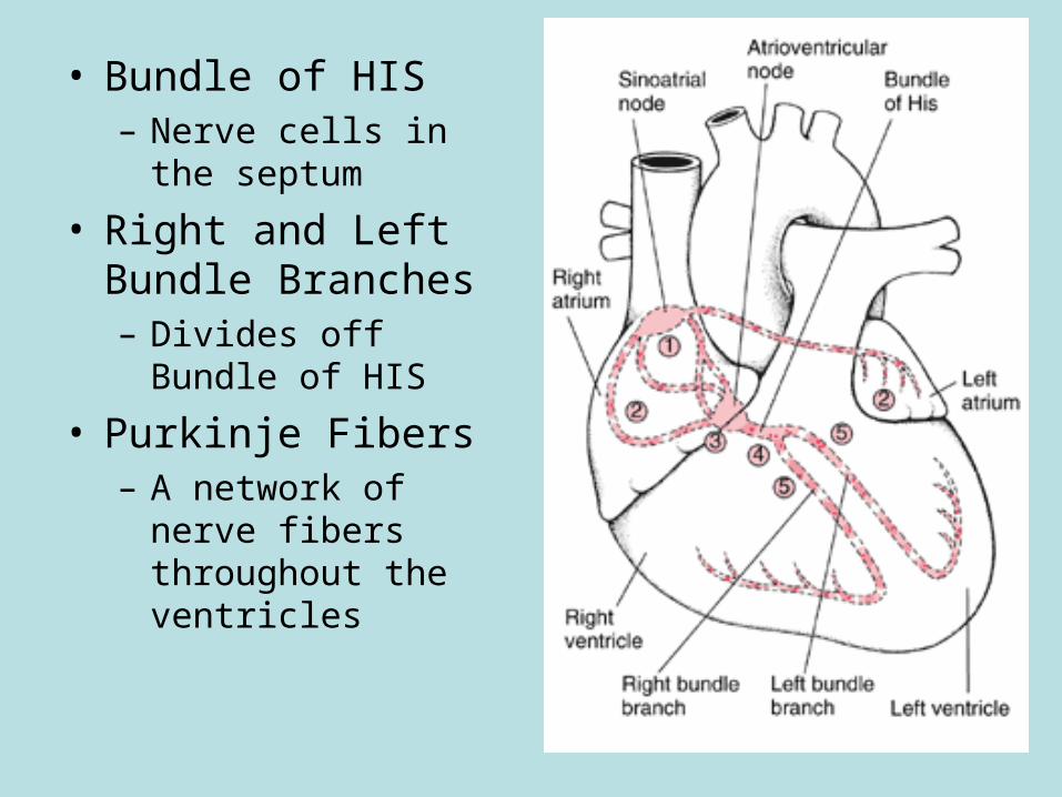
• Bundle of HIS– Nerve cells in the
septum
• Right and Left Bundle Branches– Divides off Bundle of
HIS
• Purkinje Fibers– A network of nerve
fibers throughout the ventricles
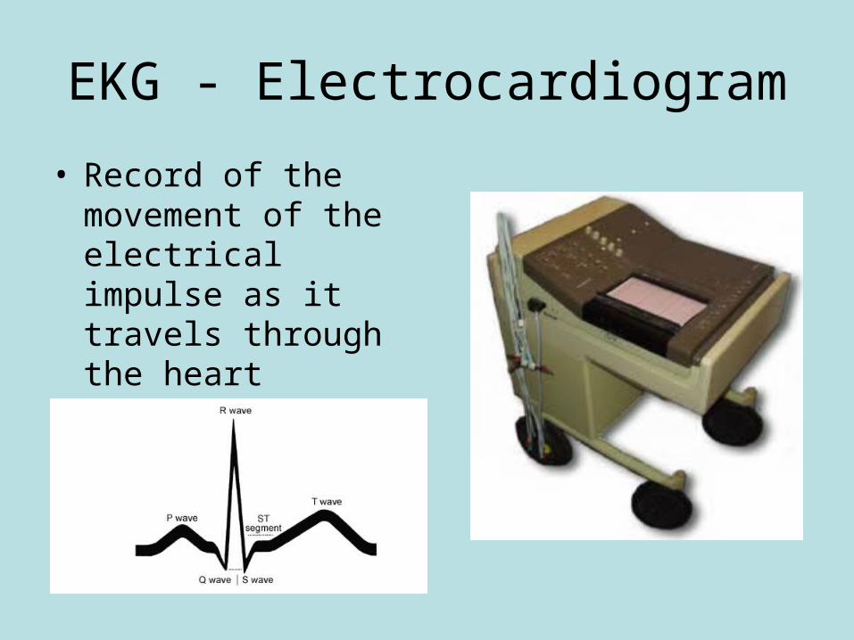
EKG - Electrocardiogram
• Record of the movement of the electrical impulse as it travels through the heart
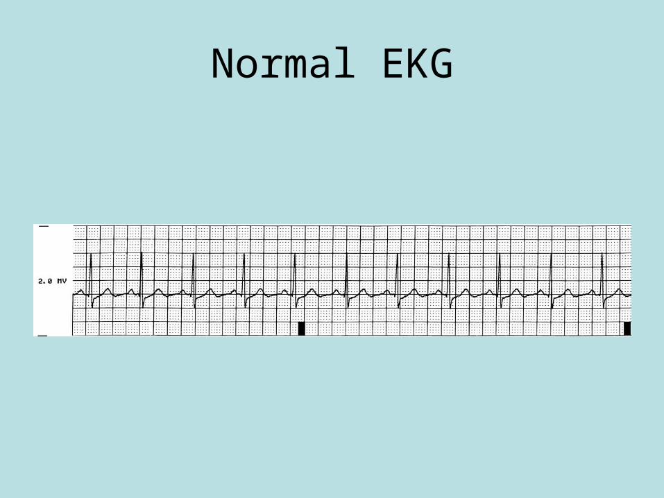
Normal EKG
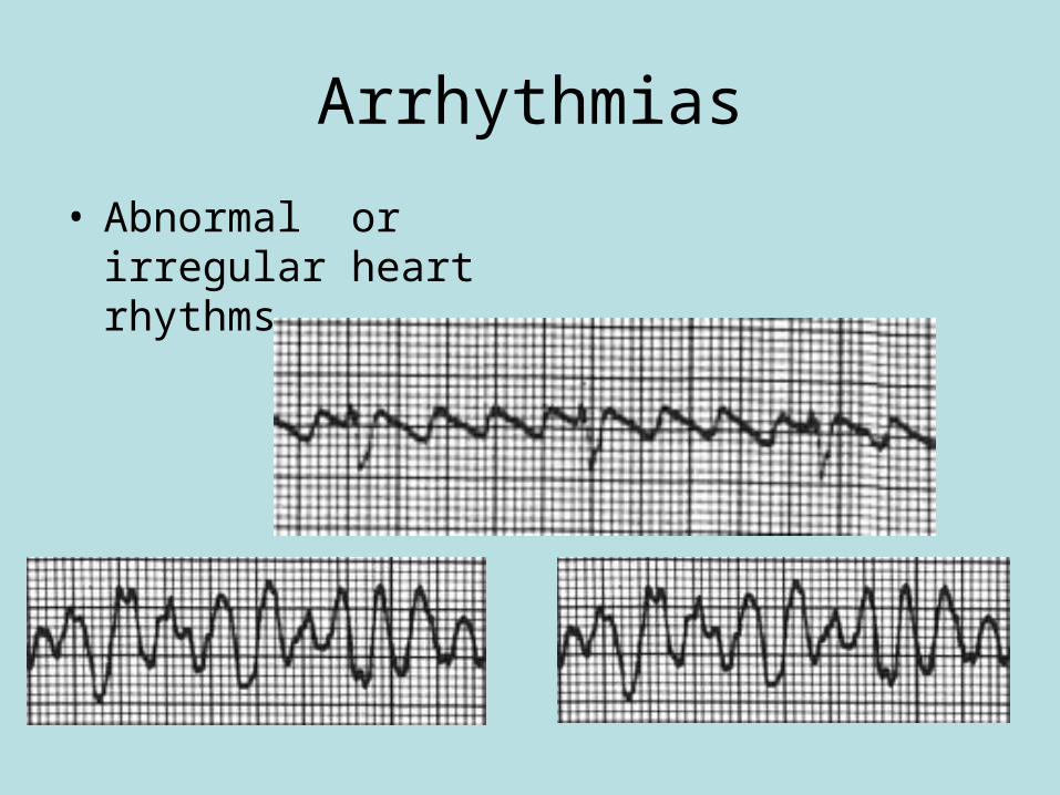
Arrhythmias
• Abnormal or irregular heart rhythms
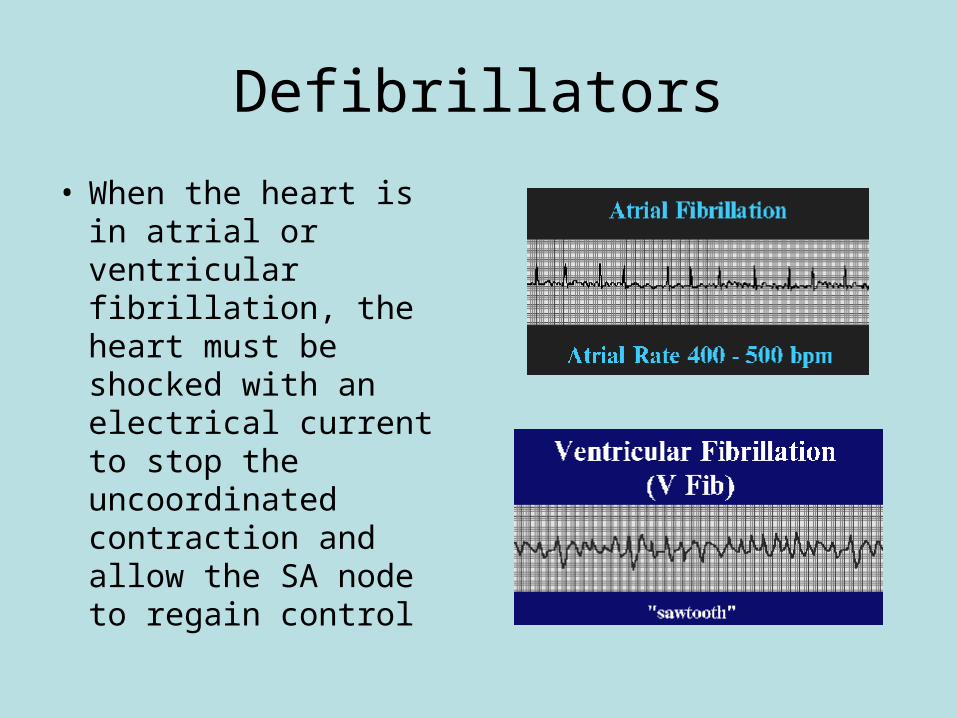
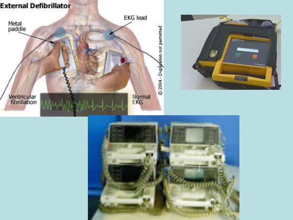
Defibrillators
• When the heart is in atrial or ventricular fibrillation, the heart must be shocked with an electrical current to stop the uncoordinated contraction and allow the SA node to regain control
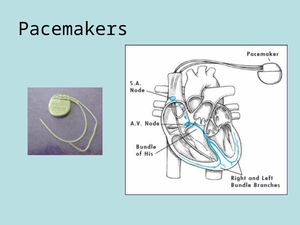
Pacemakers
• A small battery powered device with electrodes that monitors the hearts activity and delivers an electrical impulse to stimulate contraction
• Fixed and demand
• Avoid electromagnetic forces like microwaves and cellular phones
Pacemakers
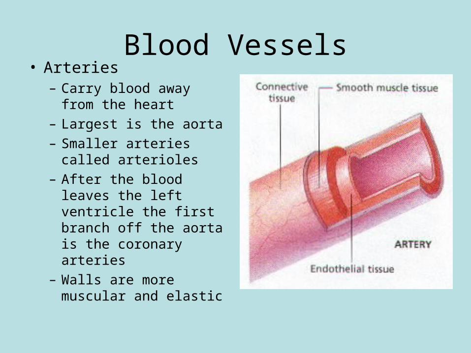
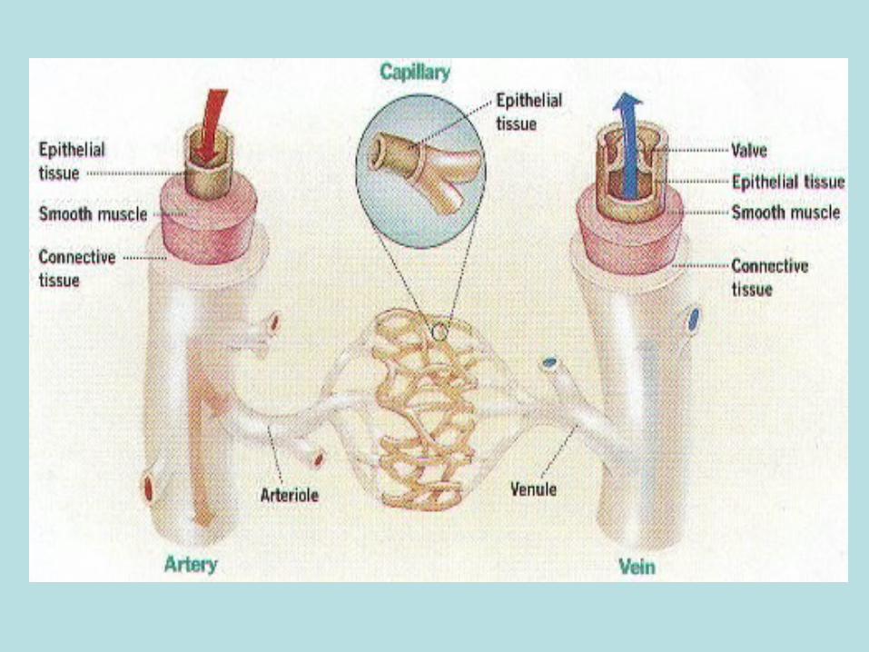
Blood Vessels• Arteries
– Carry blood away from the heart
– Largest is the aorta– Smaller arteries called
arterioles– After the blood leaves
the left ventricle the first branch off the aorta is the coronary arteries
– Walls are more muscular and elastic
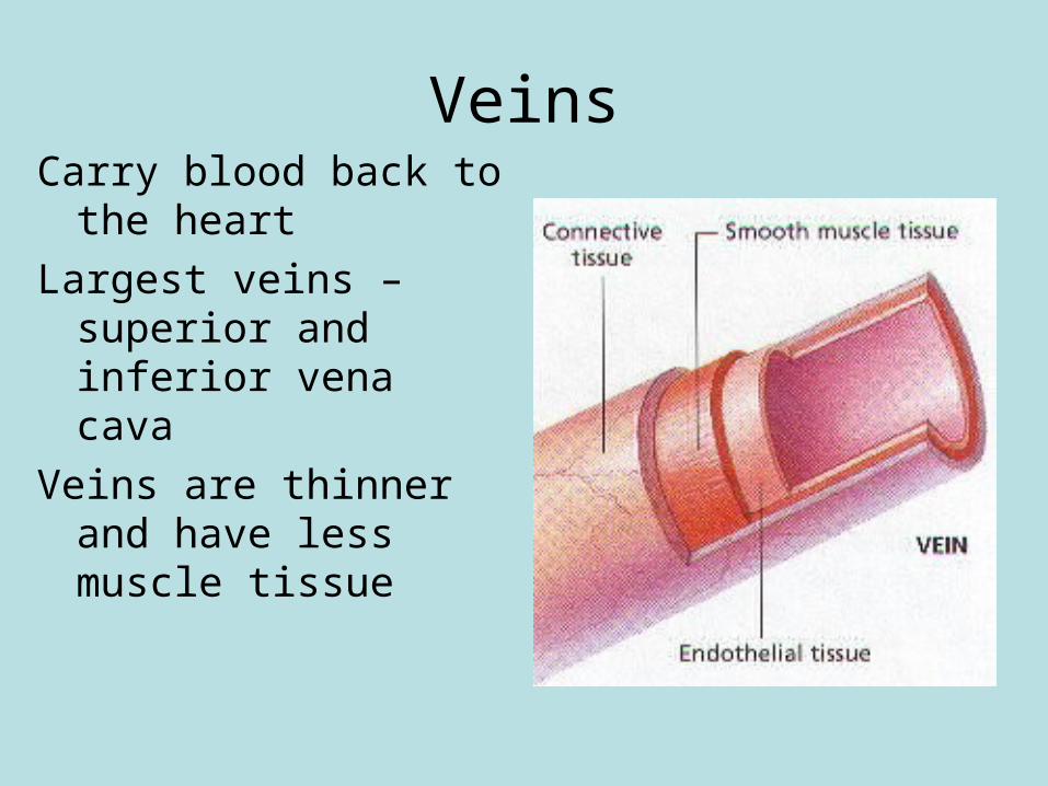
VeinsCarry blood back to the
heart
Largest veins – superior and inferior vena cava
Veins are thinner and have less muscle tissue
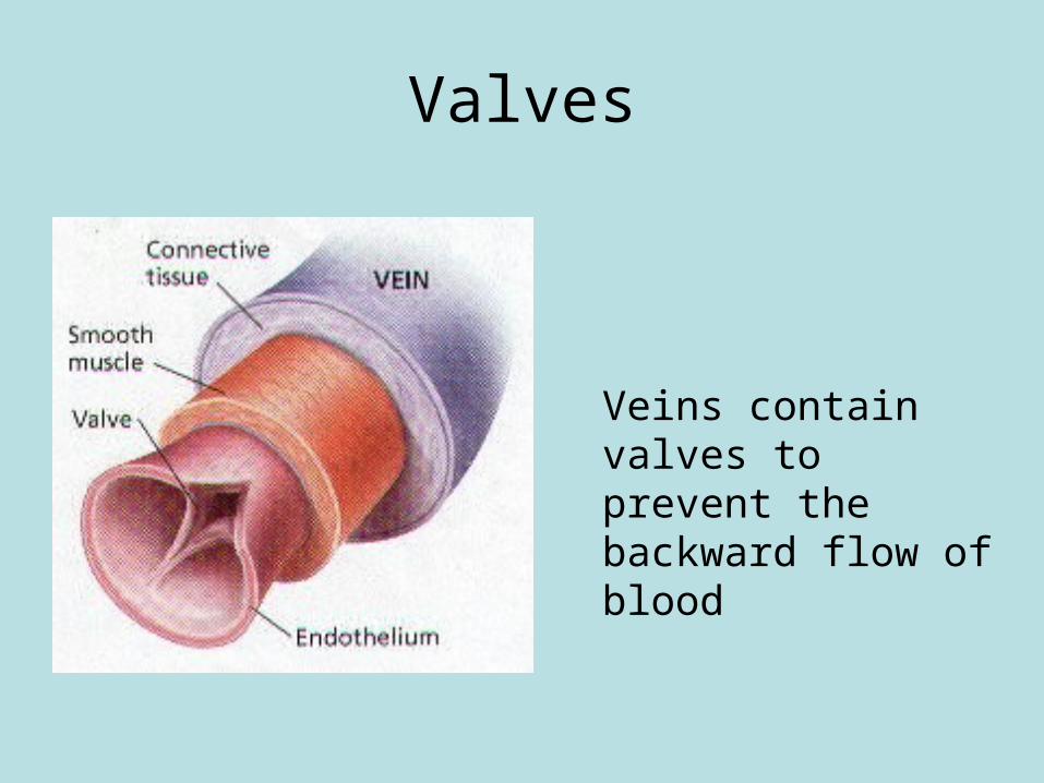
Valves
Veins contain valves to prevent the backward flow of blood
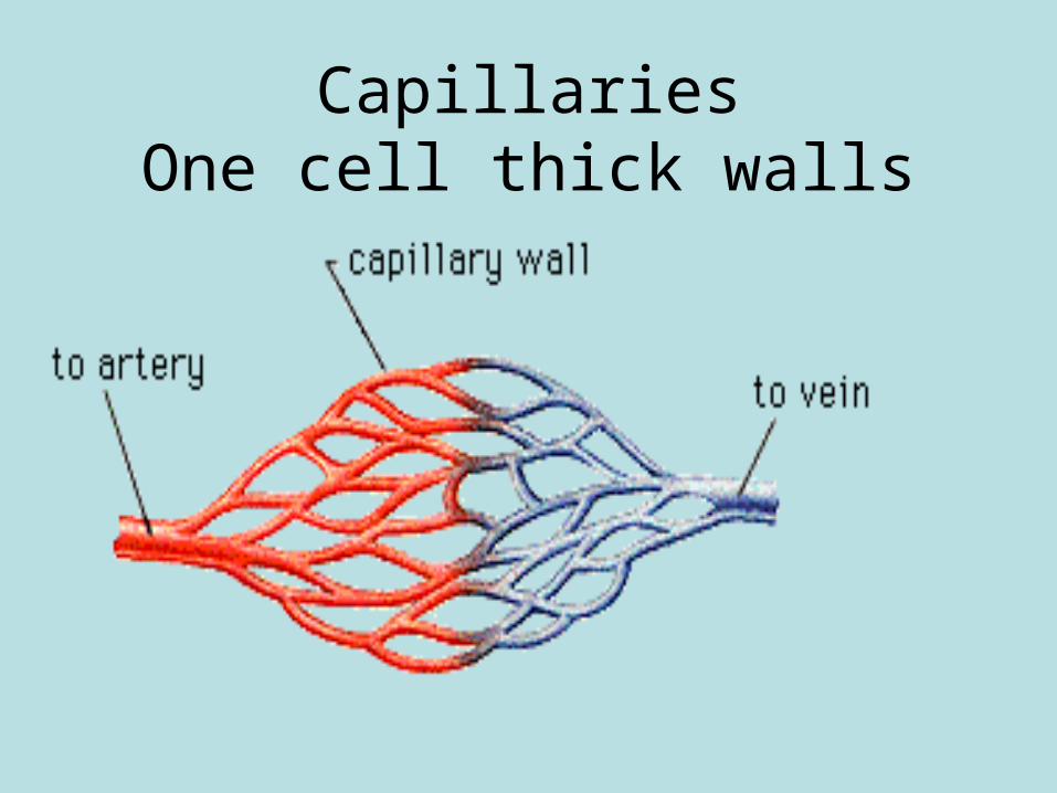
CapillariesOne cell thick walls
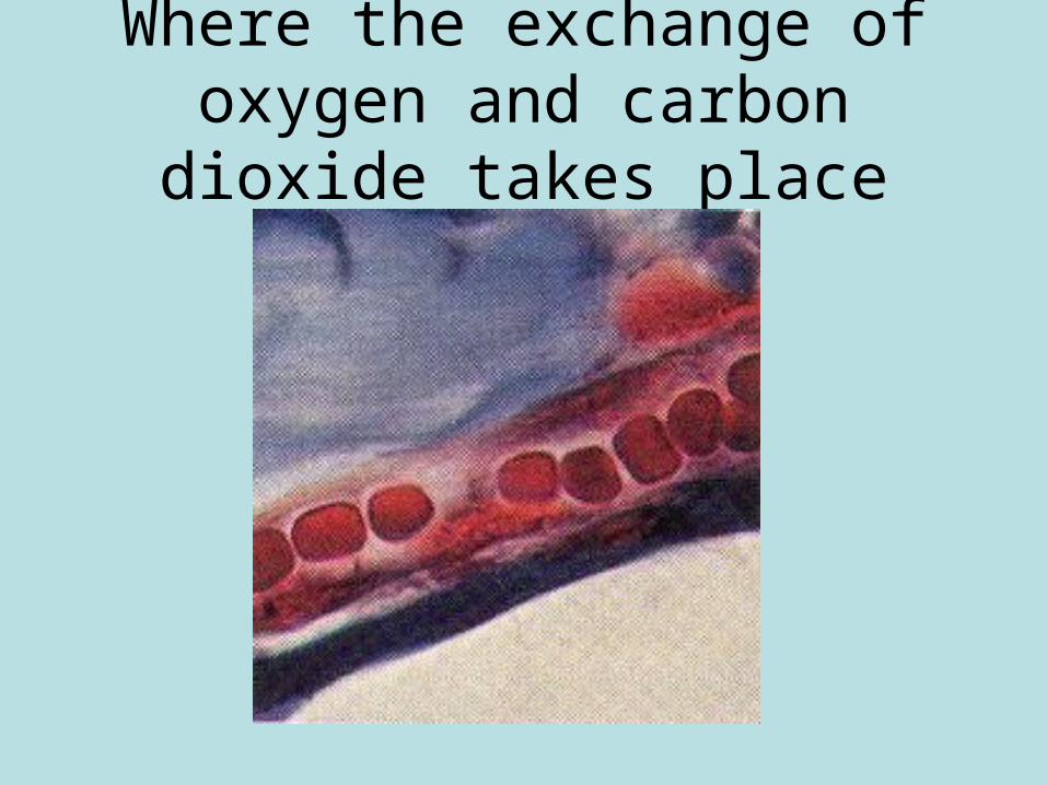
Where the exchange of oxygen and carbon dioxide takes place
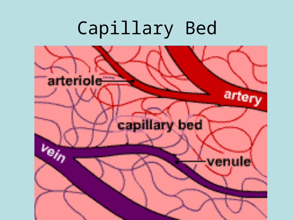
Capillary Bed
Blood
• Called a tissue because it contains many types of cells
• 4-6 quarts in the average adult
• Composed of plasma and formed elements called blood cells
• Plasma is 90% water with dissolved elements in it
Functions
• Carries oxygen from the lungs to the cells and carbon dioxide from the cells to the lungs
• Nutrients from the digestive tract to cells
• Metabolic waste from the cells to organs of excretion
• Carries heat produced by the body
• Carries hormones to body organs
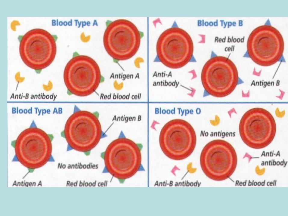
Blood Types
• O, A, B, AB
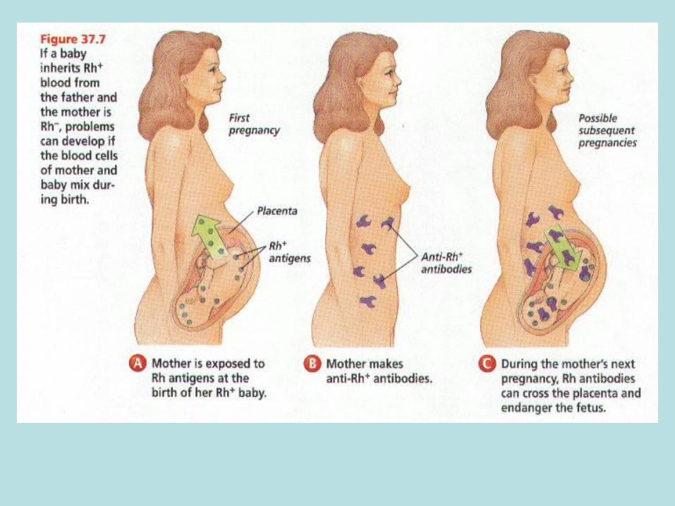
• Rh factor positive or negative
Universal DonorO+
Universal Recipient
AB+
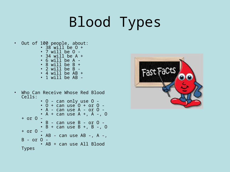
Blood Types• Out of 100 people, about:
• 38 will be O + • 7 will be O - • 34 will be A + • 6 will be A - • 8 will be B + • 2 will be B - • 4 will be AB + • 1 will be AB -
• Who Can Receive Whose Red Blood Cells: • O - can only use O - • O + can use O + or O - • A - can use A - or O - • A + can use A +, A -, O + or O - • B - can use B - or O - • B + can use B +, B -, O + or O - • AB - can use AB -, A -, B - or O - • AB + can use All Blood Types
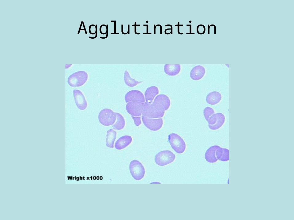
Agglutination
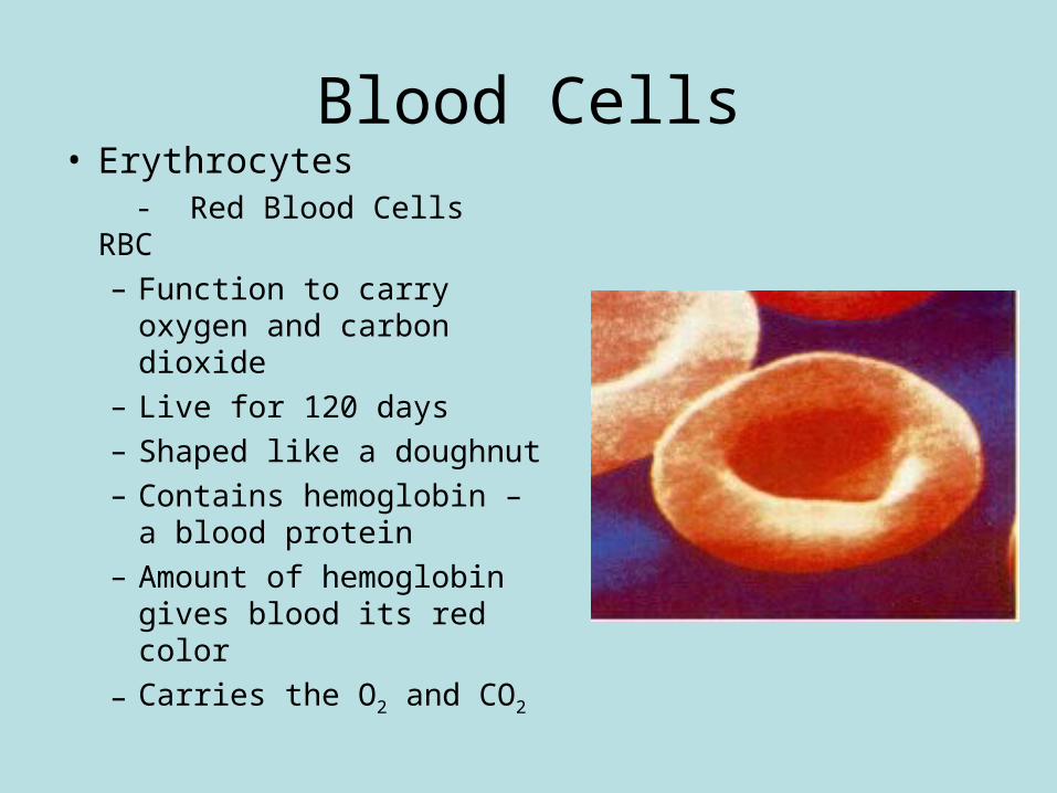
Blood Cells• Erythrocytes
- Red Blood Cells RBC– Function to carry oxygen
and carbon dioxide– Live for 120 days– Shaped like a doughnut– Contains hemoglobin – a
blood protein– Amount of hemoglobin
gives blood its red color
– Carries the O2 and CO2
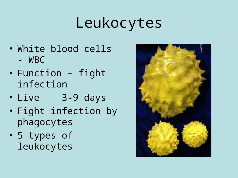
Leukocytes
• White blood cells - WBC• Function – fight infection• Live 3-9 days• Fight infection by
phagocytes• 5 types of leukocytes
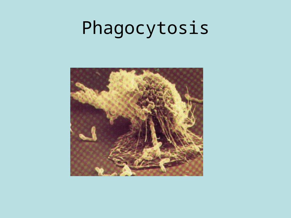
Phagocytosis
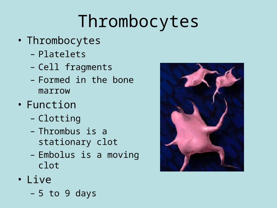
Thrombocytes• Thrombocytes
– Platelets– Cell fragments– Formed in the bone
marrow
• Function– Clotting– Thrombus is a stationary
clot– Embolus is a moving clot
• Live– 5 to 9 days
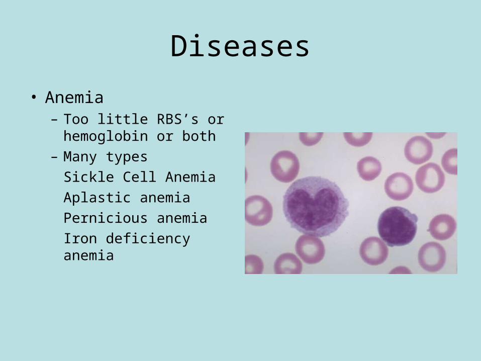
Diseases
• Anemia– Too little RBS’s or
hemoglobin or both– Many types
Sickle Cell Anemia
Aplastic anemia
Pernicious anemia
Iron deficiency anemia
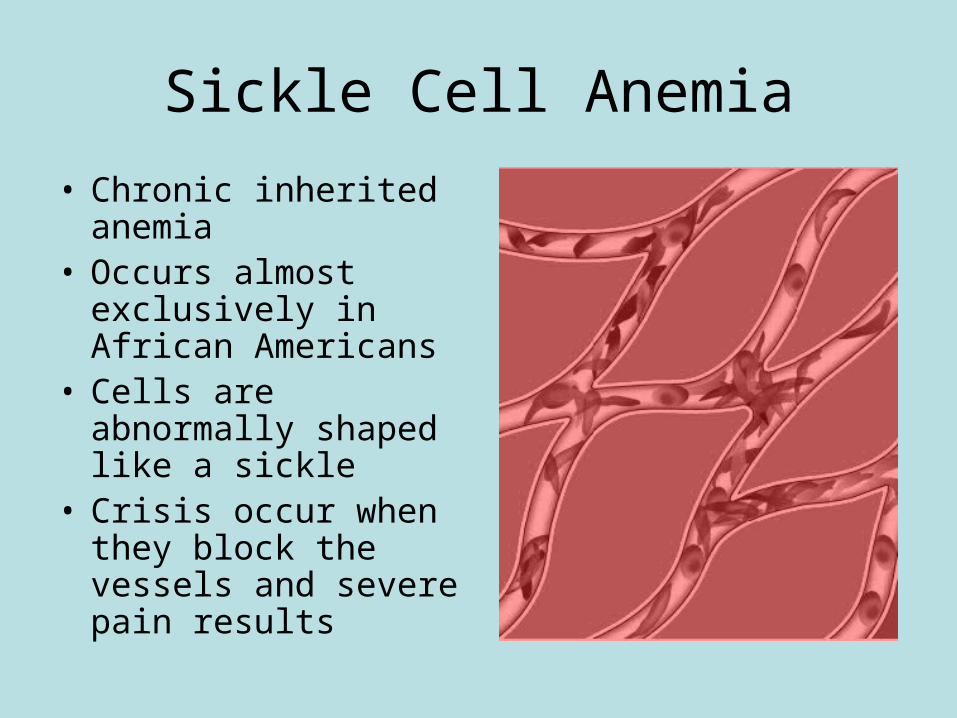
Sickle Cell Anemia
• Chronic inherited anemia
• Occurs almost exclusively in African Americans
• Cells are abnormally shaped like a sickle
• Crisis occur when they block the vessels and severe pain results
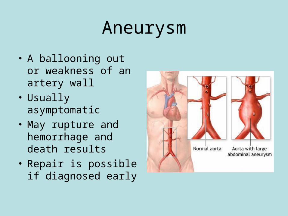
Aneurysm
• A ballooning out or weakness of an artery wall
• Usually asymptomatic• May rupture and
hemorrhage and death results
• Repair is possible if diagnosed early
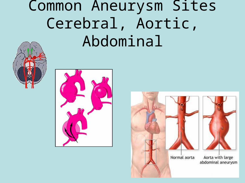
Common Aneurysm SitesCerebral, Aortic, Abdominal
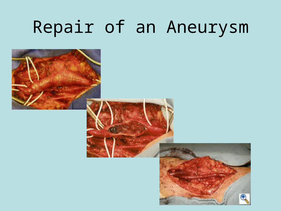
Repair of an Aneurysm
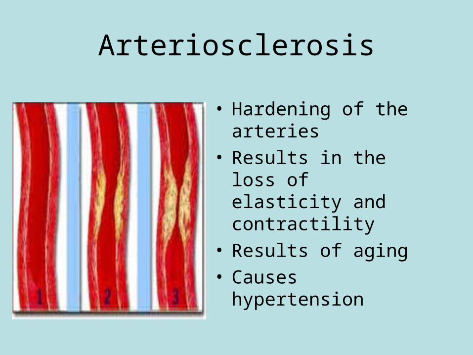
Arteriosclerosis
• Hardening of the arteries
• Results in the loss of elasticity and contractility
• Results of aging• Causes hypertension
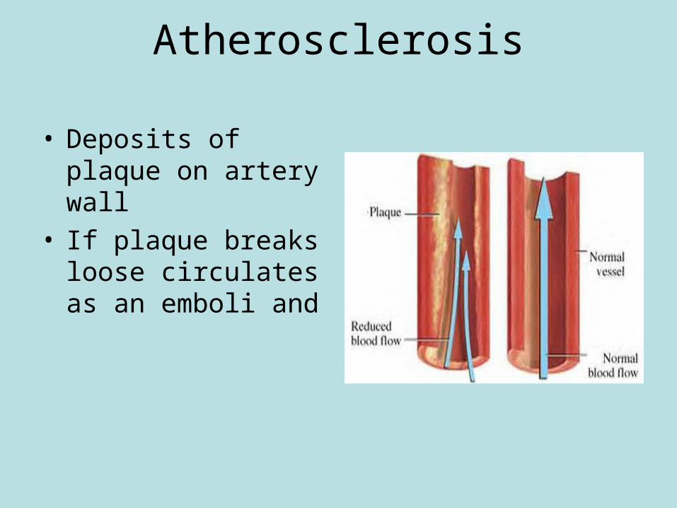
Atherosclerosis
• Deposits of plaque on artery wall
• If plaque breaks loose circulates as an emboli and
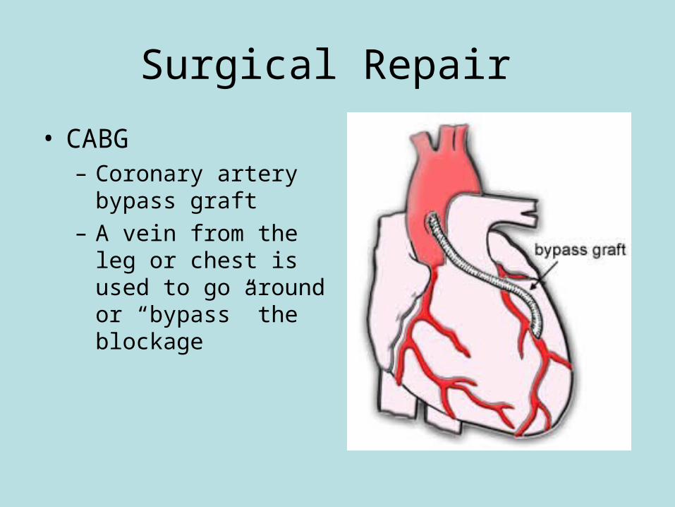
Surgical Repair
• CABG– Coronary artery
bypass graft– A vein from the leg or
chest is used to go around or “bypass” the blockage
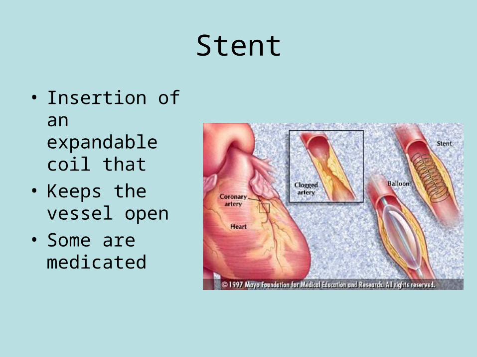
Stent
• Insertion of an expandable coil that
• Keeps the vessel open
• Some are medicated
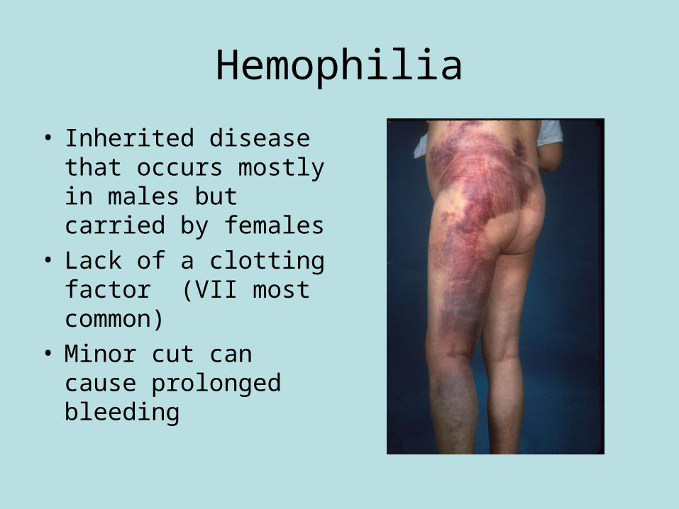
Hemophilia
• Inherited disease that occurs mostly in males but carried by females
• Lack of a clotting factor (VII most common)
• Minor cut can cause prolonged bleeding
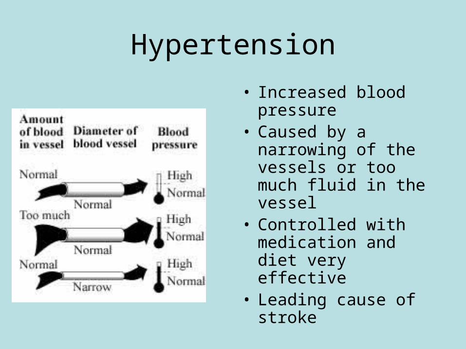
Hypertension
• Increased blood pressure
• Caused by a narrowing of the vessels or too much fluid in the vessel
• Controlled with medication and diet very effective
• Leading cause of stroke
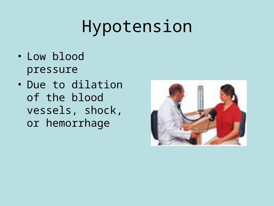
Hypotension
• Low blood pressure• Due to dilation of the
blood vessels, shock, or hemorrhage
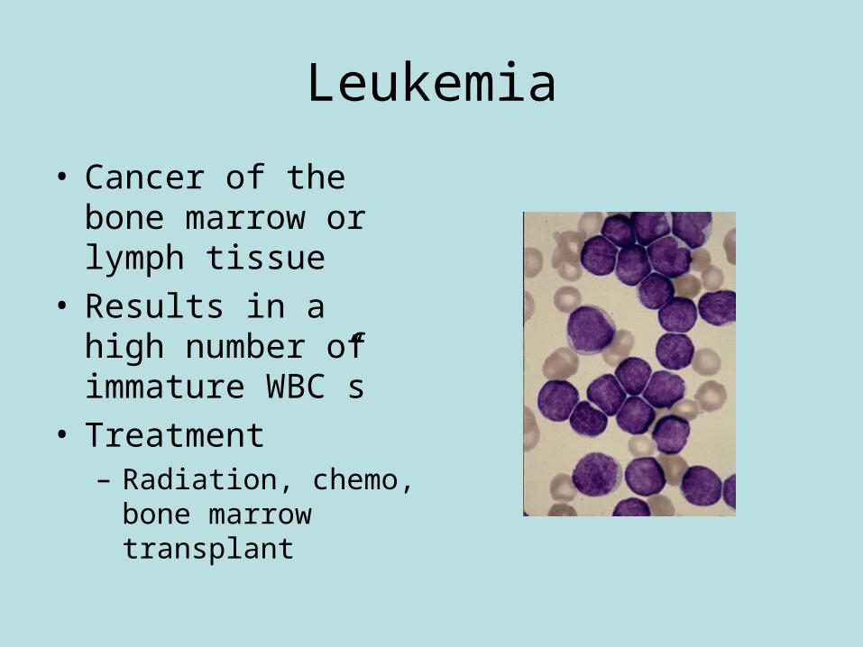
Leukemia
• Cancer of the bone marrow or lymph tissue
• Results in a high number of immature WBC”s
• Treatment– Radiation, chemo,
bone marrow transplant
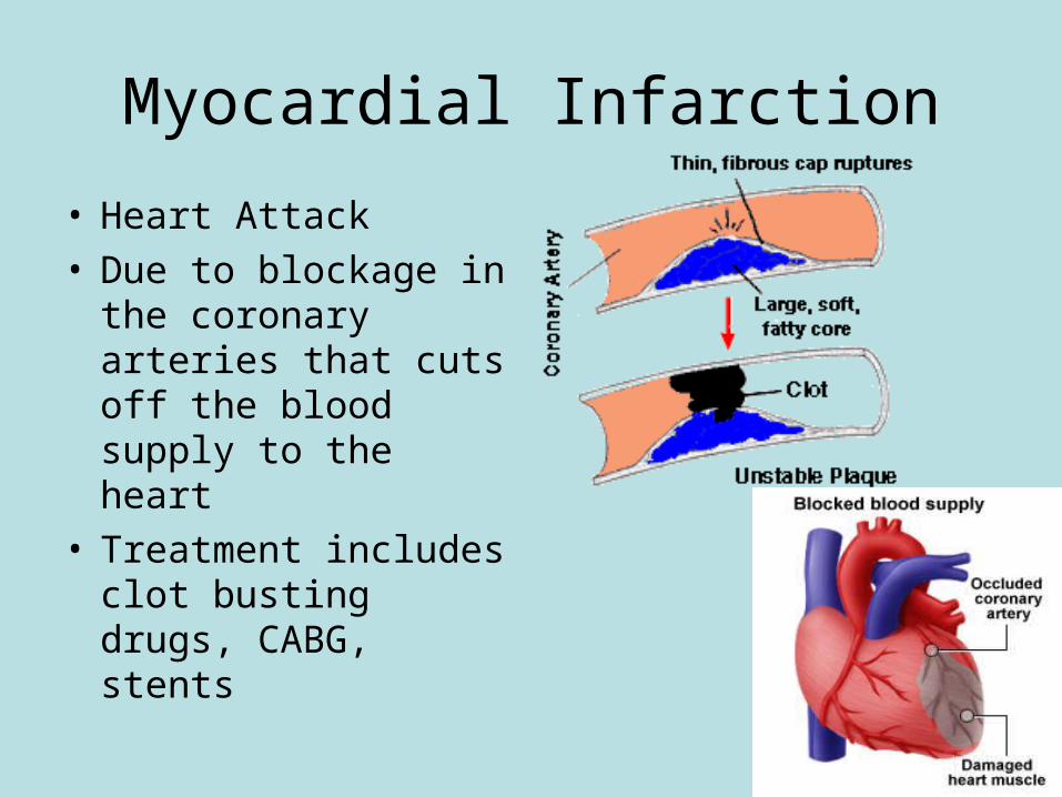
Myocardial Infarction
• Heart Attack• Due to blockage in
the coronary arteries that cuts off the blood supply to the heart
• Treatment includes clot busting drugs, CABG, stents
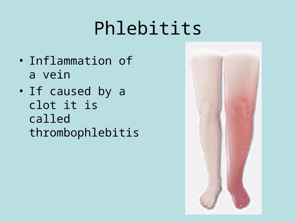
Phlebitits
• Inflammation of a vein• If caused by a clot it is
called thrombophlebitis
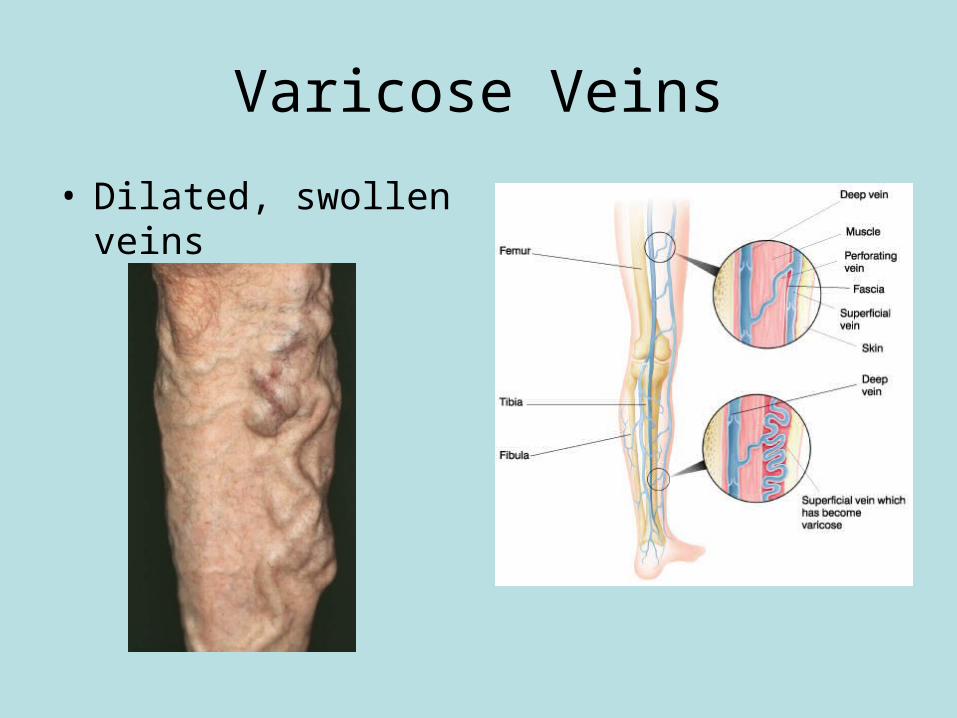
Varicose Veins
• Dilated, swollen veins