Embed Size (px)
DESCRIPTION
inflamasi
Citation preview

INFLAMMATION

Acute inflammation
Chronic inflammation
RepairResolution
Abscess
Injury

• “Inflame” – to set fire.
• Inflammation is “A dynamic response of
vascularised tissue to injury.”
• It is a protective response.
• It serves to bring defense & healing mechanisms
to the site of injury.

What is Inflammation?
• A reaction of a living tissue & its micro-
circulation to a pathogenic insult.
• A defense mechanism for survival .

• Reaction of tissues to injury, characterized clinically
by: heat, swelling, redness, pain, and loss of function.
• Pathologically by : vasoconstriction followed by
vasodilatation, hyperemia, accumulation of leukocytes,
exudation of fluid, stasis and deposition of fibrin.
• The processes followed by repair, the production of
new capillaries and fibroblasts, organization, and
cicatrization.
How Does It Occur?
• The vascular & cellular responses of
inflammation are mediated by chemical factors
(derived from blood plasma or some cells) &
triggered by inflammatory stimulus.
• Tissue injury or death ---> Release mediators

Etiologies
• Microbial infections: bacterial, viral, fungal, etc.
• Physical agents: burns, trauma--like cuts, radiation
• Chemicals: drugs, toxins, or caustic substances like
battery acid.
• Immunologic reactions: rheumatoid arthritis.

Cardinal Signs of Inflammation
• Redness : Hyperaemia.
• Warm : Hyperaemia.
• Pain : Nerve, Chemical
mediators.
• Swelling : Exudation
• Loss of Function


• Time course
– Acute inflammation: Less than 48 hours
– Chronic inflammation: Greater than 48 hours
(weeks, months, years)
• Cell type
– Acute inflammation: Polymorphonuclear
leukocyte (PMN)
– Chronic inflammation: Mononuclear cells
(Macrophages, Lymphocytes, Plasma cells).

PATHOGENESIS: Three main processes occur at the
site of inflammation, due to the release of chemical
mediators :
• Increased blood flow (redness and warmth).
• Increased vascular permeability (swelling,
pain & loss of function).
• Leukocytic Infiltration.

Mechanism of Inflammation
1. Vaso dilatation
2. Exudation - Edema
3. Emigration of cells
4. Chemotaxis

The major local manifestations
of acute inflammation,
compared to normal.
(1)Vascular dilation and
increased blood flow
(causing erythema and
warmth)
(2)Extravasation and
deposition of plasma fluid
and proteins (edema)
(3) leukocyte emigration and
accumulation in the site of
injury.

Changes in vascular flow (hemodynamic
changes)
Slowing of the circulation
outpouring of albumin rich fluid into the extravascular
tissues results in the concentration of RBCs in small
vessels and increased viscosity of blood.
Leukocyte margination
PMNs become oriented at the periphery of vessels and
start to stick.

Lymphatics in inflammation:
Lymphatics are responsible for draining edema.
Edema: An excess of fluid in the interstitial tissue
exudateor an transudateor serous cavities; either a

Transudate:
• An ultrafiltrate of blood plasma
–permeability of endothelium is usually normal.
– low protein content ( mostly albumin)

Exudate:
• A filtrate of blood plasma mixed with
inflammatory cells and cellular debris.
–permeability of endothelium is usually altered
–high protein content.
Pus:
• A purulent exudate: an inflammatory exudate
rich in leukocytes (mostly neutrophils) and
parenchymal cell debris.

Leukocyte exudation
Divided into 4 steps
Margination, rolling, and adhesion to endothelium
Diapedesis (trans-migration across endothelium)
Migration toward a chemotactic stimuli from the
source of tissue injury.
Phagocytosis


Defects in leukocyte function:
• Margination and adhesion
– steroids, leukocyte adhesion deficiency
• Emigration toward a chemotactic stimulus
• drugs
• chemotaxis inhibitors
• Phagocytosis
• Chronic granulomatous disease (CGD)

Phagocytosis
3 distinct steps
Recognition and attachment
Engulfment
Killing or degradation


Mechanism of Inflammation

Neutrophil Margination

Vascular changes
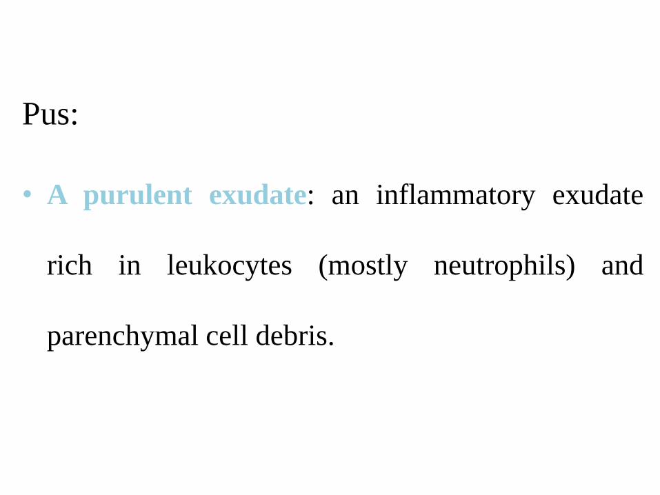
Exudation: Pneumonia
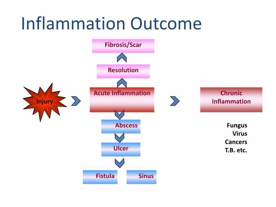
Inflammation Outcome
Acute Inflammation
Resolution
Chronic Inflammation
Abscess
SinusFistula
Fibrosis/Scar
Ulcer
Injury
FungusVirus
CancersT.B. etc.

Chemical Mediators:
Chemical substances synthesised or released and
mediate the changes in inflammation.
Histamine by mast cells - vasodilatation.
Prostaglandins – Cause pain & fever.
Bradykinin - Causes pain.

Chemical mediators of inflammation
• Platelet activating factor (PAF)
• Cytokines (IL-1, TNF, IL-8, IL-12)
• Nitric oxide (vasodilator, cytotoxin)
• Lysosomal constituents of leukocytes
• Oxygen derived free radicals
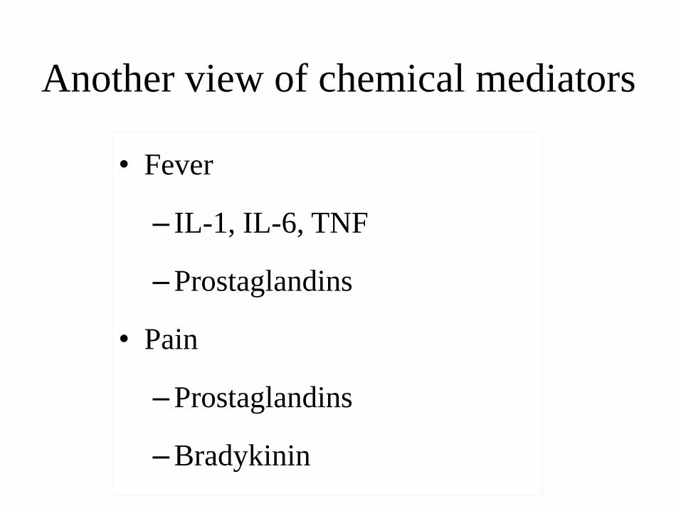
Another view of chemical mediators
• Fever
– IL-1, IL-6, TNF
–Prostaglandins
• Pain
–Prostaglandins
–Bradykinin

Morphologic types of acute inflammation
– Exudative or catarrhal Inflammation: excess fluid. TB
lung.
– Fibrinous – pneumonia – fibrin
– Membranous (fibrino-necrotic) inflammation
– Suppuration/Purulent – Bacterial - neutrophils
– Serous – excess clear fluid – Heart, lung
– Allergic inflammation
– Haemorrhagic – b.v. damage - anthrax.
– Necrotising inflammation.

Acute inflammation has one of four outcomes:
• Abscess formation
• Progression to chronic inflammation
• Resolution--tissue goes back to normal
• Repair--healing by scarring or fibrosis

Abscess formation:
• "A circumscribed collection of pus (suppurative
inflammation) appearing in an acute or chronic
localized infection, and associated with tissue
destruction, and frequently, swelling.“
• It is usually the result of a pyogenic organism.
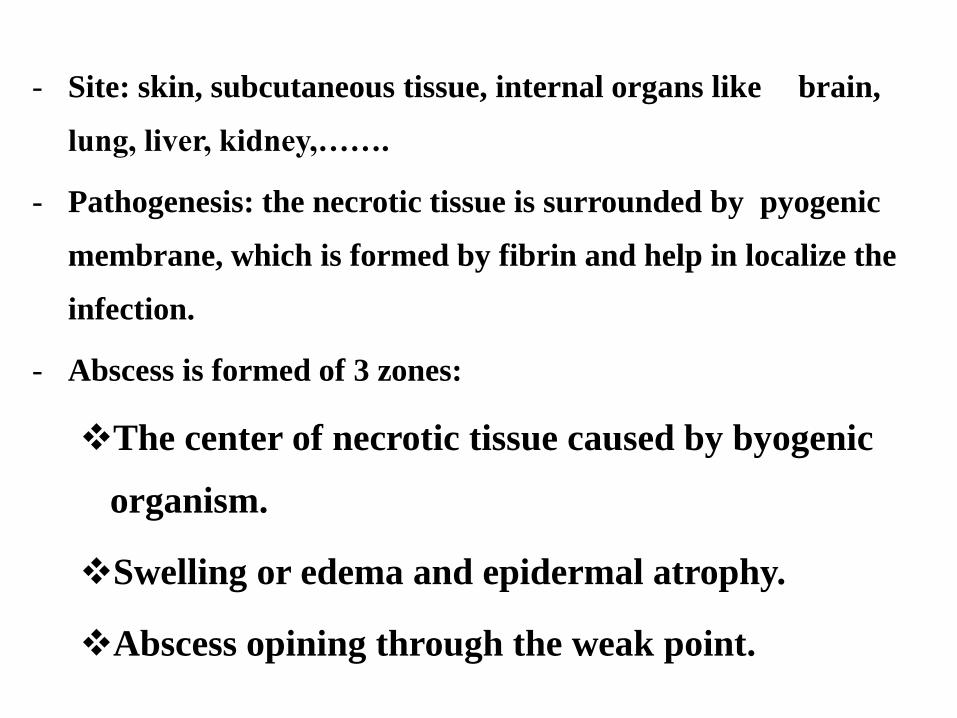
- Site: skin, subcutaneous tissue, internal organs like brain,
lung, liver, kidney,…….
- Pathogenesis: the necrotic tissue is surrounded by pyogenic
membrane, which is formed by fibrin and help in localize the
infection.
- Abscess is formed of 3 zones:
The center of necrotic tissue caused by byogenic
organism.
Swelling or edema and epidermal atrophy.
Abscess opining through the weak point.

Carbuncle
- It is an extensive form of abscess in which pus
is present in multiple loci open at the surface
by sinuses.
- Occur in the back of the neck and the scalp.

Furuncle or boil
- It is a small abscess related to hair
follicles or sebaceous glands, could
be multiple furunclosis.

Cellulitis - It is an acute diffuse suppurative inflammation caused
by streptococci, which secrete hyaluronidase &
streptokinase enzymes that dissolve the ground
substances and facilitate the spread of infection.
- Sites:
- - Areolar tissue; orbit, pelvis, …
- - Lax subcutaneous tissue

Sinus
- It is a tract of granulation tissue a cavity
(abscess) to the outside (skin), and has a
blind end

Fistula
- It is a tract of granulation tissue connecting two
epithelial surfaces; i.e. two ends open.
- Types of fistula: - Congenital or Acquired.
- The acquired form could be: traumatic;
inflammatory; neoplastic.