Embed Size (px)
Citation preview
Inflammtion
• Vascularized connective tissue response to cell injury
Acute Inflammation
What is inflammation?
• Inflammation -– protective response intended to eliminate the initial
cause of cell injury and the necrotic cells and tissues arising from the injury
• Inflammation is intimately associated with the repair process which includes parenchymal cell regeneration and scarring
• Acute - minutes to days– Characterized by fluid and protein – PMN’s
• Chronic - weeks to years– Lymphocytes and macrophages
• ACUTE Inf - PMN’s (Polymorphonuclear Cells)
• CHRONIC Inf - Mononuclear Cells
Inflammation
EXUDATE
Acute inflammation
• The immediate and early response to injury
• The point? Get the pmn’s to the site as fast as possible
• Vasodilatation
• Endothelial permeability
• Extravasation of pmn’s
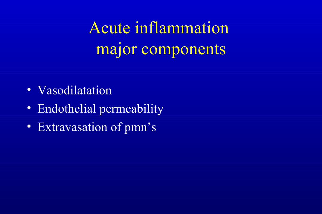
Acute inflammation major components
• Vasodilatation
• Endothelial permeability
• Extravasation of pmn’s
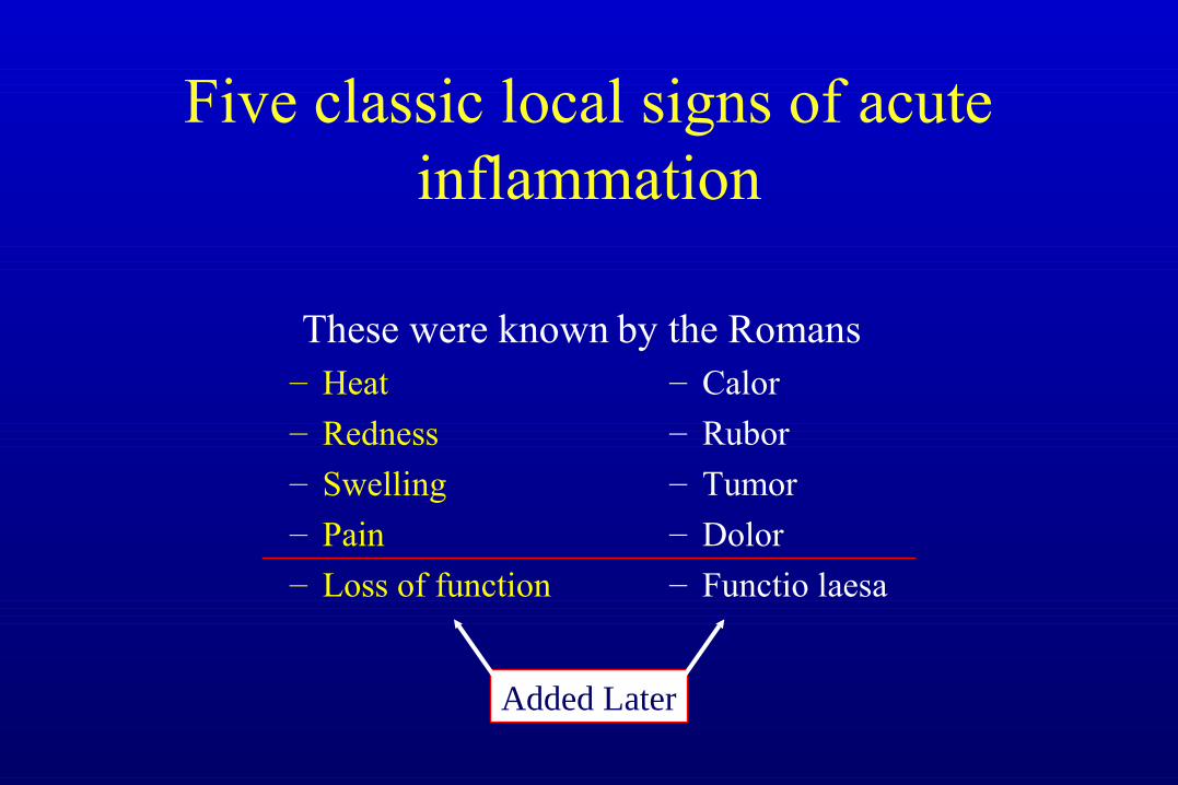
Five classic local signs of acute inflammation
These were known– Heat– Redness– Swelling– Pain
– Loss of function
by the Romans– Calor– Rubor
– Tumor– Dolor– Functio laesa
Added Later
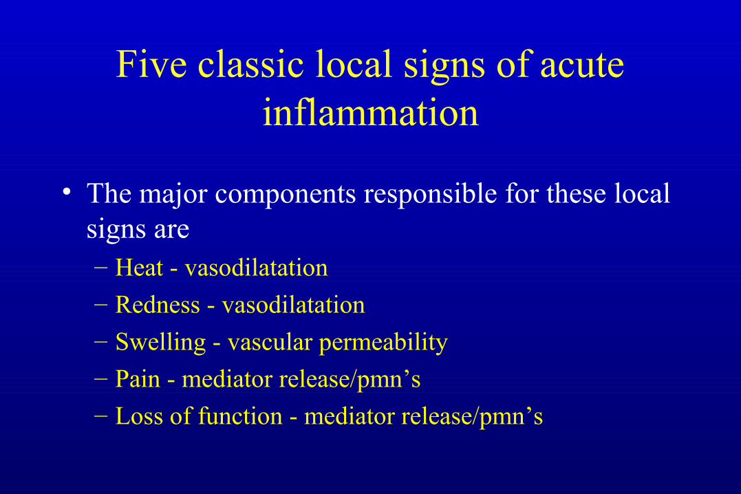
Five classic local signs of acute inflammation
• The major components responsible for these local signs are – Heat - vasodilatation– Redness - vasodilatation– Swelling - vascular permeability– Pain - mediator release/pmn’s– Loss of function - mediator release/pmn’s
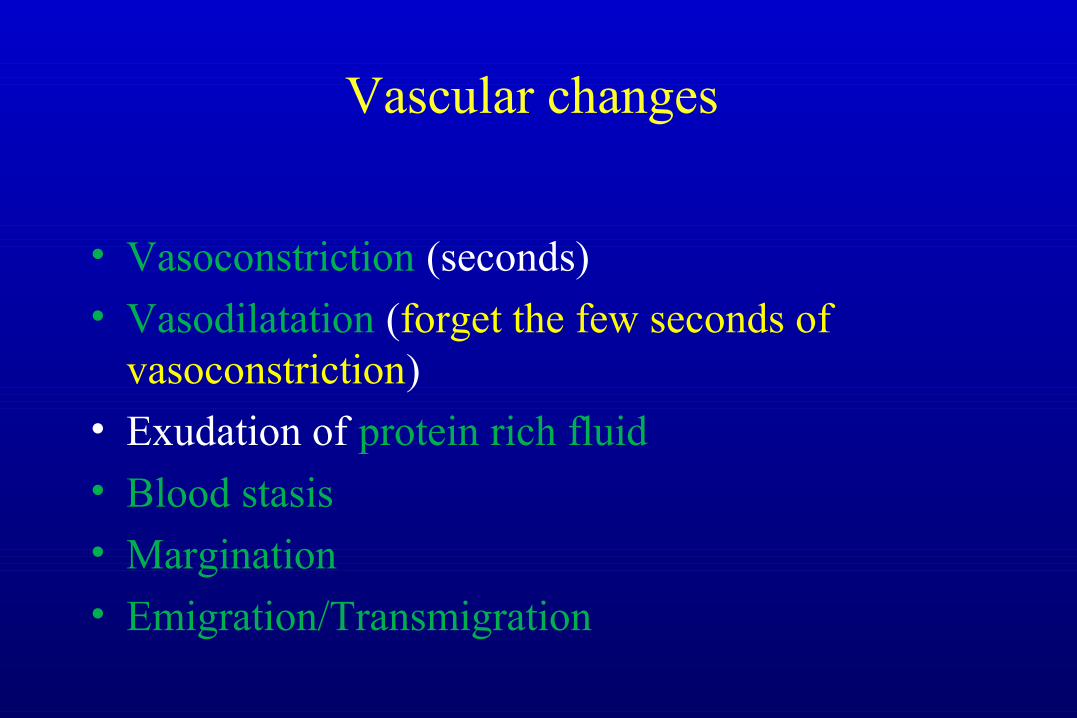
Vascular changes
• Vasoconstriction (seconds)
• Vasodilatation (forget the few seconds of vasoconstriction)
• Exudation of protein rich fluid
• Blood stasis
• Margination
• Emigration/Transmigration
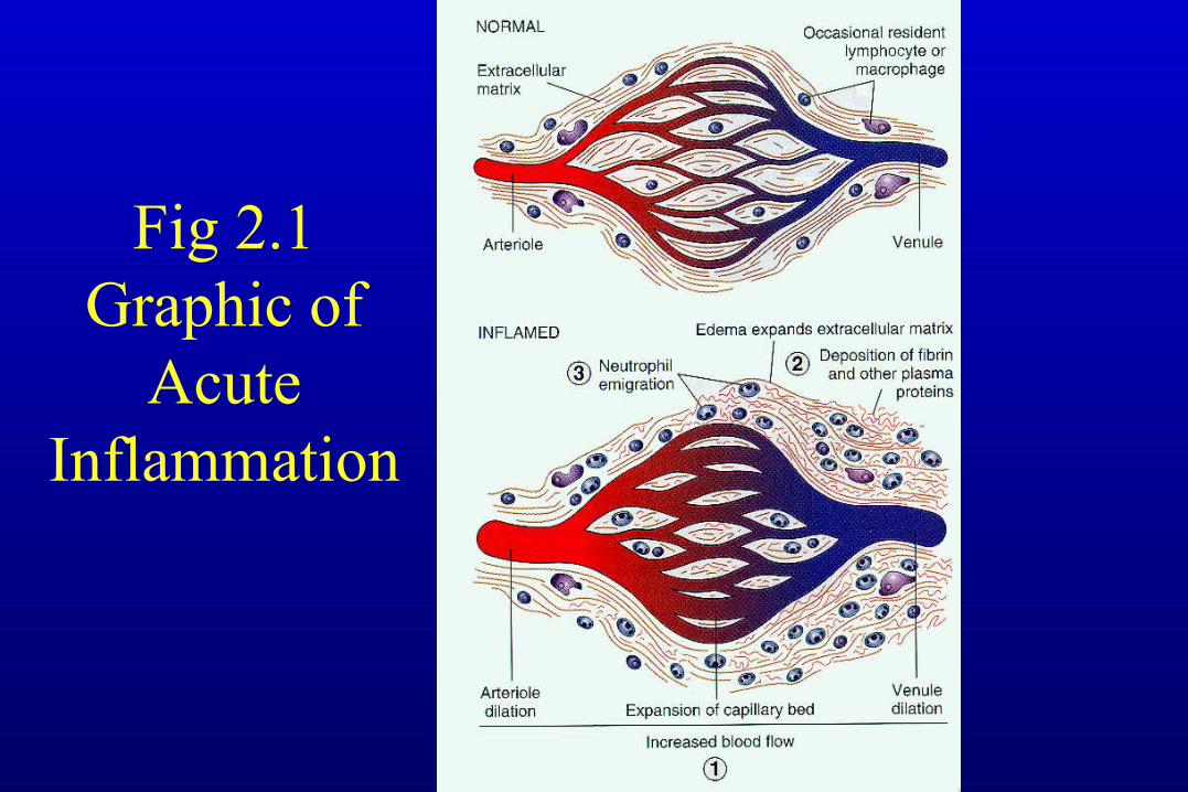
Fig 2.1 Graphic of
Acute Inflammation
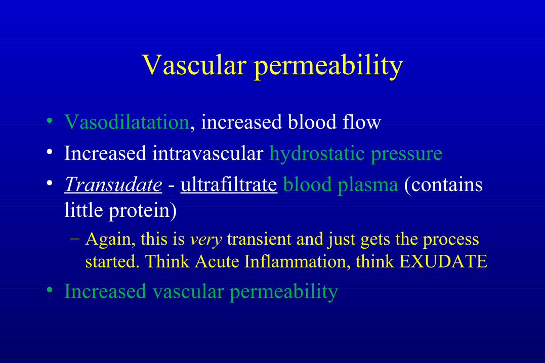
Vascular permeability
• Vasodilatation, increased blood flow
• Increased intravascular hydrostatic pressure
• Transudate - ultrafiltrate blood plasma (contains little protein)– Again, this is very transient and just gets the process
started. Think Acute Inflammation, think EXUDATE
• Increased vascular permeability
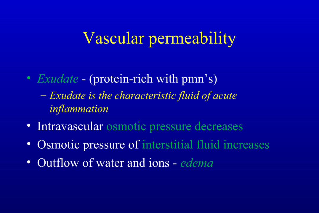
Vascular permeability
• Exudate - (protein-rich with pmn’s)– Exudate is the characteristic fluid of acute
inflammation
• Intravascular osmotic pressure decreases
• Osmotic pressure of interstitial fluid increases
• Outflow of water and ions - edema
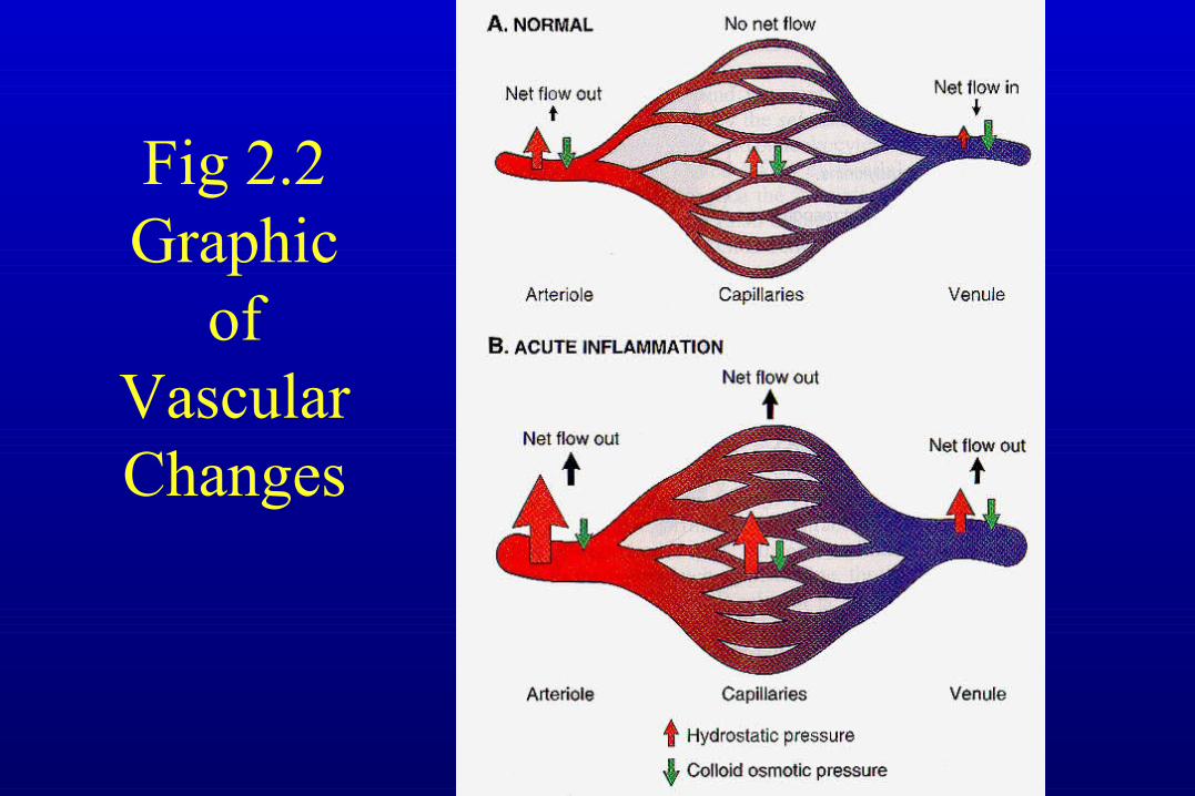
Fig 2.2 Graphic
of Vascular Changes
How do endothelial cellsbecome leaky?
• Endothelial cell contraction
• Junctional retraction
• Direct endothelial injury (immediate sustained response)
• Leukocyte-dependent endothelial injury
• Increased transcytosis of fluid
Direct endothelial injury (immediate sustained response)
• Endothelial cell necrosis and detachment
• Result of severe injury or burn
• Occurs immediately and lasts until vessel repaired
• Occurs at sites of leukocyte accumulation
• Due to leukocyte activation which releases proteolytic enzymes and toxic oxygen
Leukocyte-dependent endothelial injury
Leukocyte Cellular Events
• Margination and Rolling
• Adhesion and Transmigration
• Migration into interstitial tissue
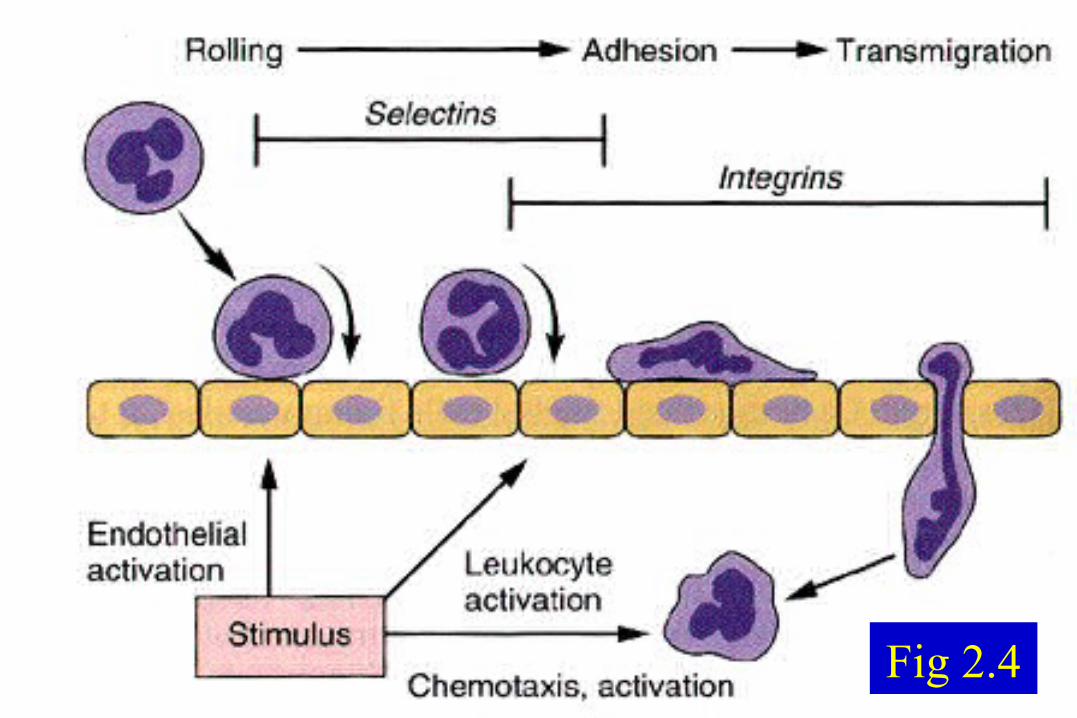
Fig 2.4
Margination
• Normal flow - RBC’s and WBC’s flow in the center of the vessel
• A cell poor plasma is flowing adjacent to endothelium
• As blood flow slows, WBC’s collect along the endothelium (this is “Margination”)
Endothelial Activation
• The underlying stimulus causes release of inflammatory mediators which activate the endothelium causing selectins and other mediators to be moved quickly to the surface of the cell
Selectins
• Selectins bind selected sugars – Selected + Lectins (sugars) = Selectins
• Some selectins are present on endothelial cells (E-Selectin)• Some selectins are present on leukocytes (L-Selectin)• Some selectins are present on platelets and endothelial cells
(P-Selectin)
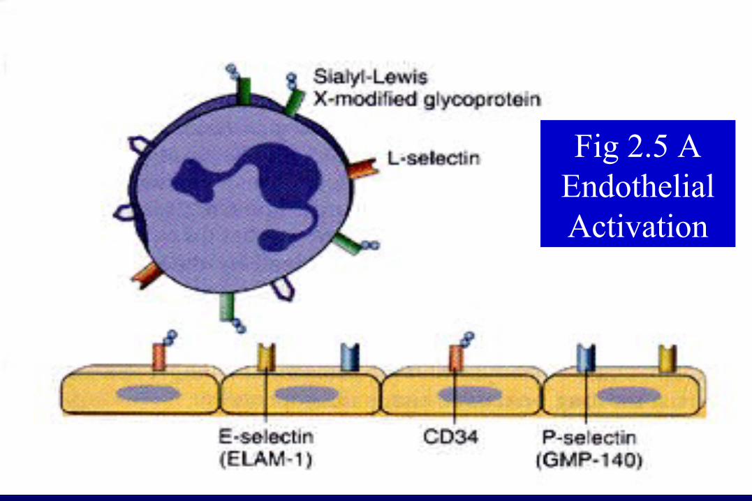
Fig 2.5 A Endothelial Activation
Rolling
• Selectins transiently bind to receptors
• PMN’s bounce or roll along
• This is Rolling
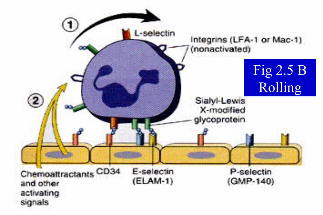
Fig 2.5 B Rolling
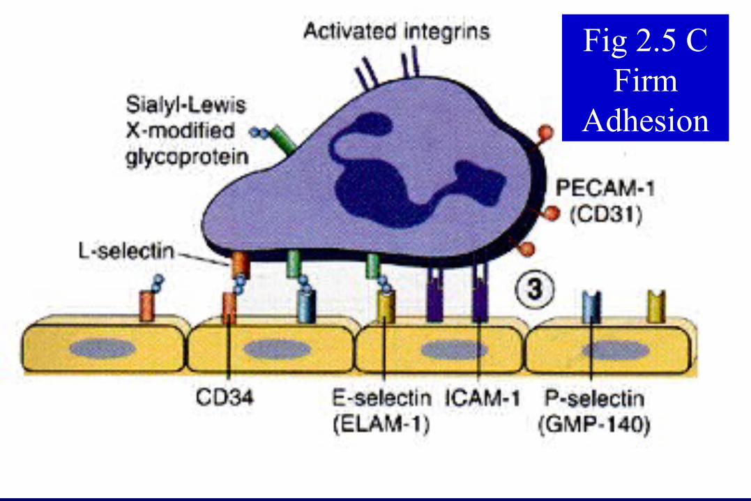
Fig 2.5 C Firm
Adhesion
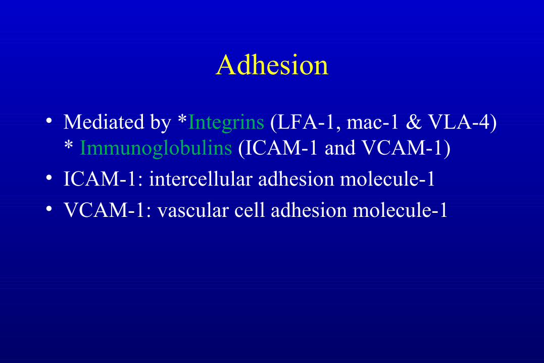
Adhesion
• Mediated by *Integrins (LFA-1, mac-1 & VLA-4) * Immunoglobulins (ICAM-1 and VCAM-1)
• ICAM-1: intercellular adhesion molecule-1
• VCAM-1: vascular cell adhesion molecule-1
Transmigration
• Mediated by PECAM-1
• Diapedesis
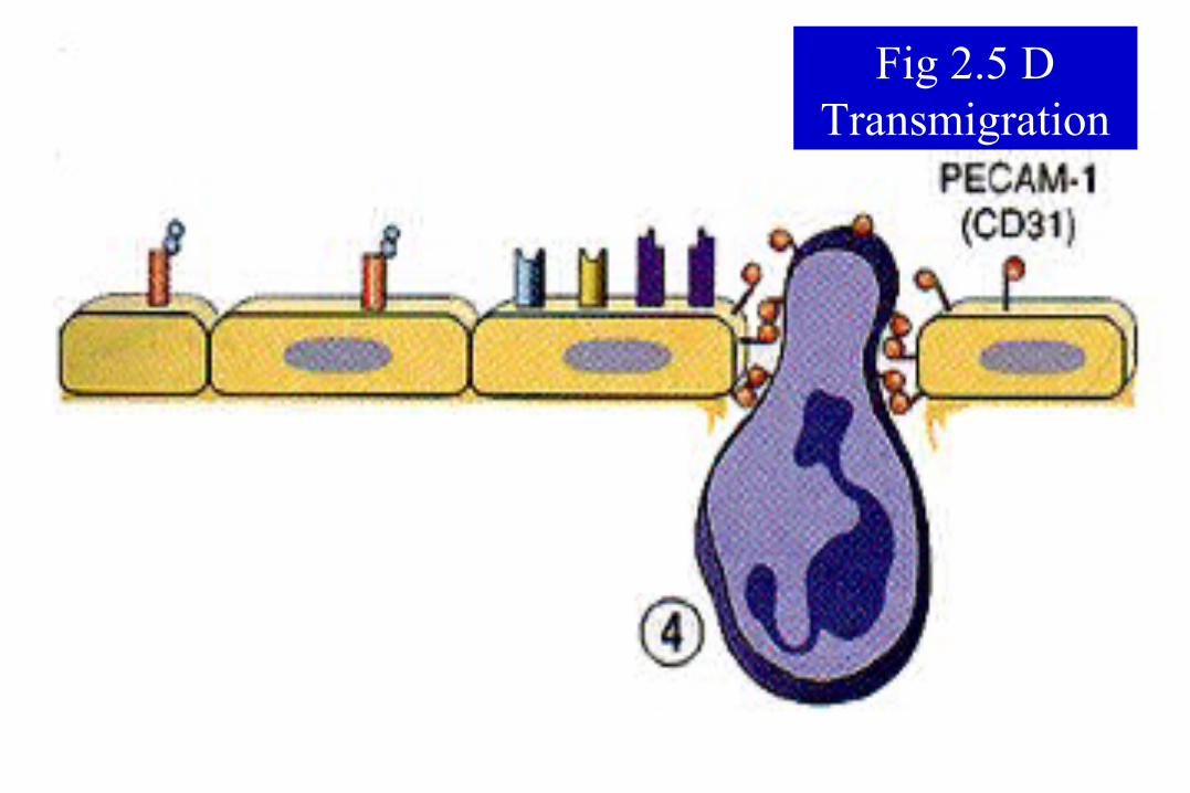
Fig 2.5 DTransmigration
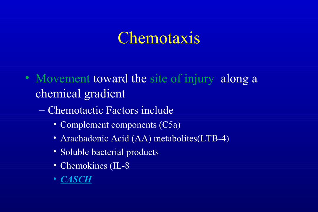
Chemotaxis
• Movement toward the site of injury along a chemical gradient– Chemotactic Factors include
• Complement components (C5a)• Arachadonic Acid (AA) metabolites(LTB-4)• Soluble bacterial products• Chemokines (IL-8
• CASCH
Leukocyte Activation
• Chemokines also “activate” PMN’s– AA metabolite production
– Degranulation and Secretion of lysosomal enzymes– Oxidative burst (free radicals)– Modulation of adhesion molecules
Phagocytosis & Degranulation
• Phagocytosis (to eat and destroy)– Attach– Engulf– Kill
• Degranulation and the oxidative burst destroy the engulfed particle
Leukocyte-induced tissue injury
• Lysosomal enzymes are released into the extracellular space during phagocytosis causing cell injury and matrix degradation
• Activated leukocytes release reactive oxygen species and products of arachidonic acid metabolism which can injure tissue and endothelial cells
• These events underlie many human diseases (e.g. Rheumatoid arthritis)
Leukocyte adhesion deficiency 1 (LAD-1)
• Recurrent bacterial infections
• Inflammatory lesions lack neutrophil infiltrate
• High numbers of neutrophils in the circulation
• Neutrophils from patients can roll but do not stick
• Transfuse patients with normal neutrophils and they can emigrate
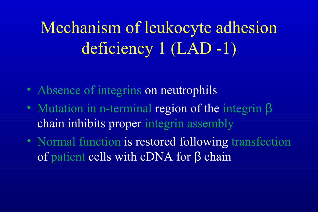
Mechanism of leukocyte adhesion deficiency 1 (LAD -1)
• Absence of integrins on neutrophils
• Mutation in n-terminal region of the integrin β chain inhibits proper integrin assembly
• Normal function is restored following transfection of patient cells with cDNA for β chain
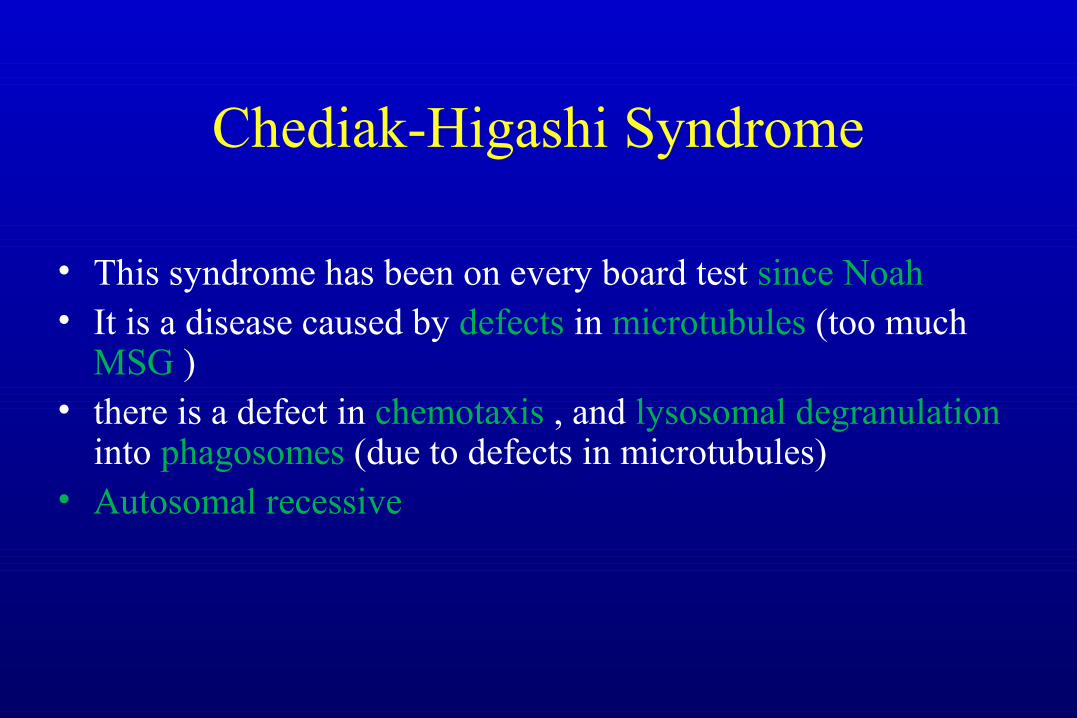
Chediak-Higashi Syndrome
• This syndrome has been on every board test since Noah• It is a disease caused by defects in microtubules (too much
MSG ) • there is a defect in chemotaxis , and lysosomal degranulation
into phagosomes (due to defects in microtubules)• Autosomal recessive
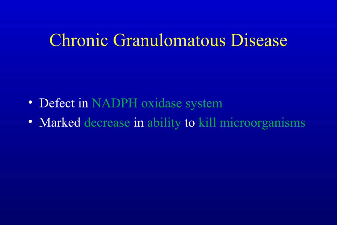
Chronic Granulomatous Disease
• Defect in NADPH oxidase system
• Marked decrease in ability to kill microorganisms
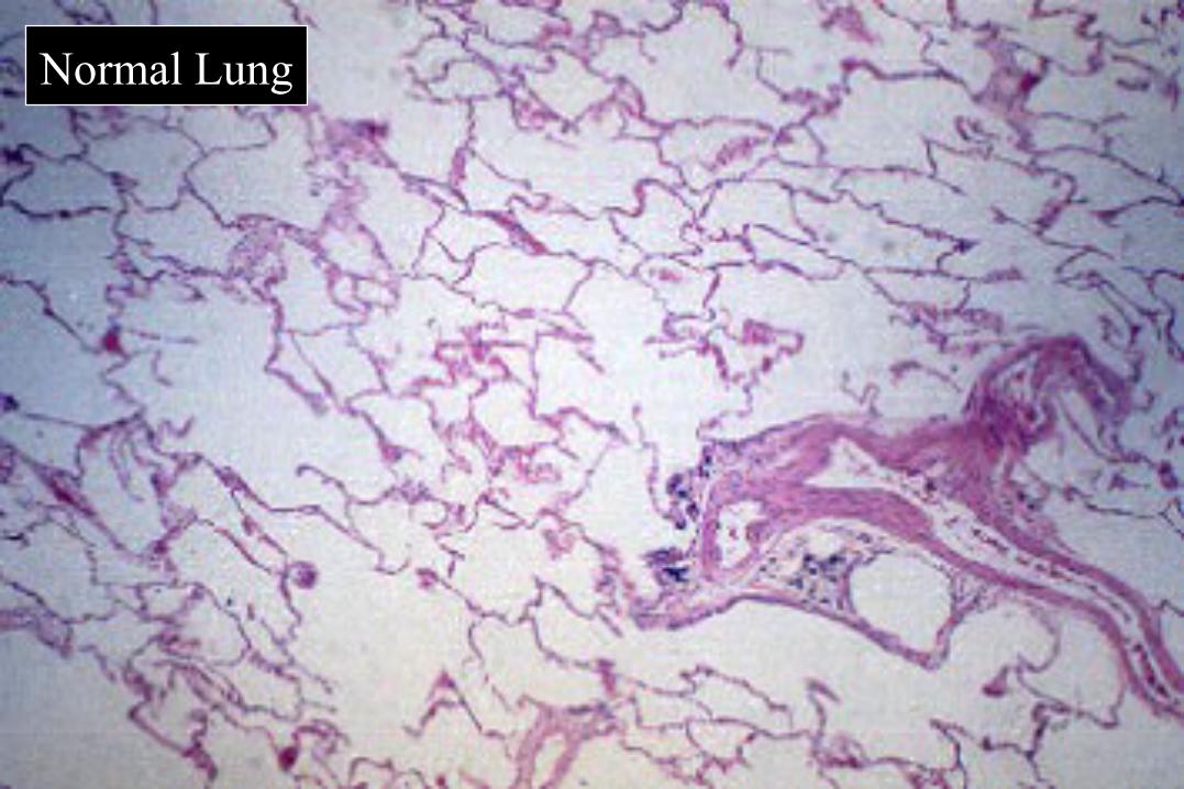
Normal Lung
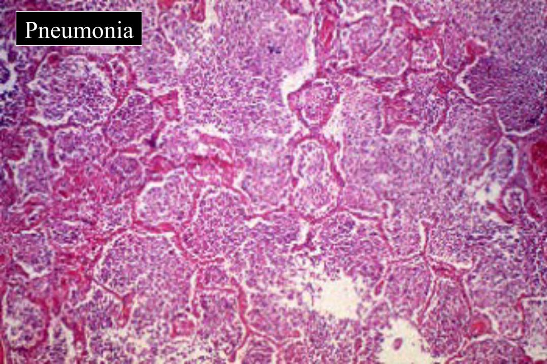
Pneumonia
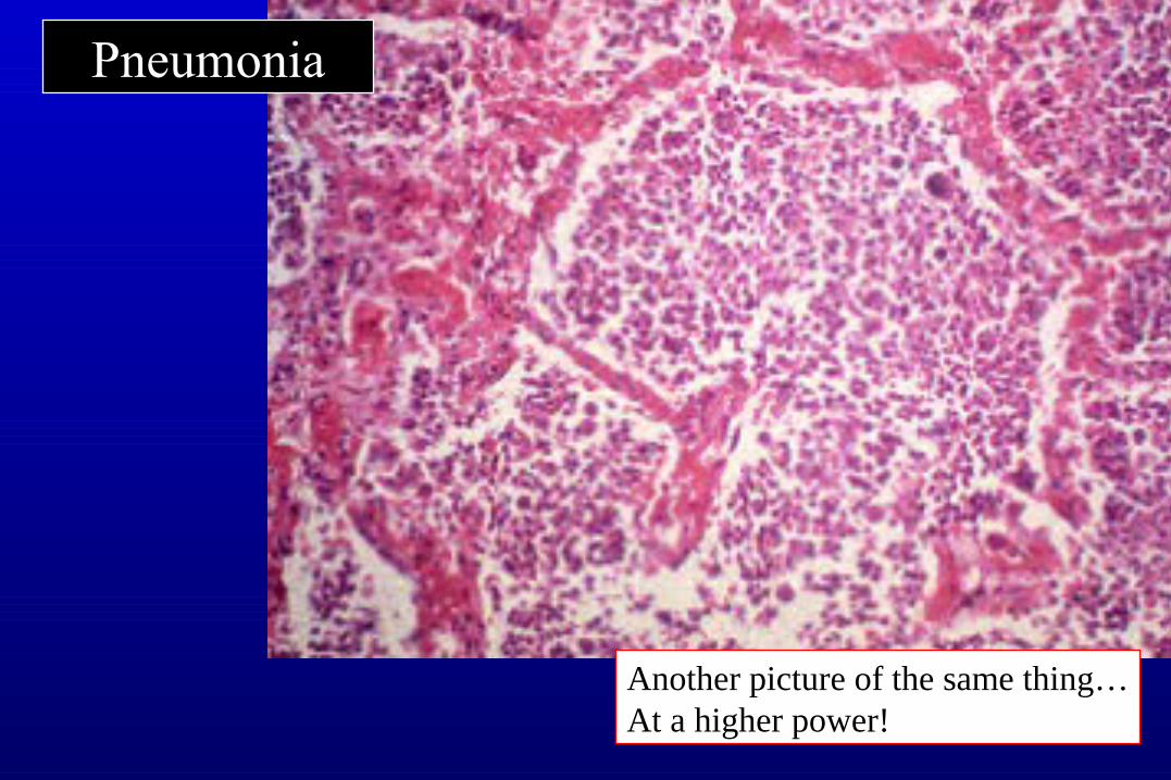
Pneumonia
Another picture of the same thing…At a higher power!
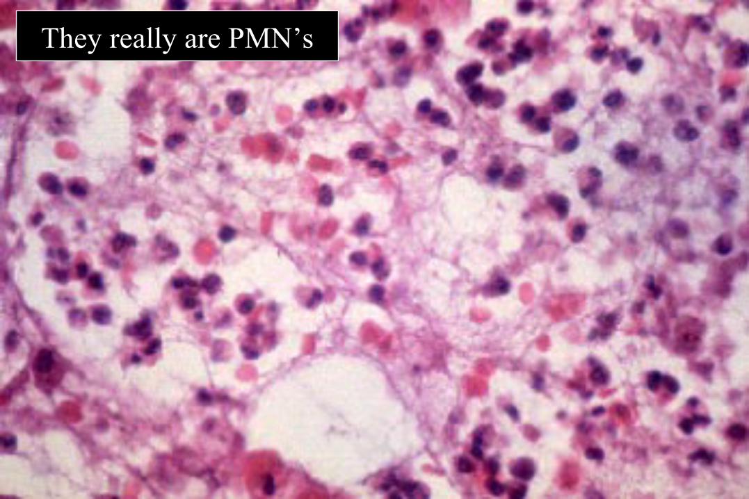
They really are PMN’s
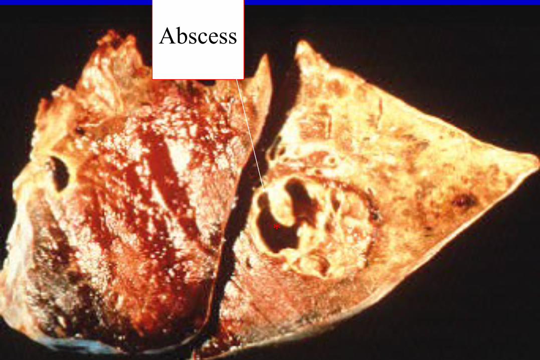
Abscess
*
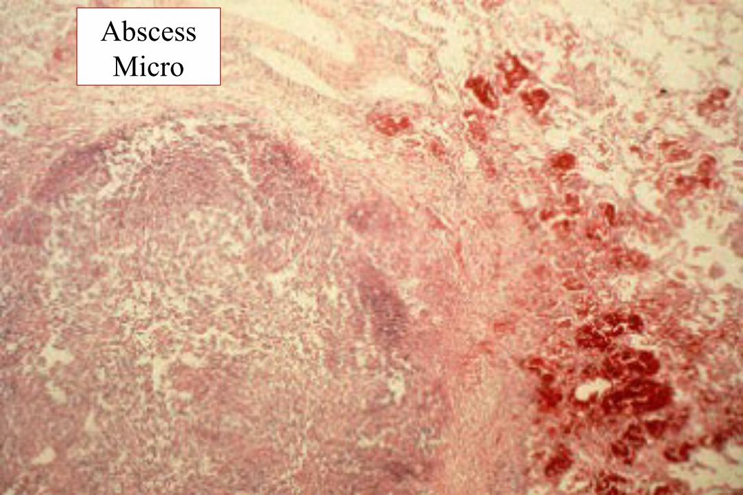
AbscessMicro
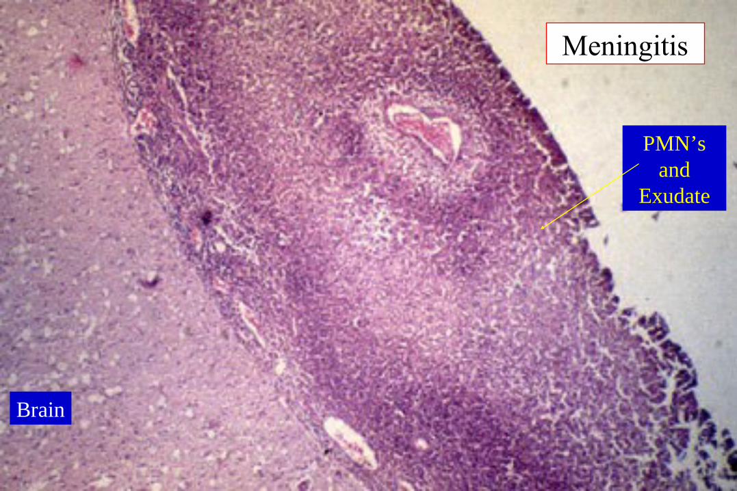
Meningitis
Brain
PMN’s and
Exudate
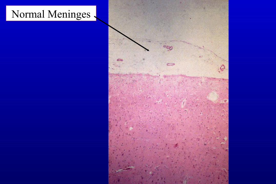
Normal Meninges