Embed Size (px)
Citation preview

Functional Census ofMutation Sequence Spaces:
The Example of p53 Cancer Rescue MutantsSamuel A. Danziger, S. Joshua Swamidass, Jue Zeng, Lawrence R. Dearth, Qiang Lu,
Jonathan H. Chen, Jianlin Cheng, Vinh P. Hoang, Hiroto Saigo, Ray Luo, Pierre Baldi,
Rainer K. Brachmann, and Richard H. Lathrop
Abstract—Many biomedical problems relate to mutant functional properties across a sequence space of interest, e.g., flu, cancer, and
HIV. Detailed knowledge of mutant properties and function improves medical treatment and prevention. A functional census of
p53 cancer rescue mutants would aid the search for cancer treatments from p53 mutant rescue. We devised a general methodology
for conducting a functional census of a mutation sequence space by choosing informative mutants early. The methodology was tested
in a double-blind predictive test on the functional rescue property of 71 novel putative p53 cancer rescue mutants iteratively predicted
in sets of three (24 iterations). The first double-blind 15-point moving accuracy was 47 percent and the last was 86 percent; r = 0.01
before an epiphanic 16th iteration and r = 0.92 afterward. Useful mutants were chosen early (overall r = 0.80). Code and data are freely
available (http://www.igb.uci.edu/research/research.html, corresponding authors: R.H.L. for computation and R.K.B. for biology).
Index Terms—Biology and genetics, feature extraction or construction, machine learning, medicine and science.
�
1 INTRODUCTION
MUTATIONS and their functional effects drive evolution,drug resistance, genetic disorders, viral evasion of the
immune system, and other important biomedical processes.In pharmacogenomics [1] and drug resistant HIV [2], [3],[4], detailed knowledge of functionally important mutationsleads directly to better patient treatment. In flu [5], knowl-edge of important mutations leads directly to better diseaseprevention, by way of better vaccine design. In cancer, theconcern of this paper, the effect of functionally importantmutations causes the disease.
Medical practice is often advanced by knowing mutantfunctional properties across a mutation sequence space ofspecific interest. One difficulty is that mutation spaces growto be combinatorially large, while experimental time andresources remain bounded. Computational analysis ischallenging because subtle effects on structure and functionresult in broad and diverse changes.
1.1 p53 Overview
Cancer is caused by the accumulation of genetic mutationsin two critical regulatory pathways: normal cell growth and
programmed cell death (apoptosis). Defects in the cell
growth pathway can result in uncontrolled cellular pro-
liferation. Tumor suppressor proteins such as p53 normally
trigger apoptosis in affected cells and destroy the tumor.p53 exerts its tumor suppressor activity mainly as a
transcription factor that induces cell cycle arrest, apoptosis,
DNA repair, and/or senescence. It is stabilized and
activated in response to cell stress by a complicated series
of posttranslational modifications [6], [7], [8], [9]. Activated
p53 suppresses tumors through one of the following
mechanisms:
1. Induction—p53 directly targets and induces geneswith tumor suppressor functions [10], [11]. There areapproximately 100 known genes with p53 bindingsites [12] and several hundred genes are directly orindirectly upregulated by activated p53 [13], [14].
2. Repression—p53 also represses the expression ofgenes. As most of the repressed genes lack a distinctp53 binding site, the mechanism is currentlyunknown [15].
3. Nontranscriptional Mechanisms—p53 translocatesto the mitochondria in response to DNA damageand causes cytochrome c release [16], [17].
p53 mutations that disrupt these mechanisms are
complicit in human cancers. The International Agency for
Research on Cancer (IARC) TP53 Mutation Database1 (R10)
lists 21,588 p53 mutations found in human cancer patients
[18]. Seventy-one percent of the entries (15,387) result in
full-length protein with a single amino acid change in the
DNA binding p53 core domain. The top eight mutants
114 IEEE/ACM TRANSACTIONS ON COMPUTATIONAL BIOLOGY AND BIOINFORMATICS, VOL. 3, NO. 2, APRIL-JUNE 2006
. S.A. Danziger, S.J. Swamidass, J. Zeng, L.R. Dearth, J.H. Chen, J. Cheng,V.P. Hoang, R. Luo, P. Baldi, R.K. Brachmann, and R.H. Lathrop are withthe University of California, Irvine, CA 92697-3435.E-mail: {sdanzige, sswamida, jzeng, ldearth, chenjh, jianlinc, vphoang,rluo, pfbaldi, rbrachma, rickl}@uci.edu.
. Q. Lu is with the State University of New York, Stony Brook, NY 11794.E-mail: [email protected].
. H. Saigo is with the Bioinformatics Center, Institute for ChemicalResearch, Kyoto University, Uji, Kyoto 611-0011, Japan.E-mail: [email protected].
Manuscript received 1 June 2005; revised 26 Sept. 2005; accepted 30 Sept.2005; published online 1 May 2006.For information on obtaining reprints of this article, please send e-mail to:[email protected], and reference IEEECS Log Number TCBB-0057-0605. 1. http://www-p53.iarc.fr/index.html.
1545-5963/06/$20.00 � 2006 IEEE Published by the IEEE CS, CI, and EMB Societies & the ACM

account for 30 percent and the top 50 account for 54 percentof these single amino acid change mutants [19].
The structure of full-length wild-type p53 is unknown,but the crystal structure of the core domain [20], inconjunction with biophysical and NMR studies [21], [22],[23], has made it possible to construct homology models.p53 has 393 amino acids and three important domains: anamino-terminal transactivation domain, a core domainconsisting of amino acids 96-292 which recognizes p53DNA binding sites, and a carboxy-terminal tetramerizationdomain [24], [25], [26], [27], [28].
1.2 Novel Cancer Treatments and p53 FunctionalRescue
A long-held medical goal for anticancer therapy is achiev-ing functional rescue of p53 cancer mutants by stabilizingthe wild-type conformation, thereby activating apoptosis incancerous cells and shrinking or killing the tumor. Severalpromising drug-like small molecules have been identified[29], [30], but their mechanisms of action and their spectraof activity are not known. This has led to intense scientificinterest in the basic mechanisms of p53 functional rescue.
1.2.1 p53 Cancer Rescue Mutants
We established the existence of global functional rescuemechanisms for p53 cancer mutants [31] through studies ofintragenic second-site suppressor mutations that restorenative p53 function (“cancer rescue,” “cancer suppressor,”among other names). Surprisingly, a second-site p53suppressor mutation can cooccur with a p53 cancermutation such that functional effects cancel and the doublemutant protein has normal p53 function.
A search for such suppressor mutations resulted inidentification of a “global suppressor motif” involving coredomain amino acids 235, 239, and 240 [32]. Specific aminoacid changes of one or more of these restored p53 functionto 16 of 30 of the most common p53 cancer mutants tested.
1.2.2 Terminology
In this paper, the terms active and inactive are used todescribe mutant functionality. In other literature, an activemutant may be referred to as a “functional,” “positive,” or“rescued” mutant and an inactive mutant may be referredto as a “nonfunctional,” “negative,” or “cancer” mutant.
1.3 Computational Approaches to p53
The p53 mutant classification problem is to predict whethera given set of amino acid changes to the p53 core domainresults in an active p53 protein or not. It is a difficultproblem because the p53 protein is marginally stable atphysiological temperature (37�C). p53 cancer mutants canbe destabilized by only a few kcal/mole [33]. Some p53mutants are inactive at human physiological temperature(37�C), but regain activity at 30�C. It is a substantialchallenge to predict mutant functional activity fromsequence when it depends crucially upon such subtlenuances.
The first, and previously the only, systematic integratedcomputational analysis of p53 mutation data and structuraleffects was made by Martin et al. [34]. They correlatedmutations in the IARC database [18] with structural and
evolutionary features, but did not make predictions orconsider mutant phenotypic function. In 34 percent ofdistinct cancer mutations, their analysis was able to findidentifiable underlying structural changes that might beexpected to affect protein folding or protein-DNA contacts,based on secondary structure, hydrogen bonding, backbonetorsion angles, and solvent accessibility. Possibly explain-able changes rose to 56 percent by including substitutions ofamino acids that are 100 percent conserved across manyspecies.
While their results are impressive, they highlight thedifficult case of p53. Two-thirds of all distinct p53 cancermutants lack even a single putative explanation in terms ofidentifiable underlying structural changes and nearly halfhave no putative explanation whatsoever.
2 A THEORY OF COMPUTATION ASSISTING
EXPERIMENTALISTS TO PURSUE FUNCTION
A functional census of p53 cancer and suppressor mutationsmeans a catalog of the functional effect of each mutation. Thecensus assigns active or inactive labels to every mutant, byexperimental determination or computational prediction.
Initially, experimental work would focus on selectivescreens in relevant regions of the p53 core domain, wheremost mutations that inactivate p53 occur. Hits from thescreens would provide an initial training set for computa-tional predictors of mutant p53 activity. The result of testedcomputational predictions would be a larger pool of knownmutants with experimental activities. The larger training setwould yield more accurate computational predictors,leading to a repeating cycle of improving predictions andexperiments. Once the computational predictor was suffi-ciently accurate, it would be used to guide experimentalwork by identifying interesting regions in the p53 sequence.
2.1 Functional Census of Mutation Sequence Space
This section defines procedures for
1. iterated predictions,2. informative mutant selection,3. cross-validation, and4. periodic methodology updates.
2.1.1 Iterated Predictions
Let set Ki be the mutants known to be active or inactive atstep i. Predict Ki with cross-validation using Ki as a trainingset. Select a set Xi of unknown mutants as described belowin Section 2.1.2. Predict Xi blindly using Ki as a training set,and record the predictions. Determine functions for Xiexperimentally and score the recorded predictions. PredictKi, Xi, and Ki + Xi with cross-validation using Ki + Xi as atraining set. Finally, let K{i + 1} equal Ki + Xi and advance tostep i + 1.2
2.1.2 Informative Mutant Selection
Active learning is a technique for selecting the mostinformative unlabeled examples and was previously used
DANZIGER ET AL.: FUNCTIONAL CENSUS OF MUTATION SEQUENCE SPACES: THE EXAMPLE OF P53 CANCER RESCUE MUTANTS 115
2. This abuse of notation, + as set union instead of U, was found to bemore intuitive to a wider audience.

successfully for drug discovery and cancer classification [35],[36], [37]. Here, the most informative mutant is determined byestimating its impact on classifier accuracy. First, suppose theunknown mutant is active, rebuild the classifier, anddetermine the new cross-validated accuracy on the trainingset. Then, suppose the mutant is inactive and repeat. Themaximum increase in the cross-validated correlation coeffi-cient (CC), for an unknown mutant (m), across both assumedclasses is here called “curiosity” (1), (2).
CCc;t ¼ðtpc;t � tnc;tÞ � ðfpc;t � fnc;tÞffiffiffiffiffiffiffiffiffiffiffiffiffiffiffiffiffiffiffiffiffiffiffiffiffiffiffiffiffiffiffiffiffiffiffiffiffiffiffiffiffiffiffiffiffiffiffiffiffiffiffiffiffiffiffiffiffiffiffiffiffiffiffiffiffiffiffiffiffiffiffiffiffiffiffiffiffiffiffiffiffiffiffiffiffiffiffiffiffiffiffiffiffiffiffiffiffiffiffiffiffiffiffiffiffiffiffiffi
ðtpc;t þ fpc;tÞðtpc;t þ fnc;tÞðtnc;t þ fpc;tÞðtnc;t þ fnc;tÞp ;
ð1Þ
curiositym ¼ max
PcðCCc;tþmðactiveÞ � CCc;tÞ;PcðCCc;tþmðinactiveÞ � CCc;tÞ
� �: ð2Þ
The CCc;t for a given classifier (c) in the set of all componentclassifiers with training set (t) is calculated using the truepositive (tpc;t), false positive (fpc;t), false negative (fnc;t),and true negative (tnc;t) receiver operator characteristic(ROC) statistics.
2.1.3 Overlap Exclusion Cross-Validation (OECV)
The usual cross-validation strategies may not be sufficientlystringent for mutation sequence spaces because the trainingset may contain mutants that differ in only trivial ways(irrelevant mutations) from mutants in the test set. InOECV, mutants are removed from the training set if theyshare more than one mutation with the mutant beingpredicted. Thus, no cancer/rescue pair ever occurs in bothtraining and test sets. Even so, cross-validation can be amisleading estimator. A major strength of this paper andmethodology is that all predictions are made blindly andare verified experimentally.
2.1.4 Periodically Update Methodology
During the course of the iterated mutant predictions, newinformation will become available about mutant behavior.This will lead to better theories to describe behavior andbetter classifiers to predict function. New information aboutmutant behavior is used periodically to update the classifierand framework (see Fig. 1).
2.2 Molecular Models and Statistical Learning
If molecular models and dynamics (MD) simulations couldpredict protein function correctly from one or a few aminoacid changes, then computation would face an easy task.However, atomic models are not strictly accurate in atomic-level detail due to structure prediction limitations withcurrent tools. Current computer simulations cannot accu-rately predict the functional effects nor definitively predict
the protein structure resulting from even one single keyamino acid change. This is especially so for marginallystable proteins like p53.
Our hypothesis is that: 1) Atomic models and MDsimulations encode useful information, in the form of weaktrends and tendencies that are partially correlated withmolecular function, even when the molecular modelsthemselves fail to achieve consistent, reliable, detailedatomic-level accuracy, and 2) statistical machine learningmethods can extract that information in a useful way.
2.3 This Paper
The goals of this paper are: 1) to demonstrate machinelearning and statistical predictions in synergy with mole-cular modeling (see Section 2.2) and 2) to perform a double-blind test of the functional census methodology on p53cancer rescue mutants (see Section 2.1).
To accomplish the first goal, we constructed molecularmodels of all mutants considered in this paper. Weextracted predictive features as described in Section 3.4and used the features to make the predictions described inthe second goal. As a control, we constructed and optimizedtwo purely string-based classifiers. They were used to makethe same test predictions, based on the same trainingmutants, as for the molecular model-based predictions.
To accomplish the second goal, we began with a trainingset of 123 known p53 putative cancer rescue mutantsexperimentally determined to contain 52 active and 71 in-active mutants. These constituted K1, the initial known set.The test set consisted of 71 novel p53 mutants, selected andassayed by the Brachmann laboratory. These constituted X1to X24 and were predicted by 24 iterations of Section 2.1.1 ingroups of three mutants (the last group had two mutants).The experimentalists first released mutant identities, butsequestered all other information, including summarystatistics. After each double-blind computational predictionwas made, the corresponding experimental result wasreleased.
2.3.1 Biological Advance
This paper will demonstrate a general methodology for thecomputer-aided functional census of protein mutationsequence spaces, together with its instantiation on a centralcancer protein. Other groups can use the methodology tocreate a functional census for other proteins. For example,Karchin et al. [38] provide a practical system that automatesthe model-building described below. Thus, the techniquesin this paper can be implemented on a large scale usingtools available now. After a functional census has beenachieved for several dozen cancer proteins, we will know agreat deal more about cancer systems biology than weknow now.
2.3.2 Computational Advance
This paper will demonstrate that machine learning andstatistical methods extend the utility of modeling techni-ques, while atomic modeling methods improve the powerand predictive accuracy of machine learning. This willadvance molecular computation by extending both mole-cular modeling and machine learning/statistical methods
116 IEEE/ACM TRANSACTIONS ON COMPUTATIONAL BIOLOGY AND BIOINFORMATICS, VOL. 3, NO. 2, APRIL-JUNE 2006
Fig. 1. The overall prediction strategy. The in silico predictions drive the
in vitro experiments, which in turn improve the in silico models.
into useful but poorly understood applications to molecular
function.
3 METHODS
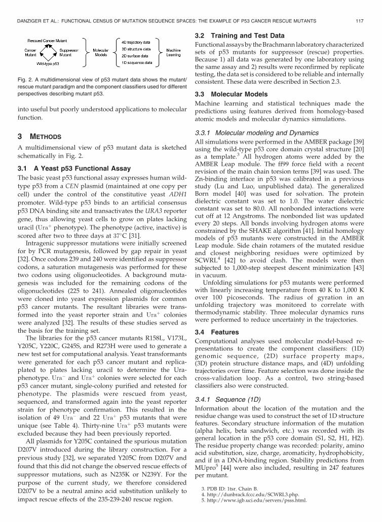
A multidimensional view of p53 mutant data is sketched
schematically in Fig. 2.
3.1 A Yeast p53 Functional Assay
The basic yeast p53 functional assay expresses human wild-
type p53 from a CEN plasmid (maintained at one copy per
cell) under the control of the constitutive yeast ADH1
promoter. Wild-type p53 binds to an artificial consensus
p53 DNA binding site and transactivates the URA3 reporter
gene, thus allowing yeast cells to grow on plates lacking
uracil (Uraþ phenotype). The phenotype (active, inactive) is
scored after two to three days at 37�C [31].Intragenic suppressor mutations were initially screened
for by PCR mutagenesis, followed by gap repair in yeast[32]. Once codons 239 and 240 were identified as suppressorcodons, a saturation mutagenesis was performed for thesetwo codons using oligonucleotides. A background muta-genesis was included for the remaining codons of theoligonucleotides (225 to 241). Annealed oligonucleotideswere cloned into yeast expression plasmids for commonp53 cancer mutants. The resultant libraries were trans-formed into the yeast reporter strain and Uraþ colonieswere analyzed [32]. The results of these studies served asthe basis for the training set.
The libraries for the p53 cancer mutants R158L, V173L,Y205C, Y220C, G245S, and R273H were used to generate anew test set for computational analysis. Yeast transformantswere generated for each p53 cancer mutant and replica-plated to plates lacking uracil to determine the Ura-phenotype. Ura� and Uraþ colonies were selected for eachp53 cancer mutant, single-colony purified and retested forphenotype. The plasmids were rescued from yeast,sequenced, and transformed again into the yeast reporterstrain for phenotype confirmation. This resulted in theisolation of 49 Ura� and 22 Uraþ p53 mutants that wereunique (see Table 4). Thirty-nine Uraþ p53 mutants wereexcluded because they had been previously reported.
All plasmids for Y205C contained the spurious mutation
D207V introduced during the library construction. For a
previous study [32], we separated Y205C from D207V and
found that this did not change the observed rescue effects of
suppressor mutations, such as N235K or N239Y. For the
purpose of the current study, we therefore considered
D207V to be a neutral amino acid substitution unlikely to
impact rescue effects of the 235-239-240 rescue region.
3.2 Training and Test Data
Functional assays by the Brachmann laboratory characterizedsets of p53 mutants for suppressor (rescue) properties.Because 1) all data was generated by one laboratory usingthe same assay and 2) results were reconfirmed by replicatetesting, the data set is considered to be reliable and internallyconsistent. These data were described in Section 2.3.
3.3 Molecular Models
Machine learning and statistical techniques made thepredictions using features derived from homology-basedatomic models and molecular dynamics simulations.
3.3.1 Molecular modeling and Dynamics
All simulations were performed in the AMBER package [39]using the wild-type p53 core domain crystal structure [20]as a template.3 All hydrogen atoms were added by theAMBER Leap module. The ff99 force field with a recentrevision of the main chain torsion terms [39] was used. TheZn-binding interface in p53 was calibrated in a previousstudy (Lu and Luo, unpublished data). The generalizedBorn model [40] was used for solvation. The proteindielectric constant was set to 1.0. The water dielectricconstant was set to 80.0. All nonbonded interactions werecut off at 12 Angstroms. The nonbonded list was updatedevery 20 steps. All bonds involving hydrogen atoms wereconstrained by the SHAKE algorithm [41]. Initial homologymodels of p53 mutants were constructed in the AMBERLeap module. Side chain rotamers of the mutated residueand closest neighboring residues were optimized bySCWRL4 [42] to avoid clash. The models were thensubjected to 1,000-step steepest descent minimization [43]in vacuum.
Unfolding simulations for p53 mutants were performedwith linearly increasing temperature from 40 K to 1,000 Kover 100 picoseconds. The radius of gyration in anunfolding trajectory was monitored to correlate withthermodynamic stability. Three molecular dynamics runswere performed to reduce uncertainty in the trajectories.
3.4 Features
Computational analyses used molecular model-based re-presentations to create the component classifiers: (1D)genomic sequence, (2D) surface property maps,(3D) protein structure distance maps, and (4D) unfoldingtrajectories over time. Feature selection was done inside thecross-validation loop. As a control, two string-basedclassifiers also were constructed.
3.4.1 Sequence (1D)
Information about the location of the mutation and theresidue change was used to construct the set of 1D structurefeatures. Secondary structure information of the mutation(alpha helix, beta sandwich, etc.) was recorded with itsgeneral location in the p53 core domain (S1, S2, H1, H2).The residue property change was recorded: polarity, aminoacid substitution, size, charge, aromaticity, hydrophobicity,and if in a DNA-binding region. Stability predictions fromMUpro5 [44] were also included, resulting in 247 featuresper mutant.
DANZIGER ET AL.: FUNCTIONAL CENSUS OF MUTATION SEQUENCE SPACES: THE EXAMPLE OF P53 CANCER RESCUE MUTANTS 117
Fig. 2. A multidimensional view of p53 mutant data shows the mutant/
rescue mutant paradigm and the component classifiers used for different
perspectives describing mutant p53.
3. PDB ID: 1tsr. Chain B.4. http://dunbrack.fccc.edu/SCWRL3.php.5. http://www.igb.uci.edu/servers/psss.html.
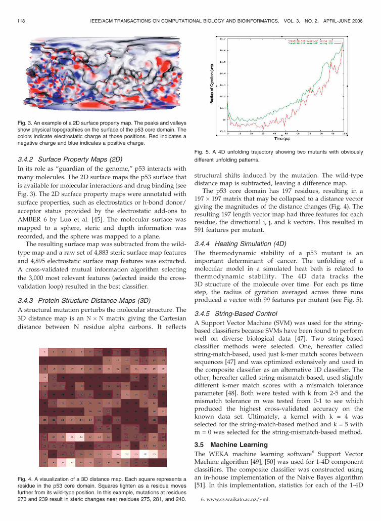
3.4.2 Surface Property Maps (2D)
In its role as “guardian of the genome,” p53 interacts with
many molecules. The 2D surface maps the p53 surface that
is available for molecular interactions and drug binding (see
Fig. 3). The 2D surface property maps were annotated with
surface properties, such as electrostatics or h-bond donor/
acceptor status provided by the electrostatic add-ons to
AMBER 6 by Luo et al. [45]. The molecular surface was
mapped to a sphere, steric and depth information was
recorded, and the sphere was mapped to a plane.The resulting surface map was subtracted from the wild-
type map and a raw set of 4,883 steric surface map features
and 4,895 electrostatic surface map features was extracted.
A cross-validated mutual information algorithm selecting
the 3,000 most relevant features (selected inside the cross-
validation loop) resulted in the best classifier.
3.4.3 Protein Structure Distance Maps (3D)
A structural mutation perturbs the molecular structure. The
3D distance map is an N�N matrix giving the Cartesian
distance between N residue alpha carbons. It reflects
structural shifts induced by the mutation. The wild-typedistance map is subtracted, leaving a difference map.
The p53 core domain has 197 residues, resulting in a197� 197 matrix that may be collapsed to a distance vectorgiving the magnitudes of the distance changes (Fig. 4). Theresulting 197 length vector map had three features for eachresidue, the directional i, j, and k vectors. This resulted in591 features per mutant.
3.4.4 Heating Simulation (4D)
The thermodynamic stability of a p53 mutant is animportant determinant of cancer. The unfolding of amolecular model in a simulated heat bath is related tothermodynamic stability. The 4D data tracks the3D structure of the molecule over time. For each ps timestep, the radius of gyration averaged across three runsproduced a vector with 99 features per mutant (see Fig. 5).
3.4.5 String-Based Control
A Support Vector Machine (SVM) was used for the string-based classifiers because SVMs have been found to performwell on diverse biological data [47]. Two string-basedclassifier methods were selected. One, hereafter calledstring-match-based, used just k-mer match scores betweensequences [47] and was optimized extensively and used inthe composite classifier as an alternative 1D classifier. Theother, hereafter called string-mismatch-based, used slightlydifferent k-mer match scores with a mismatch toleranceparameter [48]. Both were tested with k from 2-5 and themismatch tolerance m was tested from 0-1 to see whichproduced the highest cross-validated accuracy on theknown data set. Ultimately, a kernel with k = 4 wasselected for the string-match-based method and k = 5 withm = 0 was selected for the string-mismatch-based method.
3.5 Machine Learning
The WEKA machine learning software6 Support VectorMachine algorithm [49], [50] was used for 1-4D componentclassifiers. The composite classifier was constructed usingan in-house implementation of the Naive Bayes algorithm[51]. In this implementation, statistics for each of the 1-4D
118 IEEE/ACM TRANSACTIONS ON COMPUTATIONAL BIOLOGY AND BIOINFORMATICS, VOL. 3, NO. 2, APRIL-JUNE 2006
Fig. 3. An example of a 2D surface property map. The peaks and valleys
show physical topographies on the surface of the p53 core domain. The
colors indicate electrostatic charge at those positions. Red indicates a
negative charge and blue indicates a positive charge.
Fig. 4. A visualization of a 3D distance map. Each square represents a
residue in the p53 core domain. Squares lighten as a residue moves
further from its wild-type position. In this example, mutations at residues
273 and 239 result in steric changes near residues 275, 281, and 240.
Fig. 5. A 4D unfolding trajectory showing two mutants with obviously
different unfolding patterns.
6. www.cs.waikato.ac.nz/~ml.

component classifiers and all combinations thereof are usedto determine the probability of each classifier correctlypredicting a mutant. Specifically, let A be the event thatmutant m is active, c½i� be the ith component classifiertrained on set t, Ci ¼ c½i�ðmÞ be the prediction of c½i� on m,and Di ¼ ðC1&C2 . . . &CiÞ with D0 ¼ ðÞ. Then, P ðAjDNÞ, theBayesian probability that m is active given the predictionsof the N component classifiers, is estimated as follows (3),(4), (5), (6):
P ðAjD0Þ ¼ 0:5; ð3Þ
P ðAjDiÞ ¼ P ðAÞ �P ðCi&Di�1jAÞP ðCi&Di�1Þ
¼ P ðAjDi�1Þ �P ðCijAÞP ðCijDi�1Þ
;
ð4Þ
P ðCijAÞ ¼tpc½i�;t
tpc½i�;tþfnc½i�;t if c½i�ðmÞ ¼ activefnc½i�;t
tpc½i�;tþfnc½i�;t if c½i�ðmÞ ¼ inactive:
8<: ð5Þ
P ðCijAÞ estimates the probability of an active or inactiveprediction given an active mutant.
P ðCijDi�1Þ ¼tpc½i�;t
tpc½i�;tþfnc½i�;t � P ðAjDi�1Þ
þ fpc½i�;tfpc½i�;tþtnc½i�;t � ð1� P ðAjDi�1ÞÞ if c½i�ðmÞ ¼ activefnc½i�;t
tpc½i�;tþfnc½i�;t � P ðAjDi�1Þ
þ tnc½i�;tfpc½i�;tþtnc½i�;t � ð1� P ðAjDi�1ÞÞ if c½i�ðmÞ ¼ inactive:
8>>>>>>><>>>>>>>:
ð6Þ
P ðCijDi�1Þ estimates the probability that a given compo-nent classifier makes an active or inactive prediction givenall previous component classifiers. Ultimately, a mutantwith P ðAjDNÞ greater than 0.5 was predicted to be active.
4 RESULTS
This section gives results from 1) the preparatory analysis,2) the double-blind trials, and 3) the postmortem analysis.
4.1 Preparatory Analysis
Table 1 summarizes the composite classifier on K1, theinitial training set of 123 mutants. Table 2 shows eachcomponent classifier in cross-validated predictions, also on
K1. Table 3 quantifies the correlation between predictions
produced by the component classifiers.
4.2 Double-Blind Trials
Table 4 presents the raw results achieved during the
24 iterations from Section 2.1. Fig. 6 shows accuracies
derived from Table 4. Accuracy is shown for both the
predictions made in each iteration (predicting one group of
three mutants) and for a moving window of 5-iteration
moving average. As expected, prediction accuracy begins
low (47 percent for the initial 15-point moving average) and
climbs throughout the course of the experiment as the most
informative mutants are identified and added to the
training set (86 percent for the final 14-point moving
average).
Table 5 summarizes the predictive accuracy of the
classifier on the double-blind test set. Fig. 7 shows an
ROC curve and Table 6 shows a 2� 2 confusion matrix, for
the predictions shown in Table 4. Fig. 8 shows the curiosity
outlined in Section 2.1.2. As expected, the mutants selected
initially were more informative than those deferred until
later in the process.
4.3 Postmortem Analysis
Table 7 shows the cross-validated accuracy of the final
mutant set (K25) predicting the behavior of different mutant
subsets.
4.3.1 String-Based Control
We repeated the predictions using the same training and test
sets in the same order as shown in Table 4 and Fig. 6 using two
string-based controls (Section 3.4.5) as a direct comparison to
the model-based classifiers (Sections 3.4.1-3.4.4).
Fig. 9 shows the composite prediction accuracy versus
string-match-based and string-mismatch-based prediction
accuracy. While the predictive accuracy of the string-match
based k-mer predictor did increase over time, it was
substantially lower than for model-based features. The
string-mismatch-based classifier accuracy demonstrated no
clear pattern.
DANZIGER ET AL.: FUNCTIONAL CENSUS OF MUTATION SEQUENCE SPACES: THE EXAMPLE OF P53 CANCER RESCUE MUTANTS 119
TABLE 1Cross-Validated Composite Classifier Accuracy
K1 is the initial set of known mutants cross-validated using OECV(Section 2.1).
TABLE 2Cross-Validated Accuracy of the Component Classifiers
Accuracies calculated using data set K1.
TABLE 3Component Classifier Correlations
Correlation between the component classifiers (1D-4D), the string-match based control and the composite classifier (C) created usingcross-validated K1.
4.3.2 Random Control
A baseline for active learning is established by control trials
wherein mutants are selected randomly. Fig. 10 shows the
prediction progression using active learning (Section 2.1.2)
versus the prediction progression using randomly selected
mutants. Their accuracy increased slightly as more trainingdata was added, but much less than the curiosity-basedactive learning.
5 CLASSIFIER METHODOLOGY IMPROVEMENTS
As discussed in Section 2.1.4, the computational models andbiological experiments synergistically evolve while explor-ing the mutant space.
5.1 Motivation to Improve 2D Component Classifier
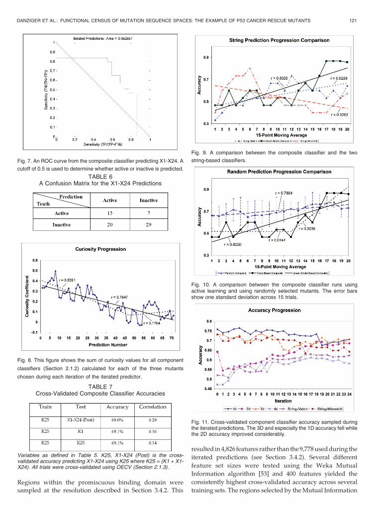
As demonstrated in Section 4.2, the composite classifieraccuracy improved considerably while performing theiterated predictions. When analyzed in terms of the cross-validated component classifier accuracy, two trends becameapparent (see Fig. 11). The 1D classifier fell slightly in cross-validated accuracy from approximately 75.8 percent to69.1 percent, while the 2D classifier rose in cross-validatedaccuracy from 64.2 percent to 72.2 percent.
5.1.1 Surface Evolution by Functional Region
DNA and almost all small molecules bind to p53 around apromiscuous binding domain [52] on the surface of aminoacids 94-160 and 264-315. The 2D surface was modified so thatregions not in the promiscuous binding region were sampledat a lower resolution: Each amino acid was reduced to onesurface position feature and one surface electrostatic feature.
120 IEEE/ACM TRANSACTIONS ON COMPUTATIONAL BIOLOGY AND BIOINFORMATICS, VOL. 3, NO. 2, APRIL-JUNE 2006
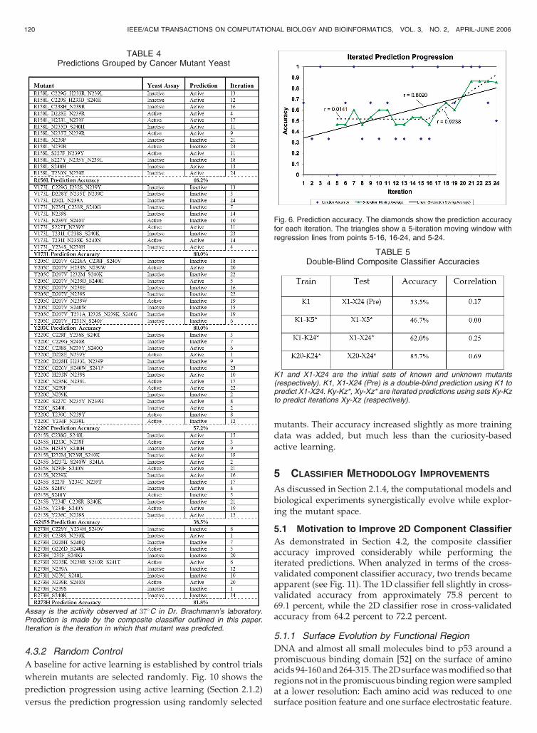
Fig. 6. Prediction accuracy. The diamonds show the prediction accuracyfor each iteration. The triangles show a 5-iteration moving window withregression lines from points 5-16, 16-24, and 5-24.
TABLE 5Double-Blind Composite Classifier Accuracies
K1 and X1-X24 are the initial sets of known and unknown mutants(respectively). K1, X1-X24 (Pre) is a double-blind prediction using K1 topredict X1-X24. Ky-Kz*, Xy-Xz* are iterated predictions using sets Ky-Kzto predict iterations Xy-Xz (respectively).
TABLE 4Predictions Grouped by Cancer Mutant Yeast
Assay is the activity observed at 37�C in Dr. Brachmann’s laboratory.Prediction is made by the composite classifier outlined in this paper.Iteration is the iteration in which that mutant was predicted.
Regions within the promiscuous binding domain were
sampled at the resolution described in Section 3.4.2. This
resulted in 4,826 features rather than the 9,778 used during the
iterated predictions (see Section 3.4.2). Several different
feature set sizes were tested using the Weka Mutual
Information algorithm [53] and 400 features yielded the
consistently highest cross-validated accuracy across several
training sets. The regions selected by the Mutual Information
DANZIGER ET AL.: FUNCTIONAL CENSUS OF MUTATION SEQUENCE SPACES: THE EXAMPLE OF P53 CANCER RESCUE MUTANTS 121
TABLE 6A Confusion Matrix for the X1-X24 Predictions
Fig. 8. This figure shows the sum of curiosity values for all component
classifiers (Section 2.1.2) calculated for each of the three mutants
chosen during each iteration of the iterated predictor.
TABLE 7Cross-Validated Composite Classifier Accuracies
Variables as defined in Table 5. K25, X1-X24 (Post) is the cross-validated accuracy predicting X1-X24 using K25 where K25 = (K1 + X1-X24). All trials were cross-validated using OECV (Section 2.1.3).
Fig. 9. A comparison between the composite classifier and the two
string-based classifiers.
Fig. 10. A comparison between the composite classifier runs usingactive learning and using randomly selected mutants. The error barsshow one standard deviation across 15 trials.
Fig. 11. Cross-validated component classifier accuracy sampled duringthe iterated predictions. The 3D and especially the 1D accuracy fell whilethe 2D accuracy improved considerably.
Fig. 7. An ROC curve from the composite classifier predicting X1-X24. A
cutoff of 0.5 is used to determine whether active or inactive is predicted.
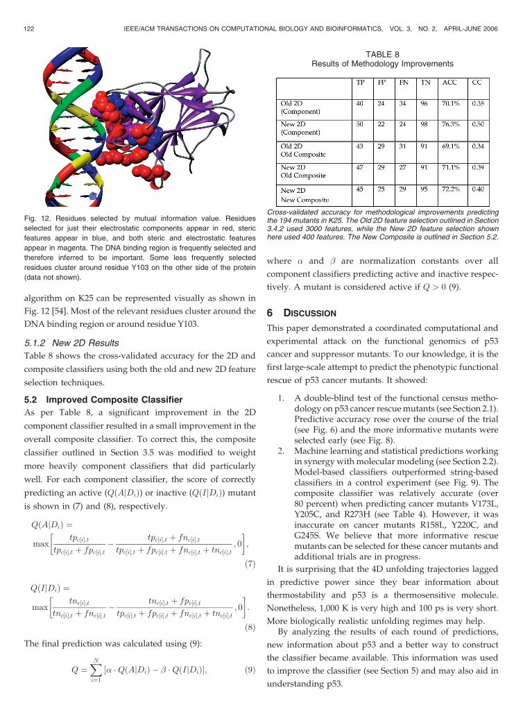
algorithm on K25 can be represented visually as shown in
Fig. 12 [54]. Most of the relevant residues cluster around the
DNA binding region or around residue Y103.
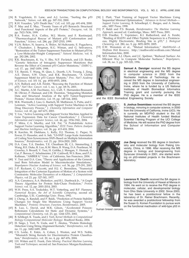
5.1.2 New 2D Results
Table 8 shows the cross-validated accuracy for the 2D and
composite classifiers using both the old and new 2D feature
selection techniques.
5.2 Improved Composite Classifier
As per Table 8, a significant improvement in the 2D
component classifier resulted in a small improvement in the
overall composite classifier. To correct this, the composite
classifier outlined in Section 3.5 was modified to weight
more heavily component classifiers that did particularly
well. For each component classifier, the score of correctly
predicting an active (QðAjDiÞ) or inactive (QðIjDiÞ) mutant
is shown in (7) and (8), respectively.
QðAjDiÞ ¼
maxtpc½i�;t
tpc½i�;t þ fpc½i�;t�
tpc½i�;t þ fnc½i�;ttpc½i�;t þ fpc½i�;t þ fnc½i�;t þ tnc½i�;t
; 0
� �;
ð7Þ
QðIjDiÞ ¼
maxtnc½i�;t
tnc½i�;t þ fnc½i�;t�
tnc½i�;t þ fpc½i�;ttpc½i�;t þ fpc½i�;t þ fnc½i�;t þ tnc½i�;t
; 0
� �:
ð8Þ
The final prediction was calculated using (9):
Q ¼XNi¼1
½� �QðAjDiÞ � � �QðIjDiÞ�; ð9Þ
where � and � are normalization constants over all
component classifiers predicting active and inactive respec-
tively. A mutant is considered active if Q > 0 (9).
6 DISCUSSION
This paper demonstrated a coordinated computational and
experimental attack on the functional genomics of p53
cancer and suppressor mutants. To our knowledge, it is the
first large-scale attempt to predict the phenotypic functional
rescue of p53 cancer mutants. It showed:
1. A double-blind test of the functional census metho-dology on p53 cancer rescue mutants (see Section 2.1).Predictive accuracy rose over the course of the trial(see Fig. 6) and the more informative mutants wereselected early (see Fig. 8).
2. Machine learning and statistical predictions workingin synergy with molecular modeling (see Section 2.2).Model-based classifiers outperformed string-basedclassifiers in a control experiment (see Fig. 9). Thecomposite classifier was relatively accurate (over80 percent) when predicting cancer mutants V173L,Y205C, and R273H (see Table 4). However, it wasinaccurate on cancer mutants R158L, Y220C, andG245S. We believe that more informative rescuemutants can be selected for these cancer mutants andadditional trials are in progress.
It is surprising that the 4D unfolding trajectories lagged
in predictive power since they bear information about
thermostability and p53 is a thermosensitive molecule.
Nonetheless, 1,000 K is very high and 100 ps is very short.
More biologically realistic unfolding regimes may help.By analyzing the results of each round of predictions,
new information about p53 and a better way to construct
the classifier became available. This information was used
to improve the classifier (see Section 5) and may also aid in
understanding p53.
122 IEEE/ACM TRANSACTIONS ON COMPUTATIONAL BIOLOGY AND BIOINFORMATICS, VOL. 3, NO. 2, APRIL-JUNE 2006
Fig. 12. Residues selected by mutual information value. Residues
selected for just their electrostatic components appear in red, steric
features appear in blue, and both steric and electrostatic features
appear in magenta. The DNA binding region is frequently selected and
therefore inferred to be important. Some less frequently selected
residues cluster around residue Y103 on the other side of the protein
(data not shown).
TABLE 8Results of Methodology Improvements
Cross-validated accuracy for methodological improvements predictingthe 194 mutants in K25. The Old 2D feature selection outlined in Section3.4.2 used 3000 features, while the New 2D feature selection shownhere used 400 features. The New Composite is outlined in Section 5.2.

6.1 Implications
The biological advance is a general method to catalogmutation sequence spaces across important proteins ofmedical interest, which may eventually extend to medicalknowledge of entire pathways and networks. The computa-tional advance is a method whereby robust statisticalmethods applied to noisy, biased, imperfect molecularmodels help experimentalists to pursue function in areaswhere, previously, the techniques were believed not to apply.
The broad goal is a comprehensive census of thefunctional rescue of p53 cancer mutants by second-sitesuppressor mutations. A functional census of suppressormutations for p53 cancer mutants will significantly furtherour knowledge of p53 rescue mechanisms. Knowledge of allregions of the p53 core domain that improve stability whenaltered will provide guidance in choosing possible dockingsites for small molecules.
The methodology generalizes to other mutational sys-tems where mutants can be classified as active or inactive.Computational classifiers that predict mutant function willallow experimentalists to map structure/function relation-ships for proteins in other mutation-related diseases.
6.2 Conclusion
Central to the goal of cancer treatment by p53 functionalrescue is better knowledge of p53 rescue mechanisms.Intragenic suppressor mutations pinpoint key regions of thep53 core domain that may be modified to increase stabilityof or restore binding domains in the p53 protein. This givesa validated point of control that restores native p53 functionand identifies the cancer mutants that are amenable tofunctional rescue and, thus, the most likely drug targets.Our long-term goal is to exploit these findings for thedesign of drug compounds that can restore p53 functionand preliminary small molecule studies are underway withour collaborators.
ACKNOWLEDGMENTS
The authors thank Richard Chamberlin, Melanie Cocco,Richard Colman, John Coroneus, Hartmut Luecke, DonSenear, and Ying Wang for contributions to the largercollaborative p53 functional rescue project. Thanks to theUniversity of California Irvine (UCI) office of Research andGraduate Studies and the UCI Institute for Genomics andBioinformatics for financial support. Work supported inpart by US National Institutes of Health (NIH) BiomedicalInformatics Training grant (LM-07443-01), US NationalScience Foundation (NSF) MRI grant (EIA-0321390) toPierre Baldi, NIH BISTI grant (CA-112560) and NSF ITRgrant (0326037) to Richard H. Lathrop, NIH grant(CA81511) to Rainer K. Brachmann, Harvey Fellowship toS. Joshua Swamidass, and the UCI Medical ScientistTraining Program.
REFERENCES
[1] D.L. Rubin, F. Shafa, D.E. Oliver, M. Hewett, and R.B. Altman,“Representing Genetic Sequence Data for Pharmacogenomics: AnEvolutionary Approach Using Ontological and Relational Mod-els,” Bioinformatics, vol. 18, supplement 1, pp. S207-215, 2002.
[2] R.H. Lathrop and M.J. Pazzani, “Combinatorial Optimization inRapidly Mutating Drug-Resistant Viruses,” J. Combinatorial Opti-mization, vol. 3, pp. 301-320, 1999.
[3] R.H. Lathrop, N.R. Steffen, M. Raphael, S. Deeds-Rubin, M.J.Pazzani, P.J. Cimoch, D.M. See, and J.G. Tilles, “Knowledge-BasedAvoidance of Drug-Resistant HIV Mutants,” AI Magazine, vol. 20,pp. 13-25, 1999.
[4] N. Beerenwinkel, T. Lengauer, J. Selbig, B. Schmidt, H. Walter, K.Korn, R. Kaiser, and D. Hoffman, “Geno2pheno: InterpretingGenotypic HIV Drug Resistance Tests,” IEEE Intelligent Systems,vol. 16, pp. 35-41, 2001.
[5] M.R. Bush, C.A. Bender, K.C. Subbarao, J. Nancy, and W.M. Fitch,“Predicting the Evolution of Human Influenza A,” Science, vol. 286,pp. 1921-1925, 1999.
[6] G.M. Wahl and A.M. Carr, “The Evolution of Diverse BiologicalResponses to DNA Damage: Insights from Yeast and p53,” NaturalCell Biology, vol. 3, pp. E277-286, 2001.
[7] Y. Xu, “Regulation of p53 Responses by Post-TranslationalModifications,” Cell Death Differentiation vol. 10, pp. 400-403, 2003.
[8] E. Appella and C.W. Anderson, “Post-Translational Modificationsand Activation of p53 by Genotoxic Stresses,” European J.Biochemistry, vol. 268, pp. 2764-2772, 2001.
[9] C.L Brooks and W. Gu, “Ubiquitination, Phosphorylation andAcetylation: The Molecular Basis for p53 Regulation,” CurrentOpinions in Cell Biology, vol. 15, pp. 164-171, 2003.
[10] W.S. el-Deiry, S.E. Kern, J.A. Pietenpol, K.W. Kinzler, and B.Vogelstein, “Definition of a Consensus Binding Site for p53,”Nature and Genetics, vol. 1, pp. 45-49, 1992.
[11] W.D. Funk, D.T. Pak, R.H. Karas, W.E. Wright, and J.W. Shay, “ATranscriptionally Active DNA-Binding Site for Human p53Protein Complexes,” Molecular Cell Biology, vol. 12, pp. 2866-2871, 1992.
[12] H. Qian, T. Wang, L. Naumovski, C.D. Lopez, and R.K.Brachmann, “Groups of p53 Target Genes Involved in Specificp53 Downstream Effects Cluster into Different Classes of DNABinding Sites,” Oncogene, vol. 21, pp. 7901-7911, 2002.
[13] K. Kannan, N. Amariglio, G. Rechavi, J. Jakob-Hirsch, I. Kela, N.Kaminski, G. Getz, E. Domany, and D. Givol, “DNA MicroarraysIdentification of Primary and Secondary Target Genes Regulatedby p53,” Oncogene, vol. 20, pp. 2225-2234, 2001.
[14] K. Polyak, Y. Xia, J.L. Zweier, K.W. Kinzler, and B. Vogelstein, “AModel for p53-Induced Apoptosis,” Nature, vol. 389, pp. 300-305,1997.
[15] C. Caelles, A. Helmberg, and M. Karin, “p53-Dependent Apop-tosis in the Absence of Transcriptional Activation of p53-TargetGenes,” Nature, vol. 370, pp. 220-223, 1994.
[16] J.J Manfredi, “p53 and Apoptosis: It’s Not Just in the NucleusAnymore,” Molecular Cell, vol. 11, pp. 552-554, 2003.
[17] M. Mihara, S. Erster, A. Zaika, O. Petrenko, T. Chittenden, P.Pancoska, and U.M. Moll, “p53 Has a Direct Apoptogenic Role atthe Mitochondria,” Molecular Cell, vol. 11, pp. 577-590, 2003.
[18] M. Olivier, R. Eeles, M. Hollstein, M.A. Khan, C.C. Harris, and P.Hainaut, “The IARC TP53 Database: New Online MutationAnalysis and Recommendations to Users,” Human Mutations,vol. 19, no. 6, pp. 607-14, June 2002.
[19] C. Beroud and T. Soussi, “The UMD-p53 Database: NewMutations and Analysis Tools,” Human Mutations, vol. 21,pp. 176-181, 2003.
[20] Y. Cho, S. Gorina, P.D. Jeffrey, and N.P. Pavletich, “CrystalStructure of a p53 Tumor Suppressor-DNA Complex: Under-standing Tumorigenic Mutations,” Science, vol. 265, p. 346, 1994.
[21] A.N. Bullock, J. Henckel, B.S. DeDecker, C.M. Johnson, P.V.Nikolova, M.R. Proctor, D.P. Lane, and A.R. Fersht, “Thermo-dynamic Stability of Wild-Type and Mutant p53 Core Domain,”Proc. Nat’l Academy of Science USA, vol. 94, pp. 14338-14342, 1997.
[22] A.N. Bullock, J. Henckel, and A.R. Fersht, “Quantitative Analysisof Residual Folding and DNA Binding in Mutant p53 CoreDomain: Definition of Mutant States for Rescue in CancerTherapy,” Oncogene, vol. 19, pp. 1245-1256, 2000.
[23] K.B. Wong, B.S. DeDecker, S.M. Freund, M.R. Proctor, M. Bycroft,and A.R. Fersht, “Hot-Spot Mutants of p53 Core Domain EvinceCharacteristic Local Structural Changes,” Proc. Nat’l Academy ofScience USA, vol. 96, pp. 8438-8442, 1999.
[24] A.J. Levine, “p53, the Cellular Gatekeeper for Growth andDivision,” Cell, vol. 88, pp. 323-331, 1997.
[25] C. Prives and P.A. Hall, “The p53 Pathway,” J. Pathology, vol. 187,pp. 112-126, 1999.
DANZIGER ET AL.: FUNCTIONAL CENSUS OF MUTATION SEQUENCE SPACES: THE EXAMPLE OF P53 CANCER RESCUE MUTANTS 123
[26] B. Vogelstein, D. Lane, and A.J. Levine, “Surfing the p53Network,” Nature, vol. 408, pp. 307-310, 2000.
[27] K.H. Vousden, “p53: Death Star,” Cell, vol. 103, pp. 691-694, 2000.[28] P. May and E. May, “Twenty Years of p53 Research: Structural
And Functional Aspects of the p53 Protein,” Oncogene, vol. 18,pp. 7621-7636, 1999.
[29] B.A. Foster, H.A. Coffey, M.J. Morin, and F. Rastinejad,“Pharmacological Rescue of Mutant p53 Conformation andFunction,” Science, vol. 286, pp. 2507-2510, 1999.
[30] V.J.N. Bykov, N. Issaeva, A. Shilov, M Hultcrantz, E. Pugacheva,P. Chumakov, J. Bergman, K.G. Wiman, and G. Selivanova,“Restoration of the Tumor Suppressor Function to Mutant p53 bya Low-Molecular-Weight Compound,” Natural Medicine, vol. 8,pp. 282-288, 2002.
[31] R.K. Brachmann, K. Yu, Y. Eby, N.P. Pavletich, and J.D. Boeke,“Genetic Selection of Intragenic Suppressor Mutations thatReverse the Effect of Common p53 Cancer Mutations,” EMBO J.,vol. 17, pp. 1847-1859, 1998.
[32] T.E. Baroni, T. Wang, H. Qian, L. Dearth, L.N. Troung, J. Zeng,A.E. Denes, S.W. Chen, and R.K. Brachmann, “A GlobalSuppressor Motif for p53 Cancer Mutants,” Proc. Nat’l Academyof Sciences, vol. 101 14, pp. 4930-4935, 2004.
[33] A.N. Bullock and A.R. Fersht, “Rescuing the Function of Mutantp53,” Nat’l Rev. Cancer, vol. 1, no. 1, pp. 68-76, 2001.
[34] A.C. Martin, A.M. Facchiano, A.L. Cuff, T. Hernandez-Boussard,M. Olivier, P. Hainaut, and J.M. Thornton, “Integrating MutationData and Structural Analysis of the tp53 Tumor-SuppressorProtein,” Human Mutation, vol. 19, pp. 149-164, 2002.
[35] M.K. Warmuth, J. Liao, G. Raetsch, M. Mathieson, S. Putta, and C.Lemmen, “Active Learning with Support Vector Machines in theDrug Discovery Process,” J. Chemistry Information and ComputerScience, vol. 43, pp. 667-673, 2003.
[36] Y. Liu, “Active Learning with Support Vector Machine Applied toGene Expression Data for Cancer Classification,” J. ChemistryInformation and Computer Science, vol. 44, pp. 1936-1941, 2004.
[37] P. Mitra, C.A. Murthy, and S.K. Pal, “A Probabilistic ActiveSupport Vector Learning Algorithm,” IEEE Trans. Pattern Analysisand Machine Intelligence, vol. 26, pp. 413-418, 2004.
[38] R. Karchin, M. Diekhans, L. Kelly, D.J. Thomas, U. Pieper, N.Eswar, D. Haussler, and A. Sali, “LS-SNP: Large-Scale Annotationof Coding Non-Synonymous SNPs Based on Multiple InformationSources,” Bioinformatics, vol. 21, pp. 2814-2820, 2005.
[39] D.A. Case, T.A. Darden, T.E. Cheatham III, C.L. Simmerling, J.Wang, R.E. Duke, R. Luo, K.M. Merz, B. Wang, D.A. Pearlman, M.Crowley, S. Brozell, V. Tsui, H. Gohlke, J. Mongan, V. Hornak, G.Cui, P. Beroza, C. Schafmeister, J.W. Caldwell, W.S. Ross, and P.A.Kollman, “AMBER 8,” Univ. of California, San Francisco, 2004.
[40] V. Tsui and D.A. Case, “Theory and Applications of the General-ized Born Solvation Model in Macromolecular Simulations,”Biopolymers (Nuclear Academy of Science, vol. 56, pp. 275-291, 2001.
[41] J.-P. Ryckaert, G. Ciccotti, and H.J. C. Berendsen, “NumericalIntegration of the Cartesian Equations of Motion of a System withConstraints: Molecular Dynamics of n-Alkanes,” J. ComputationalPhysics, vol 23, pp. 327-341, 1977.
[42] A.A. Canutescu, A.A. Shelenkov, and R.L. Dunbrack Jr., “A GraphTheory Algorithm for Protein Side-Chain Prediction,” ProteinScience, vol. 12, pp. 2001-2014, 2003.
[43] W.H. Press, S.A. Teukolsky, W.T. Vetterling, and B.P. Flannery,Numerical Recipes, the Art of Scientific Computing, second ed.Cambridge, U.K.: Cambridge Univ. Press, 1992.
[44] J. Cheng, A. Randall, and P. Baldi, “Prediction of Protein StabilityChanges for Single Site Mutations Using Support VectorMachines,” Proteins: Structure, Function, Bioinformatics, 2005.
[45] R. Luo, L. David, and M.K. Gilson, “Accelerated Poisson-Boltzmann Calculations for Static and Dynamic Systems,” J.Computational Chemistry, vol. 23, pp. 1244-1253, 2002.
[46] B. Schlkopf, K. Tsuda, and J. Vert, Kernel Methods in ComputationalBiology (Computational Molecular Biology). Bradford Books, 2004.
[47] H. Saigo, J. Vert, N. Ueda, and T. Akutsu, “Protein HomologyDetection Using String Alignment Kernels,” Bioinformatics, vol. 20,no. 11, pp. 1682-1689, 2004.
[48] C.S. Leslie, E. Eskin, A. Cohen, J. Weston, and W.S. Noble,“Mismatch String Kernels for Discriminative Protein Classifica-tion,” Bioinformatics, vol. 20, no. 4, pp. 467-476, 2004.
[49] I.H. Witten and E. Frank, Data Mining: Practical Machine LearningTools and Techniques, second ed. San Francisco: Morgan Kaufmann,2005.
[50] J. Platt, “Fast Training of Support Vector Machines UsingSequential Minimal Optimization,” Advances in Kernel Methods—-Support Vector Learning, B. Schoelkopf, C. Burges, and A. Smola,eds., MIT Press, 1998.
[51] P. Baldi and P.S. Brunak, Bioinformatics: The Machine LearningApproach, second ed. Cambridge, Mass.: MIT Press, 2001.
[52] D.B. Friedler, T. Veprintsev, K.I. Rutherford, and A. Fersht,“Binding of RAD51 and Other Peptide Sequences to a Promiscu-ous, Highly Electrostatic, Binding Site in p53,” J. BiologicalChemistry, vol. 280, no. 9, pp. 8051-8059, 2005.
[53] E.W. Weisstein et al., “Mutual Information,” MathWorld—AWolfram Web Resource, http://mathworld.wolfram.com/MutualInformation.htm, 2000.
[54] M.F. Sanner, A.J. Olson, and J.C. Spehner, “Reduced Surface: AnEfficient Way to Compute Molecular Surfaces,” Biopolymers,vol. 38, no. 3, pp. 305-320, 1996.
Samuel A. Danziger received the BS degreein electrical engineering and the MS degreein computer science in 2002 from theRochester Institute of Technology. He re-ceived the MS degree in biomedical engineer-ing in 2003 from the University of California,Irvine, where he is funded by a US NationalInstitutes of Health Biomedical InformaticsTraining grant and currently pursuing thePhD degree. He is a member of the IEEE
and the IEEE Biomedical Engineering Society.
S. Joshua Swamidass received the BS degreein biology, minoring in computer science, in 2000from the University of California, Irvine (UCI). Heis currently an MD/PhD student in the USNational Institutes of Health funded MedicalScientist Training Program of the UCI Collegeof Medicine. He will receive the PhD degree fromthe School of Information and ComputerScience.
Jue Zeng received the BS degree in biochem-istry and molecular biology from Peking Uni-versity, China, in 1996. After receiving the MSdegree in biology and bioengineering fromSyracuse University in 2001, she started work-ing on p53-related projects in the Brachmannlaboratory.
Lawrence R. Dearth received the BA degree inbiology from the University of Hawaii at Manoa in1994. He went on to receive the PhD degree inmolecular, cellular, and developmental biologyfrom Ohio State University in 2002. Since 2003,he has been a postdoctoral fellow in thelaboratory of Dr. Rainer Brachmann. Recently,he was awarded a postdoctoral fellowship fromthe Susan G. Komen Foundation to pursue workon the functional inactivation of wild-type p53 in
human breast cancers.
124 IEEE/ACM TRANSACTIONS ON COMPUTATIONAL BIOLOGY AND BIOINFORMATICS, VOL. 3, NO. 2, APRIL-JUNE 2006

Qiang Lu received the BS degree in physics in1994 from Northeast University, the MS degreein physics from the Dalian Institute of Technol-ogy, and the PhD degree in theoretical physicsin 2000 from Nankai University. He was alecturer in the Department of Physics at NankaiUniversity from 2000 to 2002. He continued hisresearch training in biophysics at the Universityof California, Irvine, and the State University ofNew York, Stony Brook. He is a member of the
American Chemical Society.
Jonathan H. Chen received the BS degree incybernetics with a specialization in computerstudies in 2000 from the University of California,Los Angeles. He is currently an MD/PhD studentat the University of California, Irvine, pursuing ajoint degree with the department of Informationand Computer Science, supported by the USNational Institutes of Health Medical ScientistTraining Program. Outside of academia, he hasbeen employed as a software developer at
Trilogy Software in Austin, Texas, and the Information TechnologyDepartment at 20th Century Fox. His current research interests focus onlarge-scale chemical informatics to aid tasks ranging from drugdiscovery to broad explorations of chemical space.
Jianlin Cheng received the BS degree incomputer science from the Huazhong Universityof Science and Technology in China in 1994. Hereceived the MS degree in computer sciencefrom Utah State University in 2001. He iscurrently a PhD candidate in computer scienceat the Institute for Genomics and Bioinformaticsin the School of Information and ComputerSciences of the University of California, Irvine.His research interests include algorithms and
applications for bioinformatics, systems biology, and machine learning.
Vinh P. Hoang received the BS degree incomputer science from the Donald Bren Schoolof Computer Science from the University ofCalifornia, Irvine in 2004. He is currently workingas a software developer at Raytheon andpursuing the MS degree at the University ofCalifornia, Irvine, in the field of bioinformatics.
Hiroto Saigo received the BS degree inelectrical and electronics engineering from So-phia University in 2001 and the master’s degreein informatics from Kyoto University in 2003. Heis currently a PhD candidate in the Departmentof Intelligence Science and Technology inGraduate School of Informatics at Kyoto Uni-versity. His research interests are in algorithmsand applications in the bioinformatics field,especially in machine learning methods.
Ray Luo received the BS degree in biophysicsin 1990 from Beijing University and the PhDdegree in chemistry in 1998 from University ofMaryland, College Park. He continued hisresearch training at the University of California,San Francisco, as a postdoctoral fellow beforestarting his own research group at the Universityof California, Irvine, in 2001. His research issupported in part by the US National Institutes ofHealth (R01 GM069620) and CRCC (35140). He
is a member of the American Chemical Society and Biophysical Society.
Pierre Baldi received the PhD degree inmathematics from the California Institute ofTechnology (Caltech) in 1986. He has heldpostdoctoral, faculty, and member of the techni-cal staff positions at the University of California,San Diego, and Caltech, in the Division ofBiology and the Jet Propulsion Laboratory. Hewas CEO of a startup company for a few yearsand joined the University of California, Irvine(UCI) in 1999. He is now a professor in the
School of Information and Computer Sciences with a joint appointmentin the Department of Biological Chemistry and the director of the UCIInstitute for Genomics and Bioinformatics. He received the Lew AllenAward at JPL in 1993 and the Laurel Wilkening Faculty InnovationAward at UCI. He has published four books and more than 100 scientificarticles. His research focuses on the application of AI and machinelearning methods to problems in the life sciences. His main contributionshave been in the area of statistical machine learning and bioinformatics,including the development of Hidden Markov Models (HMMPro) forsequence analysis, recursive neural networks for de novo proteinstructure prediction (SCRATCH), Bayesian statistical methods for DNAmicroarray analysis (Cyber-T), and, more recently, kernel methods inchemical informatics. The work of his group has resulted in severaldatabases, software, and Web servers that are widely used (www.igb.uci.edu/servers/servers.html).
Rainer K. Brachmann received the MD degreefrom the Ludwig-Maximilians-University in Mu-nich, Germany, and is a medical oncologist. Hislaboratory studies the pathway of the tumorsuppressor protein p53 with particular emphasison the novel therapeutic strategy of restoringfunction to p53 cancer mutants. He is a memberof the AACR.
Richard H. Lathrop received the BA degree inmathematics from Reed College (1978) and themaster’s degree in computer science (1983), thegraduate degree of electrical engineer (1983),and the PhD degree in artificial intelligence(1990) from the Massachusetts Institute ofTechnology. He is a professor in the BrenSchool of Information and Computer Sciencesat the University of California, Irvine. He is on theeditorial boards of Molecular and Cellular Pro-
teomics (2001-present) and IEEE Intelligent Systems (2002-present).He received Best Paper Awards from the International Conference onGenome Informatics (2001) and the ACM/IEEE International DesignAutomation Conference (1987) and an Innovative Application Awardfrom the AAAI/IAAI Conference (1998). He has more than 65 scientificand technical publications, and his research has appeared on the coverof AI Magazine (1999), the Journal of Molecular Biology (1996), andCommunications of the ACM (1987). He was a cofounding scientist ofArris Pharmaceutical Corp. and of CODA Genomics, Inc., served on theScientific Advisory Boards of CombiChem, Inc., and GeneFormatics,Inc., and was a founding officer (treasurer) and member of the foundingBoard of Directors of the International Society for Computational Biology(1996). His research interests are in artificial intelligence and computa-tional biology. He is a member of the IEEE Computer Society, AAAI (LifeMember), ACM, and ISCB.
DANZIGER ET AL.: FUNCTIONAL CENSUS OF MUTATION SEQUENCE SPACES: THE EXAMPLE OF P53 CANCER RESCUE MUTANTS 125