Embed Size (px)
Citation preview
1The Human Body: An Orientation
P A R T A
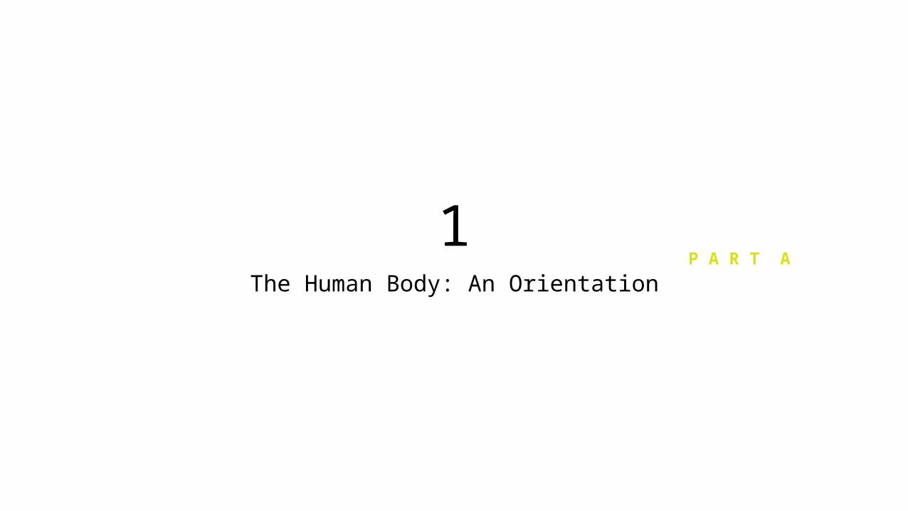
Overview of Anatomy and Physiology
• Anatomy – (Greek – to cut up) - the study of the structure of body parts and their relationships to one another
• Gross or macroscopic• Microscopic• Developmental
• Physiology – (Greek – study of nature) - the study of the function of the body’s structural machinery; explains physical & chemical processes that direct body activities
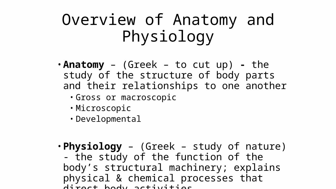
Gross Anatomy
• Regional – all structures in one part of the body (such as the abdomen or leg)
• Systemic – gross anatomy of the body studied by system• Surface – study of internal structures as they relate to the overlying
skin
Microscopic Anatomy
• Cytology – study of the cell• Histology – study of tissues
Developmental Anatomy• Traces structural changes throughout life• Embryology – study of developmental changes of the body before
birth
Specialized Branches of Anatomy
• Pathological anatomy – study of structural changes caused by disease• Radiographic anatomy – study of internal structures visualized by
specialized scanning procedures such as X-ray, MRI, and CT scans• Molecular biology – study of anatomical structures at a subcellular
level
Physiology
• Considers the operation of specific organ systems• Renal – kidney function• Neurophysiology – workings of the nervous system• Cardiovascular – operation of the heart and blood vessels
• Focuses on the functions of the body, often at the cellular or molecular level
• Understanding physiology also requires a knowledge of physics, which explains
• electrical currents• blood pressure• the way muscle uses bone for movement
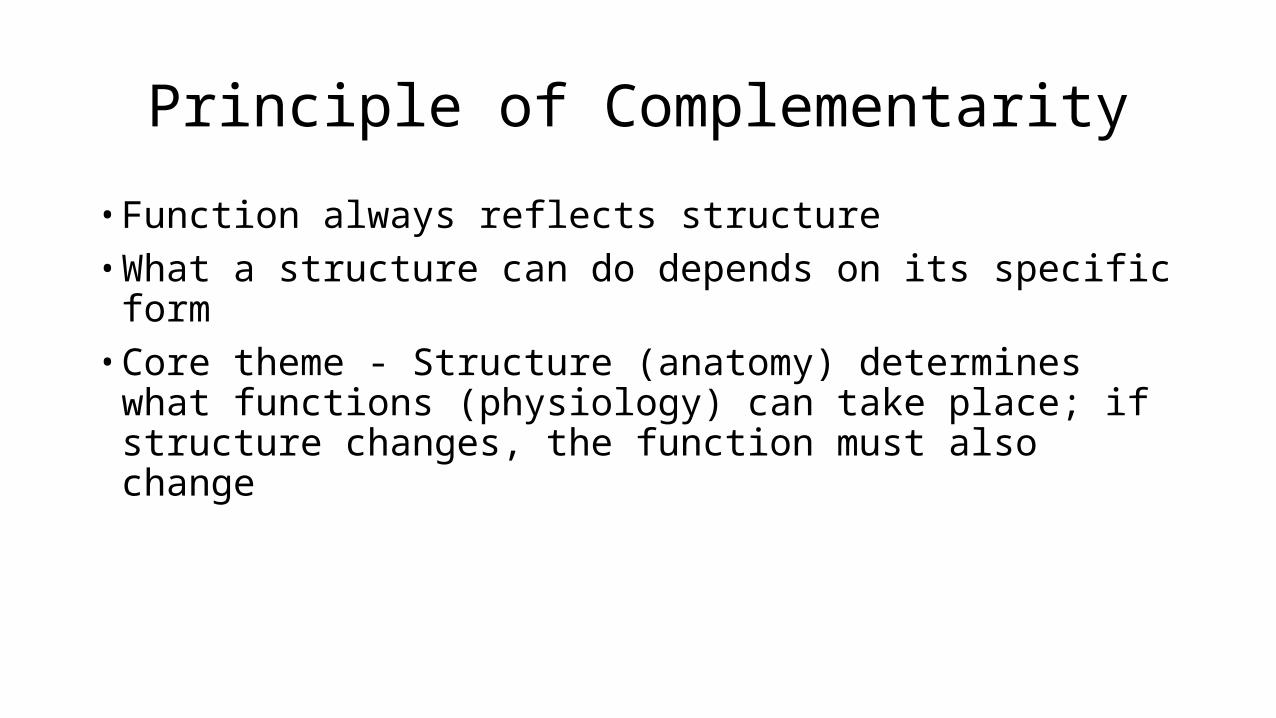
Principle of Complementarity
• Function always reflects structure• What a structure can do depends on its specific form• Core theme - Structure (anatomy) determines what functions
(physiology) can take place; if structure changes, the function must also change
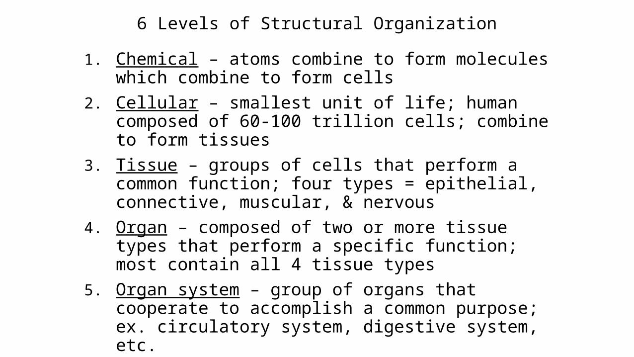
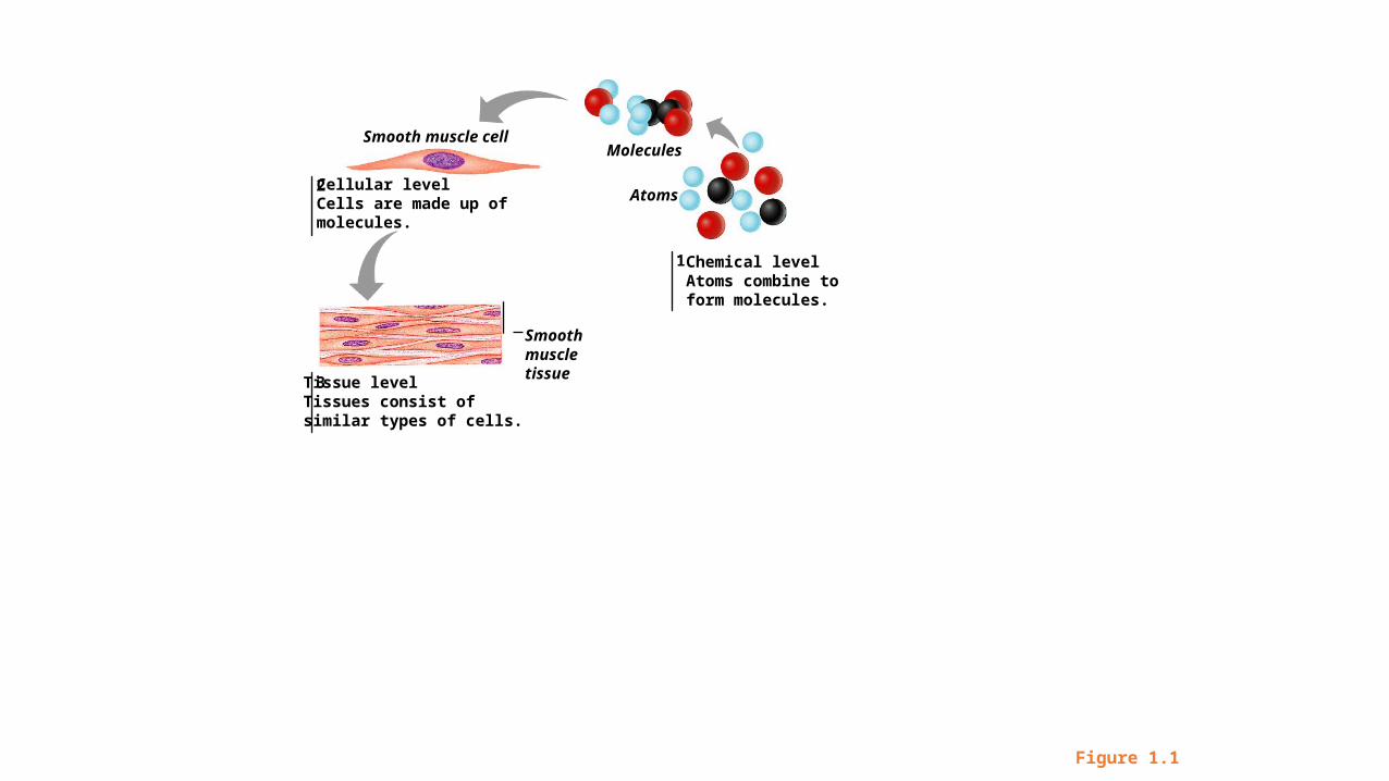
6 Levels of Structural Organization
1. Chemical – atoms combine to form molecules which combine to form cells
2. Cellular – smallest unit of life; human composed of 60-100 trillion cells; combine to form tissues
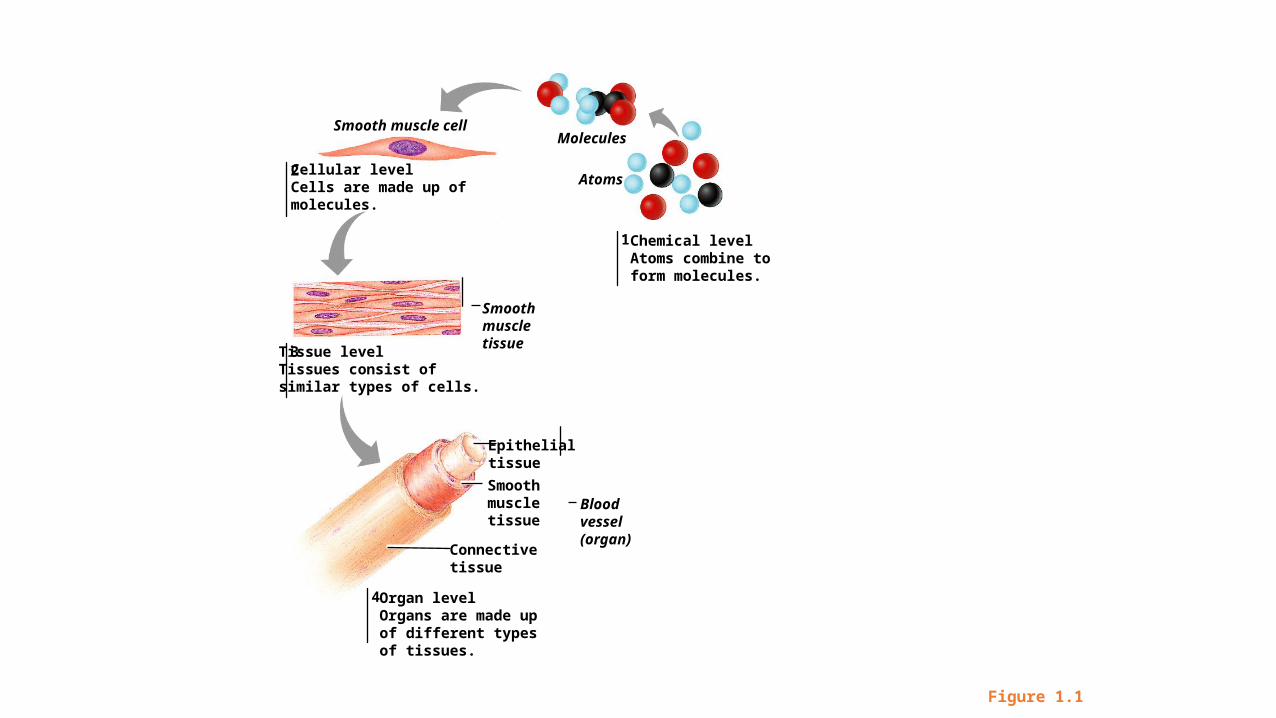
3. Tissue – groups of cells that perform a common function; four types = epithelial, connective, muscular, & nervous
4. Organ – composed of two or more tissue types that perform a specific function; most contain all 4 tissue types
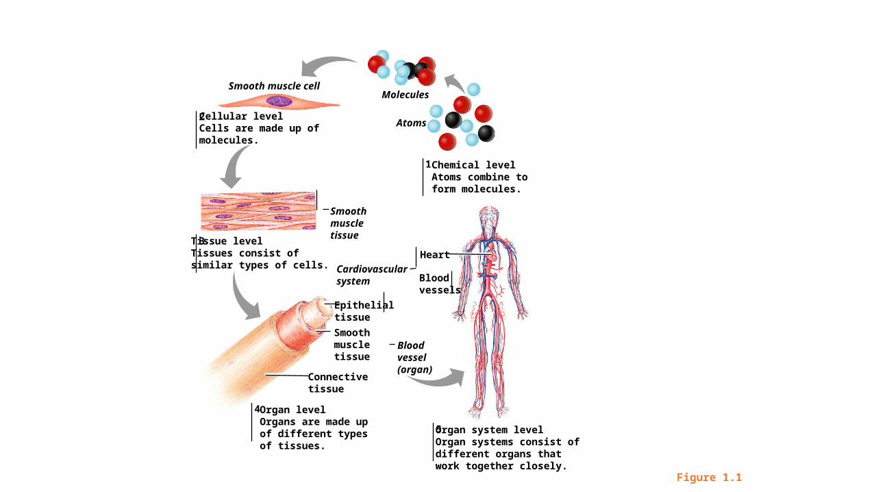
5. Organ system – group of organs that cooperate to accomplish a common purpose; ex. circulatory system, digestive system, etc.
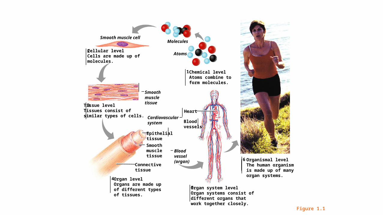
6. Organismal – highest level of structural organization; all systems of body working together
1
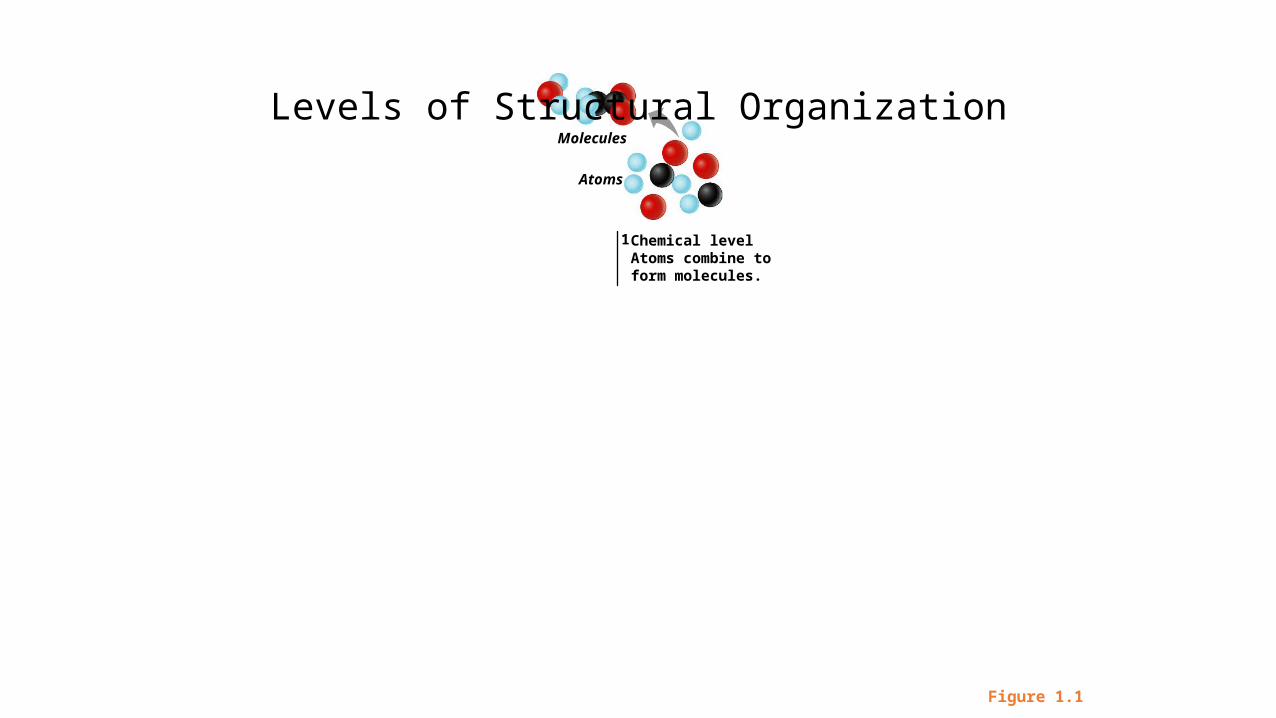
Molecules
Atoms
Chemical levelAtoms combine toform molecules.
Levels of Structural Organization
Figure 1.1
1
2
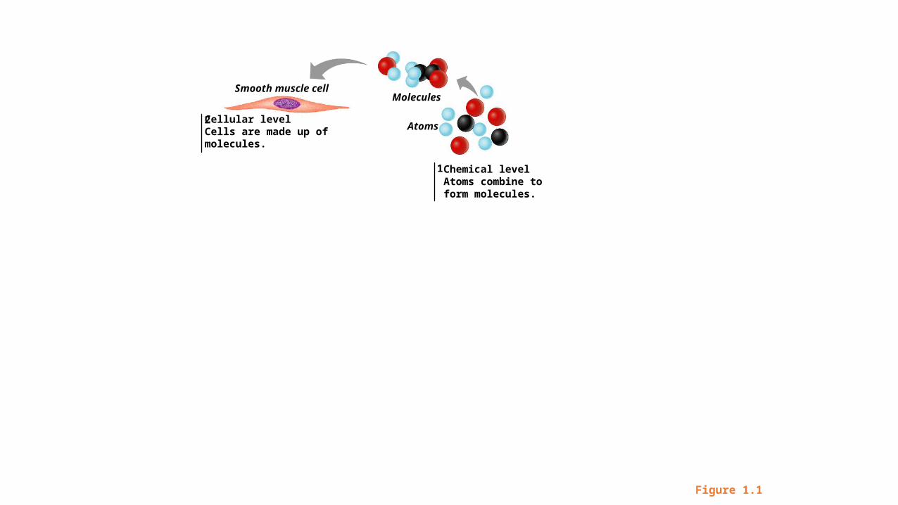
Smooth muscle cellMolecules
AtomsCellular levelCells are made up ofmolecules.
Chemical levelAtoms combine toform molecules.
Figure 1.1
1
2
3
Smooth muscle cellMolecules
Atoms
Smoothmuscletissue
Cellular levelCells are made up ofmolecules.
Tissue levelTissues consist ofsimilar types of cells.
Chemical levelAtoms combine toform molecules.
Figure 1.1
1
2
4
3
Smooth muscle cellMolecules
Atoms
Smoothmuscletissue
Epithelialtissue
Smoothmuscletissue
Connectivetissue
Bloodvessel(organ)
Cellular levelCells are made up ofmolecules.
Tissue levelTissues consist ofsimilar types of cells.
Organ levelOrgans are made upof different typesof tissues.
Chemical levelAtoms combine toform molecules.
Figure 1.1
1
2
4
5
3
Smooth muscle cellMolecules
Atoms
Smoothmuscletissue
Epithelialtissue
Heart
Bloodvessels
Smoothmuscletissue
Connectivetissue
Bloodvessel(organ)
Cardiovascularsystem
Cellular levelCells are made up ofmolecules.
Tissue levelTissues consist ofsimilar types of cells.
Organ levelOrgans are made upof different typesof tissues.
Organ system levelOrgan systems consist ofdifferent organs thatwork together closely.
Chemical levelAtoms combine toform molecules.
Figure 1.1
1
2
4
5
6
3
Smooth muscle cellMolecules
Atoms
Smoothmuscletissue
Epithelialtissue
Heart
Bloodvessels
Smoothmuscletissue
Connectivetissue
Bloodvessel(organ)
Cardiovascularsystem
Cellular levelCells are made up ofmolecules.
Tissue levelTissues consist ofsimilar types of cells.
Organ levelOrgans are made upof different typesof tissues.
Organ system levelOrgan systems consist ofdifferent organs thatwork together closely.
Organismal levelThe human organismis made up of manyorgan systems.
Chemical levelAtoms combine toform molecules.
Figure 1.1
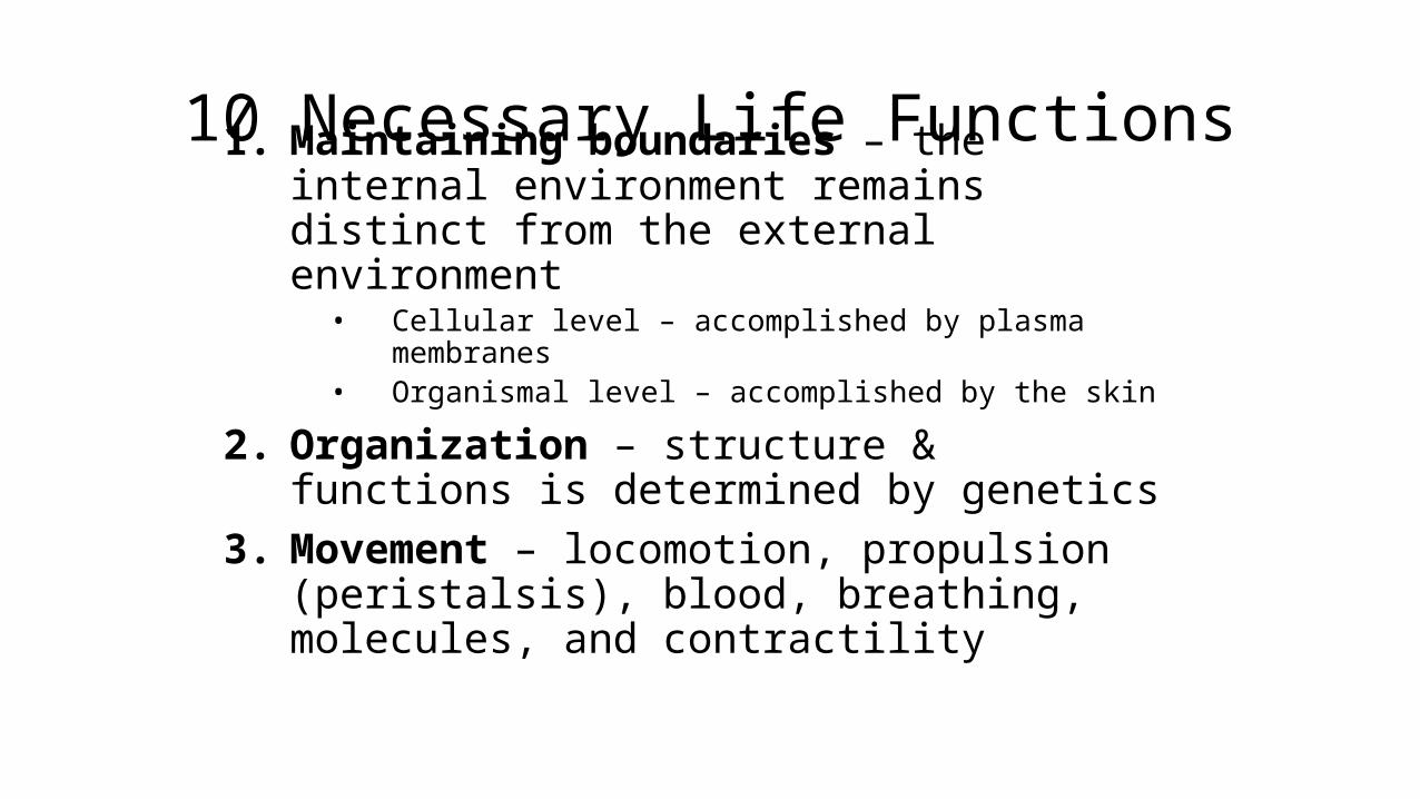
10 Necessary Life Functions1. Maintaining boundaries – the internal environment remains distinct from the external environment
• Cellular level – accomplished by plasma membranes• Organismal level – accomplished by the skin
2. Organization – structure & functions is determined by genetics
3. Movement – locomotion, propulsion (peristalsis), blood, breathing, molecules, and contractility
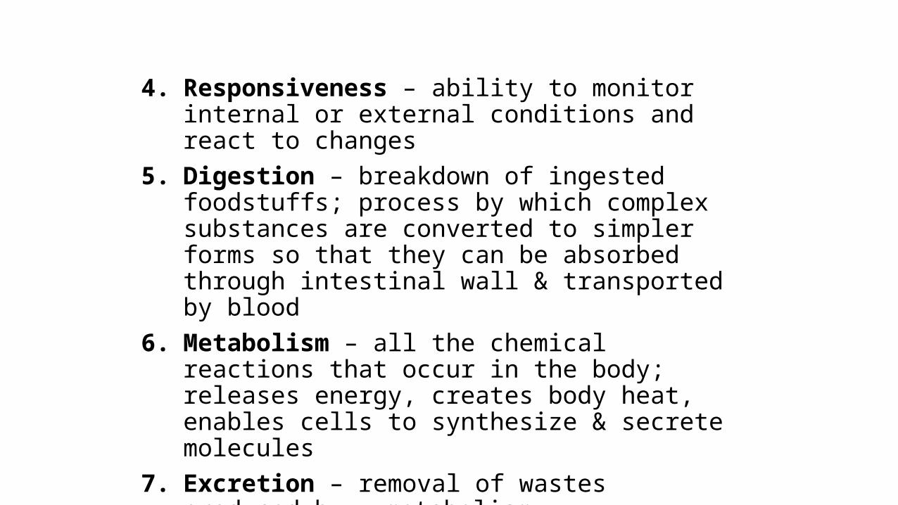
4. Responsiveness – ability to monitor internal or external conditions and react to changes
5. Digestion – breakdown of ingested foodstuffs; process by which complex substances are converted to simpler forms so that they can be absorbed through intestinal wall & transported by blood
6. Metabolism – all the chemical reactions that occur in the body; releases energy, creates body heat, enables cells to synthesize & secrete molecules
7. Excretion – removal of wastes produced by metabolism

8. Reproduction – cellular and organismal levels• Cellular – an original cell divides and produces two identical
daughter cells• Organismal – sperm and egg unite to make a whole new person
9. Growth – increase in size of a body part or of the organism
10. Differentiation – result in structurally and functionally distinct organs
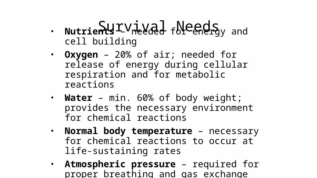
Survival Needs• Nutrients – needed for energy and cell building• Oxygen – 20% of air; needed for release of energy
during cellular respiration and for metabolic reactions
• Water – min. 60% of body weight; provides the necessary environment for chemical reactions
• Normal body temperature – necessary for chemical reactions to occur at life-sustaining rates
• Atmospheric pressure – required for proper breathing and gas exchange in the lungs
Homeostasis
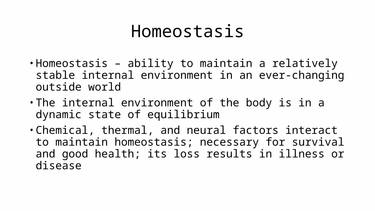
• Homeostasis – ability to maintain a relatively stable internal environment in an ever-changing outside world
• The internal environment of the body is in a dynamic state of equilibrium
• Chemical, thermal, and neural factors interact to maintain homeostasis; necessary for survival and good health; its loss results in illness or disease
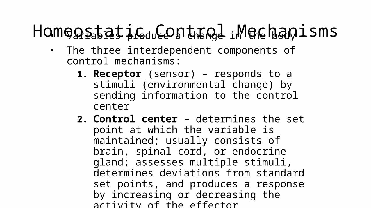
Homeostatic Control Mechanisms• Variables produce a change in the body• The three interdependent components of control
mechanisms:1. Receptor (sensor) – responds to a stimuli
(environmental change) by sending information to the control center
2. Control center – determines the set point at which the variable is maintained; usually consists of brain, spinal cord, or endocrine gland; assesses multiple stimuli, determines deviations from standard set points, and produces a response by increasing or decreasing the activity of the effector
3. Effector – muscles or glands; provides the means to respond to stimuli
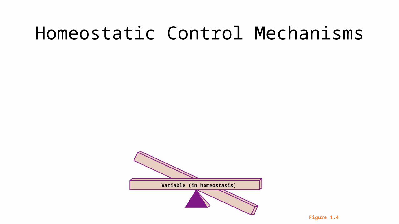
Homeostatic Control Mechanisms
Figure 1.4
Variable (in homeostasis)
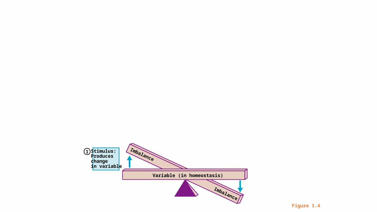
Figure 1.4
Stimulus: Produces changein variable
Variable (in homeostasis)
Imbalance
Imbalance
1
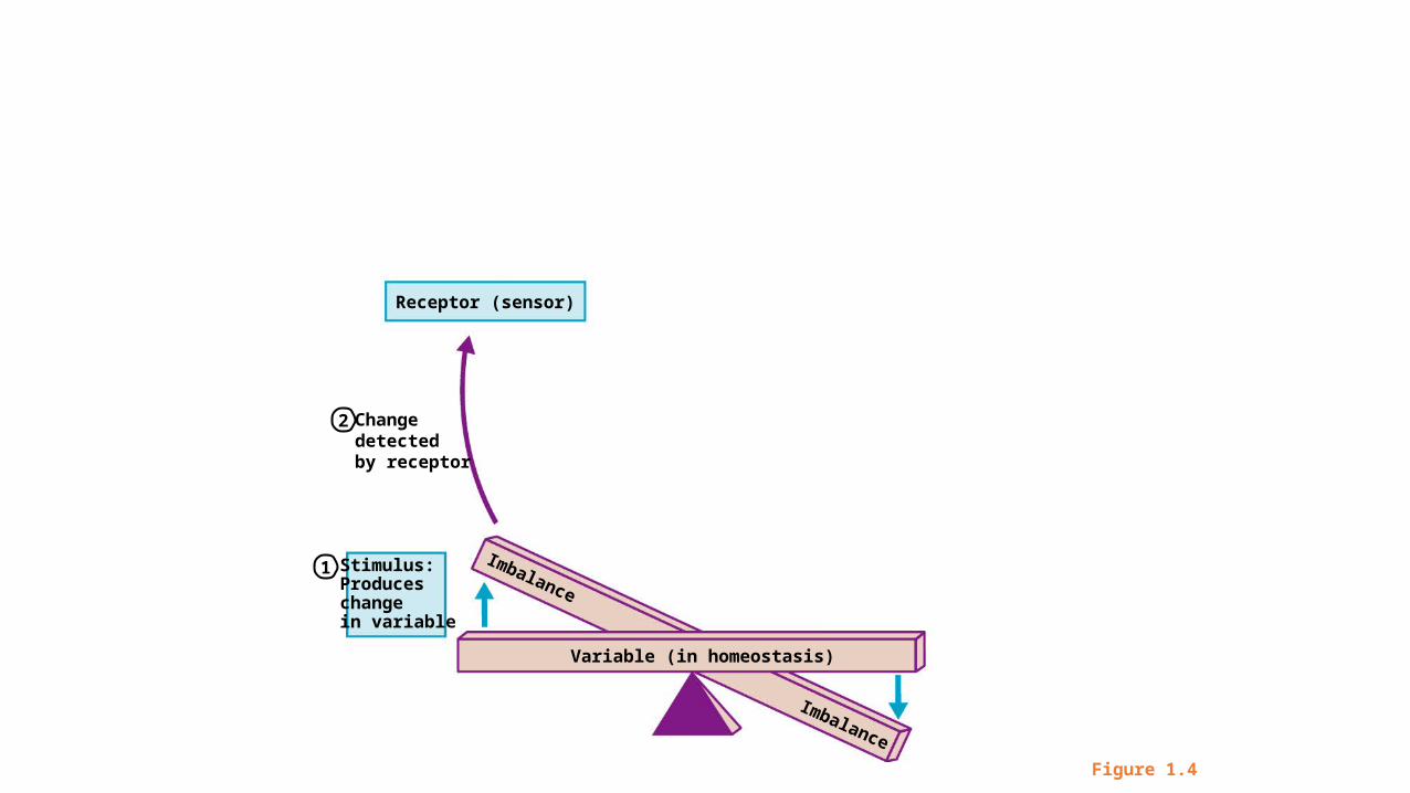
Figure 1.4
Change detected by receptor
Stimulus: Produces changein variable
Receptor (sensor)
Variable (in homeostasis)
Imbalance
Imbalance
2
1
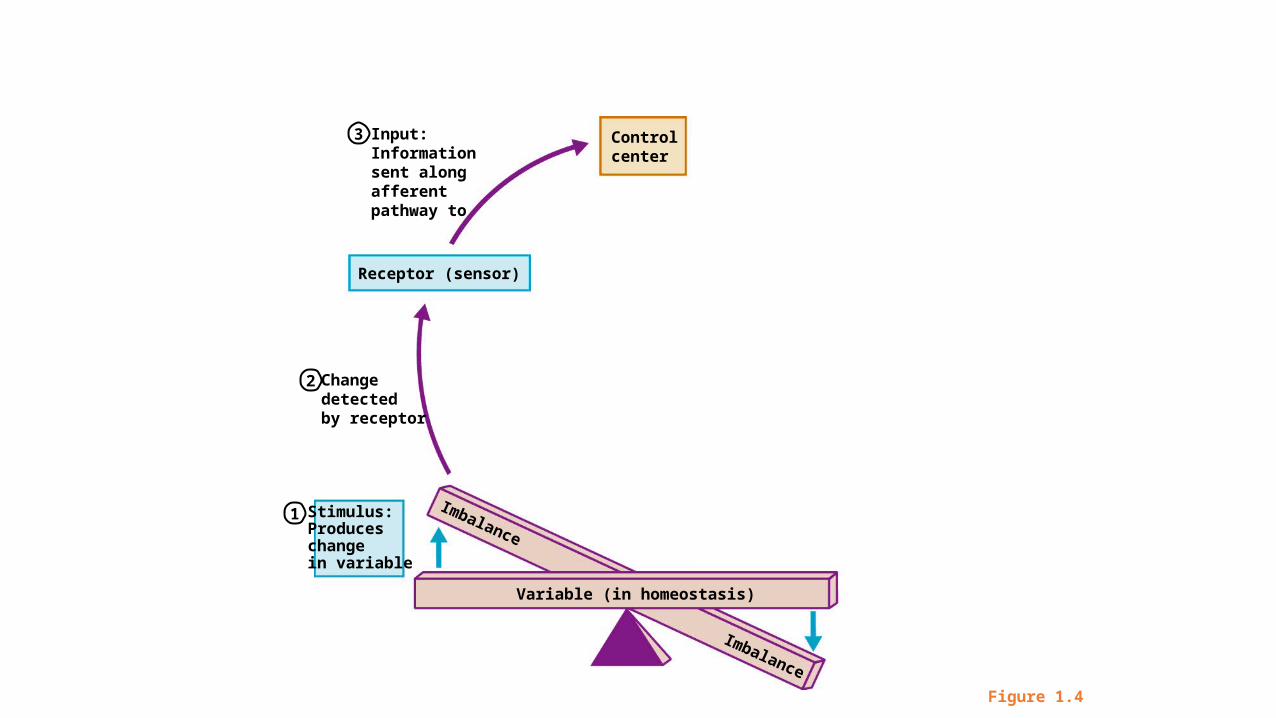
Figure 1.4
Change detected by receptor
Stimulus: Produces changein variable
Input:Informationsent along afferentpathway to
Receptor (sensor)
Controlcenter
Variable (in homeostasis)
Imbalance
Imbalance
2
3
1
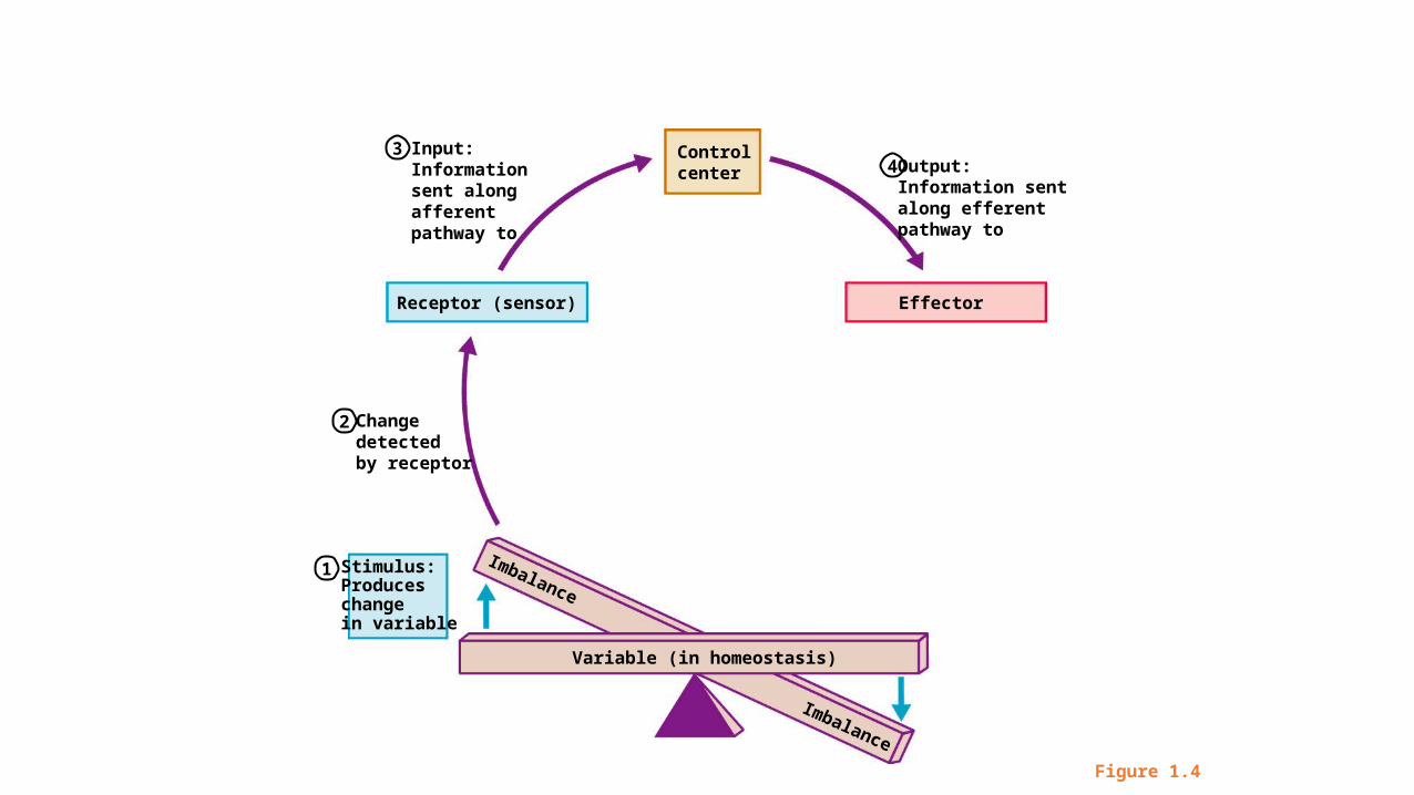
Figure 1.4
Change detected by receptor
Stimulus: Produces changein variable
Input:Informationsent along afferentpathway to
Receptor (sensor) Effector
Controlcenter
Variable (in homeostasis)
Output:Information sentalong efferentpathway to
Imbalance
Imbalance
2
34
1
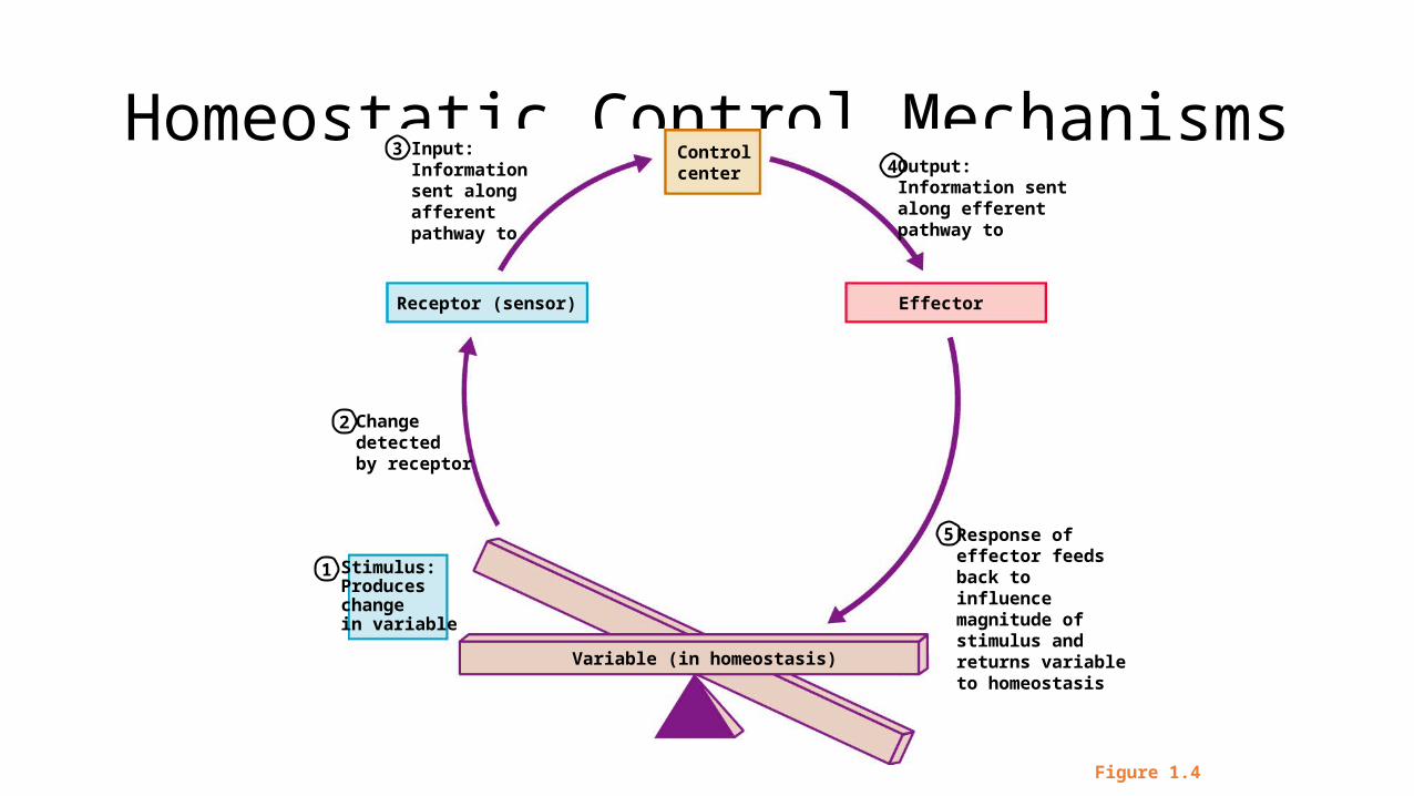
Homeostatic Control Mechanisms
Figure 1.4
Change detected by receptor
Stimulus: Produces changein variable
Input:Informationsent along afferentpathway to
Receptor (sensor) Effector
Controlcenter
Variable (in homeostasis)
Response ofeffector feedsback toinfluencemagnitude ofstimulus andreturns variableto homeostasis
Output:Information sentalong efferentpathway to
2
34
5
1
Negative Feedback
• In negative feedback systems, the output shuts off the original stimulus
• Most control systems involve negative feedback systems which act to reduce or stop the initial stimulus; ex. maintenance of body temperature
- antagonistic actions – opposite actions; allow for fine tuning of homeostatic conditions
• Example: Regulation of room temperature
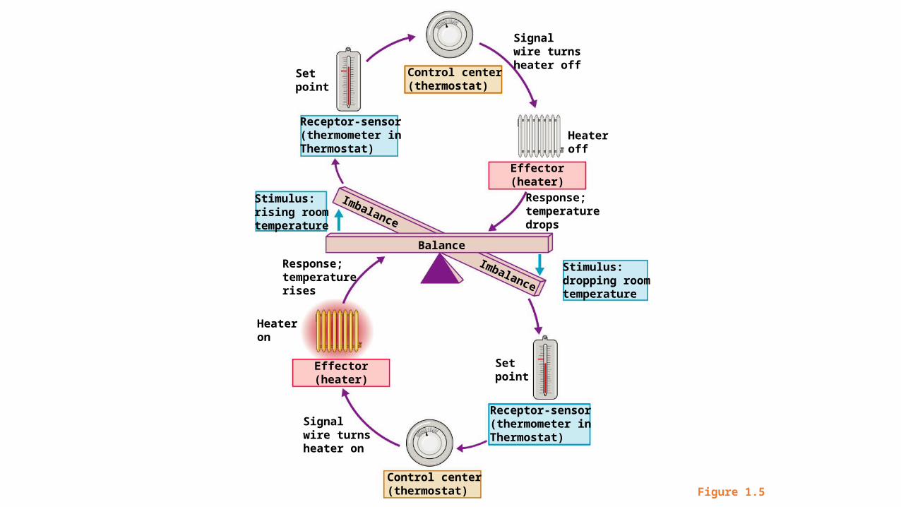
Figure 1.5
Signalwire turnsheater on
Signalwire turnsheater off
Response;temperaturerises
Response;temperaturedrops
Stimulus: rising roomtemperature
Stimulus: dropping roomtemperature
Balance
Effector(heater)
Effector(heater)
Setpoint
Control center(thermostat)
Heateroff
Setpoint
Receptor-sensor(thermometer inThermostat)
Control center(thermostat)
Heateron
Imbalance
Imbalance
Receptor-sensor(thermometer inThermostat)
Figure 1.5
Balance
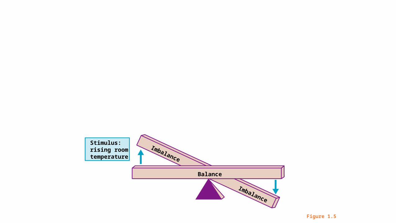
Figure 1.5
Stimulus: rising roomtemperature
Balance
Imbalance
Imbalance
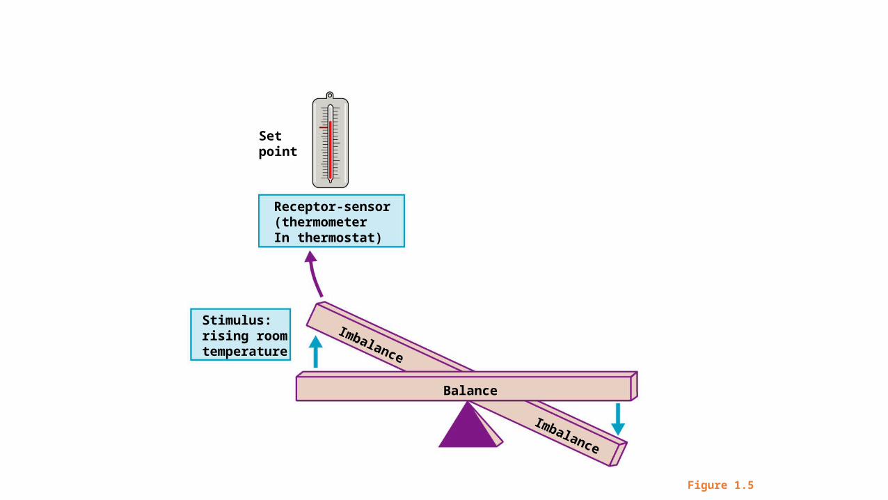
Figure 1.5
Stimulus: rising roomtemperature
Balance
Imbalance
Imbalance
Receptor-sensor(thermometerIn thermostat)
Setpoint
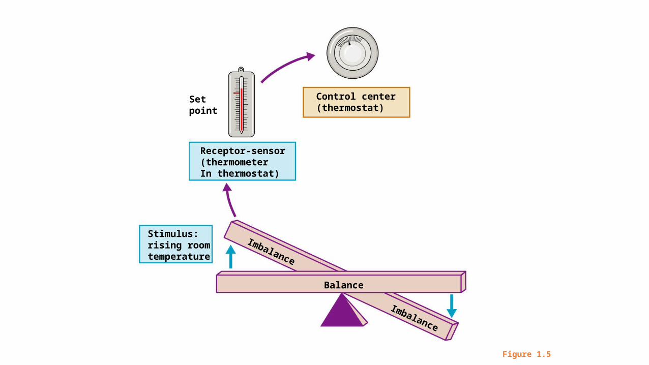
Figure 1.5
Stimulus: rising roomtemperature
Balance
Imbalance
Imbalance
Receptor-sensor(thermometerIn thermostat)
Setpoint
Control center(thermostat)
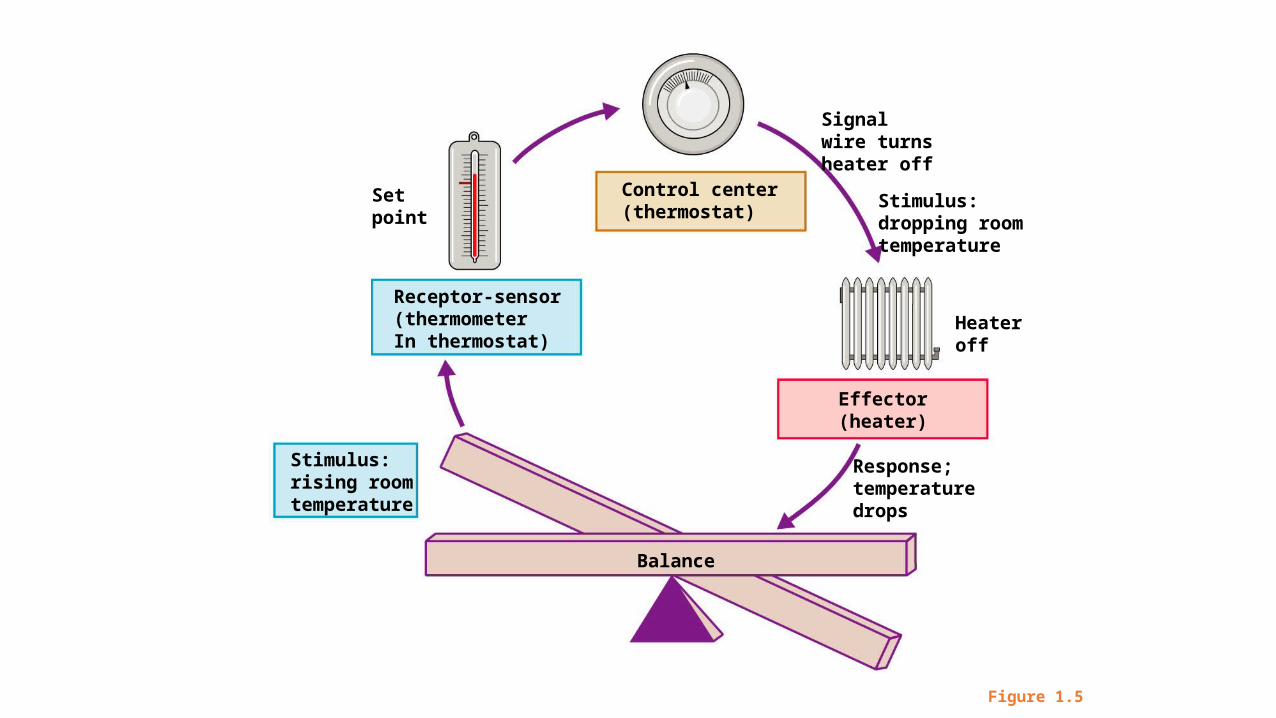
Figure 1.5
Signalwire turnsheater off
Response;temperaturedrops
Stimulus: rising roomtemperature
Balance
Effector(heater)
Stimulus: dropping roomtemperature
Receptor-sensor(thermometerIn thermostat)
Setpoint
Control center(thermostat)
Heateroff
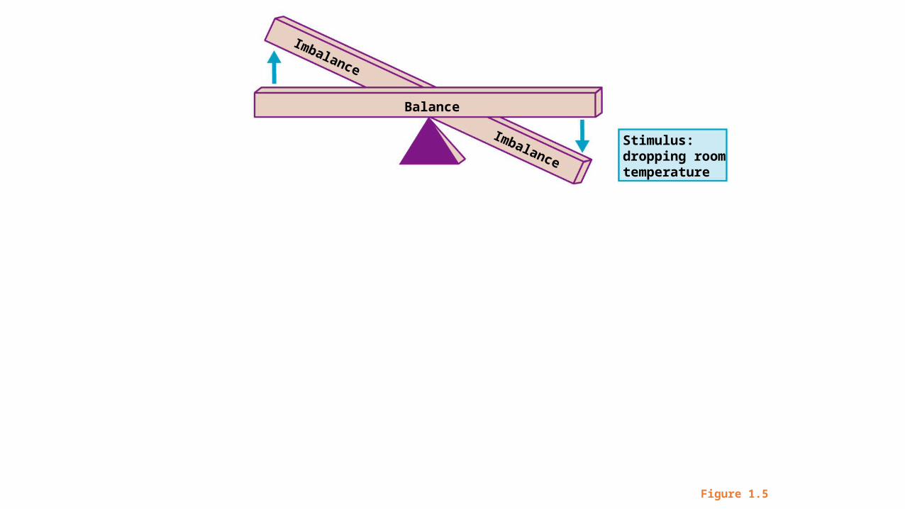
Figure 1.5
Stimulus: dropping roomtemperature
Balance
Imbalance
Imbalance
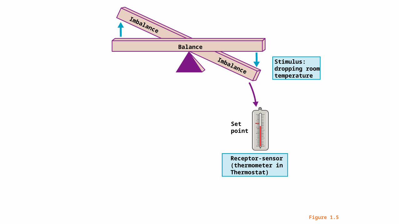
Figure 1.5
Stimulus: dropping roomtemperature
Balance
Setpoint
Receptor-sensor(thermometer inThermostat)
Imbalance
Imbalance
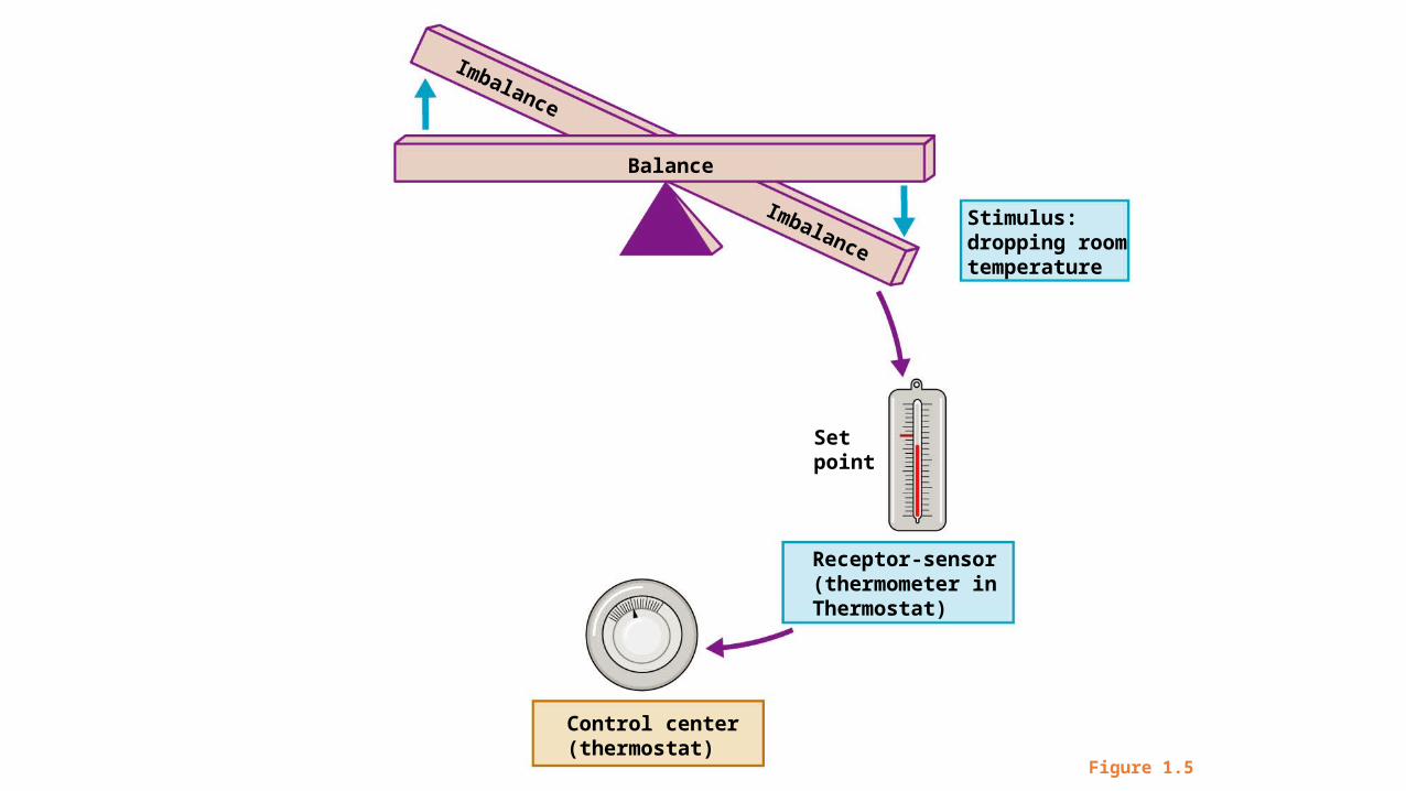
Figure 1.5
Stimulus: dropping roomtemperature
Balance
Setpoint
Receptor-sensor(thermometer inThermostat)
Control center(thermostat)
Imbalance
Imbalance
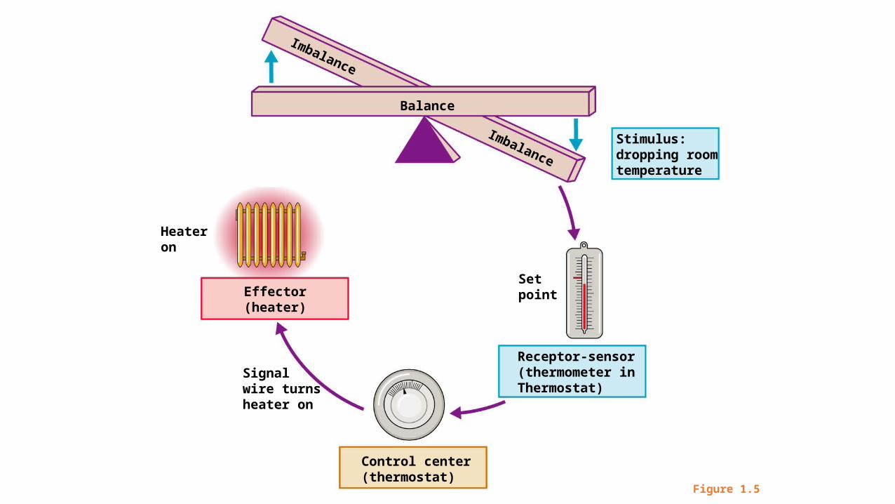
Figure 1.5
Signalwire turnsheater on
Stimulus: dropping roomtemperature
Balance
Effector(heater)
Setpoint
Receptor-sensor(thermometer inThermostat)
Control center(thermostat)
Heateron
Imbalance
Imbalance
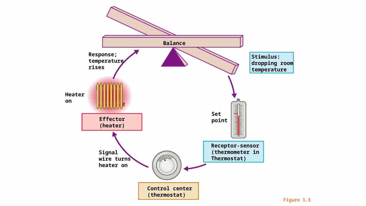
Figure 1.5
Signalwire turnsheater on
Response;temperaturerises
Stimulus: dropping roomtemperature
Balance
Effector(heater)
Setpoint
Receptor-sensor(thermometer inThermostat)
Control center(thermostat)
Heateron

Figure 1.5
Signalwire turnsheater on
Signalwire turnsheater off
Response;temperaturerises
Response;temperaturedrops
Stimulus: rising roomtemperature
Stimulus: dropping roomtemperature
Balance
Effector(heater)
Effector(heater)
Setpoint
Control center(thermostat)
Heateroff
Setpoint
Receptor-sensor(thermometer inThermostat)
Control center(thermostat)
Heateron
Imbalance
Imbalance
Receptor-sensor(thermometer inThermostat)
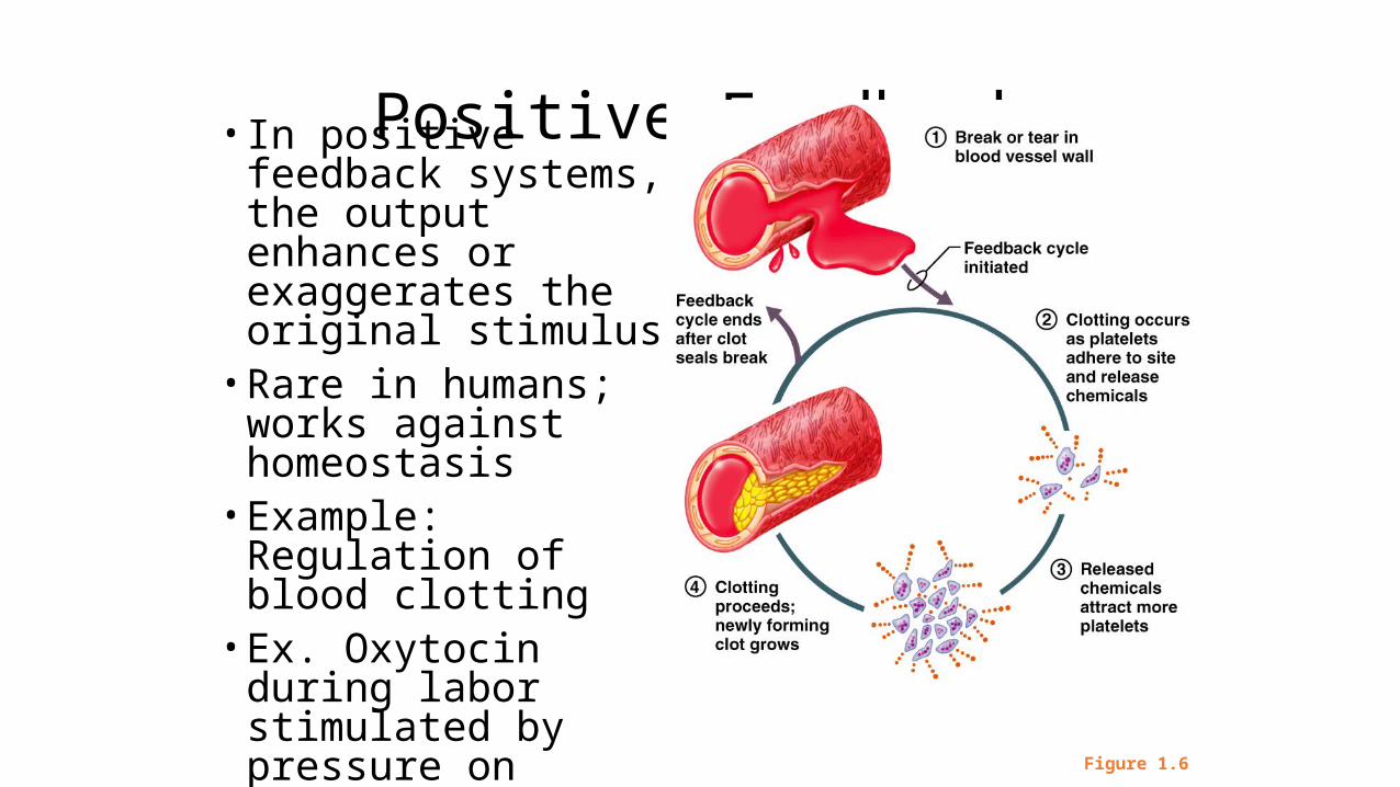
Positive Feedback• In positive feedback systems, the output enhances or exaggerates the original stimulus
• Rare in humans; works against homeostasis
• Example: Regulation of blood clotting
• Ex. Oxytocin during labor stimulated by pressure on cervix; causes greater uterine contractions & more pressure on cervix
Figure 1.6

Figure 1.6
Break or tear inblood vessel wall
Feedback cycleinitiated
1
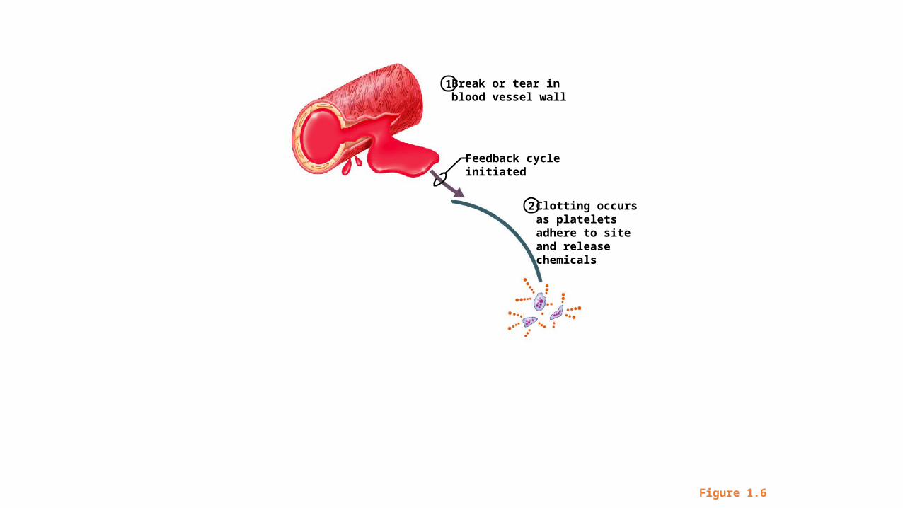
Figure 1.6
Clotting occursas platelets adhere to site and releasechemicals
Break or tear inblood vessel wall
Feedback cycleinitiated
2
1

Figure 1.6
Releasedchemicalsattract moreplatelets
Clotting occursas platelets adhere to site and releasechemicals
Break or tear inblood vessel wall
Feedback cycleinitiated
2
1
3
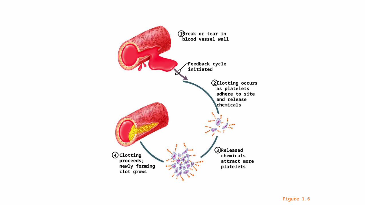
Figure 1.6
Releasedchemicalsattract moreplatelets
Clotting occursas platelets adhere to site and releasechemicals
Break or tear inblood vessel wall
Feedback cycleinitiated
Clotting proceeds; newly formingclot grows
2
1
34
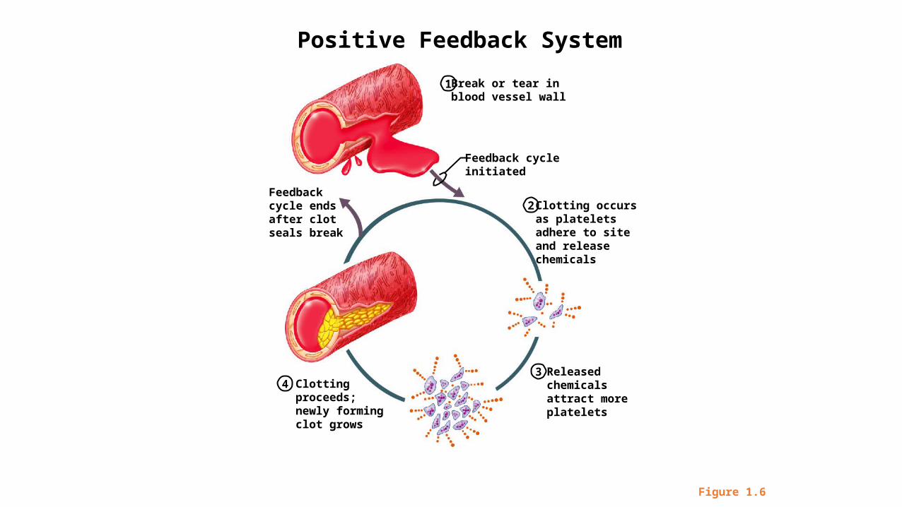
Figure 1.6
Releasedchemicalsattract moreplatelets
Clotting occursas platelets adhere to site and releasechemicals
Break or tear inblood vessel wall
Feedback cycleinitiated
Feedbackcycle endsafter clotseals break
Clotting proceeds; newly formingclot grows
2
1
34
Positive Feedback System
Homeostatic Imbalance
• Disturbance of homeostasis or the body’s normal equilibrium• Overwhelming the usual negative feedback mechanisms allows
destructive positive feedback mechanisms to take over
Directional Terms
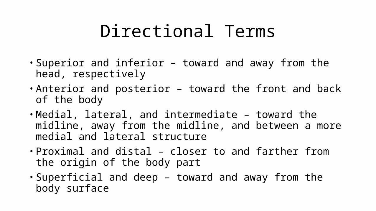
• Superior and inferior – toward and away from the head, respectively• Anterior and posterior – toward the front and back of the body• Medial, lateral, and intermediate – toward the midline, away from the
midline, and between a more medial and lateral structure• Proximal and distal – closer to and farther from the origin of the body part• Superficial and deep – toward and away from the body surface
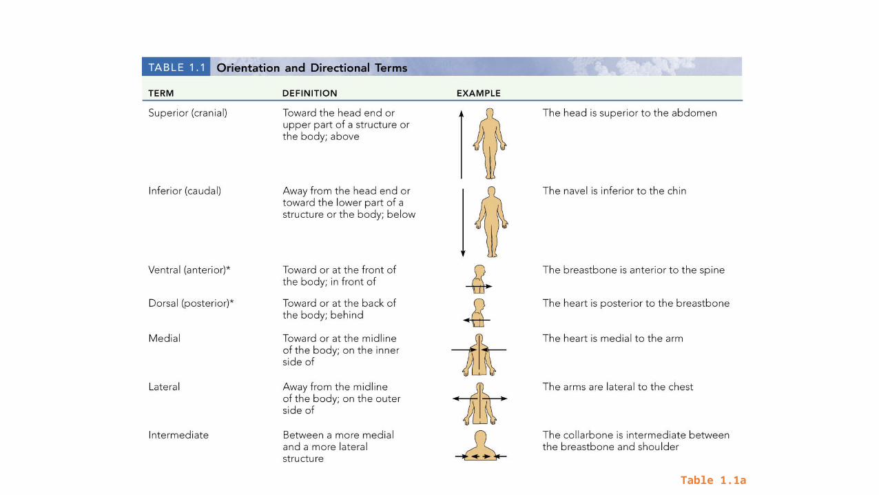
Directional Terms
Table 1.1a
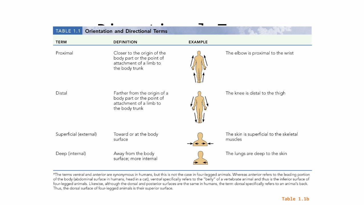
Directional Terms
Table 1.1b
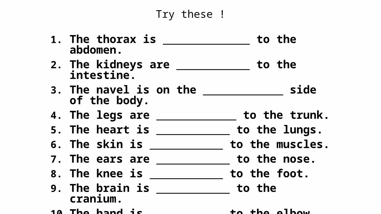
Try these !
1. The thorax is to the abdomen.2. The kidneys are to the intestine.3. The navel is on the side of the body.4. The legs are to the trunk. 5. The heart is to the lungs.6. The skin is to the muscles.7. The ears are to the nose.8. The knee is to the foot.9. The brain is to the cranium.10. The hand is to the elbow.
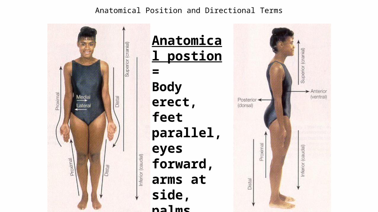
Anatomical Position and Directional Terms
Anatomical postion = Body erect, feet parallel, eyes forward, arms at side, palmsforward, fingers downward
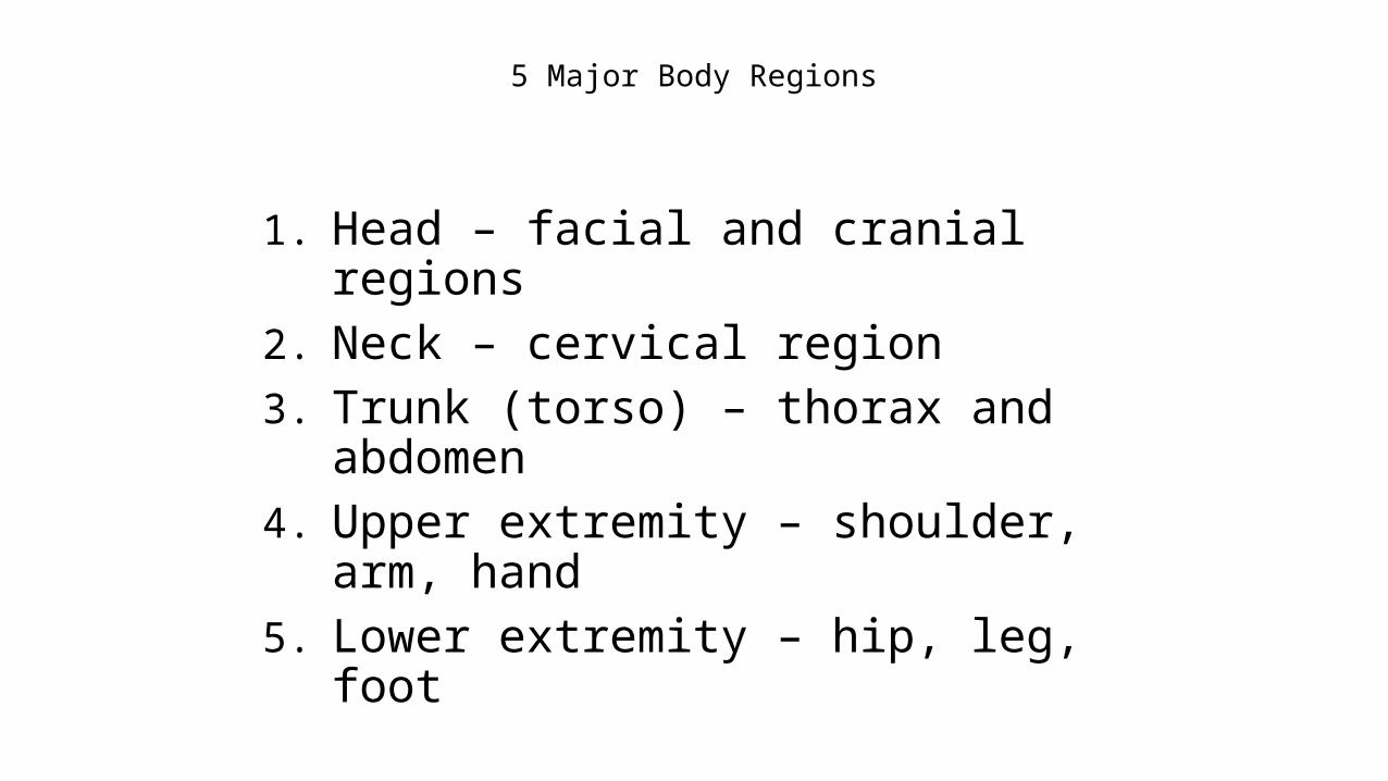
5 Major Body Regions
1. Head – facial and cranial regions2. Neck – cervical region3. Trunk (torso) – thorax and abdomen4. Upper extremity – shoulder, arm, hand5. Lower extremity – hip, leg, foot
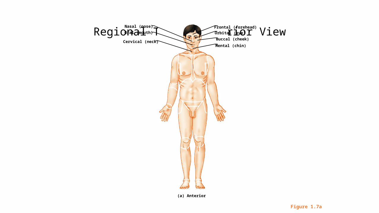
Regional Terms: Anterior View
Figure 1.7a
Nasal (nose)
Oral (mouth)
Cervical (neck)
Frontal (forehead)
Orbital (eye)
Buccal (cheek)
Mental (chin)
(a) Anterior
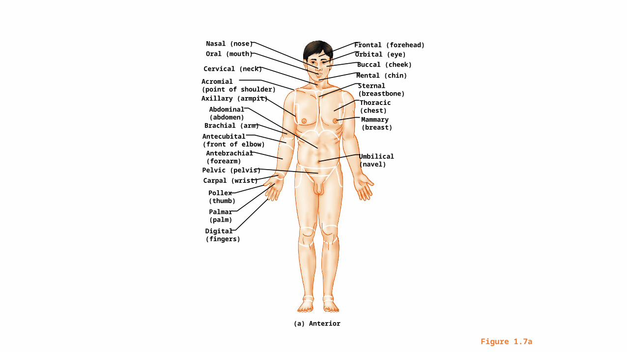
Figure 1.7a
Nasal (nose)
Oral (mouth)
Cervical (neck)
Acromial(point of shoulder)Axillary (armpit)
Brachial (arm)
Antecubital(front of elbow)
Abdominal(abdomen)
Pelvic (pelvis)
Antebrachial(forearm)
Carpal (wrist)
Palmar(palm)
Pollex(thumb)
Digital(fingers)
Mammary(breast)
Frontal (forehead)
Orbital (eye)
Buccal (cheek)
Sternal(breastbone)Thoracic(chest)
Mental (chin)
Umbilical(navel)
(a) Anterior
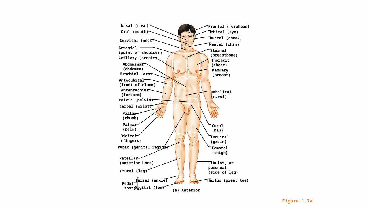
Figure 1.7a
Nasal (nose)
Oral (mouth)
Cervical (neck)
Acromial(point of shoulder)Axillary (armpit)
Brachial (arm)
Antecubital(front of elbow)
Abdominal(abdomen)
Pelvic (pelvis)
Antebrachial(forearm)
Carpal (wrist)
Palmar(palm)
Pollex(thumb)
Digital(fingers)
Pubic (genital region)
Patellar(anterior knee)
Crural (leg)
Tarsal (ankle)Pedal(foot) Digital (toes)
Inguinal(groin)
Coxal(hip)
Femoral(thigh)
Fibular, orperoneal(side of leg)
Hallux (great toe)
Mammary(breast)
Frontal (forehead)
Orbital (eye)
Buccal (cheek)
Sternal(breastbone)Thoracic(chest)
Mental (chin)
Umbilical(navel)
(a) Anterior
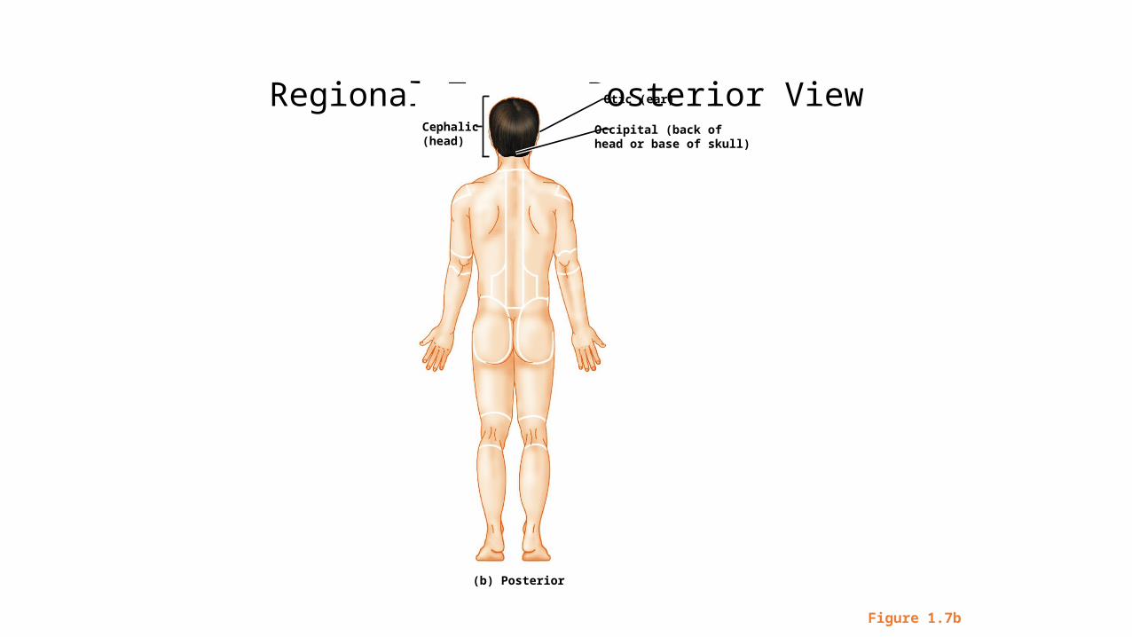
Regional Terms: Posterior View
Figure 1.7b
Otic (ear)
Occipital (back ofhead or base of skull)
Cephalic(head)
(b) Posterior
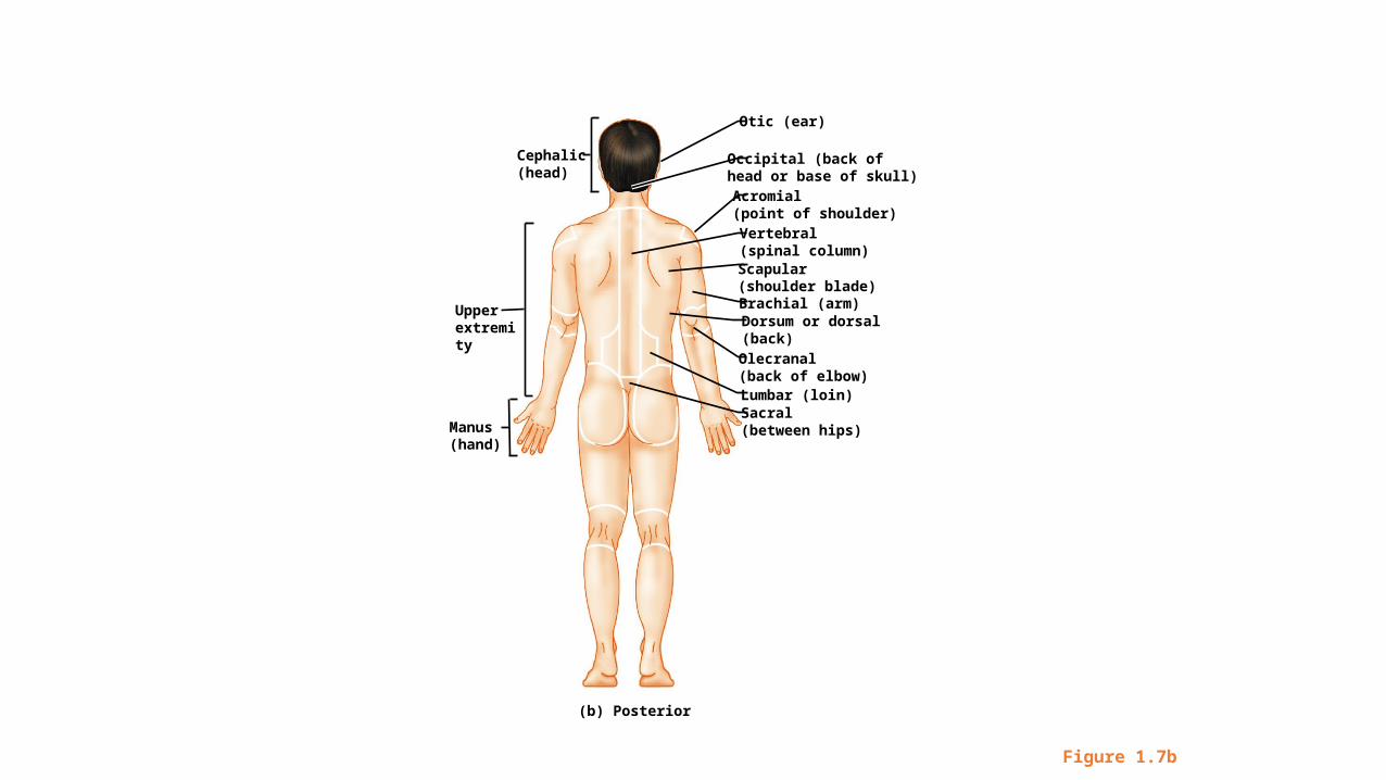
Figure 1.7b
Brachial (arm)
Otic (ear)
Occipital (back ofhead or base of skull) Acromial(point of shoulder)Vertebral(spinal column)Scapular(shoulder blade)
Dorsum or dorsal(back)
Olecranal(back of elbow)Lumbar (loin)Sacral(between hips)Manus
(hand)
Upperextremity
Cephalic(head)
(b) Posterior
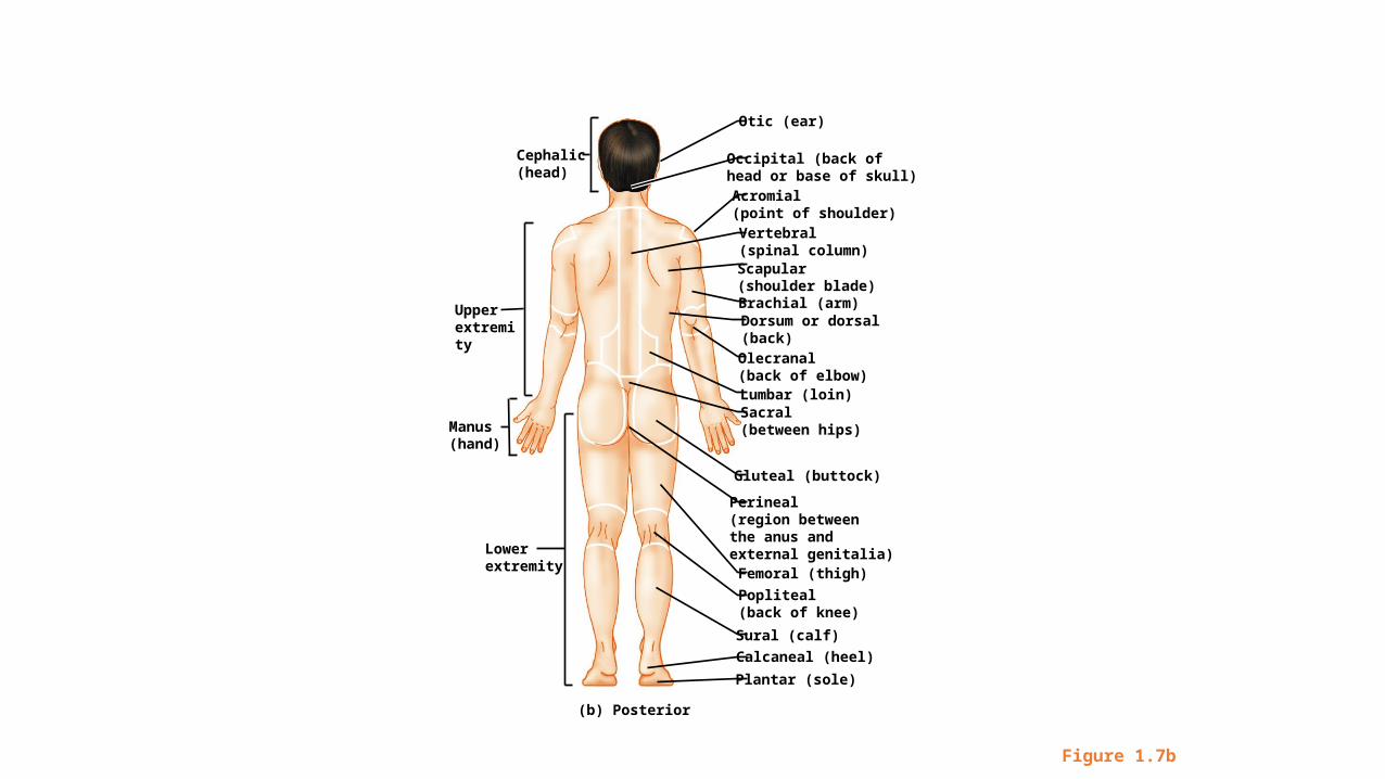
Figure 1.7b
Brachial (arm)
Otic (ear)
Occipital (back ofhead or base of skull) Acromial(point of shoulder)Vertebral(spinal column)Scapular(shoulder blade)
Dorsum or dorsal(back)
Olecranal(back of elbow)Lumbar (loin)Sacral(between hips)
Gluteal (buttock)
Perineal(region betweenthe anus and external genitalia)Femoral (thigh)
Popliteal(back of knee)
Sural (calf)
Calcaneal (heel)
Plantar (sole)
Manus(hand)
Upperextremity
Cephalic(head)
Lowerextremity
(b) Posterior
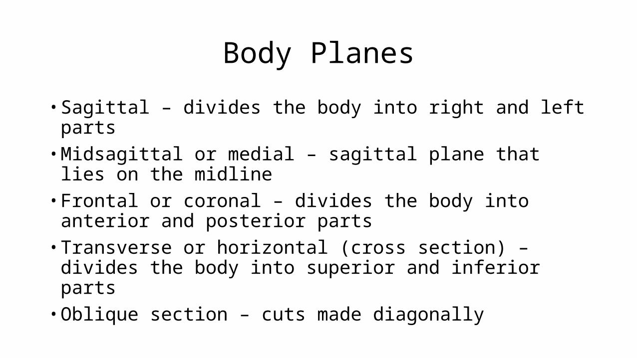
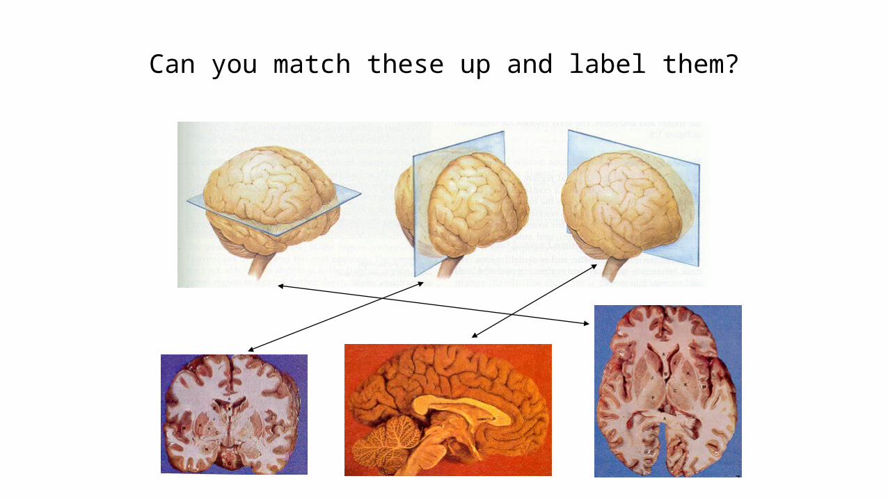
Body Planes
• Sagittal – divides the body into right and left parts• Midsagittal or medial – sagittal plane that lies on the midline• Frontal or coronal – divides the body into anterior and posterior parts• Transverse or horizontal (cross section) – divides the body into
superior and inferior parts• Oblique section – cuts made diagonally
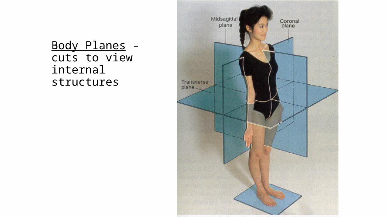
Body Planes – cuts to view internal structures
Human Anatomy and Physiology, 7eby Elaine Marieb & Katja Hoehn
Copyright © 2007 Pearson Education, Inc.,publishing as Benjamin Cummings.
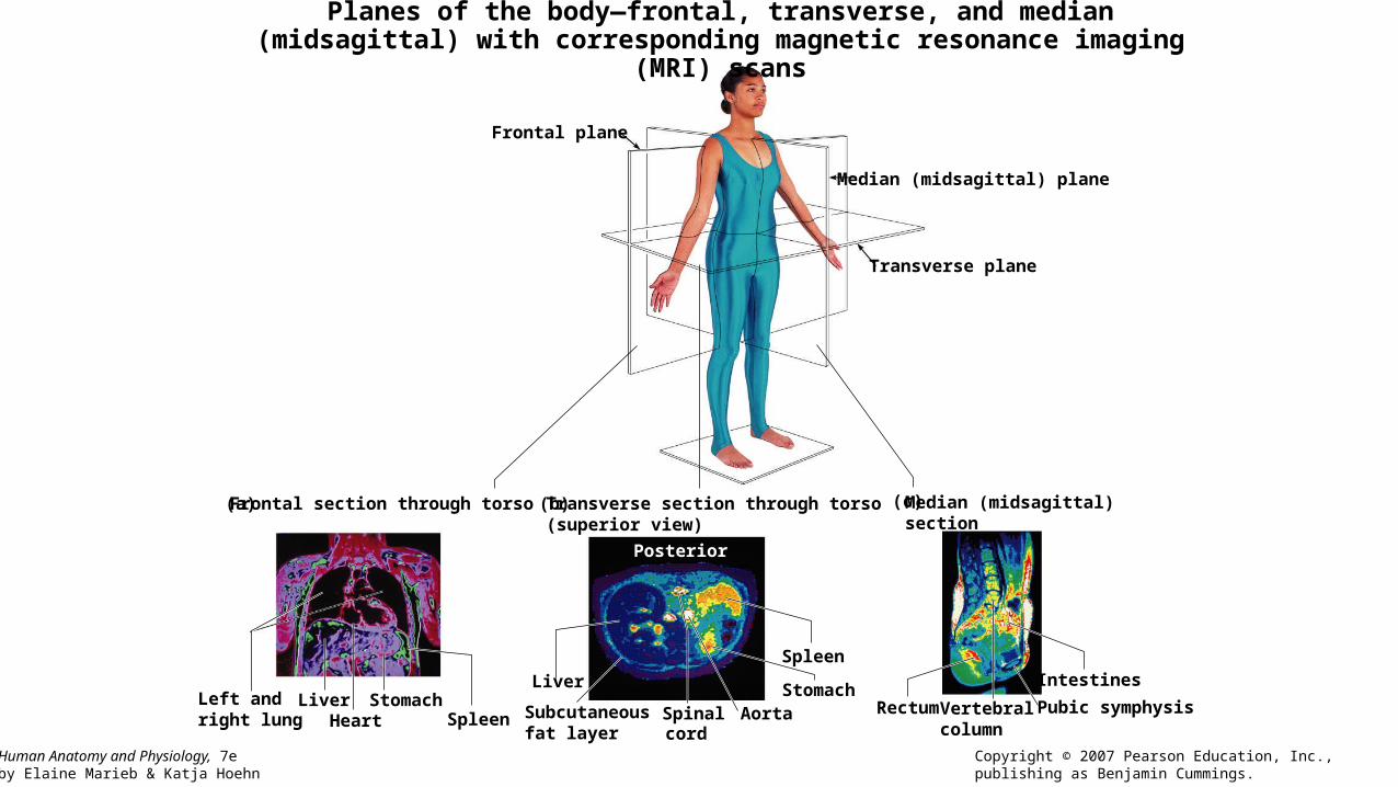
Planes of the body—frontal, transverse, and median (midsagittal) with corresponding magnetic resonance imaging (MRI) scans
Transverse plane
Median (midsagittal) plane
Frontal plane
Frontal section through torso Transverse section through torso(superior view)
Median (midsagittal)section
Posterior
Left andright lung
LiverHeart
StomachSpleen
Liver
Spleen
StomachAorta Vertebral
columnSpinalcord
Subcutaneous fat layer
Rectum
Intestines
Pubic symphysis
(a) (b) (c)
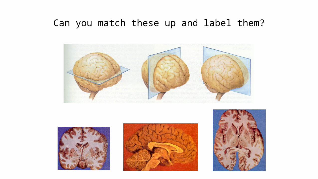
Can you match these up and label them?
Can you match these up and label them?
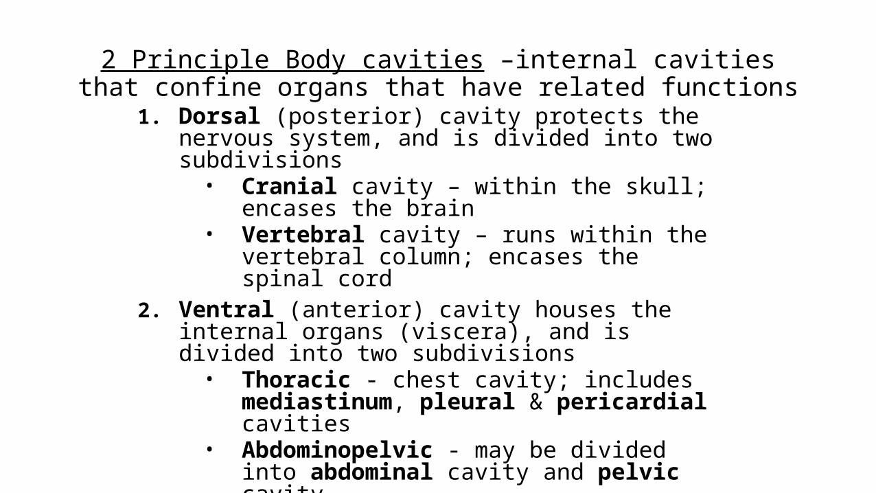
2 Principle Body cavities –internal cavities that confine organs that have related functions
1. Dorsal (posterior) cavity protects the nervous system, and is divided into two subdivisions
• Cranial cavity – within the skull; encases the brain• Vertebral cavity – runs within the vertebral column;
encases the spinal cord2. Ventral (anterior) cavity houses the internal organs
(viscera), and is divided into two subdivisions• Thoracic - chest cavity; includes mediastinum,
pleural & pericardial cavities• Abdominopelvic - may be divided into abdominal
cavity and pelvic cavity
Body Cavities
• Thoracic cavity is subdivided into two pleural cavities, the mediastinum, and the pericardial cavity
• Pleural cavities – each houses a lung• Mediastinum – contains the pericardial cavity; surrounds the remaining
thoracic organs• Pericardial cavity – encloses the heart
Body Cavities
• The abdominopelvic cavity is separated from the superior thoracic cavity by the dome-shaped diaphragm
• It is composed of two subdivisions• Abdominal cavity – contains the stomach, intestines, spleen, liver, and other
organs• Pelvic cavity – lies within the pelvis and contains the bladder, reproductive
organs, and rectum
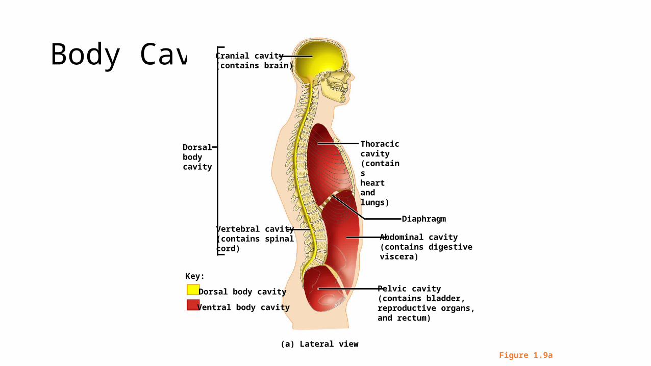
Body Cavities
Figure 1.9a
Cranial cavity(contains brain)
Dorsalbodycavity
Diaphragm
Abdominal cavity(contains digestiveviscera)
Pelvic cavity(contains bladder,reproductive organs,and rectum)
Vertebral cavity(contains spinal cord)
Key:
Dorsal body cavity
Ventral body cavity
Thoraciccavity(containsheartand lungs)
(a) Lateral view
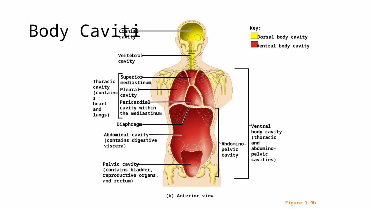
Body Cavities
Figure 1.9b
Ventral body cavity(thoracic and abdomino-pelviccavities)
Abdomino-pelviccavity
Superiormediastinum
Pleuralcavity
Cranialcavity
Vertebralcavity
Pericardialcavity withinthe mediastinum
Diaphragm
Abdominal cavity(contains digestiveviscera)
Pelvic cavity(contains bladder,reproductive organs,and rectum)
Thoraciccavity(containsheartand lungs)
(b) Anterior view
Key:
Dorsal body cavity
Ventral body cavity
Body membranes – thin layers of connective and epithelial tissue that cover, separate, and support
viscera and line body cavities2 types
1. Mucous membranes – secrete thick liquid substance called mucus; lubricates or protects organs; lines cavities that enter or exit the body
2. Serous membranes – line thoracic and adominopelvic cavities and cover visceral organs; secrete watery serous fluid; include: pleurae (lung), pericardial (heart) membranes, & peritoneal (abdominal) membranes
Ventral Body Cavity Membranes
• Parietal serosa lines internal body walls• Visceral serosa covers the internal organs• Serous fluid separates the serosae
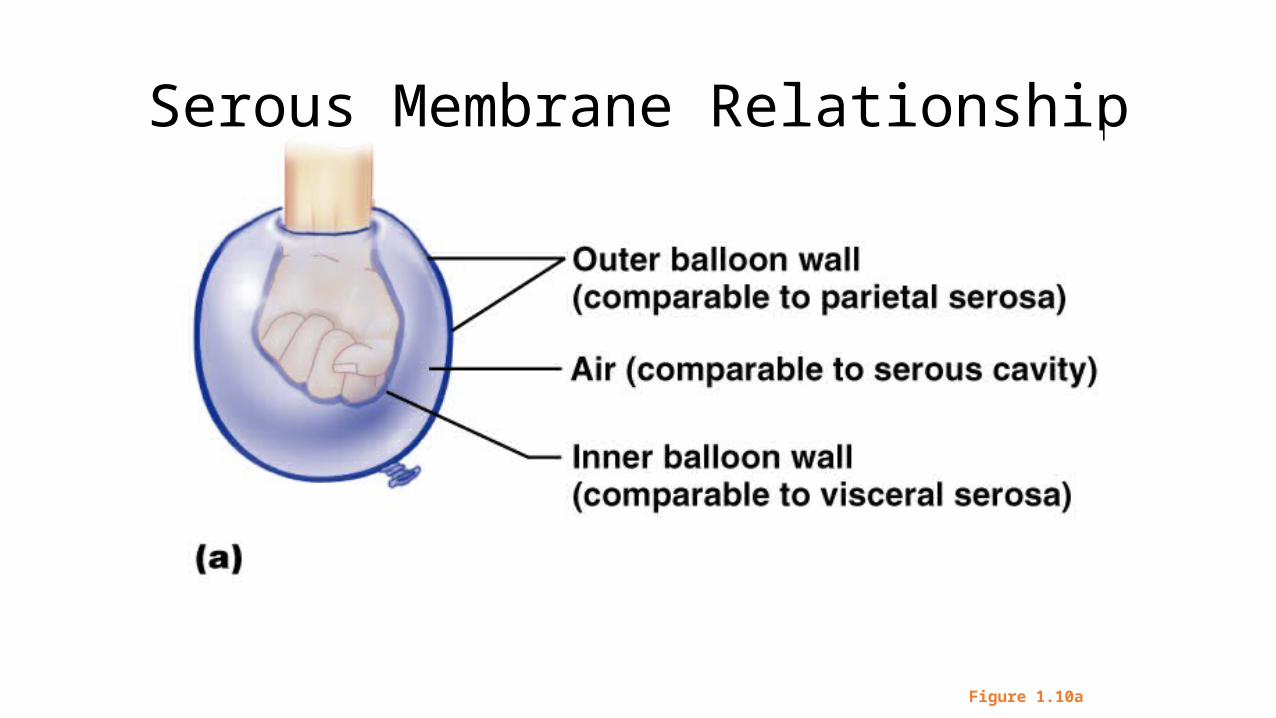
Serous Membrane Relationship
Figure 1.10a
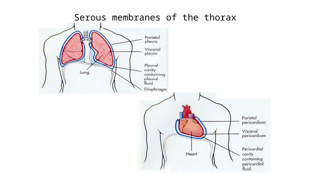
Serous membranes of the thorax
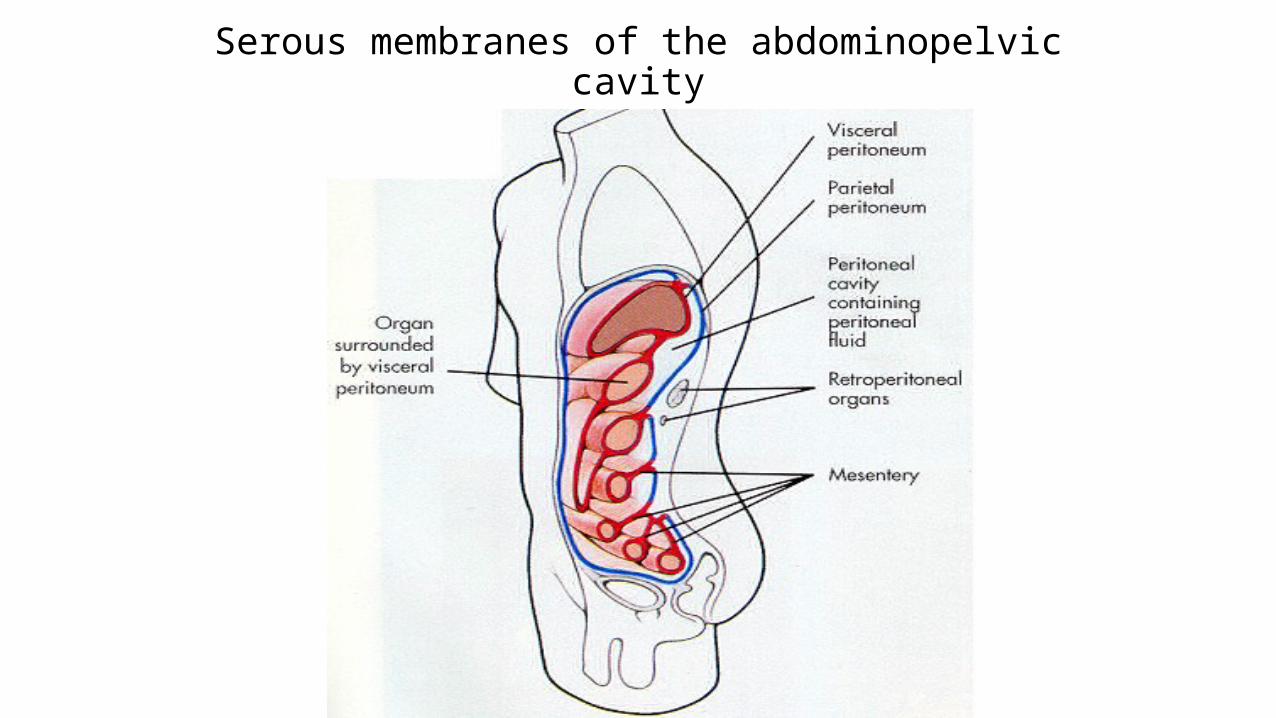
Serous membranes of the abdominopelvic cavity
Other Body Cavities
• Oral and digestive – mouth and cavities of the digestive organs• Nasal –located within and posterior to the nose• Orbital – house the eyes• Middle ear – contains bones (ossicles) that transmit sound vibrations• Synovial – joint cavities
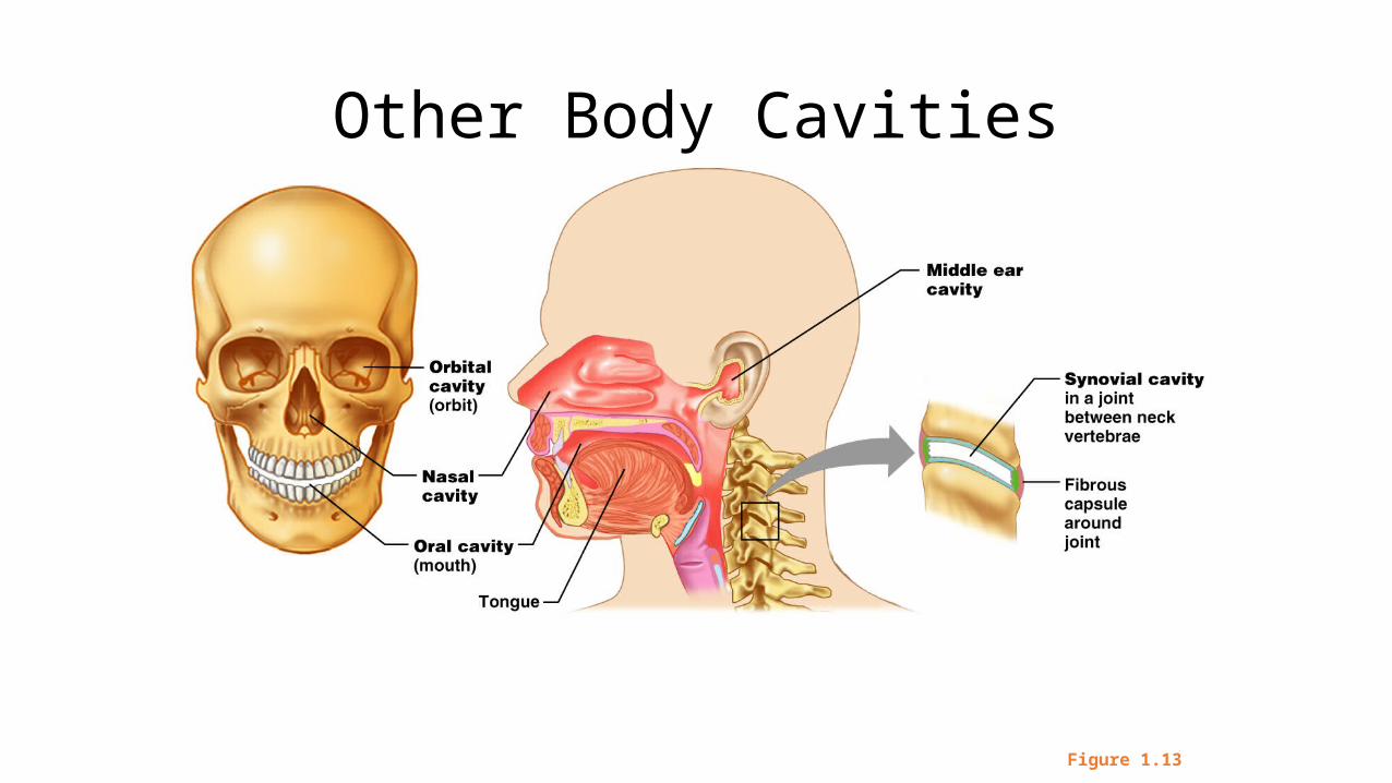
Other Body Cavities
Figure 1.13
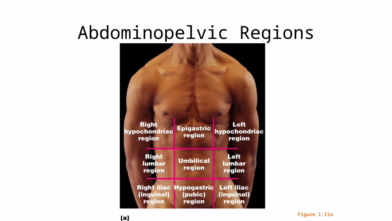
Abdominopelvic Regions
Figure 1.11a
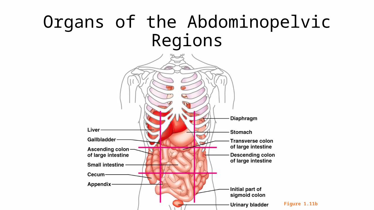
Organs of the Abdominopelvic Regions
Figure 1.11b
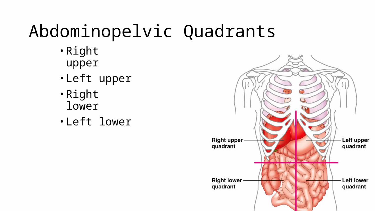
Abdominopelvic Quadrants• Right upper• Left upper• Right lower• Left lower
Figure 1.12
Clinical procedures used to determine anatomical structure and function
1. Inspection – observing body for swelling, rashes, needle marks, irregular breathing, etc.
2. Palpation – applying fingers with firm pressure to surface of body to feel lumps, tender spots, body landmarks, etc.
3. Percussion – tapping sharply on body walls to detect vibrations; used to locate excess fluid or organ abnormalities
4. Auscultation – listening to sounds of various organs; breathing, heartbeat, etc.
5. Reflex testing – observing automatic response to stimulus