Embed Size (px)
Citation preview
© 2015. Published by The Company of Biologists Ltd.
This is an Open Access article distributed under the terms of the Creative Commons Attribution License
(http://creativecommons.org/licenses/by/3.0), which permits unrestricted use, distribution and reproduction in any
medium provided that the original work is properly attributed.
Hyperactivity and attention deficits in mice with decreased levels of Stress Inducible
Phosphoprotein 1 (STIP1)
Flavio H. Beraldo1,*, Anu Thomas1, Benjamin Kolisnyk1,2, Pedro H. Hirata1, Xavier De
Jaeger1, Amanda C. Martyn1, Jue Fan1, Daniela F. Goncalves1, Matthew F. Cowan1, Talal
Masood1,2, Vilma R. Martins3, Robert Gros1,4, Vania F.Prado1,2,4,5* and Marco A.M.
Prado1,2,4,5*
1-Robarts Research Institute, The University of Western Ontario, London, ON, Canada;
2- Program in Neuroscience, The University of Western Ontario; 3- Department of
Molecular and Cell biology, International Research Center, A.C Camargo Cancer Center
and National Institute for Translational Neuroscience Research Center; 4-Department of
Physiology and Pharmacology, The University of Western Ontario, London, ON,
Canada; 5-Department of Anatomy and Cell Biology, The University of Western Ontario,
London, ON, Canada;
Keywords: Touchscreen, Autism, ASD, Stress Inducible Phosphoprotein 1, Attention
deficits, mouse models, BAC
*To whom correspondence should be addressed
Robarts Research Institute, The University of Western Ontario 1151 Richmond St,
London, Ontario Canada N6A 5B7
Dise
ase
Mod
els &
Mec
hani
sms
D
MM
Acce
pted
man
uscr
ipt
http://dmm.biologists.org/lookup/doi/10.1242/dmm.022525Access the most recent version at DMM Advance Online Articles. Posted 17 September 2015 as doi: 10.1242/dmm.022525http://dmm.biologists.org/lookup/doi/10.1242/dmm.022525Access the most recent version at
First posted online on 17 September 2015 as 10.1242/dmm.022525
Summary statement
Here, using a series of behavioural tests including touchscreen tasks we show that
decreased levels of stress inducible phosphoprotein 1 (STIP1) lead to attention deficits
and hyperactivity in mice.
Abstract
Stress-inducible phosphoprotein I (STIP1, STI1, or HOP) is a cochaperone
intermediating Hsp70/Hsp90 exchange of client proteins, but it can also be secreted to
trigger prion protein-mediated neuronal signaling. Some mothers of children with autism-
spectrum disorders (ASD) present antibodies against certain brain proteins, including
antibodies against STIP1. Maternal antibodies can cross the fetus blood brain barrier
during pregnancy, suggesting the possibility that they can interfere with STIP1 levels and
presumably functions. However, it is currently unknown whether abnormal levels of
STIP1 have any impact in ASD-related behavior. Here we used mice with reduced (50%)
or increased STIP1 levels (5-fold) to test for potential ASD-like phenotypes. We find that
increased STIP1 regulates the abundance of Hsp70 and Hsp90, whereas reduced STIP1
does not affect Hsp70, Hsp90 or the prion protein. Interestingly, BAC transgenic mice
presenting 5-fold more STIP1 have no major phenotype when examined in a series of
behavioral tasks, including locomotor activity, elevated plus maze, Morris water maze
and 5-choice serial reaction time task (5-CSRTT). In contrast, mice with reduced STIP1
levels are hyperactive and have attentional deficits on the 5-CSRTT, but have normal
performance in the other tasks. We conclude that reduced STIP1 levels can contribute
with phenotypes related to ASD. However, future experiments are needed to define
Dise
ase
Mod
els &
Mec
hani
sms
D
MM
Acce
pted
man
uscr
ipt
whether it is decreased chaperone capacity or impaired prion protein signaling that
contributes to these phenotypes.
Dise
ase
Mod
els &
Mec
hani
sms
D
MM
Acce
pted
man
uscr
ipt
Introduction
In autism spectrum disorders (ASD) genetic variance and neurodevelopmental
alterations are both thought to contribute to phenotype heterogeneity. Womb environment
and autoimmune responses have been proposed to contribute to complex behavioral
alterations observed in ASD, which include, but are not limited to, abnormal socialization,
communication and stereotyped behavior (Brimberg et al., 2013; Goldani et al., 2014).
Several distinct groups have investigated the existence of antibodies against fetal brain
tissue in mothers of ASD children (Bauman et al., 2013; Braunschweig et al., 2012b;
Braunschweig et al., 2013; Dalton et al., 2003; Nordahl et al., 2013). Passive transfer of
maternal anti-brain antibodies to pregnant experimental animal models (including mice,
rats and non-human primates) has shown that their offspring develop a number of
endophenotypes that resemble phenotypes in ASD (Bauman et al., 2013; Braunschweig
et al., 2012b; Dalton et al., 2003). Indeed, a recent study indicate that the prevalence of
antibodies against fetal brain proteins is increased four fold in mothers of an ASD child
compared to control groups (Brimberg et al., 2013). Proteomics analysis identified 6
brain proteins as targets for ASD antibodies, including Lactate dehydrogenase A and B
(LDH), Cypin, Stress-Inducible phosphoprotein Protein1 (STIP1), collapsine response
mediator protein 1 and 2 (CRMP1, CRMP2) and Y-box-binding protein (YBX1)
(Braunschweig et al., 2013). Interestingly, injection of maternal antibodies that recognize
LDH, STIP1 and CRMP1 in developing mouse embryos causes an increase in cortical
neural precursor proliferation and cortical neuron volume, with consequent increase in
brain size and weight (Martinez-Cerdeno et al., 2014). These phenotypes are consistent
Dise
ase
Mod
els &
Mec
hani
sms
D
MM
Acce
pted
man
uscr
ipt
with the notion that the presence of maternal autoantibodies can affect neuronal
development.
STIP1, also known as Heat-shock organizing protein (Hop) or STI1, is a co-
chaperone that interacts concomitantly with Heat-shock proteins 70 and 90 (Hsp70 and
HsP90) (Abbas-Terki et al., 2002; Chen et al., 1996; Nicolet and Craig, 1989; Picard,
2002; Smith et al., 1993). The chaperone machinery is thought to provide a buffer for
cells to respond to environmental challenges, and as such disturbance of Hsp70/90
chaperone activity decreases cellular resilience to stress (Chen et al., 2015; Hashimoto-
Torii et al., 2014; Taipale et al., 2010; Taipale et al., 2014). The absence of STIP1 in
mice has critical consequences for development including increased apoptosis, DNA
damage and death (Beraldo et al., 2013). These phenotypes are rescued by transgenic
BAC expression of STIP1 (Beraldo et al., 2013).
In addition to its intracellular role as a cochaperone, STIP1 is also secreted by a
variety of cells (Erlich et al., 2007; Eustace and Jay, 2004; Hajj et al., 2013; Lima et al.,
2007; Wang et al., 2010) via extracellular vesicles (Hajj et al., 2013). Extracellular STIP1
can signal via the prion protein (PrPC) to produce a myriad of effects related to brain
development (Beraldo et al., 2010; Beraldo et al., 2013; Caetano et al., 2008; Lopes et al.,
2005; Soares et al., 2013). Here we used Stip1 heterozygous mice, as well as mice
overexpresssing 4-5 fold more STIP1 to investigate the consequences for alterations of
STIP1 levels in vivo. We report that decreased, but not increased STIP1 levels, affects
attention and causes hyperactivity in mice, two phenotypes that are related to ASD-like
phenotypes. Our results suggest that interference with STIP1 functions, which
Dise
ase
Mod
els &
Mec
hani
sms
D
MM
Acce
pted
man
uscr
ipt
presumably may happen in the presence of STIP1 antibodies, has the potential to
contribute to ASD-like phenotypes.
Results
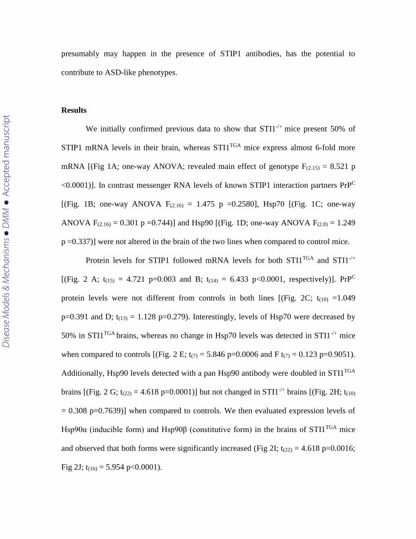
We initially confirmed previous data to show that STI1-/+ mice present 50% of
STIP1 mRNA levels in their brain, whereas STI1TGA mice express almost 6-fold more
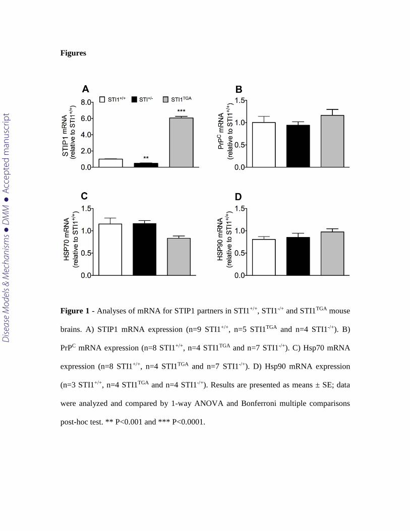
mRNA [(Fig 1A; one-way ANOVA; revealed main effect of genotype F(2.15) = 8.521 p
<0.0001)]. In contrast messenger RNA levels of known STIP1 interaction partners PrPC
[(Fig. 1B; one-way ANOVA F(2.16) = 1.475 p =0.2580], Hsp70 [(Fig. 1C; one-way
ANOVA F(2.16) = 0.301 p =0.744)] and Hsp90 [(Fig. 1D; one-way ANOVA F(2.8) = 1.249
p =0.337)] were not altered in the brain of the two lines when compared to control mice.
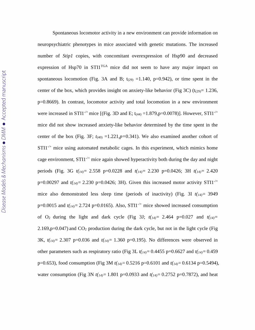
Protein levels for STIP1 followed mRNA levels for both STI1TGA and STI1-/+
[(Fig. 2 A; t(15) = 4.721 p=0.003 and B; t(14) = 6.433 p<0.0001, respectively)]. PrPC
protein levels were not different from controls in both lines [(Fig. 2C; t(10) =1.049
p=0.391 and D; t(13) = 1.128 p=0.279). Interestingly, levels of Hsp70 were decreased by
50% in STI1TGA brains, whereas no change in Hsp70 levels was detected in STI1-/+ mice
when compared to controls [(Fig. 2 E; t(7) = 5.846 p=0.0006 and F t(7) = 0.123 p=0.9051).
Additionally, Hsp90 levels detected with a pan Hsp90 antibody were doubled in STI1TGA
brains [(Fig. 2 G; t(22) = 4.618 p=0.0001)] but not changed in STI1-/+ brains [(Fig. 2H; t(10)
= 0.308 p=0.7639)] when compared to controls. We then evaluated expression levels of
Hsp90α (inducible form) and Hsp90β (constitutive form) in the brains of STI1TGA mice
and observed that both forms were significantly increased (Fig 2I; t(22) = 4.618 p=0.0016;
Fig 2J; t(16) = 5.954 p<0.0001).
Dise
ase
Mod
els &
Mec
hani
sms
D
MM
Acce
pted
man
uscr
ipt
Spontaneous locomotor activity in a new environment can provide information on
neuropsychiatric phenotypes in mice associated with genetic mutations. The increased
number of Stip1 copies, with concomitant overexpression of Hsp90 and decreased
expression of Hsp70 in STI1TGA mice did not seem to have any major impact on
spontaneous locomotion (Fig. 3A and B; t(29) =1.140, p=0.942), or time spent in the
center of the box, which provides insight on anxiety-like behavior (Fig 3C) (t(29)= 1.236,
p=0.8669). In contrast, locomotor activity and total locomotion in a new environment
were increased in STI1-/+ mice [(Fig. 3D and E; t(44) =1.879,p=0.0078)]. However, STI1-/+
mice did not show increased anxiety-like behavior determined by the time spent in the
center of the box (Fig. 3F; t(40) =1.221,p=0.341). We also examined another cohort of
STI1-/+ mice using automated metabolic cages. In this experiment, which mimics home
cage environment, STI1-/+ mice again showed hyperactivity both during the day and night
periods (Fig. 3G t(14)= 2.558 p=0.0228 and t(14)= 2.230 p=0.0426; 3H t(14)= 2.420
p=0.00297 and t(14)= 2.230 p=0.0426; 3H). Given this increased motor activity STI1-/+
mice also demonstrated less sleep time (periods of inactivity) (Fig. 3I t(14)= 3949
p=0.0015 and t(14)= 2.724 p=0.0165). Also, STI1-/+ mice showed increased consumption
of O2 during the light and dark cycle (Fig 3J; t(14)= 2.464 p=0.027 and t(14)=
2.169,p=0.047) and CO2 production during the dark cycle, but not in the light cycle (Fig
3K, t(14)= 2.307 p=0.036 and t(14)= 1.360 p=0.195). No differences were observed in
other parameters such as respiratory ratio (Fig 3L t(14)= 0.4455 p=0.6627 and t(14)= 0.459
p=0.653), food consumption (Fig 3M t(14)= 0.5216 p=0.6101 and t(14)= 0.6134 p=0.5494),
water consumption (Fig 3N t(14)= 1.801 p=0.0933 and t(14)= 0.2752 p=0.7872), and heat
Dise
ase
Mod
els &
Mec
hani
sms
D
MM
Acce
pted
man
uscr
ipt
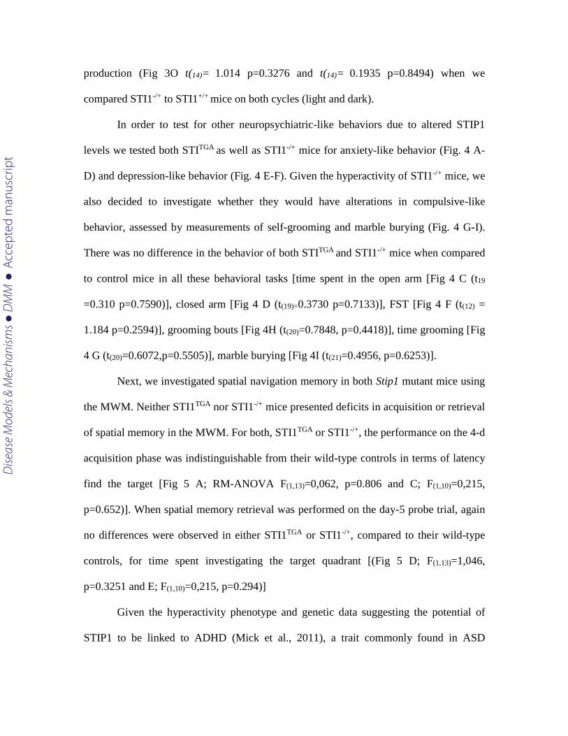
production (Fig 3O t(14)= 1.014 p=0.3276 and t(14)= 0.1935 p=0.8494) when we
compared STI1-/+ to STI1+/+ mice on both cycles (light and dark).
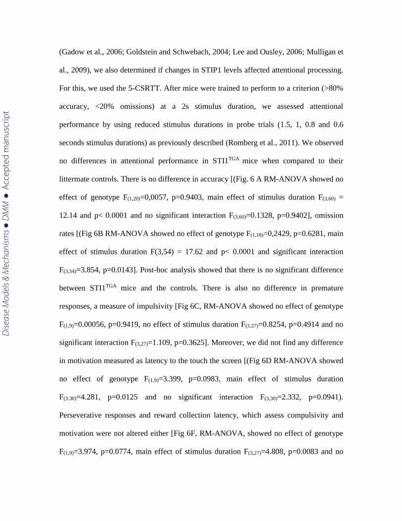
In order to test for other neuropsychiatric-like behaviors due to altered STIP1
levels we tested both STITGA as well as STI1-/+ mice for anxiety-like behavior (Fig. 4 A-
D) and depression-like behavior (Fig. 4 E-F). Given the hyperactivity of STI1-/+ mice, we
also decided to investigate whether they would have alterations in compulsive-like
behavior, assessed by measurements of self-grooming and marble burying (Fig. 4 G-I).
There was no difference in the behavior of both STITGA and STI1-/+ mice when compared
to control mice in all these behavioral tasks [time spent in the open arm [Fig 4 C (t19
=0.310 p=0.7590)], closed arm [Fig 4 D (t(19)=0.3730 p=0.7133)], FST [Fig 4 F (t(12) =
1.184 p=0.2594)], grooming bouts [Fig 4H (t(20)=0.7848, p=0.4418)], time grooming [Fig
4 G (t(20)=0.6072,p=0.5505)], marble burying [Fig 4I (t(21)=0.4956, p=0.6253)].
Next, we investigated spatial navigation memory in both Stip1 mutant mice using
the MWM. Neither STI1TGA nor STI1-/+ mice presented deficits in acquisition or retrieval
of spatial memory in the MWM. For both, STI1TGA or STI1-/+, the performance on the 4-d
acquisition phase was indistinguishable from their wild-type controls in terms of latency
find the target [Fig 5 A; RM-ANOVA F(1,13)=0,062, p=0.806 and C; F(1,10)=0,215,
p=0.652)]. When spatial memory retrieval was performed on the day-5 probe trial, again
no differences were observed in either STI1TGA or STI1-/+, compared to their wild-type
controls, for time spent investigating the target quadrant [(Fig 5 D; F(1,13)=1,046,
p=0.3251 and E; F(1,10)=0,215, p=0.294)]
Given the hyperactivity phenotype and genetic data suggesting the potential of
STIP1 to be linked to ADHD (Mick et al., 2011), a trait commonly found in ASD
Dise
ase
Mod
els &
Mec
hani
sms
D
MM
Acce
pted
man
uscr
ipt
(Gadow et al., 2006; Goldstein and Schwebach, 2004; Lee and Ousley, 2006; Mulligan et
al., 2009), we also determined if changes in STIP1 levels affected attentional processing.
For this, we used the 5-CSRTT. After mice were trained to perform to a criterion (>80%
accuracy, <20% omissions) at a 2s stimulus duration, we assessed attentional
performance by using reduced stimulus durations in probe trials (1.5, 1, 0.8 and 0.6
seconds stimulus durations) as previously described (Romberg et al., 2011). We observed
no differences in attentional performance in STI1TGA mice when compared to their
littermate controls. There is no difference in accuracy [(Fig. 6 A RM-ANOVA showed no
effect of genotype F(1,20)=0,0057, p=0.9403, main effect of stimulus duration F(3,60) =
12.14 and p< 0.0001 and no significant interaction F(3,60)=0.1328, p=0.9402], omission
rates [(Fig 6B RM-ANOVA showed no effect of genotype F(1,18)=0,2429, p=0.6281, main
effect of stimulus duration F(3,54) = 17.62 and p< 0.0001 and significant interaction
F(3,54)=3.854, p=0.0143]. Post-hoc analysis showed that there is no significant difference
between STI1TGA mice and the controls. There is also no difference in premature
responses, a measure of impulsivity [Fig 6C, RM-ANOVA showed no effect of genotype
F(1,9)=0.00056, p=0.9419, no effect of stimulus duration F(3.27)=0.8254, p=0.4914 and no
significant interaction F(3,27)=1.109, p=0.3625]. Moreover, we did not find any difference
in motivation measured as latency to the touch the screen [(Fig 6D RM-ANOVA showed
no effect of genotype F(1,9)=3.399, p=0.0983, main effect of stimulus duration
F(3.30)=4.281, p=0.0125 and no significant interaction F(3,30)=2.332, p=0.0941).
Perseverative responses and reward collection latency, which assess compulsivity and
motivation were not altered either [Fig 6F, RM-ANOVA, showed no effect of genotype
F(1,9)=3.974, p=0.0774, main effect of stimulus duration F(3,27)=4.808, p=0.0083 and no
Dise
ase
Mod
els &
Mec
hani
sms
D
MM
Acce
pted
man
uscr
ipt
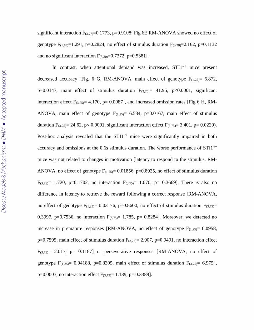
significant interaction F(3,27)=0.1773, p=0.9108; Fig 6E RM-ANOVA showed no effect of
genotype F(1,10)=1.291, p=0.2824, no effect of stimulus duration F(3,30)=2.162, p=0.1132
and no significant interaction F(3.30)=0.7372, p=0.5381].
In contrast, when attentional demand was increased, STI1-/+ mice present
decreased accuracy [Fig. 6 G, RM-ANOVA, main effect of genotype F(1,25)= 6.872,
p=0.0147, main effect of stimulus duration F(3,75)= 41.95, p<0.0001, significant
interaction effect F(3,75)= 4.170, p= 0.0087], and increased omission rates [Fig 6 H, RM-
ANOVA, main effect of genotype F(1,25)= 6.584, p=0.0167, main effect of stimulus
duration F(3,75)= 24.62, p< 0.0001, significant interaction effect F(3,75)= 3.401, p= 0.0220).
Post-hoc analysis revealed that the STI1-/+ mice were significantly impaired in both
accuracy and omissions at the 0.6s stimulus duration. The worse performance of STI1-/+
mice was not related to changes in motivation [latency to respond to the stimulus, RM-
ANOVA, no effect of genotype F(1,25)= 0.01856, p=0.8925, no effect of stimulus duration
F(3,75)= 1.720, p=0.1702, no interaction F(3,75)= 1.070, p= 0.3669]. There is also no
difference in latency to retrieve the reward following a correct response [RM-ANOVA,
no effect of genotype F(1,25)= 0.03176, p=0.8600, no effect of stimulus duration F(3,75)=
0.3997, p=0.7536, no interaction F(3,75)= 1.785, p= 0.8284]. Moreover, we detected no
increase in premature responses [RM-ANOVA, no effect of genotype F(1,25)= 0.0958,
p=0.7595, main effect of stimulus duration F(3,75)= 2.907, p=0.0401, no interaction effect
F(3,75)= 2.017, p= 0.1187] or perseverative responses [RM-ANOVA, no effect of
genotype F(1,25)= 0.04188, p=0.8395, main effect of stimulus duration F(3,75)= 6.975 ,
p=0.0003, no interaction effect F(3,75)= 1.139, p= 0.3389].
Dise
ase
Mod
els &
Mec
hani
sms
D
MM
Acce
pted
man
uscr
ipt
Discussion
The present experiments tested whether alterations in STIP1 levels have
consequences for psychiatric-like behaviors in mice. Our results suggest that decreased,
but not increased STIP1 levels, causes significant behavioral alterations in mice. Spatial
learning and memory, as well as anxiety and depression-like behavior do not seem to be
affected by reduced STIP1 levels. However, mutant mice deficient for STIP1 are
hyperactive and present attention deficits.
STIP1 has recently emerged as a protein of potential interest in ASD and
endophenotypes related to ASD. Maternal autoantibodies against STIP1 have been
identified in mothers of children with ASD (Braunschweig et al., 2013). Moreover, recent
Global-wide association study (GWAS) analysis identified a polymorphism in Stip1 (the
human gene coding for STIP1/HOP) as a potential risk factor in a population of
individuals diagnosed with attention-deficit disorder (Mick et al., 2011), a co-morbidity
often associated with ASD (Brimberg et al., 2013; Goldani et al., 2014). The
consequences of this polymorphism for STIP1 expression is unknown, but the presence
of autoantibodies against STIP1 may affect expression levels of the protein, given that
during pregnancy antibodies can penetrate the blood brain barrier in the fetus
(Braunschweig et al., 2012a; Diamond et al., 2009; Fox et al., 2012; Zhang et al., 2012).
Indeed, maternal antibodies that recognize STIP1 and other targets when injected in
pregnant rodents or developing pups can lead to offspring with abnormal neurons and
behaviors that relate to ASD (Braunschweig et al., 2012b; Camacho et al., 2014). To a
degree, STI1-/+ mice model this early developmental deficit in STIP1 levels. However, in
Dise
ase
Mod
els &
Mec
hani
sms
D
MM
Acce
pted
man
uscr
ipt
STI1-/+ mice STIP1 expression is persistently decreased through life, which could also
have important consequences for the phenotypes described.
STIP1 is a modular protein containing several tetratricopeptide (TRP) repeat
domains and aspartate-proline (DP) reach domains (Taipale et al., 2010). TRP1 and
TRP2B can interact with Hsp70 (Flom et al., 2007; Scheufler et al., 2000), whereas
TPR2A is required for interaction with Hsp90 (Flom et al., 2007; Flom et al., 2006).
Hsp90 activity is regulated by STIP1 and previous work has shown that in mice no other
co-chaperone can replace for STIP1 (Beraldo et al., 2013). Recent experiments have
indicated that the chaperone machinery, activated by the transcription factor Heat shock
factor 1 (HSF1), is responsible for preventing damaging effects from environmental
factors in the developing brain (Hashimoto-Torii et al., 2014). Indeed, the chaperone
machinery can buffer many stresses at the cellular level and therefore it is not surprising
that functional changes in its components would have physiological consequences.
In addition to its intracellular chaperone function, STIP1 is also secreted by a
myriad of cells, including astrocytes via an extracellular vesicle population, which
includes exosomes (Hajj et al., 2013). Extracellular STIP1 also mediates important
physiological responses in the brain. Acting as a trophic factor to engage PrPC to signal in
neurons, it regulates neuritogenesis and neuronal survival (Beraldo et al., 2010; Lopes et
al., 2005; Roffe et al., 2010). STIP1 has a role in functional recovery in stroke (Beraldo et
al., 2013; Lee et al., 2013). Moreover, STIP1 also modulates toxicity of Aβ peptides in
models of Alzheimer’s disease (Brehme et al., 2014; Ostapchenko et al., 2013).
It is remarkable that mice with increased levels of STIP1 up to almost 5 fold do
not present any major behavioral alteration. In the extensive evaluation of cognitive
Dise
ase
Mod
els &
Mec
hani
sms
D
MM
Acce
pted
man
uscr
ipt
phenotypes that we performed in this study, which included anxiety and depression-like
behaviors, spatial memory or attention we showed that STI1TGA mice perform as well as
littermate controls. These results suggest that strategies to increase STIP1 levels should
not cause toxicity with consequences for brain functions. This is important, given that
increased STIP1 levels may be protective against insults, such as stroke-mediated cell
death and in Alzheimer’s disease (Beraldo et al., 2013; Ostapchenko et al., 2013).
Interestingly, whereas increased levels of STIP1 seem to affect the chaperone machinery,
by decreasing the levels of Hsp70 and increasing Hsp90, prion protein expression is not
affected. These consequences of increased STIP1 seem to occur at the post-translational
level, given that mRNAs for Hsp70 and 90 were not affected. It is unknown at the
moment if increased STIP1 levels may stabilize a complex containing Hsp90
preferentially leading to increased turnover of Hsp70.
At present the exact mechanism by which decreased STIP1 levels affect
psychiatric-like behavior is still unknown. Although it is possible that decreased levels of
STIP1 early during development may have persistent effects in brain circuits, culminating
with hyperactivity and attentional deficits, we cannot discard the possibility that in the
adult brain STIP1 may play a role in regulating circuitry function. Our experiments at the
moment also do not discriminate whether the phenotypes observed in mutant mice
resulted from decreased STIP1co-chaperone function, diminished STIP1extracellular
signaling or both. Our results suggest that reduced levels of STIP1 have important
consequences for behavior and seem to affect brain circuits that regulate attention. It is
possible that exposure to STIP1 antibodies during pregnancy could reduce STIP1 levels,
which based on the present results would have important consequences. Future
Dise
ase
Mod
els &
Mec
hani
sms
D
MM
Acce
pted
man
uscr
ipt
experiments are however required to define potential mechanisms as well as
consequences of disturbed STIP1 activity in ASD.
Material and methods
Animals
STI1-/+ and STI1TGA mice were generated as described in (Beraldo et al., 2013).
Both mouse lines were in the C57BL/6J background. All experimental procedures were
conducted in compliance with the Canadian Council of Animal Care guidelines for use
and care of animals and in accordance with approved animal use protocols at the
University of Western Ontario (2008/127). Animals were housed in groups of two or four
per cage. Mice were kept in a temperature-controlled room with a 12/12-light/dark cycle
(7AM/7PM) with food and water provided ad libitum unless stated otherwise. For
behavioral studies, only male mice were used. Mice were randomized and the
experimenter was blind to genotypes. For most of the behavioral tasks, software-based
analyzes were used to score mice performance with minimum human interference.
qPCR and Western Blot
For real-time quantitative PCR (qPCR), brain tissues were homogenized in Trizol
and total RNA was extracted using the Aurum Total RNA for fatty and fibrous tissue kit
from Biorad (Bio-Rad, Hercules, CA, USA). qPCR were performed as previously
described (Martins-Silva et al., 2011). Primers sequences: STIP1-
F:GCCAAGAAAGGAGACTACCAG; STIP1-R:TCATAGGTTCGT TTGGCTTCC;
Dise
ase
Mod
els &
Mec
hani
sms
D
MM
Acce
pted
man
uscr
ipt
HsP90-F:CCACCCTGCTCTGTACTACT; HsP90- R:CCAGGGCA
TCTGAAGCATTA; HsP70-R:ACCTTGACAGTAATCGGTGC; HsP70-F:
CTCCCGGTGTGGTCTAGAAA; PRP-F:GAACCATTTCAACCGAGCTG; PRP-
R:CATAGTCACAAAGAGGGCCAG; Actin-F:TGGAATCCTGTGGCATCCATGA;
Actin-R: AATGCCTGGGTACATGGTGGTA. Immunoblot analysis was carried out as
described previously (Beraldo et al., 2013). The antibodies used were: anti-STIP1
(1:5000 – in house antibody generated by Bethyl Laboratories Montgomery, USA using
recombinant STIP1) (Beraldo et al., 2013); anti-Hsp90 (1:1000), anti-Hsp70 (1:1000),
anti-Hsp90α (1:1000), anti Hsp90β (1:1000) (Cell Signalling, Danvers, USA); anti-PrP
8H4 (1:2000) (Abcam, Cambrige, UK).
Locomotor activity
Mice were acclimated to the testing room for 30 min prior to beginning the test
and locomotor activity was automatically recorded (Omnitech Electronics, Inc. Columbus,
USA). Mice were placed in the center of the apparatus and locomotor activity was
measured at 5 min intervals for 1 hour as described previously (Martyn et al., 2012).
Elevated Plus-Maze
To access anxiety-like behavior, mice were acclimated to the testing room for 30
min prior to beginning the test and then placed in the center of the elevated plus maze
(Med Associates, Inc, St. Albans, USA). The activity was recorded and videos were
analyzed using ANY-maze software (Stoelting Co. USA) to determine the amount of
time spent in the close and in the open section of the maze.
Dise
ase
Mod
els &
Mec
hani
sms
D
MM
Acce
pted
man
uscr
ipt
Forced-swimming test
Depressive-like behavior was assessed by forced swim test (FST) as described
previously (Martyn et al., 2012). Briefly, mice were placed in a 2L beaker containing 1.7
L of water at 25 - 27oC for 6 min. Experimental sessions were recorded and immobility
time was evaluated using ANY-Maze Software (Stoelting Co. USA). Data was obtained
from the last 4 min of testing were used for the analysis.
Morris water maze
The spatial version of Morris water maze (MWM) was conducted as described
previously (Kolisnyk et al., 2013; Martyn et al., 2012; Vorhees and Williams, 2006).
Briefly, the task was performed in a 1.5-m-diammeter/1-m-deep pool filled with water at
25oC. Spatial cues, 40 by 40 cm boards containing black symbols (vertical and horizontal
stripes, triangles, squares and circles), were placed on the wall distributed around the pool
and the platform was submerged 1 cm below the surface of the water. Mice were
submitted to four training trails a day (90s each) for 4 consecutive days with a 15 min
intertrial interval. On day 5, memory was assessed by single 60 s trial on which the
platform was removed and the time spent in the target quadrant was evaluated. All the
experimental sessions were recorded and analyzed using the ANY-Maze Software.
Five-Choice Serial Reaction time task
The five choice serial reaction time task (5-CSRTT) was used to evaluate
attention in mice as described previously (Kolisnyk et al., 2013; Romberg et al., 2011).
Dise
ase
Mod
els &
Mec
hani
sms
D
MM
Acce
pted
man
uscr
ipt
Mice were trained in the 5-CSRTT in automated Bussey-SaksidaTouch screen Systems
(Campden Instruments Limited, Loughborough, EN) and the data were collected using
ABET II Touch software V.2.18 (Lafayette Instruments, Lafayette, USA). Mice were
submitted to a pre-training program which consists of first habituating the mouse to the
testing chamber with the lights off for 10 min. The next day, the mouse was put in the
chamber with the lights off for 20 min. After two days of habituation with no reward
been offered, the reward tray was primed with 11% fat strawberry milkshake (Nielson -
Saputo Dairy Products) and a tone was played when the mouse entered the reward tray.
This was repeated for the next 2 days for 40 min sessions and whenever the mouse
returned to the reward tray, the reward was offered and paired with a tone (Phase I). The
following training phase consisted in pairing the reward with the presentation of a
random stimulus (flash of light in one of the five windows), which is removed after 30s.
At this phase if the mouse touched the screen, when the stimulus was displayed, it
received a reward. This cycle was repeated until the mouse completed 30 trials or 60 min
timeout (Phase II). At Phase III of the training, the stimulus is displayed randomly in one
to the five windows. The mouse had to touch the window where the stimulus was
displayed to receive the reward paired with a tone. Similar to the phase II, this cycle was
repeated until the mouse completed 30 trials or 60 min time out. The next step (Phase IV)
is identical to Phase III except by the fact that the mouse had to poke its nose in the
reward trail to initiate the task. This process was repeated in the last phase of the pre-
training (Phase V), however if the mouse touched an incorrect screen, it received a 5s
timeout and the light in the chamber was turned on. After the mouse finished pre-training
and reached criterion at 4s and 2s stimulus duration (80% accuracy, 20% omission for 3
Dise
ase
Mod
els &
Mec
hani
sms
D
MM
Acce
pted
man
uscr
ipt
consecutive days), mice were probed for attention deficits following probe trial
schedules: each mouse was tested over two sessions at 1.5, 1.0, 0.8 and 0.6 s stimulus
duration (the order of the probe trial sessions was randomized and the groups
counterbalanced). Between each different stimulus duration each mouse was returned to a
2s stimulus for 2 consecutive sessions. Number of trial to criterion, accuracy, omission,
reward collection latency and perseverative response were analyzed.
Metabolic assessments
Oxygen consumption, carbon dioxide production, respiratory exchange ratio
(RER), carbon dioxide production, water and food and physical activity were
simultaneously measured for adult STI1+/+ and STI1+/- mice by using the Comprehensive
Lab Animal Monitoring System (CLAMS) interfaced with Oxymax Software (Columbus
Instruments, Columbus, OH, USA) as previously described in detail (Guzman et al.,
2013; Kolisnyk et al., 2013). Briefly, mice were individually housed in the metabolic
chambers with ad libitum access to water and food. Following a 16 h of habituation
period, all measurements were obtained every 10 min for 24h (12h light/12h dark).
Marble Burying Task
A marble burying task was used to assess repetitive and anxiety-like behavior as
previously described (Deacon, 2006).
Dise
ase
Mod
els &
Mec
hani
sms
D
MM
Acce
pted
man
uscr
ipt
Assessment of Self-grooming
Self-grooming was assessed to evaluate repetitive behavior, as previously described
(McFarlane et al., 2008). Briefly, each mouse was placed individually in a clean, empty,
cage and it was given a 10 minute habituation period, after which they were filmed for
another 10 minutes. Cumulative time spent grooming and grooming bouts were counted
by an experimenter blinded to the genotypes of the mice.
Statistical analyses
Data are presented as mean ± SEM. Statistical analyses were performed using SigmaStat
3.5 software. Student’s t test was used to compare two experimental groups and for
comparison of several experimental groups, two-way ANOVA or two-way repeated-
measures ANOVA were used as required. Tukey’s post hoc comparison was used when
required.
Competing interests - Authors have read and understood DMM police and declare there
is no conflict of interests.
Author contribution: F.H.B., M.A.M.P., V.F.P., and R.G conceived and designed
experiments. F.H.B, A.T., B.K., P.H.H., R.G.X.D.J. A.C.M., J.F. D.F.G., M.F.C. and
T.M. performed the experiments. V.R.M. Contributed with specific reagents. F.H.B.,
A.T., B.K., A.C.M, V.F.P, R.G., V.R.M and M.A.M.P. analysed the data. F.H.B, V.F.P
and M.A.M.P wrote the paper.
Dise
ase
Mod
els &
Mec
hani
sms
D
MM
Acce
pted
man
uscr
ipt
Funding: This work was supported by the Canadian Institute of Health Research (MOP
136930, MOP 126000 and MOP 89919, M.A.M.P. V.F.P), Canadian Foundation for
Innovation (M.A.M.P., V.F.P., and R.G.) and Fundação de Amparo a Pesquisa do Estado
de São Paulo (FAPESP- 2009/14027-2; São Paulo, Brazil; V.R.M.).
References
Abbas-Terki, T., Briand, P. A., Donze, O. and Picard, D. (2002). The Hsp90
co-chaperones Cdc37 and Sti1 interact physically and genetically. Biol Chem 383, 1335-
42.
Abrahams, B. S. and Geschwind, D. H. (2008). Advances in autism genetics: on
the threshold of a new neurobiology. Nat Rev Genet 9, 341-55.
Bauman, M. D., Iosif, A. M., Ashwood, P., Braunschweig, D., Lee, A.,
Schumann, C. M., Van de Water, J. and Amaral, D. G. (2013). Maternal antibodies
from mothers of children with autism alter brain growth and social behavior development
in the rhesus monkey. Transl Psychiatry 3, e278.
Beraldo, F. H., Arantes, C. P., Santos, T. G., Queiroz, N. G., Young, K.,
Rylett, R. J., Markus, R. P., Prado, M. A. and Martins, V. R. (2010). Role of alpha7
nicotinic acetylcholine receptor in calcium signaling induced by prion protein interaction
with stress-inducible protein 1. J Biol Chem 285, 36542-50.
Beraldo, F. H., Soares, I. N., Goncalves, D. F., Fan, J., Thomas, A. A., Santos,
T. G., Mohammad, A. H., Roffe, M., Calder, M. D., Nikolova, S. et al. (2013). Stress-
inducible phosphoprotein 1 has unique cochaperone activity during development and
regulates cellular response to ischemia via the prion protein. FASEB J 27, 3594-607.
Braunschweig, D., Duncanson, P., Boyce, R., Hansen, R., Ashwood, P.,
Pessah, I. N., Hertz-Picciotto, I. and Van de Water, J. (2012a). Behavioral correlates
of maternal antibody status among children with autism. J Autism Dev Disord 42, 1435-
45.
Braunschweig, D., Golub, M. S., Koenig, C. M., Qi, L., Pessah, I. N., Van de
Water, J. and Berman, R. F. (2012b). Maternal autism-associated IgG antibodies delay
development and produce anxiety in a mouse gestational transfer model. J Neuroimmunol
252, 56-65.
Braunschweig, D., Krakowiak, P., Duncanson, P., Boyce, R., Hansen, R. L.,
Ashwood, P., Hertz-Picciotto, I., Pessah, I. N. and Van de Water, J. (2013). Autism-
Dise
ase
Mod
els &
Mec
hani
sms
D
MM
Acce
pted
man
uscr
ipt
specific maternal autoantibodies recognize critical proteins in developing brain. Transl
Psychiatry 3, e277.
Brehme, M., Voisine, C., Rolland, T., Wachi, S., Soper, J. H., Zhu, Y., Orton,
K., Villella, A., Garza, D., Vidal, M. et al. (2014). A chaperome subnetwork safeguards
proteostasis in aging and neurodegenerative disease. Cell Rep 9, 1135-50.
Brimberg, L., Sadiq, A., Gregersen, P. K. and Diamond, B. (2013). Brain-
reactive IgG correlates with autoimmunity in mothers of a child with an autism spectrum
disorder. Mol Psychiatry 18, 1171-7.
Caetano, F. A., Lopes, M. H., Hajj, G. N., Machado, C. F., Pinto Arantes, C.,
Magalhaes, A. C., Vieira Mde, P., Americo, T. A., Massensini, A. R., Priola, S. A. et
al. (2008). Endocytosis of prion protein is required for ERK1/2 signaling induced by
stress-inducible protein 1. J Neurosci 28, 6691-702.
Camacho, J., Jones, K., Miller, E., Ariza, J., Noctor, S., Van de Water, J. and
Martinez-Cerdeno, V. (2014). Embryonic intraventricular exposure to autism-specific
maternal autoantibodies produces alterations in autistic-like stereotypical behaviors in
offspring mice. Behav Brain Res 266, 46-51.
Chen, S., Prapapanich, V., Rimerman, R. A., Honore, B. and Smith, D. F.
(1996). Interactions of p60, a mediator of progesterone receptor assembly, with heat
shock proteins hsp90 and hsp70. Mol Endocrinol 10, 682-93.
Chen, X., Zhao, C., Li, X., Wang, T., Li, Y., Cao, C., Ding, Y., Dong, M.,
Finci, L., Wang, J. H. et al. (2015). Terazosin activates Pgk1 and Hsp90 to promote
stress resistance. Nat Chem Biol 11, 19-25.
Dalton, P., Deacon, R., Blamire, A., Pike, M., McKinlay, I., Stein, J., Styles, P.
and Vincent, A. (2003). Maternal neuronal antibodies associated with autism and a
language disorder. Ann Neurol 53, 533-7.
Deacon, R. M. (2006). Digging and marble burying in mice: simple methods for
in vivo identification of biological impacts. Nat Protoc 1, 122-4.
Diamond, B., Huerta, P. T., Mina-Osorio, P., Kowal, C. and Volpe, B. T.
(2009). Losing your nerves? Maybe it's the antibodies. Nat Rev Immunol 9, 449-56.
Erlich, R. B., Kahn, S. A., Lima, F. R., Muras, A. G., Martins, R. A., Linden,
R., Chiarini, L. B., Martins, V. R. and Moura Neto, V. (2007). STI1 promotes glioma
proliferation through MAPK and PI3K pathways. Glia 55, 1690-8.
Eustace, B. K. and Jay, D. G. (2004). Extracellular roles for the molecular
chaperone, hsp90. Cell Cycle 3, 1098-100.
Flom, G., Behal, R. H., Rosen, L., Cole, D. G. and Johnson, J. L. (2007).
Definition of the minimal fragments of Sti1required for dimerization, interaction with
Hsp70 and Hsp90 and in vivo functions. Biochem J 404, 159-67.
Flom, G., Weekes, J., Williams, J. J. and Johnson, J. L. (2006). Effect of
mutation of the tetratricopeptide repeat and asparatate-proline 2 domains of Sti1 on
Hsp90 signaling and interaction in Saccharomyces cerevisiae. Genetics 172, 41-51.
Dise
ase
Mod
els &
Mec
hani
sms
D
MM
Acce
pted
man
uscr
ipt
Fox, E., Amaral, D. and Van de Water, J. (2012). Maternal and fetal antibrain
antibodies in development and disease. Dev Neurobiol 72, 1327-34.
Gadow, K. D., DeVincent, C. J. and Pomeroy, J. (2006). ADHD symptom
subtypes in children with pervasive developmental disorder. J Autism Dev Disord 36,
271-83.
Geschwind, D. H. (2011). Genetics of autism spectrum disorders. Trends Cogn
Sci 15, 409-16.
Goldani, A. A., Downs, S. R., Widjaja, F., Lawton, B. and Hendren, R. L.
(2014). Biomarkers in autism. Front Psychiatry 5, 100.
Goldstein, S. and Schwebach, A. J. (2004). The comorbidity of Pervasive
Developmental Disorder and Attention Deficit Hyperactivity Disorder: results of a
retrospective chart review. J Autism Dev Disord 34, 329-39.
Guzman, M. S., De Jaeger, X., Drangova, M., Prado, M. A., Gros, R. and
Prado, V. F. (2013). Mice with selective elimination of striatal acetylcholine release are
lean, show altered energy homeostasis and changed sleep/wake cycle. J Neurochem 124,
658-69.
Hajj, G. N., Arantes, C. P., Dias, M. V., Roffe, M., Costa-Silva, B., Lopes, M.
H., Porto-Carreiro, I., Rabachini, T., Lima, F. R., Beraldo, F. H. et al. (2013). The
unconventional secretion of stress-inducible protein 1 by a heterogeneous population of
extracellular vesicles. Cell Mol Life Sci 70, 3211-27.
Hashimoto-Torii, K., Torii, M., Fujimoto, M., Nakai, A., El Fatimy, R.,
Mezger, V., Ju, M. J., Ishii, S., Chao, S. H., Brennand, K. J. et al. (2014). Roles of
heat shock factor 1 in neuronal response to fetal environmental risks and its relevance to
brain disorders. Neuron 82, 560-72.
Kolisnyk, B., Guzman, M. S., Raulic, S., Fan, J., Magalhaes, A. C., Feng, G.,
Gros, R., Prado, V. F. and Prado, M. A. (2013). ChAT-ChR2-EYFP mice have
enhanced motor endurance but show deficits in attention and several additional cognitive
domains. J Neurosci 33, 10427-38.
Lee, D. O. and Ousley, O. Y. (2006). Attention-deficit hyperactivity disorder
symptoms in a clinic sample of children and adolescents with pervasive developmental
disorders. J Child Adolesc Psychopharmacol 16, 737-46.
Lee, S. D., Lai, T. W., Lin, S. Z., Lin, C. H., Hsu, Y. H., Li, C. Y., Wang, H. J.,
Lee, W., Su, C. Y., Yu, Y. L. et al. (2013). Role of stress-inducible protein-1 in
recruitment of bone marrow derived cells into the ischemic brains. EMBO Mol Med 5,
1227-46.
Lima, F. R., Arantes, C. P., Muras, A. G., Nomizo, R., Brentani, R. R. and
Martins, V. R. (2007). Cellular prion protein expression in astrocytes modulates
neuronal survival and differentiation. J Neurochem 103, 2164-76.
Lopes, M. H., Hajj, G. N., Muras, A. G., Mancini, G. L., Castro, R. M.,
Ribeiro, K. C., Brentani, R. R., Linden, R. and Martins, V. R. (2005). Interaction of
Dise
ase
Mod
els &
Mec
hani
sms
D
MM
Acce
pted
man
uscr
ipt
cellular prion and stress-inducible protein 1 promotes neuritogenesis and neuroprotection
by distinct signaling pathways. J Neurosci 25, 11330-9.
Martinez-Cerdeno, V., Camacho, J., Fox, E., Miller, E., Ariza, J., Kienzle, D.,
Plank, K., Noctor, S. C. and Van de Water, J. (2014). Prenatal Exposure to Autism-
Specific Maternal Autoantibodies Alters Proliferation of Cortical Neural Precursor Cells,
Enlarges Brain, and Increases Neuronal Size in Adult Animals. Cereb Cortex.
Martins-Silva, C., De Jaeger, X., Guzman, M. S., Lima, R. D., Santos, M. S.,
Kushmerick, C., Gomez, M. V., Caron, M. G., Prado, M. A. and Prado, V. F. (2011).
Novel strains of mice deficient for the vesicular acetylcholine transporter: insights on
transcriptional regulation and control of locomotor behavior. PLoS One 6, e17611.
Martyn, A. C., De Jaeger, X., Magalhaes, A. C., Kesarwani, R., Goncalves, D.
F., Raulic, S., Guzman, M. S., Jackson, M. F., Izquierdo, I., Macdonald, J. F. et al.
(2012). Elimination of the vesicular acetylcholine transporter in the forebrain causes
hyperactivity and deficits in spatial memory and long-term potentiation. Proc Natl Acad
Sci U S A 109, 17651-6.
McFarlane, H. G., Kusek, G. K., Yang, M., Phoenix, J. L., Bolivar, V. J. and
Crawley, J. N. (2008). Autism-like behavioral phenotypes in BTBR T+tf/J mice. Genes
Brain Behav 7, 152-63.
Mick, E., McGough, J., Loo, S., Doyle, A. E., Wozniak, J., Wilens, T. E.,
Smalley, S., McCracken, J., Biederman, J. and Faraone, S. V. (2011). Genome-wide
association study of the child behavior checklist dysregulation profile. J Am Acad Child
Adolesc Psychiatry 50, 807-17 e8.
Mulligan, A., Anney, R. J., O'Regan, M., Chen, W., Butler, L., Fitzgerald, M.,
Buitelaar, J., Steinhausen, H. C., Rothenberger, A., Minderaa, R. et al. (2009).
Autism symptoms in Attention-Deficit/Hyperactivity Disorder: a familial trait which
correlates with conduct, oppositional defiant, language and motor disorders. J Autism Dev
Disord 39, 197-209.
Nicolet, C. M. and Craig, E. A. (1989). Isolation and characterization of STI1, a
stress-inducible gene from Saccharomyces cerevisiae. Mol Cell Biol 9, 3638-46.
Nordahl, C. W., Braunschweig, D., Iosif, A. M., Lee, A., Rogers, S., Ashwood,
P., Amaral, D. G. and Van de Water, J. (2013). Maternal autoantibodies are associated
with abnormal brain enlargement in a subgroup of children with autism spectrum disorder.
Brain Behav Immun 30, 61-5.
Ostapchenko, V. G., Beraldo, F. H., Mohammad, A. H., Xie, Y. F., Hirata, P.
H., Magalhaes, A. C., Lamour, G., Li, H., Maciejewski, A., Belrose, J. C. et al. (2013).
The prion protein ligand, stress-inducible phosphoprotein 1, regulates amyloid-beta
oligomer toxicity. J Neurosci 33, 16552-64.
Picard, D. (2002). Heat-shock protein 90, a chaperone for folding and regulation.
Cell Mol Life Sci 59, 1640-8.
Dise
ase
Mod
els &
Mec
hani
sms
D
MM
Acce
pted
man
uscr
ipt
Roffe, M., Beraldo, F. H., Bester, R., Nunziante, M., Bach, C., Mancini, G.,
Gilch, S., Vorberg, I., Castilho, B. A., Martins, V. R. et al. (2010). Prion protein
interaction with stress-inducible protein 1 enhances neuronal protein synthesis via mTOR.
Proc Natl Acad Sci U S A 107, 13147-52.
Romberg, C., Mattson, M. P., Mughal, M. R., Bussey, T. J. and Saksida, L.
M. (2011). Impaired attention in the 3xTgAD mouse model of Alzheimer's disease:
rescue by donepezil (Aricept). J Neurosci 31, 3500-7.
Scheufler, C., Brinker, A., Bourenkov, G., Pegoraro, S., Moroder, L.,
Bartunik, H., Hartl, F. U. and Moarefi, I. (2000). Structure of TPR domain-peptide
complexes: critical elements in the assembly of the Hsp70-Hsp90 multichaperone
machine. Cell 101, 199-210.
Smith, D. F., Sullivan, W. P., Marion, T. N., Zaitsu, K., Madden, B.,
McCormick, D. J. and Toft, D. O. (1993). Identification of a 60-kilodalton stress-
related protein, p60, which interacts with hsp90 and hsp70. Mol Cell Biol 13, 869-76.
Soares, I. N., Caetano, F. A., Pinder, J., Rodrigues, B. R., Beraldo, F. H.,
Ostapchenko, V. G., Durette, C., Pereira, G. S., Lopes, M. H., Queiroz-
Hazarbassanov, N. et al. (2013). Regulation of stress-inducible phosphoprotein 1
nuclear retention by protein inhibitor of activated STAT PIAS1. Mol Cell Proteomics 12,
3253-70.
Taipale, M., Jarosz, D. F. and Lindquist, S. (2010). HSP90 at the hub of protein
homeostasis: emerging mechanistic insights. Nat Rev Mol Cell Biol 11, 515-28.
Taipale, M., Tucker, G., Peng, J., Krykbaeva, I., Lin, Z. Y., Larsen, B., Choi,
H., Berger, B., Gingras, A. C. and Lindquist, S. (2014). A quantitative chaperone
interaction network reveals the architecture of cellular protein homeostasis pathways.
Cell 158, 434-48.
Vorhees, C. V. and Williams, M. T. (2006). Morris water maze: procedures for
assessing spatial and related forms of learning and memory. Nat Protoc 1, 848-58.
Wang, T. H., Chao, A., Tsai, C. L., Chang, C. L., Chen, S. H., Lee, Y. S.,
Chen, J. K., Lin, Y. J., Chang, P. Y., Wang, C. J. et al. (2010). Stress-induced
phosphoprotein 1 as a secreted biomarker for human ovarian cancer promotes cancer cell
proliferation. Mol Cell Proteomics 9, 1873-84.
Zhang, Y., Bolivar, V. J. and Lawrence, D. A. (2012). Developmental exposure
to mercury chloride does not impair social behavior of C57BL/6 x BTBR F(1) mice. J
Immunotoxicol 9, 401-10.
Dise
ase
Mod
els &
Mec
hani
sms
D
MM
Acce
pted
man
uscr
ipt
Figures
Figure 1 - Analyses of mRNA for STIP1 partners in STI1+/+, STI1-/+ and STI1TGA mouse
brains. A) STIP1 mRNA expression (n=9 STI1+/+, n=5 STI1TGA and n=4 STI1-/+). B)
PrPC mRNA expression (n=8 STI1+/+, n=4 STI1TGA and n=7 STI1-/+). C) Hsp70 mRNA
expression (n=8 STI1+/+, n=4 STI1TGA and n=7 STI1-/+). D) Hsp90 mRNA expression
(n=3 STI1+/+, n=4 STI1TGA and n=4 STI1-/+). Results are presented as means ± SE; data
were analyzed and compared by 1-way ANOVA and Bonferroni multiple comparisons
post-hoc test. ** P<0.001 and *** P<0.0001.
Dise
ase
Mod
els &
Mec
hani
sms
D
MM
Acce
pted
man
uscr
ipt
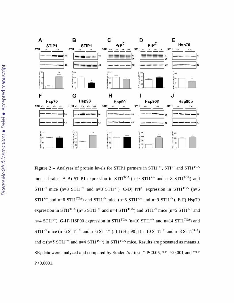
Figure 2 – Analyses of protein levels for STIP1 partners in STI1+/+, STI-/+ and STI1TGA
mouse brains. A-B) STIP1 expression in STI1TGA (n=9 STI1+/+ and n=8 STI1TGA) and
STI1-/+ mice (n=8 STI1+/+ and n=8 STI1-/+). C-D) PrPC expression in STI1TGA (n=6
STI1+/+ and n=6 STI1TGA) and STI1-/+ mice (n=6 STI1+/+ and n=9 STI1-/+). E-F) Hsp70
expression in STI1TGA (n=5 STI1+/+ and n=4 STI1TGA) and STI1-/+ mice (n=5 STI1+/+ and
n=4 STI1-/+). G-H) HSP90 expression in STI1TGA (n=10 STI1+/+ and n=14 STI1TGA) and
STI1-/+ mice (n=6 STI1+/+ and n=6 STI1-/+). I-J) Hsp90 β (n=10 STI1+/+ and n=8 STI1TGA)
and α (n=5 STI1+/+ and n=4 STI1TGA) in STI1TGA mice. Results are presented as means ±
SE; data were analyzed and compared by Student’s t test. * P<0.05, ** P<0.001 and ***
P<0.0001.
Dise
ase
Mod
els &
Mec
hani
sms
D
MM
Acce
pted
man
uscr
ipt
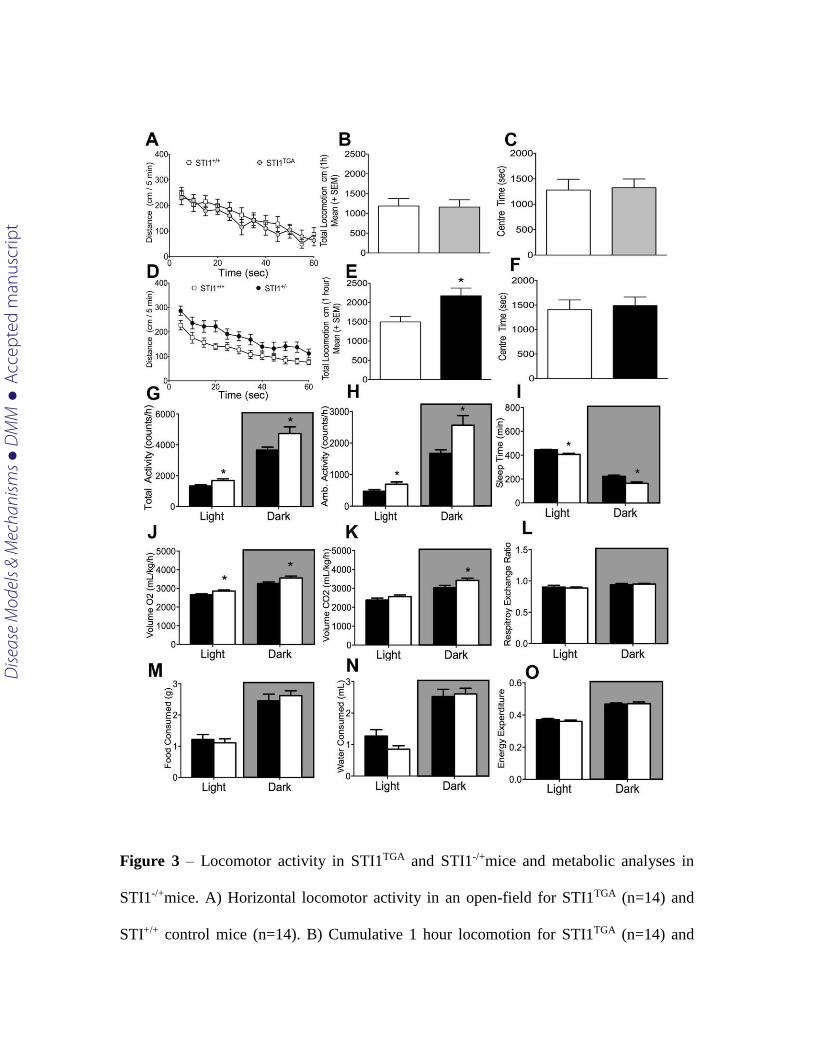
Figure 3 – Locomotor activity in STI1TGA and STI1-/+mice and metabolic analyses in
STI1-/+mice. A) Horizontal locomotor activity in an open-field for STI1TGA (n=14) and
STI+/+ control mice (n=14). B) Cumulative 1 hour locomotion for STI1TGA (n=14) and
Dise
ase
Mod
els &
Mec
hani
sms
D
MM
Acce
pted
man
uscr
ipt
STI+/+ control mice (n=14). C) Time spent in the center of the locomotion boxes for
STI1TGA (n=14) and STI+/+ control mice (n=14). D) Horizontal locomotor activity in an
open-field for STI1-/+ (n=22) and STI+/+ control mice (n=24). E) Cumulative 1 hour
locomotion for STI1-/+ (n=22) and STI+/+ control mice (n=24). F) Time spent in the center
of the locomotion boxes for STI1-/+ (n=22) and STI+/+ control mice (n=24). G) Total
activity in metabolic cages for STI1-/+ (n=22) and STI+/+ control mice (n=24). H)
Ambulatory activity in metabolic cages for STI1-/+ (n=8) and STI+/+ control mice (n=8).
I) Sleep time for STI1-/+ (n=8) and STI+/+ control mice (n=8). J) VO2 for STI1-/+ (n=8)
and STI+/+ control mice (n=8). K) VCO2 for STI1-/+ (n=8) and STI+/+ control mice (n=8).
L) Respiratory exchange ratio for STI1-/+ (n=8) and STI+/+ control mice (n=8). M) Food
Consumption for STI1-/+ (n=8) and STI+/+ control mice (n=8). N) Water consumption for
STI1-/+ (n=8) and STI+/+ control mice (n=8). O) Energy expenditure for STI1-/+ (n=8) and
STI+/+ control mice (n=8). Results are presented as means ± SE; data were analyzed and
compared by Student’s t test * P<0.05.
Dise
ase
Mod
els &
Mec
hani
sms
D
MM
Acce
pted
man
uscr
ipt
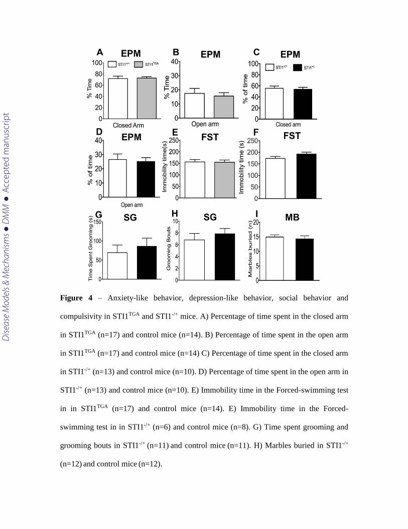
Figure 4 – Anxiety-like behavior, depression-like behavior, social behavior and
compulsivity in STI1TGA and STI1-/+ mice. A) Percentage of time spent in the closed arm
in STI1TGA (n=17) and control mice (n=14). B) Percentage of time spent in the open arm
in STI1TGA (n=17) and control mice (n=14) C) Percentage of time spent in the closed arm
in STI1-/+ (n=13) and control mice (n=10). D) Percentage of time spent in the open arm in
STI1-/+ (n=13) and control mice (n=10). E) Immobility time in the Forced-swimming test
in in STI1TGA (n=17) and control mice (n=14). E) Immobility time in the Forced-
swimming test in in STI1-/+ (n=6) and control mice (n=8). G) Time spent grooming and
grooming bouts in STI1-/+ (n=11) and control mice (n=11). H) Marbles buried in STI1-/+
(n=12) and control mice (n=12).
Dise
ase
Mod
els &
Mec
hani
sms
D
MM
Acce
pted
man
uscr
ipt
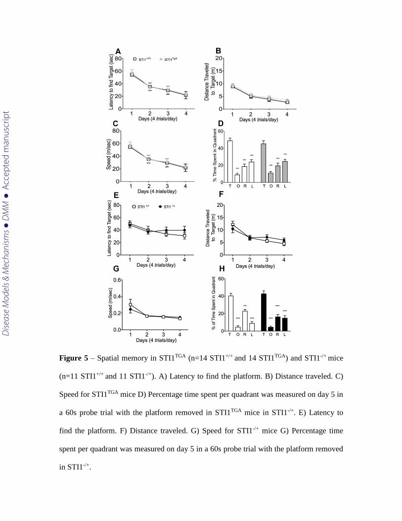
Figure 5 – Spatial memory in STI1TGA (n=14 STI1+/+ and 14 STI1TGA) and STI1-/+ mice
(n=11 STI1+/+ and 11 STI1-/+). A) Latency to find the platform. B) Distance traveled. C)
Speed for STI1TGA mice D) Percentage time spent per quadrant was measured on day 5 in
a 60s probe trial with the platform removed in STI1TGA mice in STI1-/+. E) Latency to
find the platform. F) Distance traveled. G) Speed for STI1-/+ mice G) Percentage time
spent per quadrant was measured on day 5 in a 60s probe trial with the platform removed
in STI1-/+.
Dise
ase
Mod
els &
Mec
hani
sms
D
MM
Acce
pted
man
uscr
ipt
Figure 6 – Five-choice serial reaction time task used to measure attention in STI1TGA
(n=10 STI1+/+ and 10 STI1TGA ) and STI1-/+ (n=13 STI1+/+ and 14 STI1-/+). A) Accuracy
during probe trial sessions. B) Rate of omission. C) Premature responses. D) Response
Latency. E) Reward Collection Latency. F) Perseverative responses for STI1TGA mice. G)
Accuracy during probe trial sessions. H) Rate of omission. I) Premature responses. J)
Response Latency. K) Reward Collection Latency. L) Perseverative response for STI1-
/+mice.
Dise
ase
Mod
els &
Mec
hani
sms
D
MM
Acce
pted
man
uscr
ipt
Translational impact
Clinical Issue - Autism spectrum disorders (ASD) represent a range of
neurodevelopmental disorders with no cure. ASD is characterized by difficulties in
communication and socialization, repetitive movements, hyperactivity, impulsivity and
impaired ability to concentrate and attend to simple tasks. Recent studies have
demonstrated that a number of mothers of ASD children can produce antibodies against
specific proteins present in the fetus brain and presumably the antibodies can interfere
with protein function. One of the antibodies targets a protein known as Stress Inducible
Phosphoprotein 1 or STIP1. In fact, a polymorphism for STIP1 was recently suggested as
a potential risk factor in attention deficit hyperactivity disorder, which shares some
common phenotypes with ASD.
Results – In this work, we show that mice with reduced levels of STIP1 present attention
deficits and are hyperactive. Attention deficits and hyperactivity are present in ASD,
suggesting that interference with STIP1 functions can contribute to ASD-like phenotypes.
Implications and future directions – Changes in STIP1 levels may interfere with brain
circuit development affecting ASD-like behaviour. Future experiments are required in
order to define potential mechanisms as well as the consequences of disturbed STIP1
activity in autism.
Dise
ase
Mod
els &
Mec
hani
sms
D
MM
Acce
pted
man
uscr
ipt