Embed Size (px)
Citation preview
Dis
ease
Mo
dels
& M
echa
nism
s •
DM
M •
Adv
ance
art
icle
© 2016. Published by The Company of Biologists Ltd.
This is an Open Access article distributed under the terms of the Creative Commons Attribution License
(http://creativecommons.org/licenses/by/3.0), which permits unrestricted use, distribution and reproduction
in any medium provided that the original work is properly attributed.
Peroxisome proliferator-activated receptor alpha acts as a mediator of
endoplasmic reticulum stress-induced hepatocyte apoptosis in acute liver
failure
Li Zhang1,2*, Feng Ren2,3*, Xiangying Zhang3, Xinxin Wang4, Hongbo Shi3, Li Zhou2,
Sujun Zheng2, Yu Chen2, Dexi Chen3, Liying Li5, Caiyan Zhao1#, Zhongping Duan2#
1 Department of Infectious Diseases, The Third Affiliated Hospital of Hebei Medical University,
Shijiazhuang, China
2 Beijing Artificial Liver Treatment & Training Center, Beijing YouAn Hospital, Capital Medical
University, Beijing, China
3 Beijing Institute of Hepatology, Beijing YouAn Hospital, Capital Medical University, Beijing, China
4 Department of pathology, Beijing YouAn Hospital, Capital Medical University, Beijing, China
5 Department of Cell Biology, Municipal Laboratory for Liver Protection and Regulation of
Regeneration, Capital Medical University, Beijing, China.
*These authors contributed equally to this work.
# Correspondence author:
Caiyan Zhao, Department of Infectious Diseases, the Third Affiliated Hospital of Hebei
Medical University, Shijiazhuang, China. Phone: +86-311-66776874; Fax:
+86-311-667776875; E-mail address: [email protected].
Zhongping Duan, Beijing Artificial Liver Treatment & Training Center, Beijing YouAn
Hospital, Capital Medical University, Beijing, China. Phone: +86-10-63291007; Fax:
+86-10-63295285; E-mail address: [email protected].
http://dmm.biologists.org/lookup/doi/10.1242/dmm.023242Access the most recent version at DMM Advance Online Articles. Posted 26 May 2016 as doi: 10.1242/dmm.023242http://dmm.biologists.org/lookup/doi/10.1242/dmm.023242Access the most recent version at
First posted online on 26 May 2016 as 10.1242/dmm.023242
Dis
ease
Mo
dels
& M
echa
nism
s •
DM
M •
Adv
ance
art
icle
KEY WORDS: Peroxisome proliferator-activated receptor α, Endoplasmic reticulum
stress, Acute liver failure, Hepatotoxicity, Apoptosis
Summary statement: PPARα can ameliorate hepatic injury via inhibiting ER stress
mediated hepatocyte apoptosis in a mouse model of D-GalN/LPS-induced ALF.
ABSTRACT
Peroxisome proliferator-activated receptor α (PPARα) is a key regulator to
ameliorate liver injury in cases of acute liver failure (ALF). However, its
regulatory mechanisms remain largely undetermined. Endoplasmic reticulum
stress (ER stress) plays an important role in a number of liver diseases. This
study aimed to investigate whether PPARα activation inhibit ER
stress-induced hepatocyte apoptosis, thereby protecting against ALF. In a
murine model of D-galactosamine (D-GalN) and lipopolysaccharide
(LPS)-induced ALF, Wy-14643 was administered to activate PPARα, and
4-phenylbutyric acid (4-PBA) was administered to attenuate ER stress.
PPARα activation ameliorated liver injury, because pre-administration of its
specific inducer, Wy-14643, reduced the serum aminotransferase levels and
preserved liver architecture compared with that of controls. The protective
effect of PPARα activation resulted from the suppression of ER stress-induced
hepatocyte apoptosis. Indeed, (1) PPARα activation decreased the expression
of glucose-regulated protein 78 (Grp78), Grp94 and C/EBP-homologous
Dis
ease
Mo
dels
& M
echa
nism
s •
DM
M •
Adv
ance
art
icle
protein (CHOP) in vivo; (2) the liver protection by 4-PBA was due to the
induction of PPARα expression, because 4-PBA pretreatment promoted
up-regulation of PPARα, and inhibition of PPARα by small interfering RNA
(siRNA) treatment reversed liver protection and increased hepatocyte
apoptosis; (3) in vitro PPARα activation by Wy-14643 decreased the
hepatocyte apoptosis induced by severe ER stress, and PPARα inhibition by
siRNA treatment decreased the hepatocyte survival induced by mild ER stress.
Here, we demonstrated that PPARα activation contributes to liver protection
and decreases hepatocyte apoptosis in ALF, particularly through regulating
ER stress. Therefore, trageting PPARα could be a potential therapeutic
strategy to ameliorate ALF.

Dis
ease
Mo
dels
& M
echa
nism
s •
DM
M •
Adv
ance
art
icle
INTRODUCTION
Acute liver failure (ALF) is a clinical syndrome defined by the sudden onset of severe
liver injury and is characterized by encephalopathy and coagulopathy in patients with
previously normal liver function (Khan et al., 2006). The causes of ALF are diverse
including toxins, infections, or metabolic and genetic diseases, but irrespective of etiology,
ALF results from rapid and extensive hepatic apoptosis and necrosis (Riordan and
Williams, 2003). Despite developments in treatment, orthotopic liver transplantation (OLT)
is still considered the most effective therapy. Unfortunately, the feasibility of OLT is
extremely limited by the rapid progression of the disease and the shortage of donor livers;
therefore, the pathogenesis of ALF needs to be further explored.
Peroxisome proliferator-activated receptors (PPARs) are members of the nuclear
hormone receptor superfamily of ligand-inducible transcription factors. To date, three
subtypes of PPARs (α, β, γ) have been identified in many species, including humans
(Desvergne and Wahli, 1999; Kota et al., 2005). PPARα has been reported to regulate lipid
metabolism (Staels et al., 1998), inflammation (Devchand et al., 1996; Delerive et al.,
1999), cell differentiation and apoptosis (Roberts et al., 2002). Studies have demonstrated
that PPARα plays a different role in cancer cells than in normal cells. PPARα activation is
commonly implicated in hepatocarcinogenesis protocols for rodents in which its
anti-apoptotic action is assumed to play a critical role (Misra et al., 2013; Misra and Reddy,
2014); however, activation of PPARα by exogenous agonists reduces tumor cell growth in
cell lines derived from colorectal cancer (Grau et al., 2006). In non-cancerous renal tubular
cells, a lack of PPARα exacerbates gentamicin-induced apoptosis (Hsu et al., 2008).
Dis
ease
Mo
dels
& M
echa
nism
s •
DM
M •
Adv
ance
art
icle
Additionally, Wy-14643, a potent exogenous PPARα ligand and a selective PPARα
agonist (Cuzzocrea et al, 2004; Briguglio et al., 2010), decreases the apoptosis of
cardiomyocytes via reducing the nuclear translocation of nuclear factor-κB (NF-κB) and
reducing caspase-3 activation, thus preserving myocardial function and maintaining
cardiac contractility (Yeh et al., 2006). In a third normal cell system, PPARα agonist
treatment has been shown to increase trefoil factor family-3 expression and attenuate
apoptosis in the liver tissue of bile duct-ligated rats (Karakan et al., 2013). Our recent study
has shown that PPARα activation protects the liver from acute injury by promoting the
autophagy pathway in the D-galactosamine (D-GalN) and lipopolysaccharide
(LPS)-induced ALF mouse model (Jiao et al., 2014). However, whether PPARα plays a
protective role in the liver by inhibiting hepatocyte apoptosis is yet to be determined.
The endoplasmic reticulum (ER) is a vital cellular organelle for protein folding and
trafficking, lipid synthesis and calcium homeostasis that are required for cell survival and
functions. Endoplasmic reticulum stress (ER stress) is induced by physiological and/or
pathological stress signals, leading to the accumulation of unfolded or misfolded proteins
in the ER, and activates three ER-localized transmembrane protein sensors (Ron and
Walter, 2007; Lin et al., 2008). The chaperone proteins glucose-regulated protein 78
(Grp78) and glucose-regulated protein 94 (Grp94) are master regulators of ER homeostasis
and are hallmarks for ER stress responses (Little et al., 1994). The coordinated adaptive
response is known as the unfolded protein response (UPR), and the pathological response
is known as the ER stress response. The UPR signaling pathways act rapidly to mitigate
the stressed state of the ER and enhance cell survival. However, if severe and prolonged
Dis
ease
Mo
dels
& M
echa
nism
s •
DM
M •
Adv
ance
art
icle
ER stress cannot be resolved, the signaling switches from a pro-survival to a pro-apoptotic
ER stress response (Xu et al., 2005). Compelling evidence has suggested that C/EBP–
homologous protein (CHOP)/growth arrest and DNA damage-inducible protein 153
(GADD153) and caspase-12 in rodents (caspase-4 in humans) are activated and become
involved in ER stress-induced cell apoptosis (Kim et al., 2008). As reported previously,
PPARα protects HepG2 cells against H2O2-induced ER stress-mediates apoptosis through
the down-regulation of CHOP (Tang et al., 2014). Additionally, activation of PPARα
ameliorates hepatic insulin resistance to increased ER stress (Chan et al., 2013). However,
a PPARα agonist has also been shown to induce apoptosis of triple-negative breast cancer
cells via activation of the transcription factor NF-κB, which is connected with the ER
stress response (Zhao et al., 2007). Thus, these studies have demonstrated that PPARα
plays a complicated role in ER stress.
Although our studies have demonstrated that PPARα activation effectively protects
mice from ALF, and severe ER stress promotes liver injury by inducing hepatocyte
apoptosis in D-GalN/LPS treated mice (Jiao et al., 2014; Ren et al., 2015), the underlying
mechanisms of the effects of PPARα and ER stress in vivo required further elucidation.
Thus, this study sought to address the hypothesis that PPARα can protect mice from ALF
by inhibiting ER stress-induced hepatocyte apoptosis. Indeed, we found that inhibition of
ER stress enhanced the expression of PPARα, and PPARα activation attenuated ER
stress-mediated hepatocyte apoptosis in the D-GalN/LPS-induced mouse model of ALF.
Dis
ease
Mo
dels
& M
echa
nism
s •
DM
M •
Adv
ance
art
icle
RESULTS
PPARα activation decreases hepatocyte apoptosis, thus protecting against ALF
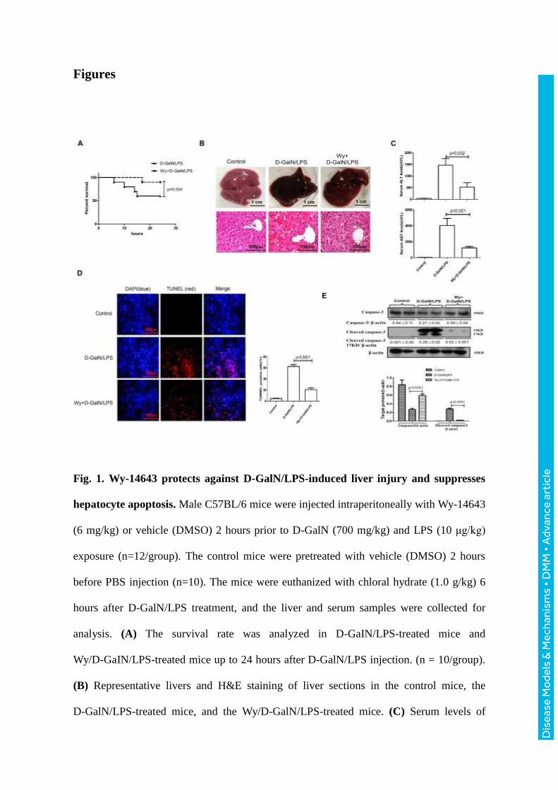
We first evaluated whether PPARα activation could rescue liver injury by applying
Wy-14643, a PPARα ligand activator. In the survival analysis (Fig. 1A), the mice in the
D-GalN/LPS group began to die 6 hours after D-GalN/LPS administration, and the
survival rate stabilized at 60% (6 of 10 mice) at 24 h; however, pretreatment with
Wy-14643 before D-GaIN/LPS administration reduced the mortality, and the survival rate
was 90% (9 of 10 mice). With respect to liver damage, compared with the D-GaIN/LPS
administration group, the gross morphology of the liver appeared to be substantially better
and the liver histopathological damages were ameliorated in the Wy-14643 treatment
group (Fig. 1B); Liver function showed significantly lower alanine aminotransferase (ALT)
and aspartic aminotransferase (AST) levels and lower total bilirubin (TBIL), alkaline
phosphatase (ALP) and prothrombin time (PT) in the Wy-14643 pretreatment group
compared with the D-GaIN/LPS administration group (Fig. 1C, Supplementary figure and
Supplementary table 2). To explore the potential protective mechanism of PPARα against
ALF induced by D-GaIN/LPS, we measured apoptotic cells in the three groups. As shown
in Fig. 1D, in the D-GaIN/LPS-treated group, a large number of TUNEL-positive cells
were observed; however, the Wy-14643 pretreatment group displayed significantly fewer
apoptotic hepatocytes. Moreover, consistently with the TUNEL data, the levels of cleaved
caspase-3 (17 and 19 kDa) increased after D-GaIN/LPS injection, but this increase was
attenuated by Wy-14643 pretreatment (Fig. 1E). Thus, these results suggested that PPARα
Dis
ease
Mo
dels
& M
echa
nism
s •
DM
M •
Adv
ance
art
icle
activation significantly reduced apoptotic cells and thereby protected mice from ALF
induced by D-GaIN/LPS.
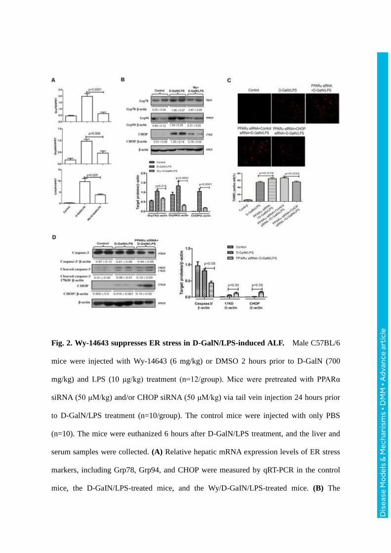
PPARα activation relieves ER stress in D-GaIN/LPS-induced ALF
Our previous paper has shown that severe ER stress promotes liver injury in the
D-GaIN/LPS-induced ALF mouse model (Ren et al., 2015). To examine the effects of
PPARα on D-GaIN/LPS-induced ER stress in mice, we measured the levels of mRNA and
protein for ER stress mediators. The expression of Grp78, Grp94 and CHOP, which are the
classical ER stress markers, was increased significantly after D-GaIN/LPS administration
but was significantly attenuated by pretreatment with Wy-14643 (Fig. 2A). These
alterations were confirmed by western blot analyses (Fig. 2B). We also used siRNA to
knock down the expression of PPARα in mice and found that, compared with
D-GaIN/LPS-treatment, PPARα siRNA treatment further increased the levels of
hepatocyte apoptosis (TUNEL) and promoted the cleavage of caspase-3 and the expression
of CHOP in D-GaIN/LPS-treated ALF mice (Figure 2C, 2D). Furthermore, we further used
siRNA to knockdown CHOP and analysis the hepatocyte apoptosis of liver. The results
showed that, compared to the mice pretreated by PPARα siRNA, the intervention of CHOP
siRNA decreased again the number of hepatocyte apoptosis (Fig. 2C). The results showed
that PPARα activation suppressed ER stress during D-GaIN/LPS-induced ALF.
Inhibition of ER stress increases the expression of PPARα in D-GaIN/LPS-induced
ALF
A small chemical chaperone, 4-phenylbutyric acid (4-PBA), has been shown to
alleviate ER stress both in vivo and in vitro (Ozcan et al., 2006; Zode et al., 2011), and
Dis
ease
Mo
dels
& M
echa
nism
s •
DM
M •
Adv
ance
art
icle
inhibition of ER stress by 4-PBA protects mice from ALF induced by D-GaIN/LPS (Ren et
al., 2015). Thus, we evaluated whether ER stress inhibition could promote the expression
of PPARα in the context of ALF. The qRT-PCR and western blotting results showed that,
compared with D-GalN/LPS treatment alone, pretreatment with 4-PBA promoted the
expression of PPARα (Fig. 3A,B). Similar results were obtained by immunofluorescence
staining of liver tissue. Moreover, our results also showed that the expression of PPARα
was cytoplasmic rather than nuclear in the three groups (Fig. 3C). These results indicated
that the expression of PPARα is promoted by 4-PBA pretreatment in D-GaIN/LPS-induced
ALF.
Inhibition of ER stress protects mice from ALF through PPARα mechanisms
Next, we sought to confirm whether the inhibition of ER stress protects the liver from
injury by inducing PPARα expression in mice. We used siRNA to knock down the
expression of PPARα in mice. The specific inhibition of PPARα in the liver by siRNA in
vivo was confirmed by the reduced levels of PPARα in mice (Fig. 4A). The results
indicated that liver in mice receiving 4-PBA treatment suffered less liver injury and the
hepatic protection was abolished by knockdown of PPARα, which was evidenced by the
decreased survival rate (Fig. 4B), abnormal gross morphology and less preserved liver
architecture as observed from histology (Fig. 4C) and the significantly higher levels of
ALT, AST, TBIL and ALP (Fig. 4D, Supplementary figure and Supplementary table 2).
Meanwhile, the knockdown of PPARα reversed the expression levels of Grp78, Grp94 and
CHOP in 4-PBA-pretreatment ALF mice (Fig. 4E, F). Thus, these results demonstrated
Dis
ease
Mo
dels
& M
echa
nism
s •
DM
M •
Adv
ance
art
icle
that the mechanism of hepatoprotection by ER stress inhibition depends on PPARα
activity.
The expression profile of PPARα in the progression of ER stress-induced hepatocyte
apoptosis in vitro
Here, we further examined how PPARα is regulated in the progression of ER
stress-induced primary hepatocyte apoptosis in vitro. The qRT-PCR and western blot
results showed that the expression of PPARα was significantly up-regulated in the early
stage of tunicamycin- (TM) or thapsigargin (TG)-induced ER stress and was significantly
down-regulated in the later time points of TM or TG treatment compared with the control
group (Fig. 5A-D). Moreover, there was a difference in responses at different doses of TM
or TG, compared with the control group. The low dose of TM or TG markedly
up-regulated PPARα expression, whereas the high dose of TM or TG reduced the
expression of PPARα (Fig. 5E-H). Moreover, for the longer time and higher dose of TM or
TG treatment, the cleavage of caspase-3 was increased (Fig. 5B,D,F,H). Therefore, these
results showed that mild ER stress promotes the expression of PPARα, and severe ER
stress reduces the expression of PPARα.
The effect of PPARα regulation on ER stress-induced primary hepatocyte apoptosis
in vitro
PPARα had been shown to be differentially regulated in the progression of ER stress.
Therefore, we further analyzed the impact of PPARα on the intrinsic potential of primary
hepatocyte apoptosis triggered by ER stress in vitro. Under the conditions of mild ER
stress, we used specific siRNA to knock down the expression of PPARα. TM or TG
Dis
ease
Mo
dels
& M
echa
nism
s •
DM
M •
Adv
ance
art
icle
treatment for 6 hours increased the release of lactate dehydrogenase (LDH) from the
hepatocytes and decreased hepatocyte viability; down-regulation of PPARα by siRNA
further increased the LDH levels from the hepatocytes and further decreased hepatocyte
viability (Fig. 6A). To evaluate the role of CHOP in the ER stress-PPARα pathway, we
used siRNA to knock down CHOP and analyzed the level of LDH release and MTT in the
different groups. The results indicated that, compared with the combination of PPARα
siRNA and TM or TG treatment, the silencing of CHOP with siRNA partially reversed the
levels of LDH and cell viability (Fig. 6A). Western blot analysis revealed that PPARα
siRNA increased the levels of CHOP and cleaved caspase-3 compared with TM or
TG-treated cells (Fig. 6B). Under conditions of severe ER stress, we used Wy-14643 to
activate PPARα. Compared with 24 hour treatment of hepatocytes with TM or TG,
activation of PPARα by Wy-14643 significantly decreased the hepatocyte levels of LDH
and increased hepatocyte viability (Fig. 6C). Western blot analysis also indicated that
Wy-14643 decreased the levels of CHOP and cleaved caspase-3, as compared with TM or
TG-treated cells (Fig. 6D). Therefore, the activation or expression of PPARα was a key
point of balance between hepatocyte survival promoted by mild ER stress and hepatocyte
apoptosis induced by severe ER stress.
The expression of CHOP and PPARα in the liver of ALF patients with HBV infection
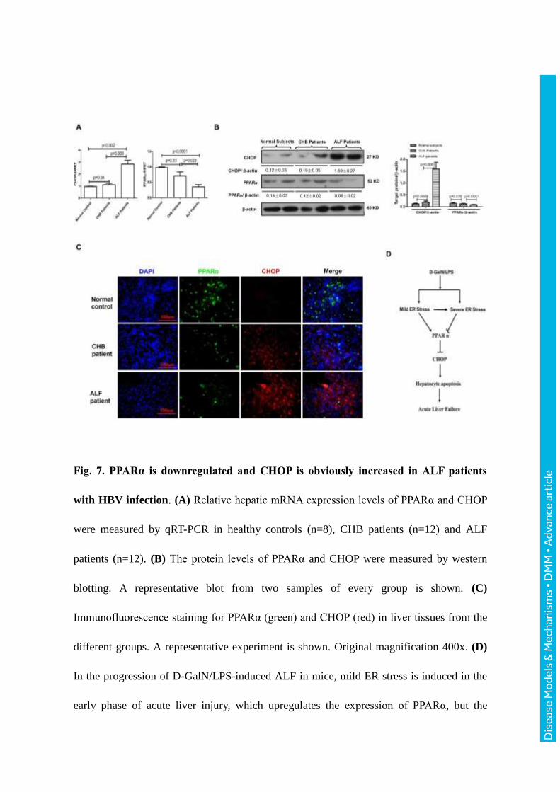
To investigate whether CHOP and PPARα associate with the progression of ALF in
patients with HBV infection, we quantified the expression of CHOP and PPARα in liver
tissues of normal subjects, chronic hepatitis B (CHB) patients and HBV-related ALF
patients. The qRT-PCR results revealed that CHOP gene expression increased significantly
Dis
ease
Mo
dels
& M
echa
nism
s •
DM
M •
Adv
ance
art
icle
in ALF patients compared to the normal subjects, but in the patients with CHB, no
significant changes were observed. PPARα gene expression gradually decreased in the
progression of CHB to ALF (Fig. 7A); similar results were observed for protein levels by
western blot analysis (Fig. 7B). Interestingly, the immunofluorescence staining revealed
that the expression level of CHOP was low in the hepatocytes in which PPARα was highly
expressed (Fig. 7C). Thus, these results indicated that CHOP expression is up-regulated,
and PPARα expression is decreased in patients with HBV-related ALF compared with
normal livers.
Dis
ease
Mo
dels
& M
echa
nism
s •
DM
M •
Adv
ance
art
icle
DISCUSSION
In the present study, we demonstrated that PPARα activation significantly decreased
hepatocellular apoptosis, thereby protecting mice from D-GalN/LPS-induced ALF. The
protective mechanism of PPARα activation was that PPARα activation regulates ER stress
and thus relieves liver injury caused by ALF in mice; moreover, PPARα could be a pivotal
molecule that facilitates the transition from mild ER stress to progressively severe ER
stress in ALF. Hence, the ER stress-PPARα pathway is necessary to the pathological
mechanism of the ALF immune response cascade (depicted in Fig. 7D).
Acute liver failure (ALF) has a variety of etiologies including viral infection,
acetaminophen damage, excessive alcohol, metabolic liver disease and uncertain causes. It
is associated with massive hepatocellular death. The mode of hepatocyte death includes
apoptosis or necrosis, but is still controversial. Traditionally, apoptosis or programmed cell
death (PCD), is actively induced by specific signaling cascades, including the intrinsic and
extrinsic apoptosis signaling pathways, and occurs in a highly controlled fashion. Necrosis
is viewed as a largely unregulated consequence of physicochemical stress characterized by
mitochondrial impairment, depletion of adenosine triphosphate (ATP), and subsequent
failure of ATP-dependent ion pumps. Recent evidence has indicated that PCD can also
trigger a specific form of necrosis, termed necroptosis (Heike et al., 2012, Tom et al., 2014).
The regulated nature of multiple cell death modes not only affects our understanding of the
underlying pathophysiology but also suggests the possibility of therapeutic treatment in
diseases.
The first novel finding in this paper is that PPARα activation protects mice from liver
Dis
ease
Mo
dels
& M
echa
nism
s •
DM
M •
Adv
ance
art
icle
injury by inhibiting ER stress-induced hepatocyte apoptosis in ALF. Prolonged or severe
ER stress triggers cell apoptosis. Several mediators of apoptosis are associated with ER
stress-induced cell death. Some of the mediators are linked to the UPR sensors, but others
are implicated in calcium and redox homeostasis. The transcription factor CHOP functions
as the most well-characterized pro-apoptotic regulator. Previous studies have demonstrated
that CHOP is significantly upregulated in GaIN/LPS induced ALF and is critical in
mediating ER stress-induced apoptosis (Rao et al., 2015). Silencing of CHOP reduces
hepatocyte apoptosis in alcohol induced liver disease (Ji et al., 2005; Tamaki et al., 2008).
Our previous research has also shown that the expression levels of Grp78, Grp94 and
CHOP are increased significantly in D-GaIN/LPS-induced ALF, demonstrating the critical
role of ER stress-mediated hepatocyte apoptosis in the mechanisms of ALF (Chen et al.,
2012). The studies have shown that PPARα plays a complex role in cell apoptosis. For
example, PPARα shows duality in liver cancer: low amounts of PPARα activation increase
cell apoptosis by changing the tumor microenvironment, and continued high levels of
PPARα activation promote the growth of hepatoma carcinoma cells (Kimura et al., 2012).
For normal cells, such as hepatocytes, vascular smooth muscle cells or kidney cells,
PPARα activation suppresses apoptosis induced by various stimuli (Chung et al., 2012;
Chen et al., 2013; Karakan et al., 2013). In the present study, we demonstrated that PPARα
activation, through its agonist Wy-14643, down-regulated expression of Grp78, Grp94 and
CHOP and reduced D-GaIN/LPS-induced ER stress-mediated cell apoptosis. Moreover,
our results in vitro also indicated that knockdown of PPARα by siRNA or activation of
PPARα by Wy-14643 promoted or inhibited ER stress-induced hepatocyte apoptosis,
Dis
ease
Mo
dels
& M
echa
nism
s •
DM
M •
Adv
ance
art
icle
respectively. Furthermore, inhibition of ER stress directly up-regulated the expression of
PPARα in the ALF mouse model, and knockdown of PPARα reversed the protective effect
of ER stress inhibition in the ALF mouse model. Together with the results reported here,
these findings support a mechanism whereby severe ER stress promotes the progression of
D-GaIN/LPS-induced ALF in mice by decreasing PPARα activation.
Another novel finding in this paper is that PPARα acts as a switch from mild ER stress
to severe ER stress. ER stress and UPR have been linked to the pathophysiology of liver
diseases. However, the UPR signaling pathways also play a crucial role in restoring ER
homeostasis via PERK, IRE1, and ATF6. One set of effectors regulated by the UPR
activates three adaptive signaling cascades to amoliorate ER stress. These adiptive
mechanisms involve global attenuation of mRNA translation, which reduces the ER
workload by blocking synthesis of new proteins; the upregulation of molecular chaperones,
which expands the protein folding capacity of the ER; and the increase in ER-associated
protein degradation (ERAD), which removes misfolded proteins from the ER. (Treglia et
al., 2012). Under sustained or massive ER stress, the UPR switches from an adaptive
program to a pro-apoptotic program. The CHOP protein is thought to be a critical mediator
of ER stress-associated apoptosis (Kim et al., 2008). Therefore, the UPR activation elicits
adaptive and pro-apoptotic effectors, and the UPR signaling serves as a binary switch
between adaptation and death. What are the molecular mechanisms to govern this
transition? Chan et al. have shown that JNK functions as a key factor that regulates β-cells
fate (Chan et al., 2015). In this paper, our findings suggest that PPARα could be a pivotal
molecule that facilitates the transition from mild ER stress-induced cell survival to
Dis
ease
Mo
dels
& M
echa
nism
s •
DM
M •
Adv
ance
art
icle
progressively severe ER stress-induced cell apoptosis. Our research has found that PPARα
is expressed in normal hepatocytes and that mild ER stress upregulates the expression of
PPARα, whereas severe ER stress downregulates the expression of PPARα. Knockdown of
PPARα decreases the mild ER stress-promoted hepatocyte survival, whereas the activation
of PPARα decreases the severe ER stress-induced hepatocyte apoptosis. Therefore, we
believe that PPARα is a new a mediator involved in the balance between adaptive and
apoptotic factors regulated by the UPR.
In conclusion, we found that PPARα prevents ALF by suppressing ER stress-induced
hepatocyte apoptosis. PPARα may be useful as a potential therapeutic agent to attenuate
ALF. Further preclinical studies targeting PPARα agonists are warranted for the
development of a clinically applicable treatment strategy to treat ALF.
Dis
ease
Mo
dels
& M
echa
nism
s •
DM
M •
Adv
ance
art
icle
MATERIALS AND METHODS
Animal experiments
Male C57BL/6 mice at the age of 8-12 weeks were purchased from the Capital
Medical University (Beijing, China) and fed freely with a standard chow diet and water;
they were housed under specific pathogen-free conditions for 1 week before the
experiments. All animals received humane care according to the Capital Medical
University Animal Care Committee guidelines.
The mice were intraperitoneally injected with D-GalN (700 mg/kg; Sigma, St. Louis,
MO, USA) and LPS (10 μg/kg; InvivoGen, San Diego, CA, USA) to induce ALF or with
saline in the control animals. The PPARα activator Wy-14643 (6 mg/kg; Sigma) was
administered via injection into the tail vein 2 hours prior to D-GalN/LPS exposure. The
downregulation of PPARα and CHOP were achieved by tail vein injection of specific
siRNA (50 μM/kg; Jima, Suzhou). A chemical chaperone that relieves ER stress, 4-PBA
(100 mg/kg; Sigma, St Luis, MO), was dissolved in PBS and administered
intraperitoneally 6 hours prior to D-GalN/LPS exposure. The mice were sacrificed at 6
hours after D-GalN/LPS treatment, and liver and serum samples were collected for future
analysis.
Human specimens.
Normal liver samples were collected from eight patients undergoing hepatic resection
for liver transplantation. CHB samples were obtained from the livers of 12 patients
undergoing liver puncture biopsy. ALF liver samples were obtained from the livers of 12
patients with HBV infection undergoing liver transplantation, which caused by acute
Dis
ease
Mo
dels
& M
echa
nism
s •
DM
M •
Adv
ance
art
icle
exacerbation of chronic hepatitis B. This study was conducted in compliance with the 1975
Declaration of Helsinki, and the study protocol was approved by the Medical Ethics
Committee of the Beijing YouAn Hospital. Written informed consent was obtained from
all patients or their families prior to enrollment. The clinical characteristics and details of
the patients included in the study are shown in the supplementary material Table1.
Liver function tests and liver histological examination
Liver injury was estimated by biochemical serum markers such as albumin (ALB),
ALT, AST, TBIL, ALP and by coagulation index such as PT and by pathological
examination. Blood biochemical indicators were measured by using a multi-parametric
analyzer (AU 5400, Olympus, Japan), according to an automated procedure. PT was
detected using fully Automatic Coagulometer (Ac.T 5diff AL, Beckman-Coulter Inc., Brea,
CA, USA). Liver tissue was fixed with 10% neutral formaldehyde and then embedded in
paraffin. The specimens were cut into 5 μm sections, which were then stained with
hematoxylin and eosin (H&E) and observed under light microscopy.
Quantitative real-time polymerase chain reaction
Total RNA was isolated from 50 mg of liver tissue with TRIzol reagent, following
the manufacturer’s protocol. The RNA was reverse transcribed into cDNA using the
SuperScript Ⅲ First-Strand Synthesis System (Invitrogen, Carlsbad, CA, USA).
Quantitative-PCR was performed using the DNA Engine with Chromo 4 Detector (MJ
Research, Waltham, MA). The reactions were set up in 20μl total volumes with 1x
SuperMix (Platinum SYBR Green qPCR Kit; Invitrogen), cDNA (2 μl) and 0.5 μM of each
primer. The PCR cycle was as follows: 50℃ for 2 minutes and 95℃ for 5 minutes,
Dis
ease
Mo
dels
& M
echa
nism
s •
DM
M •
Adv
ance
art
icle
followed by 50 cycles of 95℃ for 15 seconds and 60℃ for 30 seconds. The relative
mRNA levels were normalized to the level of hypoxanthine-guanine
phosphoribosyltransferase (HPRT) and calculated by using the 2-△△Ct method. All samples
were run in duplicate to ensure amplification integrity.
Western blot analyses
Liver tissue samples were lysed in Radio Immunoprecipitation Assay (RIPA) buffer
containing phosphatase and protease inhibitors. After heat denaturation at 95℃ for 5
minutes, proteins in SDS-loading buffer were subjected to electrophoresis in an SDS-12%
polyacrylamide gel and subsequently transferred onto a PVDF membrane (Bio-Rad,
Hercules, CA, USA). Primary antibodies against PPARα (Abcam, Cambridge, MA, USA),
Grp78, Grp94, CHOP, caspase-3, cleaved caspase-3 and β-actin (Cell Signaling
Technology Inc., Santa Cruz, CA, USA) were used. The membranes were incubated with
primary antibodies (1:500-1:1000) in TBST with 5% skim milk at 4℃ overnight. The
membranes were washed with TBST three times and then were incubated with horseradish
peroxidase-conjugated secondary antibodies (1:2000) at room temperature for 1 hour. The
bands were visualized with SuperSignal West Pico chemiluminescent substrate (Thermo
Fisher Scientific, Rockford, IL, USA) and developed by exposure on an X-ray film.
TUNEL assay
Apoptosis in liver sections was detected by terminal deoxynucleotidyl
transferase-mediated dUTP nick-end labeling (TUNEL, red fluorescence) using the In Situ
Cell Death Detection Kit (Roche, Indianapolis, IN). Negative controls were prepared
through omission of the terminal transferase. Positive controls were generated by treatment
Dis
ease
Mo
dels
& M
echa
nism
s •
DM
M •
Adv
ance
art
icle
with DNase. Nuclei were stained with 4’,6-diamidino-2-phenylindole (DAPI; 1 μg/ml;
Shizebio, Shanghai, China) for 10 minutes. Images were obtained on a Nikon Eclipse E800
fluorescent microscope (Nikon Corp., Tokyo, Japan). After four fields were randomly
selected from each section, 100 cells were successively counted for each field by an
observer who did not identify the slides. The ratio of TUNEL-positive cell number to the
total cell number is shown.
Isolation and treatment of primary mouse hepatocytes
The livers of 7-week old mice were perfused with collagenase-containing Hank’s
solution, and viable hepatocytes were isolated by Percoll isodensity centrifugation as
described (Klaunig et al., 1981). To study the effects of PPARα regulation on hepatocyte
apoptosis induced by ER stress, the cells were treated with TM (10 μg/ml; Sigma) or TG (1
μg/ml; Sigma), which increases ER stress, and the indicated conditions including
co-treatment with Wy-14643 (50μM), and/or PPARα siRNA (5 nM), and/or CHOP siRNA
(5 nM). The MTT assay (Amersco, Solon, OH, USA) was used as a qualitative index of
cell proliferation. Hepatocyte apoptosis was evaluated by western blotting for cleaved
caspase-3 and by the LDH assay (Biochain Institute, Hayward, CA) of culture supernatants.
The processing was conducted according to the manufacturer’s instructions.
Immunofluorescence staining
Paraffin sections were treated with xylene for 10 minutes three times. The sections
were hydrated through a graded alcohol series and then rinsed three times with distilled
water. After the sections were blocked for 20 minutes in 10% goat serum in PBS, they
were incubated overnight at 4 °C with the PPARα-specific rabbit polyclonal antibody

Dis
ease
Mo
dels
& M
echa
nism
s •
DM
M •
Adv
ance
art
icle
(Abcam, Cambridge, MA, USA) and the CHOP-specific mouse monoclonal antibody (Cell
Signaling Technology Inc., Santa Cruz, CA, USA). The slides were then incubated with
Alexa Fluor® 488 goat anti-rabbit IgG or Alexa Fluor® 568 goat anti-mouse IgG (1:200;
Invitrogen, Grand Island, NY, USA) for 45 minutes. After three washes with PBS, the
nuclei were stained with DAPI (1 μg/ml; Shizebio, Shanghai, China) for 10 minutes. The
images were examined on a Nikon Eclipse E800 fluorescent microscope (Nikon Corp.,
Tokyo, Japan).
Statistical analyses
The results are expressed as the means±standard deviation (SD). Statistical analyses
were performed using the unpaired Student’s t test or single-factor analysis of variance,
and a value of P < 0.05 (two-tailed) was considered significant.
Dis
ease
Mo
dels
& M
echa
nism
s •
DM
M •
Adv
ance
art
icle
Competing interests
The authors declare no conflicts of interest.
Author contributions
C.Z. and Z.D. designed the experiments; L. Zhang and F.R. performed the experiments and wrote the
manuscript; X.W. supervised the pathological observation; L. Zhang, F.R., X.Z., H.S. and L. Zhou
prepared the samples and collected the data. L. Zhang, F.R., S.Z., Y.C., D.C. and L.L. performed
statistical analyses. All authors have read and approved the submission of the manuscript.
Funding
This study was supported by the China National Key Project of the Twelfth Five-year Plan
(2012ZX10002004-006, 2012ZX10004904-003-001, 2013ZX10002002-006-001), the
National Natural Science Foundation of China (81270532, 81372094,81300349), the Wang
Boen Liver Fibrosis Research Foundation of CFHPC (CFHPC20131031), the Natural
Science Foundation of Beijing (7162085), Beijing Municipal Science & Technology
Commission (Z161100000516113), The Project of Construction of Innovative Teams and
Teacher Career Development for Universities and Colleges Under Beijing Municipality
(IDHT20150502), the High-level Technical Personnel Training Plan of the Beijing Health
System (2013-3-075) and the Innovation Project Fund Designated for Graduate Student of
Academic Degree Commission of Hebei Provincial Education Department.
Dis
ease
Mo
dels
& M
echa
nism
s •
DM
M •
Adv
ance
art
icle
References
Briguglio, E., Di, P.R., Paterniti, I., Mazzon, E., Oteri, G., Cordasco, G. and Cuzzocrea, S.
(2010).WY-14643, a potent peroxisome proliferator pctivator receptor-α agonist ameliorates the
inflammatory process associated to experimental periodontitis. PPAR Res. 2010, 193019.
Chan, J. Y., Luzuriaga, J., Maxwell, E. L., West, P. K., Bensellam, M. and Laybutt, D. R. (2015). The
balance between adaptive and apoptotic unfolded protein responses regulates beta-cell death under ER
stress conditions through XBP1, CHOP and JNK. Mol. Cell. Endocrinol.413, 189-201.
Chan, S. M., Sun, R. Q., Zeng, X. Y., Choong, Z. H., Wang, H., Watt, M. J. and Ye, J. M. (2013).
Activation of PPARalpha ameliorates hepatic insulin resistance and steatosis in high fructose-fed mice
despite increased endoplasmic reticulum stress. Diabetes.62, 2095-2105.
Chen, L., Ren, F., Zhang, H., Wen, T., Piao, Z., Zhou, L., Zheng, S., Zhang, J., Chen, Y., Han, Y. et al.
(2012). Inhibition of glycogen synthase kinase 3beta ameliorates D-GalN/LPS-induced liver injury by
reducing endoplasmic reticulum stress-triggered apoptosis. PLoS One.7, e45202.
Chen, Y. C., Chu, L. Y., Yang, S. F., Chen, H. L., Yet, S. F. and Wu, K. K. (2013). Prostacyclin and
PPARalpha agonists control vascular smooth muscle cell apoptosis and phenotypic switch through distinct
14-3-3 isoforms. PLoS One.8, e69702.
Chung, H. W., Lim, J. H., Kim, M. Y., Shin, S. J., Chung, S., Choi, B. S., Kim, H. W., Kim, Y. S., Park,
C. W. and Chang, Y. S. (2012). High-fat diet-induced renal cell apoptosis and oxidative stress in
spontaneously hypertensive rat are ameliorated by fenofibrate through the
PPARalpha-FoxO3a-PGC-1alpha pathway. Nephrol. Dial. Transplant.27, 2213-2225.
Cuzzocrea, S., Di, P.R, Mazzon, E. Genovese, T., Muià, C. and Caputi, A.P. (2004). WY 14643, a potent
exogenous PPAR-alpha ligand, reduces intestinal injury associated with splanchnic artery occlusion
shock.Shock. 22, 340-346.
Delerive, P., De Bosscher, K., Besnard, S., Vanden Berghe, W., Peters, J. M., Gonzalez, F. J., Fruchart,
J. C., Tedgui, A., Haegeman, G. and Staels, B. (1999). Peroxisome proliferator-activated receptor alpha
negatively regulates the vascular inflammatory gene response by negative cross-talk with transcription
factors NF-kappaB and AP-1. J. Biol. Chem.274, 32048-32054.
Desvergne, B. and Wahli, W. (1999). Peroxisome proliferator-activated receptors: nuclear control of
metabolism. Endocr. Rev.20, 649-688.
Devchand, P. R., Keller, H., Peters, J. M., Vazquez, M., Gonzalez, F. J. and Wahli, W. (1996). The
PPARalpha-leukotriene B4 pathway to inflammation control. Nature.384, 39-43.
Grau, R., Punzon, C., Fresno, M. and Iniguez, M. A. (2006). Peroxisome-proliferator-activated receptor
alpha agonists inhibit cyclo-oxygenase 2 and vascular endothelial growth factor transcriptional activation
in human colorectal carcinoma cells via inhibition of activator protein-1. Biochem. J.395, 81-88.
Heike, B. and Klaus, S.O. (2012). Mechanisms of cell death in acute liver failure. Front. Physiol. 3: 79.
Hsu, Y. H., Chen, C. H., Hou, C. C., Sue, Y. M., Cheng, C. Y., Cheng, T. H., Lin, H., Tsai, W. L., Chan,
P. and Chen, T. H. (2008). Prostacyclin protects renal tubular cells from gentamicin-induced apoptosis
via a PPARalpha-dependent pathway. Kidney Int.73, 578-587.
Ji, C., Mehrian-Shai, R., Chan, C., Hsu, Y. H. and Kaplowitz, N. (2005). Role of CHOP in hepatic
apoptosis in the murine model of intragastric ethanol feeding. Alcohol. Clin. Exp. Res.29, 1496-1503.
Jiao, M., Ren, F., Zhou, L., Zhang, X., Zhang, L., Wen, T., Wei, L., Wang, X., Shi, H., Bai, L. et al.
(2014). Peroxisome proliferator-activated receptor alpha activation attenuates the inflammatory response
to protect the liver from acute failure by promoting the autophagy pathway. Cell Death Dis.5, e1397.
Dis
ease
Mo
dels
& M
echa
nism
s •
DM
M •
Adv
ance
art
icle
Karakan, T., Kerem, M., Cindoruk, M., Engin, D., Alper, M. and Akin, O. (2013). PPAR-alpha agonist
treatment increases trefoil factor family-3 expression and attenuates apoptosis in the liver tissue of bile
duct-ligated rats. Turk J Gastroenterol.24, 134-140.
Khan, S. A., Shah, N., Williams, R. and Jalan, R. (2006). Acute liver failure: a review. Clin Liver Dis.10,
239-258, vii-viii.
Kim, I., Xu, W. and Reed, J. C. (2008). Cell death and endoplasmic reticulum stress: disease relevance and
therapeutic opportunities. Nat Rev Drug Discov.7, 1013-1030.
Kimura, O., Kondo, Y. and Shimosegawa, T. (2012). PPAR Could Contribute to the Pathogenesis of
Hepatocellular Carcinoma. PPAR Res.2012, 574180.
Klaunig, J. E., Goldblatt, P. J., Hinton, D. E., Lipsky, M. M., Chacko, J. and Trump, B. F. (1981).
Mouse liver cell culture. I. Hepatocyte isolation. In Vitro.17, 913-925.
Kota, B. P., Huang, T. H. and Roufogalis, B. D. (2005). An overview on biological mechanisms of PPARs.
Pharmacol. Res.51, 85-94.
Lin, J. H., Walter, P. and Yen, T. S. (2008). Endoplasmic reticulum stress in disease pathogenesis. Annu
Rev Pathol.3, 399-425.
Little, E., Ramakrishnan, M., Roy, B., Gazit, G. and Lee, A. S. (1994). The glucose-regulated proteins
(GRP78 and GRP94): functions, gene regulation, and applications. Crit. Rev. Eukaryot. Gene Expr.4, 1-18.
Misra, P. and Reddy, J. K. (2014). Peroxisome proliferator-activated receptor-alpha activation and excess
energy burning in hepatocarcinogenesis. Biochimie.98, 63-74.
Misra, P., Viswakarma, N. and Reddy, J. K. (2013). Peroxisome proliferator-activated receptor-alpha
signaling in hepatocarcinogenesis. Subcell. Biochem.69, 77-99.
Ozcan, U., Yilmaz, E., Ozcan, L., Furuhashi, M., Vaillancourt, E., Smith, R. O., Gorgun, C. Z. and
Hotamisligil, G. S. (2006). Chemical chaperones reduce ER stress and restore glucose homeostasis in a
mouse model of type 2 diabetes. Science.313, 1137-1140.
Rao, J., Zhang, C., Wang, P., Lu, L., Qian, X., Qin, J., Pan, X., Li, G., Wang, X. and Zhang, F. (2015).
C/EBP homologous protein (CHOP) contributes to hepatocyte death via the promotion of ERO1alpha
signalling in acute liver failure. Biochem. J.466, 369-378.
Ren, F., Zhou, L., Zhang, X., Wen, T., Shi, H., Xie, B., Li, Z., Chen, D., Wang, Z. and Duan, Z. (2015).
Endoplasmic reticulum stress-activated glycogen synthase kinase 3beta aggravates liver inflammation and
hepatotoxicity in mice with acute liver failure. Inflammation.38, 1151-1165.
Riordan, S. M. and Williams, R. (2003). Mechanisms of hepatocyte injury, multiorgan failure, and
prognostic criteria in acute liver failure. Semin. Liver Dis.23, 203-215.
Roberts, R. A., Chevalier, S., Hasmall, S. C., James, N. H., Cosulich, S. C. and Macdonald, N. (2002).
PPAR alpha and the regulation of cell division and apoptosis. Toxicology.181-182, 167-170.
Ron, D. and Walter, P. (2007). Signal integration in the endoplasmic reticulum unfolded protein response.
Nat. Rev. Mol. Cell Biol.8, 519-529.
Staels, B., Dallongeville, J., Auwerx, J., Schoonjans, K., Leitersdorf, E. and Fruchart, J. C. (1998).
Mechanism of action of fibrates on lipid and lipoprotein metabolism. Circulation.98, 2088-2093.
Tamaki, N., Hatano, E., Taura, K., Tada, M., Kodama, Y., Nitta, T., Iwaisako, K., Seo, S., Nakajima,
A., Ikai, I. et al. (2008). CHOP deficiency attenuates cholestasis-induced liver fibrosis by reduction of
hepatocyte injury. Am. J. Physiol. Gastrointest. Liver Physiol.294, G498-505.
Tang, W. X., Wang, L. K., Wang, Y. Q., Zong, Z. J., Gao, Z. X., Liu, X. S., Shen, Y. J., Shen, Y. X. and
Li, Y. H. (2014). Peroxisome proliferator-activated receptor-alpha activation protects against endoplasmic
reticulum stress-induced HepG2 cell apoptosis. Mol. Cell. Biochem.385, 179-190.
Dis
ease
Mo
dels
& M
echa
nism
s •
DM
M •
Adv
ance
art
icle
Tom, L., Neil, K. and Robert, F. S. (2014) Cell death and cell death responses in liver disease: mechanisms
and clinical relevance. Gastroenterology. 147, 765-783.
Treglia, A. S., Turco, S., Ulianich, L., Ausiello, P., Lofrumento, D. D., Nicolardi, G., Miele, C., Garbi,
C., Beguinot, F. and Di Jeso, B. (2012). Cell fate following ER stress: just a matter of "quo ante"
recovery or death? Histol. Histopathol.27, 1-12.
Xu, C., Bailly-Maitre, B. and Reed, J. C. (2005). Endoplasmic reticulum stress: cell life and death
decisions. J Clin Invest.115, 2656-2664.
Yeh, C. H., Chen, T. P., Lee, C. H., Wu, Y. C., Lin, Y. M. and Lin, P. J. (2006). Cardiomyocytic
apoptosis following global cardiac ischemia and reperfusion can be attenuated by peroxisome
proliferator-activated receptor alpha but not gamma activators. Shock.26, 262-270.
Zhao, W., Iskandar, S., Kooshki, M., Sharpe, J. G., Payne, V. and Robbins, M. E. (2007). Knocking out
peroxisome proliferator-activated receptor (PPAR) alpha inhibits radiation-induced apoptosis in the mouse
kidney through activation of NF-kappaB and increased expression of IAPs. Radiat. Res.167, 581-591.
Zode, G. S., Kuehn, M. H., Nishimura, D. Y., Searby, C. C., Mohan, K., Grozdanic, S. D., Bugge, K.,
Anderson, M. G., Clark, A. F., Stone, E. M. et al. (2011). Reduction of ER stress via a chemical
chaperone prevents disease phenotypes in a mouse model of primary open angle glaucoma. J Clin
Invest.121, 3542-3553.
Dis
ease
Mo
dels
& M
echa
nism
s •
DM
M •
Adv
ance
art
icle
Figures
Fig. 1. Wy-14643 protects against D-GalN/LPS-induced liver injury and suppresses
hepatocyte apoptosis. Male C57BL/6 mice were injected intraperitoneally with Wy-14643
(6 mg/kg) or vehicle (DMSO) 2 hours prior to D-GalN (700 mg/kg) and LPS (10 μg/kg)
exposure (n=12/group). The control mice were pretreated with vehicle (DMSO) 2 hours
before PBS injection (n=10). The mice were euthanized with chloral hydrate (1.0 g/kg) 6
hours after D-GalN/LPS treatment, and the liver and serum samples were collected for
analysis. (A) The survival rate was analyzed in D-GaIN/LPS-treated mice and
Wy/D-GaIN/LPS-treated mice up to 24 hours after D-GalN/LPS injection. (n = 10/group).
(B) Representative livers and H&E staining of liver sections in the control mice, the
D-GalN/LPS-treated mice, and the Wy/D-GalN/LPS-treated mice. (C) Serum levels of

Dis
ease
Mo
dels
& M
echa
nism
s •
DM
M •
Adv
ance
art
icle
ALT and AST from the different groups. (D) TUNEL staining images from the different
groups. A representative experiment is shown. Original magnification 200x. (E) The levels
of total caspase-3, cleaved caspase-3 and β-actin were measured by western blotting. A
representative blot from two samples of every group is shown. Densitometry analysis of
the protein levels was performed for each sample.
Dis
ease
Mo
dels
& M
echa
nism
s •
DM
M •
Adv
ance
art
icle
Fig. 2. Wy-14643 suppresses ER stress in D-GalN/LPS-induced ALF. Male C57BL/6
mice were injected with Wy-14643 (6 mg/kg) or DMSO 2 hours prior to D-GalN (700
mg/kg) and LPS (10 μg/kg) treatment (n=12/group). Mice were pretreated with PPARα
siRNA (50 μM/kg) and/or CHOP siRNA (50 μM/kg) via tail vein injection 24 hours prior
to D-GalN/LPS treatment (n=10/group). The control mice were injected with only PBS
(n=10). The mice were euthanized 6 hours after D-GalN/LPS treatment, and the liver and
serum samples were collected. (A) Relative hepatic mRNA expression levels of ER stress
markers, including Grp78, Grp94, and CHOP were measured by qRT-PCR in the control
mice, the D-GaIN/LPS-treated mice, and the Wy/D-GaIN/LPS-treated mice. (B) The
Dis
ease
Mo
dels
& M
echa
nism
s •
DM
M •
Adv
ance
art
icle
protein levels of Grp78, Grp94, CHOP and β-actin were measured by western blotting. A
representative blot from two samples of every group is shown. Densitometry analysis of
the proteins was performed for each sample. (C) TUNEL staining images from the control
mice, the D-GalN/LPS-treated mice, the PPARα siRNA/D-GalN/LPS-treated mice, the
PPARα siRNA/ control siRNA /D-GalN/LPS-treated mice and the PPARα siRNA/CHOP
siRNA /D-GalN/LPS-treated mice. A representative experiment is shown. Original
magnification 200x. (D) The levels of total caspase-3, cleaved caspase-3, CHOP and
β-actin were measured by western blotting. A representative blot from two samples of
every group is shown. Densitometry analysis of the protein levels was performed for each
sample.
Dis
ease
Mo
dels
& M
echa
nism
s •
DM
M •
Adv
ance
art
icle
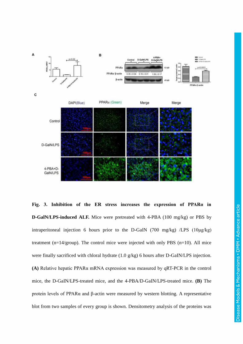
Fig. 3. Inhibition of the ER stress increases the expression of PPARα in
D-GalN/LPS-induced ALF. Mice were pretreated with 4-PBA (100 mg/kg) or PBS by
intraperitoneal injection 6 hours prior to the D-GaIN (700 mg/kg) /LPS (10μg/kg)
treatment (n=14/group). The control mice were injected with only PBS (n=10). All mice
were finally sacrificed with chloral hydrate (1.0 g/kg) 6 hours after D-GaIN/LPS injection.
(A) Relative hepatic PPARα mRNA expression was measured by qRT-PCR in the control
mice, the D-GaIN/LPS-treated mice, and the 4-PBA/D-GaIN/LPS-treated mice. (B) The
protein levels of PPARα and β-actin were measured by western blotting. A representative
blot from two samples of every group is shown. Densitometry analysis of the proteins was
Dis
ease
Mo
dels
& M
echa
nism
s •
DM
M •
Adv
ance
art
icle
performed for each sample. (C) Immunofluorescence staining for PPARα (green) in liver
tissues from the different groups. A representative experiment is shown. Original
magnification 400x.
Dis
ease
Mo
dels
& M
echa
nism
s •
DM
M •
Adv
ance
art
icle
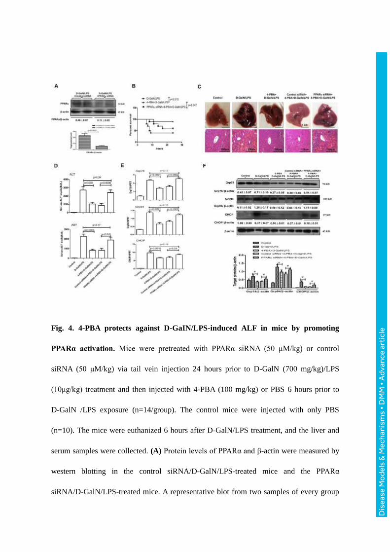
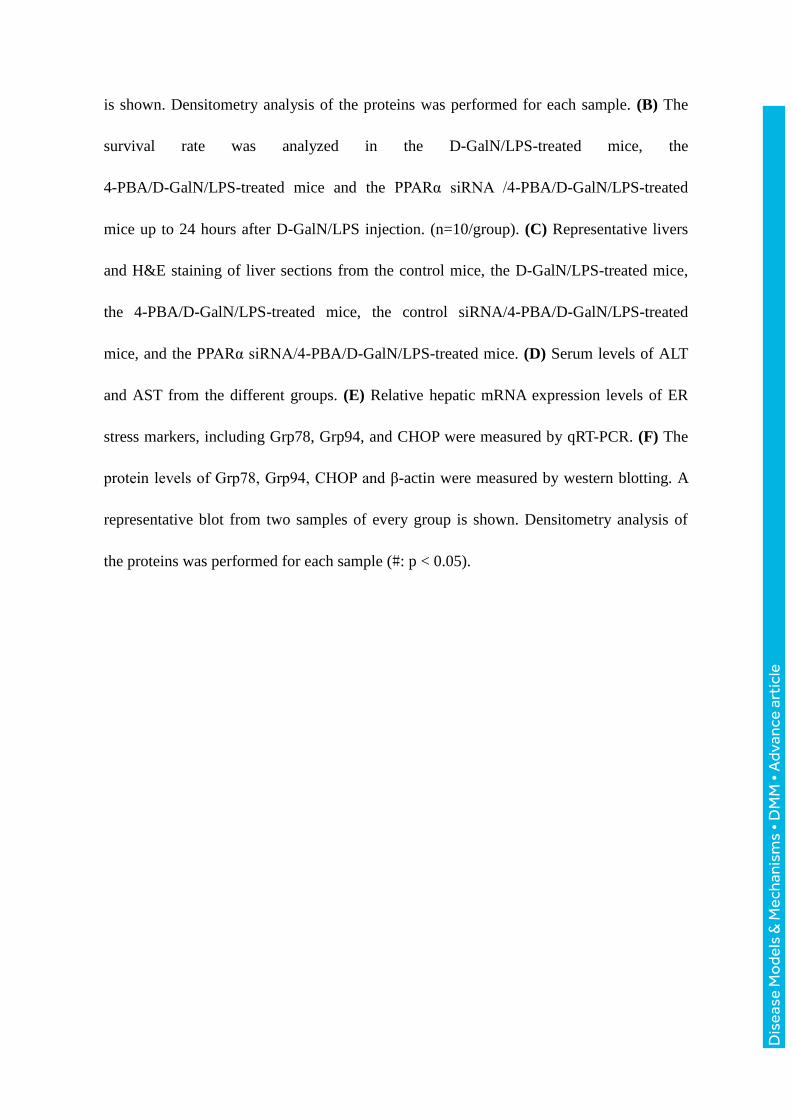
Fig. 4. 4-PBA protects against D-GaIN/LPS-induced ALF in mice by promoting
PPARα activation. Mice were pretreated with PPARα siRNA (50 μM/kg) or control
siRNA (50 μM/kg) via tail vein injection 24 hours prior to D-GalN (700 mg/kg)/LPS
(10μg/kg) treatment and then injected with 4-PBA (100 mg/kg) or PBS 6 hours prior to
D-GalN /LPS exposure (n=14/group). The control mice were injected with only PBS
(n=10). The mice were euthanized 6 hours after D-GalN/LPS treatment, and the liver and
serum samples were collected. (A) Protein levels of PPARα and β-actin were measured by
western blotting in the control siRNA/D-GalN/LPS-treated mice and the PPARα
siRNA/D-GalN/LPS-treated mice. A representative blot from two samples of every group
Dis
ease
Mo
dels
& M
echa
nism
s •
DM
M •
Adv
ance
art
icle
is shown. Densitometry analysis of the proteins was performed for each sample. (B) The
survival rate was analyzed in the D-GalN/LPS-treated mice, the
4-PBA/D-GalN/LPS-treated mice and the PPARα siRNA /4-PBA/D-GalN/LPS-treated
mice up to 24 hours after D-GalN/LPS injection. (n=10/group). (C) Representative livers
and H&E staining of liver sections from the control mice, the D-GalN/LPS-treated mice,
the 4-PBA/D-GalN/LPS-treated mice, the control siRNA/4-PBA/D-GalN/LPS-treated
mice, and the PPARα siRNA/4-PBA/D-GalN/LPS-treated mice. (D) Serum levels of ALT
and AST from the different groups. (E) Relative hepatic mRNA expression levels of ER
stress markers, including Grp78, Grp94, and CHOP were measured by qRT-PCR. (F) The
protein levels of Grp78, Grp94, CHOP and β-actin were measured by western blotting. A
representative blot from two samples of every group is shown. Densitometry analysis of
the proteins was performed for each sample (#: p < 0.05).
Dis
ease
Mo
dels
& M
echa
nism
s •
DM
M •
Adv
ance
art
icle
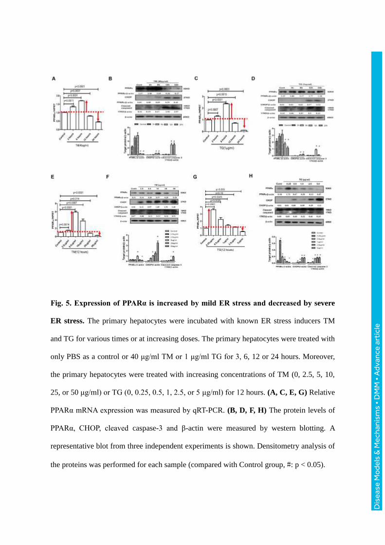
Fig. 5. Expression of PPARα is increased by mild ER stress and decreased by severe
ER stress. The primary hepatocytes were incubated with known ER stress inducers TM
and TG for various times or at increasing doses. The primary hepatocytes were treated with
only PBS as a control or 40 μg/ml TM or 1 μg/ml TG for 3, 6, 12 or 24 hours. Moreover,
the primary hepatocytes were treated with increasing concentrations of TM (0, 2.5, 5, 10,
25, or 50 μg/ml) or TG (0, 0.25, 0.5, 1, 2.5, or 5 μg/ml) for 12 hours. (A, C, E, G) Relative
PPARα mRNA expression was measured by qRT-PCR. (B, D, F, H) The protein levels of
PPARα, CHOP, cleaved caspase-3 and β-actin were measured by western blotting. A
representative blot from three independent experiments is shown. Densitometry analysis of
the proteins was performed for each sample (compared with Control group, #: p < 0.05).
Dis
ease
Mo
dels
& M
echa
nism
s •
DM
M •
Adv
ance
art
icle
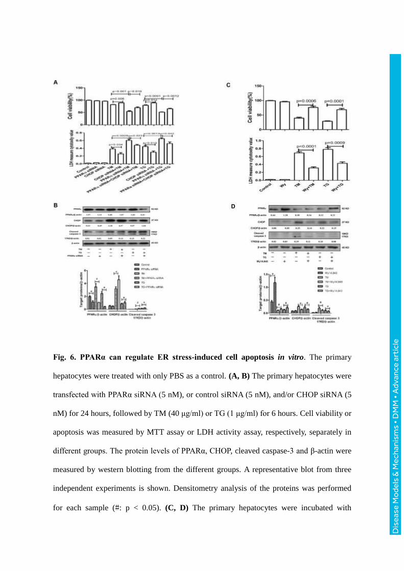
Fig. 6. PPARα can regulate ER stress-induced cell apoptosis in vitro. The primary
hepatocytes were treated with only PBS as a control. (A, B) The primary hepatocytes were
transfected with PPARα siRNA (5 nM), or control siRNA (5 nM), and/or CHOP siRNA (5
nM) for 24 hours, followed by TM (40 μg/ml) or TG (1 μg/ml) for 6 hours. Cell viability or
apoptosis was measured by MTT assay or LDH activity assay, respectively, separately in
different groups. The protein levels of PPARα, CHOP, cleaved caspase-3 and β-actin were
measured by western blotting from the different groups. A representative blot from three
independent experiments is shown. Densitometry analysis of the proteins was performed
for each sample (#: p < 0.05). (C, D) The primary hepatocytes were incubated with
Dis
ease
Mo
dels
& M
echa
nism
s •
DM
M •
Adv
ance
art
icle
Wy-14643 (50 μM) or DMSO for 2 hours and then stimulated with TM (40 μg/ml) or TG
(1 μg/ml) for 24 hours. Cell viability or apoptosis was measured by MTT assay or LDH
activity assay, respectively, separately in different group. The protein levels of PPARα,
CHOP, cleaved caspase-3 and β-actin were measured by western blotting from the different
groups. A representative blot from three independent experiments is shown. Densitometry
analysis of the proteins was performed for each sample (#: p < 0.05).
Dis
ease
Mo
dels
& M
echa
nism
s •
DM
M •
Adv
ance
art
icle
Fig. 7. PPARα is downregulated and CHOP is obviously increased in ALF patients
with HBV infection. (A) Relative hepatic mRNA expression levels of PPARα and CHOP
were measured by qRT-PCR in healthy controls (n=8), CHB patients (n=12) and ALF
patients (n=12). (B) The protein levels of PPARα and CHOP were measured by western
blotting. A representative blot from two samples of every group is shown. (C)
Immunofluorescence staining for PPARα (green) and CHOP (red) in liver tissues from the
different groups. A representative experiment is shown. Original magnification 400x. (D)
In the progression of D-GalN/LPS-induced ALF in mice, mild ER stress is induced in the
early phase of acute liver injury, which upregulates the expression of PPARα, but the
Dis
ease
Mo
dels
& M
echa
nism
s •
DM
M •
Adv
ance
art
icle
severe ER stress is induced in the late phase of ALF, which downregulates the expression
of PPARα. In ALF, the decreased PPARα triggers CHOP activity, induces extensive
hepatocyte apoptosis, and ultimately induces the development of ALF. Therefore, PPARα
is a fulcrum in the regulation of ER stress-induced liver injury.