Embed Size (px)
Citation preview

© 2016. Published by The Company of Biologists Ltd.
This is an Open Access article distributed under the terms of the Creative Commons Attribution License
(http://creativecommons.org/licenses/by/3.0), which permits unrestricted use, distribution and reproduction
in any medium provided that the original work is properly attributed.
Pharmacological and BBB-targeted genetic therapies for thyroid hormone-
dependent hypomyelination
David Zada1, Adi Tovin1, Tali Lerer-Goldshtein1 and Lior Appelbaum1
1The Faculty of Life Sciences and the Multidisciplinary Brain Research Center, Bar-
Ilan University, Ramat-Gan 5290002, Israel
To whom correspondence should be addressed: Dr. Lior Appelbaum, The Faculty of
Life Sciences, Bar-Ilan University, Ramat-Gan 5290002, Israel. Phone: +972-3-
7384536; Fax: +972-3-7384538; Email: [email protected]
Keywords: psychomotor-retardation, live-imaging, zebrafish, myelin, thyroid, Mct8
Summary statement
Expression of thyroid transporter specifically in the blood brain barrier, and
pharmacological treatments with thyroid hormone analogs and clemastine rescue
myelin deficiencies in live animal.
Dis
ease
Mo
dels
& M
echa
nism
s •
DM
M •
Adv
ance
art
icle
http://dmm.biologists.org/lookup/doi/10.1242/dmm.027227Access the most recent version at DMM Advance Online Articles. Posted 23 September 2016 as doi: 10.1242/dmm.027227http://dmm.biologists.org/lookup/doi/10.1242/dmm.027227Access the most recent version at
First posted online on 23 September 2016 as 10.1242/dmm.027227

Abstract
Hypomyelination is a key symptom of the Allan-Herndon-Dudley syndrome (AHDS),
a psychomotor retardation associated with mutations in the thyroid-hormone (TH)
transporter MCT8. AHDS is characterized by severe intellectual deficiency,
neuromuscular impairment, and brain hypothyroidism. In order to understand the
mechanism for TH-dependent hypomyelination, we developed an mct8 mutant (mct8-
/-) zebrafish model. The quantification of genetic markers for oligodendrocyte
progenitor cells (OPCs) and mature oligodendrocytes revealed reduced differentiation
of OPCs into oligodendrocytes in mct8-/- larvae and adults. Live imaging of single glial
cells showed that the number of oligodendrocytes and the length of their extensions are
reduced, and the number of peripheral Schwann cells is increased in mct8-/- larvae.
Pharmacological analysis showed that TH analogs and clemastine partially rescued the
hypomyelination in the CNS of mct8-/- larvae. Intriguingly, triiodothyronine (T3)
treatment rescued hypomyelination in mct8-/- embryos before the maturation of the
blood-brain barrier (BBB), but did not affect hypomyelination in older larvae. Thus, we
expressed Mct8-tagRFP in the endothelial cells of the vascular system and showed that
even relatively weak mosaic expression completely rescued hypomyelination in mct8-
/- larvae. These results suggest potential pharmacological treatments and BBB-targeted
gene therapy that can enhance myelination in AHDS and possibly in other TH-
dependent brain disorders.
Dis
ease
Mo
dels
& M
echa
nism
s •
DM
M •
Adv
ance
art
icle

Introduction
Leukodystrophies are a group of genetic disorders that affect the central nervous system
(CNS) by altering the development and maintenance of myelin. Hypomyelinating
leukodystrophies are caused by a deficiency in myelin deposition and are characterized
by developmental delay, hypotonia, spasticity, and various intellectual disabilities
(Boespflug-Tanguy et al., 2008; Charzewska et al., 2016). Myelination is a process in
which specialized glial cells, oligodendrocytes in the CNS, and Schwann cells in the
peripheral nervous system (PNS), send extensions of fatty substance and form myelin
sheaths that wrap axons. This insulation is vital for rapid electrical conduction and
information processing (Hartline and Colman, 2007; Raphael and Talbot, 2011;
Czopka, 2016). The functional myelin-producing cells are differentiated from
oligodendrocyte progenitor cells (OPCs), which are active primarily during embryonic
development but also in juveniles and adults (Dawson et al., 2003). Although
hypomyelination disorders are extensively studied, the pathogenic mechanism is
unclear and treatments are limited.
Among hypomyelination leukodystrophies, the X-linked Allan-Herndon-Dudley
syndrome (AHDS) is a psychomotor retardation characterized by severe intellectual
deficiency, neuromuscular impairment, and altered thyroid hormone (TH) levels in the
serum (Friesema et al., 2004; Dumitrescu et al., 2004; Brockmann et al., 2005).
Diagnosis using magnetic resonance imaging (MRI) showed a global lack of cerebral
white matter in AHDS patients (Gika et al., 2010; Holden et al., 2005; La Piana et al.,
2015). AHDS is associated with mutations in monocarboxylate transporter 8
(MCT8/SLCL6A2), which transports TH across the cell membrane (Ceballos et al.,
2009; Friesema et al., 2003). MCT8 is primarily expressed in the CNS and vascular
system, and blood-brain barrier (BBB) (Friesema et al., 2012; Pizzagalli et al., 2002;
Roberts et al., 2008). In order to study the mechanism underlying AHDS, an Mct8
knockout (Mct8-KO) mouse model was established. Similar to AHDS patients, the
Mct8-KO mice showed altered TH levels in the serum; however, neurological and
behavioral phenotypes were not apparent (Di Cosmo et al., 2013; Dumitrescu et al.,
2006; Rodrigues et al., 2013; Trajkovic et al., 2007). This may be explained by a
compensation mechanism in mice in which the organic anion transporting polypeptide
1C1 (Oatp1c1), a T4-selective transporter, is predominantly expressed in the BBB (Ito
et al., 2011; Mayerl et al., 2012; Roberts et al., 2008). Indeed, Mct8/Oatp1c1 double-
Dis
ease
Mo
dels
& M
echa
nism
s •
DM
M •
Adv
ance
art
icle

KO (dKO) mice displayed both endocrinological and neurological phenotypes found in
humans, including hypomyelination (Mayerl et al., 2014). Nevertheless, it is unclear
why a lack of MCT8 causes hypomyelination, and understanding the developmental
mechanisms could provide the groundwork to develop genetic and pharmacological
treatments for AHDS and, potentially, other hypomyelination leukodystrophies.
In order to study AHDS and hypomyelination, we used the zebrafish model, which
combines invertebrate-like genetics with vertebrate brain structures (Elbaz et al., 2015;
Levitas-Djerbi et al., 2015; Yelin-Bekerman et al., 2015), and its transparency allows
real-time imaging of myelination in a live animal (Kirby et al., 2006; Buckley et al.,
2008). In addition, zebrafish larvae have emerged as an attractive model for genetic
manipulations and high-throughput therapeutic drug screens (Kaufman et al., 2009;
MacRae and Peterson, 2015; Tsuji et al., 2014). The zebrafish mct8 gene and promoter
were isolated (Arjona et al., 2011; Vatine et al., 2013), and we have shown that
zebrafish mct8 is primarily expressed in neurons, glial cells and the vascular systems,
as in the case in humans (Vatine et al., 2013; Zada et al., 2014). Furthermore, mct8
mutant (mct8-/-) zebrafish demonstrated behavioral and neurological abnormalities,
including the altered expression of myelin-related genes (Zada et al., 2014). Here, using
transgenic zebrafish and live imaging, we studied hypomyelination in the CNS of mct8-
/- larvae, and tested the beneficial effect of putative drugs and targeted mct8 gene
therapy in the BBB on the development of oligodendrocytes in the brain and spinal cord
(SC).
Dis
ease
Mo
dels
& M
echa
nism
s •
DM
M •
Adv
ance
art
icle

Results
Loss of Mct8 alters the expression levels of markers for OPCs and mature
oligodendrocytes.
The expression of myelin-related genes in zebrafish is first detected 2 days post-
fertilization (dpf) and the onset of myelination is 3 dpf, mainly in the ventral hindbrain
and the SC (Brösamle and Halpern, 2002; Buckley et al., 2010a; Kirby et al., 2006). To
test the effect of Mct8 elimination on myelination, the transcript levels of the OPC
markers; oligodendrocyte lineage transcription factor 2 (olig2), sex determining region
Y-BOX 10 (sox10), and the mature oligodendrocyte markers; myelin basic protein
(mbp), protein zero (p0), and proteolipid protein 1b (plp1b), were quantified in 3 and 4
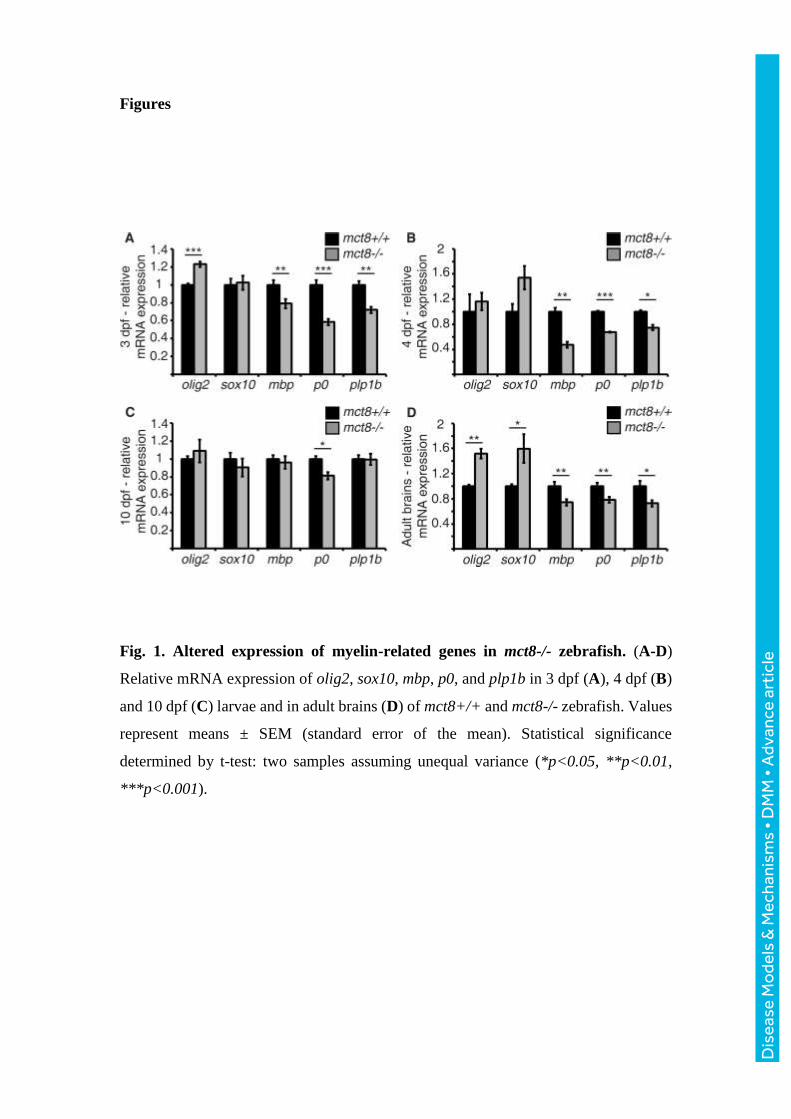
dpf mct8-/- and mct8+/+ whole embryos and larvae. While the mRNA levels of sox10
did not change in mct8-/- embryos, the mRNA levels of olig2 increased by 22% (t=-
6.69, df=14, p<0.001, Fig. 1A), and the mRNA levels of mbp, p0, and plp1b were
reduced by 21% (t=2.9, df=18, p<0.01, Fig. 1A), 42% (t=6.45, df=16, p<0.001, Fig.
1A), and 29% (t=5, df=8, p<0.01, Fig. 1A), respectively, in mct8-/- compared with
mct8+/+ embryos. Similarly, in 4 dpf larvae, the mRNA levels of mbp, p0 and plp1b
were reduced by 53% (t=6.59, df=4, p<0.01, Fig. 1B), 33% (t=19.9, df=3, p<0.001,
Fig. 1B) and 26% (t=5.18, df=3, p<0.05, Fig. 1B), respectively, in mct8-/- compared
with mct8+/+ larvae. These results suggest a global increase in the number of OPCs
alone, with a decrease in the number of mature oligodendrocytes in mct8-/- embryos.
Myelin deficiencies are found in both young and mature AHDS patients (Gika et al.,
2010; Holden et al., 2005; La Piana et al., 2015). Thus, we also measured the expression
levels of myelin marker genes in 10 dpf larvae and adult brains. Similar to 3 and 4 dpf
embryos (Fig. 1A), the mRNA levels of olig2 and sox10 increased by 51% (t=-6.4,
df=2, p<0.05, Fig. 1D) and 59% (t=-2.62, df=5, p<0.05, Fig. 1D), respectively, and
mbp, p0, and plp1b mRNA levels were reduced by 26% (t=3.12, df=16, p<0.01, Fig.
1D), 22% (t=3.08, df=13, p<0.01, Fig. 1D), and 28% (t=2.74, df=12, p<0.05, Fig. 1D),
respectively, in mct8-/- compared with mct8+/+ adult brains. In contrast, in 10 dpf
mct8-/- larvae, the loss of Mct8 did not affect the expression of the markers, excluding
p0, which is expressed specifically in the CNS (Brösamle and Halpern, 2002; Zada et
al., 2014) and reduced by 19% (t=3.1, df=4, p<0.05, Fig. 1C) in mct8-/- compared with
Dis
ease
Mo
dels
& M
echa
nism
s •
DM
M •
Adv
ance
art
icle

mct8+/+ larvae. Altogether, these results suggest reduced differentiation of OPCs into
mature oligodendrocytes in the CNS.
Visualizing myelination and glial cell dynamics in live mct8-/- larvae.
The quantification of genetic markers for glial cells in the entire body hinted for
hypomyelination in mct8-/- zebrafish. However, live imaging of specific tissues in
single-cell resolution is essential to pinpoint the spatial location of the deficiency and
to visualize oligodendrocyte and Schwann-cell developmental dynamics. Thus, we
imaged glial cell development in 3, 4, 6, 10, and 17 dpf tg(mbp:EGFP) zebrafish, which
displays EGFP expression in oligodendrocytes and Schwann cells in the CNS and PNS,
respectively (Jung et al., 2010). Double transgenic assays in 3 dpf progeny of
tg(mct8:GAL4 x uas:tagRFP) and tg(mbp:EGFP) zebrafish confirmed that mct8 is
expressed in some, but not all, mbp-positive cells (Fig. 2A). We then characterized the
development of the glial cell. In the trunk, a large number of glial cells are located in
the ventral part of the SC, and some oligodendrocytes migrate dorsally during
development (Fig. 2A). At 4 dpf, Schwann cells migrate ventrally along the motor
neuron axons, and myelin sheaths are clearly visible in 10 dpf larvae (Fig. 3A). At 3
dpf, a small number of oligodendrocytes in the brain are apparent, mainly around the
midline, at the ventral hindbrain (Fig. 5D). At 4 dpf, the number of cells increases, and
the cells are distributed primarily in the hindbrain and midbrain (Fig. 2B). This
developmental tendency persists, and a growing number of oligodendrocytes are
distributed in the midbrain, hindbrain, and SC in 6, 10, and 17 dpf larvae (Fig. 2C-E).
These results show that oligodendrocytes and Schwann cells first appear in 3 dpf and 4
dpf embryos, respectively, and robust differentiation and proliferation of glial cells, as
well as myelination, occur during development in the brain and SC.
The number of oligodendrocytes is reduced in tg(mbp:EGFP)/mct8-/- zebrafish.
The gene expression results (Fig. 1) suggest that the hypomyelination process occurs in
the CNS of mct8-/- embryos and adults. In order to determine whether the number of
oligodendrocytes is affected by loss of Mct8, we counted the number of
oligodendrocytes in the brain and SC of mct8-/- and mct8+/+ embryos and larvae. To
quantify the cell number, adult tg(mbp:EGFP)/mct8+/- and mct8+/- zebrafish were
crossed, and the number of cells counted (Fig. 2A-G). This approach assures that all
progeny carry the same transgene, and enables comparison between sibling EGFP-
Dis
ease
Mo
dels
& M
echa
nism
s •
DM
M •
Adv
ance
art
icle

positive and -negative embryos. Image analysis showed that the number of
oligodendrocytes in the dorsal SC of 3 and 4 dpf tg(mbp:EGFP)/mct8-/- larvae was
reduced by 33% (t=2.6, df=44, p<0.05, Fig. 2H) and 30% (t=4.35, df=33, p<0.001, Fig.
2H), respectively. In the brain, the number of oligodendrocytes did not change in 4 dpf
tg(mbp:EGFP)/mct8-/- embryos, presumably because OPCs only start their
differentiation at this relatively early developmental stage. In the hindbrain of 6 and 10
dpf tg(mbp:EGFP)/mct8-/- larvae, the number of oligodendrocytes was reduced by
19% (t=4.39, df=70, p<0.001, Fig. 2I) and 20% (t=3.86, df=32, p<0.001, Fig. 2I),
respectively. Similarly, in the midbrain, the number of cells was reduced by 30% (t=2.8,
df=45, p<0.01, Fig. 2J) and 23% (t=3.18, df=29, p<0.01, Fig. 2J), respectively. In older
larvae (Fig. 2E, F), the number of oligodendrocytes in the hindbrain and midbrain was
reduced by 16% (t=2.23, df=18, p<0.05, Fig. 2I) and 20% (t=2.49, df=23, p<0.05, Fig.
2J), respectively, in 17 dpf tg(mbp:EGFP)/mct8-/- larvae.
The reduced number of oligodendrocytes in the CNS suggests that loss of Mct8 causes
hypomyelination in mct8-/- zebrafish. However, since a substantial amount of
oligodendrocytes remained intact in mct8-/- larvae, they
could compensate for the loss of cells and generate either more or longer cell extensions
that produce the myelin sheaths. To study the morphology of the oligodendrocytes, we
imaged single cells in the midbrain of 10 dpf mct8-/- and mct8+/+ larvae. While the
number of extensions in a single oligodendrocyte was similar in both genotypes (Fig.
2K-M), the total length of the extensions was reduced by 23% (t=2.22, df=45, p<0.05,
Fig. 2K, L, N) in mct8-/- compared to mct8+/+ larvae. Moreover, extensions of
adjacent cells were more visible next to the cell soma of mct8+/+ compared to mct8-/-
larvae (Fig. 2K-L). These results show that a loss of Mct8 results in a reduction in the
number of oligodendrocytes during development and lower density of oligodendrocyte
extensions, and establishes the tg(mbp:EGFP)/mct8-/- zebrafish as a model for
hypomyelination.
The number of Schwann cells increased in tg(mbp:EGFP)/mct8-/- zebrafish.
The levels of mbp mRNA in the whole larvae did not change (Fig. 1B), while the
number of oligodendrocytes in the CNS decreased (Fig. 2I, J) in 10 dpf mct8-/- larvae.
Since mbp is a marker of myelin in the CNS and PNS, we imaged tg(mbp:EGFP)/mct8-
/- zebrafish and tested the effect of Mct8 elimination on the development of Schwann
Dis
ease
Mo
dels
& M
echa
nism
s •
DM
M •
Adv
ance
art
icle

cells in the periphery (Fig. 3A). While the number of Schwann cells did not change in
4 dpf tg(mbp:EGFP)/mct8-/- larvae, their number increased by 26% (t=-3.17, df=43,
p<0.05, Fig. 3B, C) in 10 dpf tg(mbp:EGFP)/mct8-/- larvae. These results suggest that
Mct8 elimination causes a hyperthyroid state in the periphery which in turn induces
Schwann cell maturation. Thus, we tested the effect of T3 treatment on the number of
Schwann cells in 10 dpf mct8+/+ larvae. Following treatment of four consecutive days,
the number of Schwann cells increased by 49% (t=-4.84, df=31, p<0.001, Fig. 3D)
compared to untreated mct8+/+ larvae. Taken together, these results show decrease in
oligodendrocyte number in the CNS and an increased Schwann cell number in the PNS
in mct8-/- larvae. These myelin deficiencies correlate with the brain hypothyroidism
and periphery hyperthyroidism found in AHDS (Ferrara et al., 2013; Trajkovic et al.,
2007). Thus, the tg(mbp:EGFP)/mct8-/- can be used to assay the effect of thyroid-
related pharmacological and genetic treatments on altered myelination in AHDS.
Thyroid analogs and clemastine rescue hypomyelination in mct8-/- zebrafish.
Establishing the myelination deficiencies in tg(mbp:EGFP)/mct8-/- larvae, we tested
putative drugs that may rescue this phenotype. In vitro and in vivo studies in mammals
have demonstrated that TH analogs, i.e., 3,3’,5-triiodothyroacetic acid (TRIAC/TA3)
and 3,5-diiodothyropropionic acid (DITPA), can cross the cell membrane even in the
absence of Mct8 (Di Cosmo et al., 2009; Kersseboom et al., 2014; Messier and
Langlois, 2000; Verge et al., 2012; Verhoeven et al., 2002). In addition, clemastine
(also named Tavist), which is an antihistamine drug, can enhance oligodendrocyte
differentiation and the wrapping of micropillars in cell lines and mice (Mei et al., 2014).
Thus, we tested the potential beneficial effect of these drugs on CNS hypomyelination
in the tg(mbp:EGFP)/mct8-/- larvae.
We exposed tg(mbp:EGFP)/mct8-/- and tg(mbp:EGFP)/mct8+/+ embryos and larvae
to 5×10⁻⁶ M control NaOH, 5 nM T3, DITPA, TRIAC, and 500 nM clemastine. These
compound concentrations were chosen based on pre-calibration assays, where the
highest doses, which did not affect pigmentation and the general morphology of the
embryos, were selected. Initially, the drugs were administered into the water at the one-
cell stage, and the treatment lasted three consecutive days. At 3 dpf, the number of
oligodendrocytes in the dorsal SC did not change in all treated tg(mbp:EGFP)/mct8+/+
embryos compared with control tg(mbp:EGFP)/mct8+/+ embryos (Fig. 4A). However,
Dis
ease
Mo
dels
& M
echa
nism
s •
DM
M •
Adv
ance
art
icle

while the oligodendrocyte number was reduced by 47% in control
tg(mbp:EGFP)/mct8-/- 3 dpf embryos (F=3.78, df=5, p<0.01, Fig. 4A`), their number
increased and they were completely rescued in T3-, DITPA-, TRIAC-, and clemastine-
treated tg(mbp:EGFP)/mct8-/- embryos compared with the control
tg(mbp:EGFP)/mct8+/+ embryos (p<0.01, Fig. 4A`). These results suggest that
treatment immediately following fertilization can prevent the myelination deficiencies
in mct8-/- larvae.
In order to determine if these drugs can repair, and not only prevent, the myelination
deficiencies at the developmental stage, when the oligodendrocyte number is already
reduced, we exposed 6 dpf larvae to these compounds for four consecutive days. At 10
dpf, the number of oligodendrocytes in the brain did not change in all treated
tg(mbp:EGFP)/mct8+/+ larvae compared with control tg(mbp:EGFP)/mct8+/+ larvae
(Fig. 4B). However, while the oligodendrocyte number was reduced by 25% in control
tg(mbp:EGFP)/mct8-/- larvae (F=5.75, df=5, p<0.001, Fig. 4B`), their number
increased and completely rescued in TRIAC-treated tg(mbp:EGFP)/mct8-/- larvae
compared with the control tg(mbp:EGFP)/mct8-/- larvae (p<0.001, Fig. 4B`).
Furthermore, DITPA and clemastine partially rescued oligodendrocyte number in
tg(mbp:EGFP)/mct8-/- larvae (Fig. 4B`). In contrast, oligodendrocyte number did not
change in T3-treated mct8-/- larvae. These results show that the TH analogs and
clemastine can enhance myelination in tg(mbp:EGFP)/mct8-/- larvae even after brain
damage occurs. In addition, these results suggest that T3 cannot cross the cell
membrane, and enter into the CNS following the 6 dpf developmental stage, and
therefore, cannot rescue myelination deficiencies in 10 dpf mct8-/- larvae.
Targeted gene therapy to the BBB rescues hypomyelination in mct8-/- zebrafish.
The BBB is vital for the maintenance and protection of the brain. This barrier is formed
by a tight junction between endothelial cells of the vascular system. In zebrafish, the
BBB is first detected in 3 dpf embryos, and its maturation occurs between 3 and 10 dpf
larvae (Fleming et al., 2013; Jeong et al., 2008). Using the endothelial cell-specific fli
promoter (Lawson and Weinstein, 2002; Yaniv et al., 2006) and the double transgenic
tg(mct8:EGFP/fli:dsRED) larvae, we showed that Mct8 is widely expressed in the
vascular system of the larval trunk and brain (Vatine et al., 2013). Considering the
efficiency and ineffectiveness of T3 treatment in 3 and 10 dpf larvae, respectively (Fig.
Dis
ease
Mo
dels
& M
echa
nism
s •
DM
M •
Adv
ance
art
icle

4A` and B`), as well as the maturation of the BBB between these developmental stages,
we reasoned that Mct8 expression in endothelial cells of the BBB is crucial for T3
transport and normal brain development. Thus, specific expression of mct8 in
endothelial cells of mct8-/- larvae may enable endogenous T3 to cross the BBB, enter
into the hypothyroidized brain, and rescue myelination deficiencies.
In order to test this assumption, the pT2-fli:Mct8-tagRFP construct, which drives
specific expression of the fusion protein Mct8-tagRFP in vascular endothelial cells, was
injected into one-cell-stage tg(mbp:EGFP)/mct8-/- and tg(mbp:EGFP)/mct8+/+
embryos, and 3 dpf Mct8-tagRFP-positive embryos were imaged and sorted. In the head
and trunk, the mosaic expression of Mct8-tagRFP was apparent in approximately 10-
20% of the vascular system (Fig. 5C-F), compared to stable expression of Mct8-EGFP
in the entire vascular system of 3 dpf tg(fli:Mct8-EGFP) embryos (Fig. 5A and B). At
3 dpf, as expected, the number of oligodendrocytes was reduced by 28% (F=6.99, df=2,
p<0.01, Fig. 5G) in the spinal cord of tg(mbp:EGFP)/mct8-/- compared with
tg(mbp:EGFP)/mct8+/+ larvae. Notably, the number of oligodendrocytes in the SC of
tg(mbp:EGFP)/mct8-/-/fli:Mct8-tagRFP-positive embryos did not change compared to
tg(mbp:EGFP)/mct8-/- embryos (Fig. 5G). These results show that the expression of
Mct8 in the vascular endothelial cells does not affect myelination in 3 dpf embryos.
Following the imaging in 3 dpf, the same individual embryos were raised and subjected
to live imaging at 10 dpf. Remarkably, while the number of oligodendrocytes in the
brain of tg(mbp:EGFP)/mct8-/- larvae was reduced by 19% (F=8.74, df=2, p<0.01, Fig.
5F, H), the number of oligodendrocytes in tg(mbp:EGFP)/mct8-/-/fli:Mct8-tagRFP-
positive larvae was completely recovered, and was similar to the number of
oligodendrocytes in tg(mbp:EGFP)/mct8+/+ larvae (p<0.01; Fig. 5F, H). These results
show that Mct8 expression in the vascular endothelial cells can rescue hypomyelination
in 10 dpf mct8-/- larvae. Considering that specific Mct8 expression in endothelial cells
did not rescue myelination in 3 dpf larvae before the development of the BBB, these
results demonstrate that this genetic treatment is beneficial post-damage and that Mct8
function specifically in the BBB is crucial for the brain. Altogether, the results in the
mct8-/- zebrafish model suggest that BBB-specific mct8 gene therapy is a promising
potential treatment for AHDS patients.
Dis
ease
Mo
dels
& M
echa
nism
s •
DM
M •
Adv
ance
art
icle

Discussion
Understanding the cellular mechanism underlying leukodystrophies and establishing
new animal models for these disorders are expected to enhance the discovery of novel
treatments. In this study, we characterized hypomyelination in a zebrafish model for
AHDS and used the transgenesis and imaging of live larvae in order to evaluate the
therapeutic effect of various genetic and pharmacological treatments. Quantification of
the expression of myelin-related markers in mct8-/- larvae implied at the delayed
maturation of oligodendrocytes and altered myelination. Indeed, the number of
oligodendrocytes in the brain and spinal cord was reduced while the number of
Schwann cells in the trunk increased in mct8-/- larvae. In order to test potential
treatments, both pharmacological and genetic approaches were evaluated. The
hypomyelination was partially or completely repaired by TH analogs and clemastine,
even when the drugs were administered after the deficiency was already apparent. In
addition, Mct8 expression that was specifically targeted to the endothelial cells of the
vascular system, completely restored myelination in mct8-/- larvae only at the
developmental stage, when the BBB was already established. Thus, we propose
pharmacological and BBB-targeted gene therapy as potential treatments for
hypomyelination in AHDS patients.
Myelination primarily occurs in mammals during post-natal stages, while in zebrafish,
the first OPCs emerge at 2 dpf and oligodendrocytes appear shortly after (Brösamle and
Halpern, 2002; Buckley et al., 2010a; Park et al., 2002). The myelination process
continues during development even in adults in all vertebrates, including zebrafish,
rodents, and humans (Jung et al., 2010; Miller et al., 2012). However, the techniques
by which myelination is quantified vary between species and studies. One of the most
adequate anatomical methods is electron microscopy (EM); however, EM imaging is
performed in fixed brain sections, while neurons and axons stand out in their significant
capacity to continuously change, adapt, and develop. Thus, using EM to image an entire
myelinated neuronal circuit across several time-points and in several individuals at a
large-scale is virtually impossible (Buckley et al., 2010a); therefore, an alternative
framework for studying myelination that will include time-lapse live imaging could
overcome these limitations. Here, we combined the expression assays of myelin
markers with the visualization of single oligodendrocytes or Schwann cells in live
transgenic zebrafish. These were proved to be effective and rapid methods to examine
Dis
ease
Mo
dels
& M
echa
nism
s •
DM
M •
Adv
ance
art
icle

myelination in many animals during development (Pogoda et al., 2006; Langworthy
and Appel, 2012; Buckley et al., 2010b). Using these methods, we found a gradual
increase in the number of mature oligodendrocytes that initially appear in the ventral
hindbrain and spinal cord of 3 dpf embryos and extend their distribution into other brain
regions at early and late larval stages. This pattern of development, as well as the unique
genetic and imaging tools used in the zebrafish model, provided us with a suitable
platform to understand the cellular mechanism of hypomyelination in AHDS.
Brain MRIs of AHDS patients showed profound myelination deficiencies in the central
and peripheral white matter of the frontal, parietal, and temporal lobes (Gika et al.,
2010). Moreover, the Mct8/Oatp1c1 dKO mice display a reduced MBP
immunofluorescence signal in the cerebral cortex and a reduced number of myelinated
axons in the corpus callusom. Surprisingly, although hypomyelination is evident, the
ultrastructure of the myelin sheath seems to be normal in this area (Mayerl et al., 2014).
Similar to the finding in AHDS patients and Mct8/Oatp1c1 dKO mice, we found
decreased expression of markers for myelin and a reduced number of oligodendrocytes
in the brain and spinal cord. Thus, these results suggest that hypomyelination in animal
models and AHDS patients is caused by the inhibition of OPC differentiation and,
ultimately, a reduced number of oligodendrocytes, while, in regions where the amount
of oligodendrocytes is intact, the structure and function of the myelin sheath is normal.
Thus, unlike neurodegenerative diseases in which cells are eliminated, in AHDS, the
promotion of OPC maturation and oligodendrocyte migration toward deficient regions
is a promising therapeutic approach.
An intriguing finding is the difference between the decrease in the number of
oligodendrocytes found in the CNS and the increase in the number of Schwan cells
found in the periphery in mct8-/- larvae. Furthermore, while the expression of markers
for myelin decreases in adult brains and 3 dpf mct8-/- embryos (at a stage at which
myelin appears only in the brain), it did not change in 10 dpf larvae (at a stage at which
myelin is distributed throughout the CNS and PNS). This can be explained by the
deferential TH levels found in MCT8-deficient animals; hypothyroidism in the CNS
and hyperthyroidism in the periphery (Ferrara et al., 2013; Trajkovic et al., 2007),
which correlate with a decreased number of oligodendrocytes and an increased number
of Schwann cells, respectively. Indeed, we found that T3 promotes maturation of
Schwann cells in larvae. Supporting this notion, TH regulates the timing of
Dis
ease
Mo
dels
& M
echa
nism
s •
DM
M •
Adv
ance
art
icle

oligodendrocyte differentiation (Bernal, 2000; Billon et al., 2001) and promotes
myelination by inducing the expression of genes such as mbp and p0 (Barradas et al.,
2001; Zada et al., 2014). Accordingly, normalizing TH levels in a specific tissue could
repair the myelin deficiencies in MCT8-deficient animals.
Why does the elimination of MCT8 cause hypomyelination? A reasonable explanation
is that there is a lack of the myelin-promoting TH in the CNS. Similar to the function
of transcription factors, TH binds to nuclear TH receptor which in turn binds the
regulatory region of myelin-related genes and enhances transcriptions (Zada et al.,
2014; Knipper et al., 1998; Jeannin et al., 1998; Harsan et al., 2008). Thus, in the
absence of Mct8, the expression levels of these genes are altered, resulting in the
abnormal development of myelin. Supporting this explanation, in AHDS patients, it is
assumed that lack of T3 entrance into the CNS resulted in a global lack of white matter
(Gika et al., 2010; Holden et al., 2005; La Piana et al., 2015). Furthermore, loss of
MCT8 results in hypothyroid and hyperthyroid states in the CNS and the peripheral
tissues of rodents (Trajkovic et al., 2007), and TH promotes the differentiation of OPCs
(Bernal, 2000). Furthermore, we showed that the treatment of mct8-/- zebrafish with
TH analogs, which can enter the cell in the absence of Mct8, partially or completely
rescued hypomyelination. Thus, a loss of Mct8 likely affects myelination by TH
signaling; however, an alternative role for Mct8 cannot be ruled out.
The pharmacological studies on zebrafish not only enhance understanding of the
mechanism underlying hypomyelination in AHDS, but also provide robust quantitative
assays to evaluate the efficiency of many putative drugs. In a previous study, we
showed that the TH analogs DITPA and TRIAC recovered the expression of the myelin
marker p0 in 3 dpf mct8-/- embryos (Zada et al., 2014). Here, in addition to the
quantification of genetic myelin-cell markers, we established a live imaging-based
assay that enables comparison of the effect of dozens of putative drugs on myelination.
A prominent advantage of the zebrafish model is the ability to apply each drug directly
to the water in a 96-well plate filled with embryos at various developmental stages and
genetic backgrounds. Taking advantage of this robust assay in live animals, we tested
the effect of the TH analogs DITPA and TRIAC as an potential treatment for
hypomyelination in AHDS, and found that both drugs can prevent the occurrence of
hypomyelination in mct8-/- embryos, and TRIAC can completely rescue
hypomyelination in the larvae even after damage was apparent. This suggests TRIAC
Dis
ease
Mo
dels
& M
echa
nism
s •
DM
M •
Adv
ance
art
icle

as a promising treatment for TH-dependent hypomyelination. However, considering
that DITPA partially rescued hypomyelination, further assays with various
concentrations and at various developmental stages are required. Supporting the results
in zebrafish, the administration of TRIAC for 12 post-natal consecutive days improved
cerebellar development and cortical myelination in Mct8/Oatp1c1 dKO mice
(Kersseboom et al., 2014; Visser et al., 2016). DITPA, on the other hand, improved
hypermetabolism and TH-level abnormalities in the serum and brain of Mct8-KO mice
(Ferrara et al., 2014; Ferrara et al., 2015; Verge et al., 2012).
In addition to TH analogs, the tg(mbp:EGFP)/mct8-/- model was used to show the
beneficial effect of clemastine on hypomyelination. To date, this myelin-promoting
drug was tested only in vitro and in mice with toxic injury in the spinal cord white
matter tracts (Mei et al., 2014). Clearly, further clemastine studies, including treatment
with various concentrations at several developmental stages, are needed. Nevertheless,
these findings show that clemastine enhances the differentiation of oligodendrocytes in
a zebrafish model for AHDS, and suggest that clemastine, as well as TH analog
treatments, can enhance myelination in other leukodystrophies.
In vertebrates, MCT8 is expressed in neurons, glial cells, and endothelial cells of the
vascular system, including in the BBB (Friesema et al., 2012; Müller and Heuer, 2014;
Pizzagalli et al., 2002; Roberts et al., 2008; Vatine et al., 2013). In contrast to humans,
the TH transporter OATP1C1 is profoundly expressed in the BBB of mice.
Interestingly, KO of both Mct8 and Oatp1c1 are required to mimic the brain
hypothyroidism and neurological phenotype of AHDS patients (Mayerl et al., 2014).
Thus, it was proposed that loss of MCT8 expression in the BBB is a key factor that
mediates the pathology of the disease (Bernal, 2000; Mayerl et al., 2014; Visser and
Visser, 2012). Supporting this hypothesis, we showed that T3 administration prevents
hypomyelination in 3 dpf mct8-/- embryos but not in 10 dpf mct8-/- larvae, which were
treated between 6 and 10 dpf. Taking into account that the maturation of the BBB in
zebrafish occurs between 3 and 10 dpf (Fleming et al., 2013; Jeong et al., 2008), these
results suggest that the lack of TH transport, specifically in the BBB, is a fundamental
impairment in AHDS.
The idea that Mct8 expression in the BBB is the key for brain function raised the
possibility that restricted expression of Mct8 in the BBB of mct8-/- larvae will benefit
Dis
ease
Mo
dels
& M
echa
nism
s •
DM
M •
Adv
ance
art
icle

the brain and rescue hypomyelination. Thus, we transiently express Mct8 in the
endothelial cells of the vascular system. Strikingly, the mosaic expression of Mct8
rescued the number of oligodendrocytes in the brain of 10 dpf tg(mbp:EGFP)/mct8-/-
larvae, but not in 3 dpf tg(mbp:EGFP)/mct8-/- larvae. This is probably because the
BBB forms and develops between 3 and 10 dpf in zebrafish. These results suggest that
although Mct8 is also expressed in neurons and glial cells, endothelial cell-specific
expression of Mct8 makes the BBB permeable to TH and enables TH access into the
brain, which is sufficient to induce myelination. Recently, intravenous injection of
adeno associated virus 9 that drives the expression of human MCT8 was shown to
increased T3 content in the mouse brain (Iwayama et al., 2016). Further studies that test
the effect of vascular Mct8 expression on neurons and glial cells in various brain
regions in mct8-/- larvae and mice models are required in order to evaluate this
treatment approach. Nevertheless, these results suggest that selective BBB transport
will enable TH to reach the brain and may limit the progression of the disorder or even
improve the symptoms. Thus, developing BBB-targeted Mct8 gene therapy or effective
BBB TH-delivery technology is expected to be an attractive future direction in AHDS
treatment.
This work established the mct8-/- zebrafish as a model for studying the mechanism and
treatment of hypomyelination in AHDS and possibly other leukodystrophies. Imaging
of single glial cells in live zebrafish, as well as genetic and pharmacological
manipulation, showed that TH analogs and clemastine, as well as gene therapy in the
BBB, rescue myelination deficiencies in mct8-/- larvae. Future studies that will
investigate the correlation between hypomyelination and neuronal activity in various
brain regions, are necessary to identify deficient brain regions and to understand the
link between neurological and behavioral impairments. Studies on the mct8-/- zebrafish
are expected to shed light on these brain processes, since two-photon imaging of
genetically encoded calcium indicators (GCaMP), combined with silencing and
activating neuronal circuits by optogenetic tools, are widely used technologies in
biomedical zebrafish research (Douglass et al., 2008; Leung et al., 2013; Muto et al.,
2013; Wyart and Del Bene, 2011). In addition, since zebrafish have become an ideal
platform for high-throughput screens of small molecules that affect neuropsychiatric
disorders (Bruni et al., 2016; Rennekamp et al., 2016), transgenic zebrafish that express
glial cell markers on the genetic background of mct8-/- zebrafish might reveal new
Dis
ease
Mo
dels
& M
echa
nism
s •
DM
M •
Adv
ance
art
icle

compounds that promote OPC differentiation, oligodendrocyte maintenance, and
myelin recovery in AHDS and other hypomyelination disorders.
Materials and methods
Zebrafish husbandry and transgenic lines.
Adult zebrafish were raised and maintained in fully automated zebrafish housing
systems (Aquazone, Israel; temperature 28±0.5°C, pH 7.0, conductivity 300 μS) under
14 h light/10 h dark cycles, and fed twice a day. Embryos were produced by natural
spawning and cultivated in egg water containing methylene blue (0.3 ppm) in a light-
controlled incubator at 28±0.5°C, as previously described (Elbaz et al., 2012). To
generate a tg(mbp:EGFP) transgenic line that carries the mct8 mutation, tg(mbp:EGFP)
zebrafish (kindly provided by Dr. Cheol-Hee Kim, Chungnam National University
Daejeon, Korea) was crossed with mct8-/- zebrafish. Heterozygous
tg(mbp:EGFP)/mct8+/- zebrafish were intercrossed and produced the
tg(mbp:EGFP)/mct8+/+, tg(mbp:EGFP)/mct8+/-, and tg(mbp:EGFP)/mct8-/- lines. In
live imaging experiments, tg(mbp:EGFP)/mct8+/- was crossed with mct8+/-, and the
progeny were imaged in various developmental stages and genotyped after each
experiment. In qRT-PCR and pharmacological assays, the tg(mbp:EGFP)/mct8+/+ and
tg(mbp:EGFP)/mct8-/- transgenic lines were used. To generate double transgenic lines
the tg(mbp:EGFP) zebrafish was crossed with tg(mct8:GAL4 x uas:TagRFP) or
tg(huc:GAL4 x uas:TagRFP) zebrafish (Zada et al., 2014) and their live progeny were
imaged. Establishment of the tg(fli:Mct8-EGFP) stable transgenic line was conducted
using the Tol2 system (Kawakami et al., 2004) and the pT2-fli:Mct8-EGFP vector.
Animal protocol was reviewed and approved by the Bar-Ilan University Bioethics
Committee, Protocol number 41-11-2013 (“AHDS syndrome: Mechanisms of disease
and therapeutic approaches in model organisms”).
Dis
ease
Mo
dels
& M
echa
nism
s •
DM
M •
Adv
ance
art
icle

qRT-PCR assays.
Relative mRNA expression levels of olig2, sox10, mbp, p0, and plp1b were determined
using qRT-PCR. Total RNA was extracted from 3, 4 and 10 dpf larvae and adult brains
using the Direct-zol RNA MiniPrep kit (Zymo Research Corporation, Irvine, CA)
according to the manufacturer’s instructions. For each tested gene, a total of 3-10
samples were used, and each sample contained a pool of 10-25 larvae or 2-3 brains.
One µg mRNA was reverse-transcribed using qScript cDNA SuperMix (Quanta
BioSciences, Gaithersburg, MD), and relative transcript levels were determined using
the 7900HT Fast Real-Time PCR System (Applied Biosystems, Foster City, CA).
Duplicates of each cDNA sample were PCR-amplified using the PerfeCTa SYBR
Green FastMix (Quanta BioSciences, Gaithersburg, MD) and the following specific
primers: olig2: 5'-cgagtgaactggaatagccttac-3' and 5'-gctcgtgtcagagtccatg-3'; sox10: 5'-
tcaatatccgcacctgcac-3' and 5'-cgcttatccgtctcgttcag-3'; mbp: 5'-
gaggagacaagaagagaaaggg -3' and 5'-gaaatgcacgacagggttg-3'; p0: 5'-
acctgtgatgccaagaacc-3' and 5'-ttgccacaacgaggatca-3'; plp1b: 5'-acactgttaacgtcctgtcag -
3' and 5'-ctggtgctttgcatatgttgg-3'; and β-actin: 5'-tgaatcccaaagccaacagag-3' and 5'-
ccagagtccatcacaataccag-3'. The relative quantification of each gene expression was
normalized against β-actin mRNA expression levels and subjected to the ΔΔCT
method.
Pharmacological assays.
In all assays, embryos were placed in glass Petri dishes (20-30 embryos per dish)
containing either a specific drug or 5×10⁻⁶ nM NaOH as a control. The medium was
changed once a day in all dishes. In clemastine experiments, stock solution of 1 mM
clemastine (Cat # S1847, Sellek Chemicals, Houston, TX) was prepared and diluted in
zebrafish water to the final administered concentrations. A preliminary dose-dependent
(10 nM to 1 µM) assay was performed on wild type embryos. The highest substance
concentration (500 nM) that did not substantially affect the morphology and behavior
of the larvae was selected. In TH analog experiments, 5 nM T3 (Cat # T2877, Sigma-
Aldrich, St. Louis, MO), TRIAC (Cat # T7650, Sigma-Aldrich, St. Louis, MO), and
DITPA (Cat # SC-256593, Santa Cruz Biotechnology, Dallas, TX) were used, as
previously described (Zada et al., 2014).
Dis
ease
Mo
dels
& M
echa
nism
s •
DM
M •
Adv
ance
art
icle

Mosaic expression of Mct8 in the BBB.
The fli promoter was PCR-amplified from the p5E-fliEP vector (kindly provided by Dr.
Karina Yaniv, Weizmann Institute of Science, Israel) using the 5'-
aacaagcttccttggagatctcatctttgaccc-3' and 5'-aatggatcccgcgtctgaattaattccagccc-3'
primers, and cloned into the pT2-uas:Mct8-tagRFP or pT2-uas:Mct8-EGFP vectors
using HindIII and BamHI, replacing the uas enhancer. Fifty ng/µl pT2-fli:Mct8-tagRFP
and 50 ng/µl TP mRNA were co-injected into one-cell-stage tg(mbp:EGFP)/mct8-/- or
tg(mbp:EGFP)/mct8+/+ embryos. At 3 dpf, the embryos were imaged under an
M165FC epifluorescence stereomicroscope (Leica, Wetzlar, Germany), and embryos
that showed strong Mct8-tagRFP mosaic expression in the head vascular system were
sorted out and later compared to their Mct8-tagRFP-negative 3 and 10 dpf sibling
larvae.
Imaging and image analysis.
To perform live imaging experiments, larvae were anesthetized with Tricaine (0.01%)
and placed in low-melting-point agarose (1.0-2.0%) in a 60 mm Petri dish filled with
zebrafish water. Confocal imaging was performed using a Zeiss LSM710 upright
confocal microscope (Zeiss, Oberkochen, Germany). All images were processed using
ImageJ (National Institutes of Health, Bethesda, MD). Calculation of the number of
mbp-positive cells, oligodendrocyte extensions and the length of the extensions was
performed using Cell Counter and NeuronJ plugins in ImageJ software (National
Institutes of Health, Bethesda, MD). We imaged a 283.4 × 283.4 µm area in the spinal
cord and a 425.1 × 425.1 µm area in the hindbrain and midbrain (dashed and dotted
boxes in Fig. 2G, respectively). mbp-positive cells (oligodendrocytes) were counted in
the dorsal spinal cord of 3 and 4 dpf larvae, and in the hindbrain and midbrain of 4, 6,
10, and 17 dpf larvae. In the late larval stages (10 and 17 dpf) we sorted out and imaged
larvae at similar size (~4.5mm and ~6.2mm, respectively).
Dis
ease
Mo
dels
& M
echa
nism
s •
DM
M •
Adv
ance
art
icle

Acknowledgments
We thank the Appelbaum lab members for technical assistance and helpful comments
on the manuscript. We thank Sharon Victor and Talia Levitas-Djerbi for assistance in
editing the manuscript.
Competing interests
The authors declare no competing or financial interests.
Author contributions
DZ and AT established the transgenic lines. DZ, AT and TLG performed the qRT-PCR
experiments and pharmacological assays. DZ performed the transient and stable
expression experiments, and conducted imaging and image analysis. DZ, AT, TLG and
LA conceived and designed the experiments and wrote the paper.
Funding
This work was supported by grants from the ERA-Net for Research Programmes on
Rare Diseases (E-RARE, 3-10861), the Sherman Foundation and the University of
Technology Sydney (590262), the German-Israeli Foundation for Scientific Research
and Development (GIF, I-1314-418.13/2015), and Fondation Jérôme Lejeune (1349).
Dis
ease
Mo
dels
& M
echa
nism
s •
DM
M •
Adv
ance
art
icle

References
Arjona, F. J., de Vrieze, E., Visser, T. J., Flik, G. and Klaren, P. H. M. (2011).
Identification and functional characterization of zebrafish solute carrier Slc16a2
(Mct8) as a thyroid hormone membrane transporter. Endocrinology 152, 5065–
5073.
Barradas, P. C., Vieira, R. S. and De Freitas, M. S. (2001). Selective effect of
hypothyroidism on expression of myelin markers during development. J.
Neurosci. Res. 66, 254–261.
Bernal, J. (2000). Thyroid Hormones in Brain Development and Function. In Endotext
(ed. De Groot, L. J.), Beck-Peccoz, P.), Chrousos, G.), Dungan, K.), Grossman,
A.), Hershman, J. M.), Koch, C.), McLachlan, R.), New, M.), Rebar, R.), et al.),
South Dartmouth (MA): MDText.com, Inc.
Billon, N., Tokumoto, Y., Forrest, D. and Raff, M. (2001). Role of thyroid hormone
receptors in timing oligodendrocyte differentiation. Dev. Biol. 235, 110–120.
Boespflug-Tanguy, O., Labauge, P., Fogli, A. and Vaurs-Barriere, C. (2008). Genes
involved in leukodystrophies: a glance at glial functions. Curr. Neurol.
Neurosci. Rep. 8, 217–229.
Brockmann, K., Dumitrescu, A. M., Best, T. T., Hanefeld, F. and Refetoff, S. (2005). X-linked paroxysmal dyskinesia and severe global retardation caused
by defective MCT8 gene. J. Neurol. 252, 663–666.
Brösamle, C. and Halpern, M. E. (2002). Characterization of myelination in the
developing zebrafish. Glia 39, 47–57.
Bruni, G., Rennekamp, A. J., Velenich, A., McCarroll, M., Gendelev, L., Fertsch,
E., Taylor, J., Lakhani, P., Lensen, D., Evron, T., et al. (2016). Zebrafish
behavioral profiling identifies multitarget antipsychotic-like compounds. Nat.
Chem. Biol. 12, 559–566.
Buckley, C. E., Goldsmith, P. and Franklin, R. J. M. (2008). Zebrafish myelination:
a transparent model for remyelination? Dis. Model. Mech. 1, 221–228.
Buckley, C. E., Marguerie, A., Alderton, W. K. and Franklin, R. J. M. (2010a).
Temporal dynamics of myelination in the zebrafish spinal cord. Glia 58, 802–
812.
Buckley, C. E., Marguerie, A., Roach, A. G., Goldsmith, P., Fleming, A., Alderton,
W. K. and Franklin, R. J. M. (2010b). Drug reprofiling using zebrafish
identifies novel compounds with potential pro-myelination effects.
Neuropharmacology 59, 149–159.
Ceballos, A., Belinchon, M. M., Sanchez-Mendoza, E., Grijota-Martinez, C.,
Dumitrescu, A. M., Refetoff, S., Morte, B. and Bernal, J. (2009). Importance
of monocarboxylate transporter 8 for the blood-brain barrier-dependent
availability of 3,5,3’-triiodo-L-thyronine. Endocrinology 150, 2491–2496.
Dis
ease
Mo
dels
& M
echa
nism
s •
DM
M •
Adv
ance
art
icle

Charzewska, A., Wierzba, J., Iżycka-Świeszewska, E., Bekiesińska-Figatowska,
M., Jurek, M., Gintowt, A., Kłosowska, A., Bal, J. and Hoffman-Zacharska,
D. (2016). Hypomyelinating leukodystrophies - a molecular insight into the
white matter pathology. Clin. Genet.
Czopka, T. (2016). Insights into mechanisms of central nervous system myelination
using zebrafish. Glia 64, 333–349.
Dawson, M. R. L., Polito, A., Levine, J. M. and Reynolds, R. (2003). NG2-
expressing glial progenitor cells: an abundant and widespread population of
cycling cells in the adult rat CNS. Mol. Cell. Neurosci. 24, 476–488.
Di Cosmo, C., Liao, X.-H., Dumitrescu, A. M., Weiss, R. E. and Refetoff, S. (2009).
A thyroid hormone analog with reduced dependence on the monocarboxylate
transporter 8 for tissue transport. Endocrinology 150, 4450–4458.
Di Cosmo, C., Liao, X.-H., Ye, H., Ferrara, A. M., Weiss, R. E., Refetoff, S. and
Dumitrescu, A. M. (2013). Mct8-deficient mice have increased energy
expenditure and reduced fat mass that is abrogated by normalization of serum
T3 levels. Endocrinology 154, 4885–4895.
Douglass, A. D., Kraves, S., Deisseroth, K., Schier, A. F. and Engert, F. (2008).
Escape behavior elicited by single, channelrhodopsin-2-evoked spikes in
zebrafish somatosensory neurons. Curr. Biol. CB 18, 1133–1137.
Dumitrescu, A. M., Liao, X.-H., Best, T. B., Brockmann, K. and Refetoff, S. (2004).
A novel syndrome combining thyroid and neurological abnormalities is
associated with mutations in a monocarboxylate transporter gene. Am. J. Hum.
Genet. 74, 168–175.
Dumitrescu, A. M., Liao, X.-H., Weiss, R. E., Millen, K. and Refetoff, S. (2006).
Tissue-specific thyroid hormone deprivation and excess in monocarboxylate
transporter (mct) 8-deficient mice. Endocrinology 147, 4036–4043.
Elbaz, I., Yelin-Bekerman, L., Nicenboim, J., Vatine, G. and Appelbaum, L. (2012). Genetic ablation of hypocretin neurons alters behavioral state transitions
in zebrafish. J. Neurosci. Off. J. Soc. Neurosci. 32, 12961–12972.
Elbaz, I., Lerer-Goldshtein, T., Okamoto, H. and Appelbaum, L. (2015). Reduced
synaptic density and deficient locomotor response in neuronal activity-regulated
pentraxin 2a mutant zebrafish. FASEB J. Off. Publ. Fed. Am. Soc. Exp. Biol. 29,
1220–1234.
Ferrara, A. M., Liao, X.-H., Gil-Ibáñez, P., Marcinkowski, T., Bernal, J., Weiss,
R. E., Dumitrescu, A. M. and Refetoff, S. (2013). Changes in thyroid status
during perinatal development of MCT8-deficient male mice. Endocrinology
154, 2533–2541.
Ferrara, A. M., Liao, X.-H., Gil-Ibáñez, P., Bernal, J., Weiss, R. E., Dumitrescu,
A. M. and Refetoff, S. (2014). Placenta passage of the thyroid hormone analog
DITPA to male wild-type and Mct8-deficient mice. Endocrinology 155, 4088–
4093.
Dis
ease
Mo
dels
& M
echa
nism
s •
DM
M •
Adv
ance
art
icle

Ferrara, A. M., Liao, X.-H., Ye, H., Weiss, R. E., Dumitrescu, A. M. and Refetoff,
S. (2015). The Thyroid Hormone Analog DITPA Ameliorates Metabolic
Parameters of Male Mice With Mct8 Deficiency. Endocrinology 156, 3889–
3894.
Fleming, A., Diekmann, H. and Goldsmith, P. (2013). Functional characterisation of
the maturation of the blood-brain barrier in larval zebrafish. PloS One 8,
e77548.
Friesema, E. C. H., Ganguly, S., Abdalla, A., Manning Fox, J. E., Halestrap, A. P.
and Visser, T. J. (2003). Identification of monocarboxylate transporter 8 as a
specific thyroid hormone transporter. J. Biol. Chem. 278, 40128–40135.
Friesema, E. C. H., Grueters, A., Biebermann, H., Krude, H., von Moers, A.,
Reeser, M., Barrett, T. G., Mancilla, E. E., Svensson, J., Kester, M. H. A.,
et al. (2004). Association between mutations in a thyroid hormone transporter
and severe X-linked psychomotor retardation. Lancet Lond. Engl. 364, 1435–
1437.
Friesema, E. C. H., Visser, T. J., Borgers, A. J., Kalsbeek, A., Swaab, D. F., Fliers,
E. and Alkemade, A. (2012). Thyroid hormone transporters and deiodinases in
the developing human hypothalamus. Eur. J. Endocrinol. Eur. Fed. Endocr.
Soc. 167, 379–386.
Gika, A. D., Siddiqui, A., Hulse, A. J., Edward, S., Fallon, P., McEntagart, M. E.,
Jan, W., Josifova, D., Lerman-Sagie, T., Drummond, J., et al. (2010). White
matter abnormalities and dystonic motor disorder associated with mutations in
the SLC16A2 gene. Dev. Med. Child Neurol. 52, 475–482.
Harsan, L.-A., Steibel, J., Zaremba, A., Agin, A., Sapin, R., Poulet, P., Guignard,
B., Parizel, N., Grucker, D., Boehm, N., et al. (2008). Recovery from chronic
demyelination by thyroid hormone therapy: myelinogenesis induction and
assessment by diffusion tensor magnetic resonance imaging. J. Neurosci. Off.
J. Soc. Neurosci. 28, 14189–14201.
Hartline, D. K. and Colman, D. R. (2007). Rapid conduction and the evolution of
giant axons and myelinated fibers. Curr. Biol. CB 17, R29–35.
Holden, K. R., Zuñiga, O. F., May, M. M., Su, H., Molinero, M. R., Rogers, R. C.
and Schwartz, C. E. (2005). X-linked MCT8 gene mutations: characterization
of the pediatric neurologic phenotype. J. Child Neurol. 20, 852–857.
Ito, K., Uchida, Y., Ohtsuki, S., Aizawa, S., Kawakami, H., Katsukura, Y., Kamiie,
J. and Terasaki, T. (2011). Quantitative membrane protein expression at the
blood-brain barrier of adult and younger cynomolgus monkeys. J. Pharm. Sci.
100, 3939–3950.
Dis
ease
Mo
dels
& M
echa
nism
s •
DM
M •
Adv
ance
art
icle

Iwayama, H., Liao, X.-H., Braun, L., Bárez-López, S., Kaspar, B., Weiss, R. E.,
Dumitrescu, A. M., Guadaño-Ferraz, A. and Refetoff, S. (2016). Adeno
Associated Virus 9-Based Gene Therapy Delivers a Functional
Monocarboxylate Transporter 8, Improving Thyroid Hormone Availability to
the Brain of Mct8-Deficient Mice. Thyroid Off. J. Am. Thyroid Assoc.
Jeannin, E., Robyr, D. and Desvergne, B. (1998). Transcriptional regulatory patterns
of the myelin basic protein and malic enzyme genes by the thyroid hormone
receptors alpha1 and beta1. J. Biol. Chem. 273, 24239–24248.
Jeong, J.-Y., Kwon, H.-B., Ahn, J.-C., Kang, D., Kwon, S.-H., Park, J. A. and Kim,
K.-W. (2008). Functional and developmental analysis of the blood-brain barrier
in zebrafish. Brain Res. Bull. 75, 619–628.
Jung, S.-H., Kim, S., Chung, A.-Y., Kim, H.-T., So, J.-H., Ryu, J., Park, H.-C. and
Kim, C.-H. (2010). Visualization of myelination in GFP-transgenic zebrafish.
Dev. Dyn. Off. Publ. Am. Assoc. Anat. 239, 592–597.
Kaufman, C. K., White, R. M. and Zon, L. (2009). Chemical genetic screening in the
zebrafish embryo. Nat. Protoc. 4, 1422–1432.
Kawakami, K., Takeda, H., Kawakami, N., Kobayashi, M., Matsuda, N. and
Mishina, M. (2004). A transposon-mediated gene trap approach identifies
developmentally regulated genes in zebrafish. Dev. Cell 7, 133–144.
Kersseboom, S., Horn, S., Visser, W. E., Chen, J., Friesema, E. C. H., Vaurs-
Barrière, C., Peeters, R. P., Heuer, H. and Visser, T. J. (2014). In vitro and
mouse studies supporting therapeutic utility of triiodothyroacetic acid in MCT8
deficiency. Mol. Endocrinol. Baltim. Md 28, 1961–1970.
Kirby, B. B., Takada, N., Latimer, A. J., Shin, J., Carney, T. J., Kelsh, R. N. and
Appel, B. (2006). In vivo time-lapse imaging shows dynamic oligodendrocyte
progenitor behavior during zebrafish development. Nat. Neurosci. 9, 1506–
1511.
Knipper, M., Bandtlow, C., Gestwa, L., Köpschall, I., Rohbock, K., Wiechers, B.,
Zenner, H. P. and Zimmermann, U. (1998). Thyroid hormone affects
Schwann cell and oligodendrocyte gene expression at the glial transition zone
of the VIIIth nerve prior to cochlea function. Dev. Camb. Engl. 125, 3709–3718.
Langworthy, M. M. and Appel, B. (2012). Schwann cell myelination requires Dynein
function. Neural Develop. 7, 37.
La Piana, R., Vanasse, M., Brais, B. and Bernard, G. (2015). Myelination Delay and
Allan-Herndon-Dudley Syndrome Caused by a Novel Mutation in the
SLC16A2 Gene. J. Child Neurol. 30, 1371–1374.
Lawson, N. D. and Weinstein, B. M. (2002). In vivo imaging of embryonic vascular
development using transgenic zebrafish. Dev. Biol. 248, 307–318.
Leung, L. C., Wang, G. X. and Mourrain, P. (2013). Imaging zebrafish neural
circuitry from whole brain to synapse. Front. Neural Circuits 7, 76.
Dis
ease
Mo
dels
& M
echa
nism
s •
DM
M •
Adv
ance
art
icle

Levitas-Djerbi, T., Yelin-Bekerman, L., Lerer-Goldshtein, T. and Appelbaum, L. (2015). Hypothalamic leptin-neurotensin-hypocretin neuronal networks in
zebrafish. J. Comp. Neurol. 523, 831–848.
MacRae, C. A. and Peterson, R. T. (2015). Zebrafish as tools for drug discovery. Nat.
Rev. Drug Discov. 14, 721–731.
Mayerl, S., Visser, T. J., Darras, V. M., Horn, S. and Heuer, H. (2012). Impact of
Oatp1c1 deficiency on thyroid hormone metabolism and action in the mouse
brain. Endocrinology 153, 1528–1537.
Mayerl, S., Müller, J., Bauer, R., Richert, S., Kassmann, C. M., Darras, V. M.,
Buder, K., Boelen, A., Visser, T. J. and Heuer, H. (2014). Transporters MCT8
and OATP1C1 maintain murine brain thyroid hormone homeostasis. J. Clin.
Invest. 124, 1987–1999.
Mei, F., Fancy, S. P. J., Shen, Y.-A. A., Niu, J., Zhao, C., Presley, B., Miao, E., Lee,
S., Mayoral, S. R., Redmond, S. A., et al. (2014). Micropillar arrays as a high-
throughput screening platform for therapeutics in multiple sclerosis. Nat. Med.
20, 954–960.
Messier, N. and Langlois, M. F. (2000). Triac regulation of transcription is T(3)
receptor isoform- and response element-specific. Mol. Cell. Endocrinol. 165,
57–66.
Miller, D. J., Duka, T., Stimpson, C. D., Schapiro, S. J., Baze, W. B., McArthur,
M. J., Fobbs, A. J., Sousa, A. M. M., Sestan, N., Wildman, D. E., et al. (2012). Prolonged myelination in human neocortical evolution. Proc. Natl.
Acad. Sci. U. S. A. 109, 16480–16485.
Müller, J. and Heuer, H. (2014). Expression pattern of thyroid hormone transporters
in the postnatal mouse brain. Front. Endocrinol. 5, 92.
Muto, A., Ohkura, M., Abe, G., Nakai, J. and Kawakami, K. (2013). Real-time
visualization of neuronal activity during perception. Curr. Biol. CB 23, 307–
311.
Park, H.-C., Mehta, A., Richardson, J. S. and Appel, B. (2002). olig2 is required for
zebrafish primary motor neuron and oligodendrocyte development. Dev. Biol.
248, 356–368.
Pizzagalli, F., Hagenbuch, B., Stieger, B., Klenk, U., Folkers, G. and Meier, P. J. (2002). Identification of a novel human organic anion transporting polypeptide
as a high affinity thyroxine transporter. Mol. Endocrinol. Baltim. Md 16, 2283–
2296.
Pogoda, H.-M., Sternheim, N., Lyons, D. A., Diamond, B., Hawkins, T. A., Woods,
I. G., Bhatt, D. H., Franzini-Armstrong, C., Dominguez, C., Arana, N., et
al. (2006). A genetic screen identifies genes essential for development of
myelinated axons in zebrafish. Dev. Biol. 298, 118–131.
Dis
ease
Mo
dels
& M
echa
nism
s •
DM
M •
Adv
ance
art
icle

Raphael, A. R. and Talbot, W. S. (2011). New insights into signaling during
myelination in zebrafish. Curr. Top. Dev. Biol. 97, 1–19.
Rennekamp, A. J., Huang, X.-P., Wang, Y., Patel, S., Lorello, P. J., Cade, L.,
Gonzales, A. P. W., Yeh, J.-R. J., Caldarone, B. J., Roth, B. L., et al. (2016).
σ1 receptor ligands control a switch between passive and active threat
responses. Nat. Chem. Biol. 12, 552–558.
Roberts, L. M., Woodford, K., Zhou, M., Black, D. S., Haggerty, J. E., Tate, E. H.,
Grindstaff, K. K., Mengesha, W., Raman, C. and Zerangue, N. (2008).
Expression of the thyroid hormone transporters monocarboxylate transporter-8
(SLC16A2) and organic ion transporter-14 (SLCO1C1) at the blood-brain
barrier. Endocrinology 149, 6251–6261.
Rodrigues, T. B., Ceballos, A., Grijota-Martínez, C., Nuñez, B., Refetoff, S.,
Cerdán, S., Morte, B. and Bernal, J. (2013). Increased oxidative metabolism
and neurotransmitter cycling in the brain of mice lacking the thyroid hormone
transporter SLC16A2 (MCT8). PloS One 8, e74621.
Trajkovic, M., Visser, T. J., Mittag, J., Horn, S., Lukas, J., Darras, V. M., Raivich,
G., Bauer, K. and Heuer, H. (2007). Abnormal thyroid hormone metabolism
in mice lacking the monocarboxylate transporter 8. J. Clin. Invest. 117, 627–
635.
Tsuji, N., Ninov, N., Delawary, M., Osman, S., Roh, A. S., Gut, P. and Stainier, D.
Y. R. (2014). Whole organism high content screening identifies stimulators of
pancreatic beta-cell proliferation. PloS One 9, e104112.
Vatine, G. D., Zada, D., Lerer-Goldshtein, T., Tovin, A., Malkinson, G., Yaniv, K.
and Appelbaum, L. (2013). Zebrafish as a model for monocarboxyl transporter
8-deficiency. J. Biol. Chem. 288, 169–180.
Verge, C. F., Konrad, D., Cohen, M., Di Cosmo, C., Dumitrescu, A. M.,
Marcinkowski, T., Hameed, S., Hamilton, J., Weiss, R. E. and Refetoff, S. (2012). Diiodothyropropionic acid (DITPA) in the treatment of MCT8
deficiency. J. Clin. Endocrinol. Metab. 97, 4515–4523.
Verhoeven, F. A., Van der Putten, H. H. a. G. M., Hennemann, G., Lamers, J. M.
J., Visser, T. J. and Everts, M. E. (2002). Uptake of triiodothyronine and
triiodothyroacetic acid in neonatal rat cardiomyocytes: effects of metabolites
and analogs. J. Endocrinol. 173, 247–255.
Visser, W. E. and Visser, T. J. (2012). Finding the way into the brain without MCT8.
J. Clin. Endocrinol. Metab. 97, 4362–4365.
Visser, W. E., Heuer, H. and Visser, T. J. (2016). Triiodothyroacetic Acid Treatment
in MCT8 Deficiency: A Word of Nuance. Thyroid Off. J. Am. Thyroid Assoc.
26, 615–617.
Wyart, C. and Del Bene, F. (2011). Let there be light: zebrafish neurobiology and the
optogenetic revolution. Rev. Neurosci. 22, 121–130.
Dis
ease
Mo
dels
& M
echa
nism
s •
DM
M •
Adv
ance
art
icle

Yaniv, K., Isogai, S., Castranova, D., Dye, L., Hitomi, J. and Weinstein, B. M. (2006). Live imaging of lymphatic development in the zebrafish. Nat. Med. 12,
711–716.
Yelin-Bekerman, L., Elbaz, I., Diber, A., Dahary, D., Gibbs-Bar, L., Alon, S.,
Lerer-Goldshtein, T. and Appelbaum, L. (2015). Hypocretin neuron-specific
transcriptome profiling identifies the sleep modulator Kcnh4a. eLife 4, e08638.
Zada, D., Tovin, A., Lerer-Goldshtein, T., Vatine, G. D. and Appelbaum, L. (2014).
Altered behavioral performance and live imaging of circuit-specific neural
deficiencies in a zebrafish model for psychomotor retardation. PLoS Genet. 10,
e1004615.
Dis
ease
Mo
dels
& M
echa
nism
s •
DM
M •
Adv
ance
art
icle
Figures
Fig. 1. Altered expression of myelin-related genes in mct8-/- zebrafish. (A-D)
Relative mRNA expression of olig2, sox10, mbp, p0, and plp1b in 3 dpf (A), 4 dpf (B)
and 10 dpf (C) larvae and in adult brains (D) of mct8+/+ and mct8-/- zebrafish. Values
represent means ± SEM (standard error of the mean). Statistical significance
determined by t-test: two samples assuming unequal variance (*p<0.05, **p<0.01,
***p<0.001).
Dis
ease
Mo
dels
& M
echa
nism
s •
DM
M •
Adv
ance
art
icle

Fig. 2. Hypomyelination in the brain and spinal cord (SC) of mct8-/- larvae during
development. (A) Lateral view of the trunk in 3 dpf tg(mct8:GAL4 x uas:tagRFP) x
tg(mbp:EGFP) embryo revealed co-localization of mct8-promoter driven signal (red,
middle panel) in oligodendrocytes (green, top panel) of the SC. Dashed white line in
the top picture marks the boundary between the dorsal and ventral SC. (B-F) Dorsal
view of the hindbrain (HB) and midbrain (MB; dashed white line marks the boundary
between the MB and HB) of 4 (B), 6 (C), 10 (D), and 17 dpf tg(mbp:EGFP) larvae (E),
as well as 17 dpf tg(mbp:EGFP)/mct8-/- larvae (F). Arrows and arrowheads indicate
Schwann cells and oligodendrocytes, respectively. (G) Illustration of the
tg(mbp:EGFP) larvae. Distribution of oligodendrocyte in the CNS is marked with green
Dis
ease
Mo
dels
& M
echa
nism
s •
DM
M •
Adv
ance
art
icle

spots. Dashed and dotted boxes represent the imaged areas in the SC and brain,
respectively, as shown in A-F. (H) Number of oligodendrocytes in the dorsal SC of 3
(mct8+/+: n=23, mct8-/-: n=23) and 4 (mct8+/+: n=19, mct8-/-: n=21) dpf larvae. (I)
Number of oligodendrocytes counted in the HB of 4 (mct8+/+: n=7, mct8-/-: n=14), 6
(mct8+/+: n=33, mct8-/-: n=39), 10 (mct8+/+: n=17, mct8-/-: n=18), and 17 dpf larvae
(mct8+/+: n=14, mct8-/-: n=12). (J) Number of oligodendrocytes counted in the MB
of 4 (mct8+/+: n=8, mct8-/-: n=8), 6 (mct8+/+: n=25, mct8-/-: n=30), 10 (mct8+/+:
n=17, mct8-/-: n=18), and 17 dpf larvae (mct8+/+: n=13, mct8-/-: n=13). (K, L)
Imaging of single oligodendrocyte in the MB of mct8+/+ (K) and mct8-/- (L) 10 dpf
larvae. Open arrows and arrowheads indicate self and neighboring oligodendrocyte
extensions, respectively. (M) The number of oligodendrocyte extensions per single cell
(mct8+/+: n=29 cells, mct8-/-: n=24 cells). (M) Total length of oligodendrocyte
extensions per single cell (mct8+/+: n=29 cells, mct8-/-: n=24 cells). A-F Scale bar =
50 µm, K-L Scale bar = 10 µm. Values are represented as means ± SEM (standard
error of the mean). Statistical significance was determined by t-test: two samples
assuming unequal variance (*p<0.05, **p<0.01, ***p<0.001).
Dis
ease
Mo
dels
& M
echa
nism
s •
DM
M •
Adv
ance
art
icle

Fig. 3. Increased number of Schwann cells in mct8-/- larvae during development.
(A) Lateral view of a motor neuron in 10 dpf live
tg(mbp:EGFP)/tg(huc:GAL4xuas:tagRFP) larvae shows Schwann-cell (green)
ensheathment of motor neuron axons (red). (B) Representative lateral-view images of
Dis
ease
Mo
dels
& M
echa
nism
s •
DM
M •
Adv
ance
art
icle

Schwann-cell ensheathment of motor neuron axons in the SC of 10 dpf mct8+/+ and
mct8-/- larvae. (C) Number of Schwann cells that myelinate the axons of the motor
neurons of 4 (mct8+/+: n=9, mct8-/-: n=9) and 10 (mct8+/+: n=26, mct8-/-: n=22) dpf
larvae. (D) Number of Schwann cells that myelinate the axons of the motor neurons of
untreated and T3-treated mct8+/+ 10 dpf larvae (untreated: n=26, T3-treated: n=19).
Scale bar = 10 µm. Values are represented as means ± SEM (standard error of the
mean). Statistical significance was determined by t-test: two samples assuming unequal
variance (*p<0.05, ***p<0.001).
Dis
ease
Mo
dels
& M
echa
nism
s •
DM
M •
Adv
ance
art
icle

Fig. 4. TH-analog and clemastine treatments rescue the number of
oligodendrocytes in mct8-/- larvae. (A, A`). Number of oligodendrocytes in the spinal
cord (SC) of 3 dpf mct8+/+ embryos treated with T3 (n=11), DITPA (n=12), TRIAC
(n=11), clemastine (Cle, n=10), NaOH (n=16), and mct8-/- embryos treated with T3
(n=12), DITPA (n=14), TRIAC (n=14), clemastine (Cle, n=12), and NaOH (n=16,
p<0.01). (B, B`). Number of oligodendrocytes in the brain of 10 dpf mct8+/+ larvae
treated with T3 (n=10), DITPA (n=11), TRIAC (n=11), clemastine (Cle, n=10), NaOH
(n=10), and mct8-/- larvae treated with T3 (n=10), DITPA (n=12), TRIAC (n=11),
clemastine (Cle, n=15), and NaOH (n=13, p<0.001). Values represented as means ±
SEM (standard error of the mean). Statistical significance determined by one-way
Dis
ease
Mo
dels
& M
echa
nism
s •
DM
M •
Adv
ance
art
icle

ANOVA, followed by post-hoc Tukey HSD test. The letters above the bars indicate
significant differences. The arrow represents the developmental timeline of the
experiment. Dashed bar represents the period in which the drug was administered.
Dis
ease
Mo
dels
& M
echa
nism
s •
DM
M •
Adv
ance
art
icle

Fig. 5. Specific expression of Mct8 in the vascular system and BBB rescues the
number of oligodendrocytes in the brain. Lateral view of the trunk (A, head pointing
to the right) and dorsal view of the head (B) of tg(fli:Mct8-EGFP) 3 dpf embryo. Lateral
view of the trunk (C, E, head pointing to the right) and dorsal view of the head (D, F)
of tg(mbp:EGFP) larvae injected with fli:Mct8-tagRFP. Arrows indicate the expression
of Mct8-tagRFP in the endothelial cells of the vascular system, and arrowheads indicate
oligodendrocytes in the CNS. Wide (C) and high magnification (dashed box, E) views
in 3 dpf larvae show red mosaic expression of Mct8-tagRFP and green
oligodendrocytes. Dorsal view of the head shows green oligodendrocytes in the brain
and mosaic expression of Mct8-tagRFP in 3 (D) and 10 (F) dpf larvae. (G) Number of
Dis
ease
Mo
dels
& M
echa
nism
s •
DM
M •
Adv
ance
art
icle

oligodendrocytes counted in the SC of 3 dpf larvae (mct8+/+: n=37, mct8-/-: n=42, and
mct8-/-/Mct8-tagRFP: n=26, p<0.01). (H) Number of oligodendrocytes counted in the
brain of 10 dpf larvae (mct8+/+: n=10, mct8-/-: n=10, and mct8-/-/Mct8-tagRFP: n=11,
p<0.01). Scale bar = 50 µm. Values represented as means ± SEM (standard error of the
mean). Statistical significance determined by one-way ANOVA followed by post-hoc
Tukey HSD test (**p<0.01).
Dis
ease
Mo
dels
& M
echa
nism
s •
DM
M •
Adv
ance
art
icle