Embed Size (px)
Citation preview
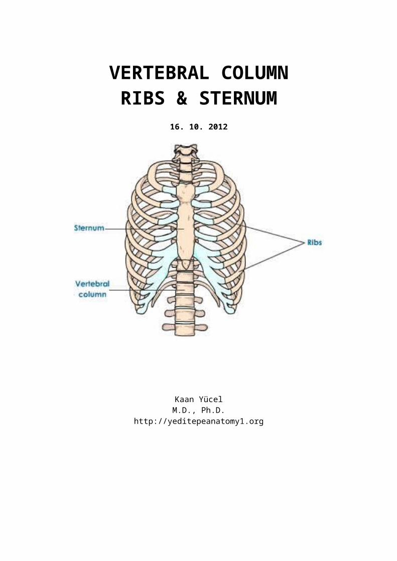
VERTEBRAL COLUMNRIBS & STERNUM
16. 10. 2012
Kaan YücelM.D., Ph.D.
http://yeditepeanatomy1.org
Dr.Kaan Yücel http://yeditepeanatomy1.org Vertebral column, ribs & sternum
http://www.youtube.com/yeditepeanatomy
VERTEBRAL COLUMNThe vertebrae and intervertebtal (IV) discs collectively make up the vertebral column (spine), the skeleton
of the neck and back that is the main part of the axial skeleton (i.e., articulated bones of the cranium, vertebral column, ribs, and sternum). The vertebral column extends from the cranium (skull) to the apex of the coccyx.
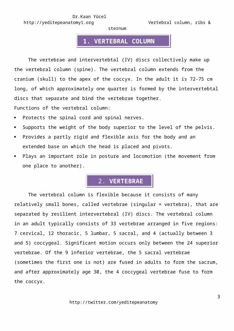
The vertebral column is flexible because it consists of many relatively small bones, called vertebrae (singular = vertebra), that are separated by resilient intervertebral (IV) discs. The vertebral column in an adult typically consists of 33 vertebrae arranged in five regions: 7 cervical, 12 thoracic, 5 lumbar, 5 sacral, and 4 coccygeal.
The vertebrae gradually become larger as the vertebral column descends to the sacrum and then become progressively smaller toward the apex of the coccyx. Vertebrae vary in size and other characteristics from one region of the vertebral column to another, and to a lesser degree within each region; however, their basic structure is the same. A typical vertebra consists of a vertebral body, a vertebral arch, and seven processes.
Regional variations in the size and shape of the vertebral canal accommodate the varying thickness of the spinal cord.
Cervical vertebrae form the skeleton of the neck. The smallest of the 24 movable vertebrae, the cervical vertebrae are located between the cranium and the thoracic vertebrae. Their smaller size reflects the fact that they bear less weight than do the larger inferior vertebrae. The most distinctive feature of each cervical vertebra is the oval foramen transversarium (transverse foramen) in the transverse process.
The thoracic vertebrae are in the upper back and provide attachment for the ribs. Thus the primary characteristic features of thoracic vertebrae are the costal facets for articulation with ribs. The middle four thoracic vertebrae (T5-T8) demonstrate all the features typical of thoracic vertebrae.
Lumbar vertebrae are in the lower back between the thorax and sacrum. Because the weight they support increases toward the inferior end of the vertebral column, lumbar vertebrae have massive bodies, accounting for much of the thickness of the lower trunk in the median plane.
The wedged-shaped sacrum (L. sacred) is usually composed of five fused sacral vertebrae in adults. It is located between the hip bones and forms the roof and posterosuperior wall of the posterior half of the pelvic cavity.
The coccyx (tail bone) is a small triangular bone that is usually formed by fusion of the four rudimentary coccygeal vertebrae, although in some people, there may be one less or one more. Coccygeal vertebra 1 (Co1) may remain separate from the fused group.
RIBSRibs (L. costae) are curved, flat bones that form most of the thoracic cage. They are remarkably light in
weight yet highly resilient. Each rib has a spongy interior containing bone marrow (hematopoietic tissue), which forms blood cells. There are three types of ribs that can be classified as typical or atypical:• True (vertebrocostal) ribs (1st-7th ribs): They attach directly to the sternum through their own costal cartilages.• False (vertebrochondral) ribs (8th, 9th, and usually 10th ribs): Their cartilages are connected to the cartilage of the rib above them; thus their connection with the sternum is indirect.• Floating (vertebral, free) ribs (11th, 12th, and sometimes 10th ribs): The rudimentary cartilages of these ribs do not connect even indirectly with the sternum; instead they end in the posterior abdominal musculature.Typical ribs (3rd-9th) have the following components:Head: wedge-shaped and has two facets.Neck: connects the head of the rib with the body at the level of the tubercle.Tubercle: located at the junction of the neck and body.Body (shaft): thin, flat, and curved, most markedly at the costal angle.
STERNUMThe sternum (G. sternon, chest) is the flat, elongated bone that forms the middle of the anterior part of the
thoracic cage. It directly overlies and affords protection for mediastinal viscera in general and much of the heart in particular. The sternum consists of three parts: manubrium, body, and xiphoid process.
2
Dr.Kaan Yücel http://yeditepeanatomy1.org Vertebral column, ribs & sternum
The vertebrae and intervertebtal (IV) discs collectively make up the vertebral column (spine). The
vertebral column extends from the cranium (skull) to the apex of the coccyx. In the adult it is 72-75 cm
long, of which approximately one quarter is formed by the intervertebtal discs that separate and bind the
vertebrae together.
Functions of the vertebral column:
Protects the spinal cord and spinal nerves.
Supports the weight of the body superior to the level of the pelvis.
Provides a partly rigid and flexible axis for the body and an extended base on which the head is placed
and pivots.
Plays an important role in posture and locomotion (the movement from one place to another).
The vertebral column is flexible because it consists of many relatively small bones, called vertebrae
(singular = vertebra), that are separated by resilient intervertebral (IV) discs. The vertebral column in an
adult typically consists of 33 vertebrae arranged in five regions: 7 cervical, 12 thoracic, 5 lumbar, 5 sacral,
and 4 (actually between 3 and 5) coccygeal. Significant motion occurs only between the 24 superior
vertebrae. Of the 9 inferior vertebrae, the 5 sacral vertebrae (sometimes the first one is not) are fused in
adults to form the sacrum, and after approximately age 30, the 4 coccygeal vertebrae fuse to form the
coccyx.
Figure 1 . Vertebral column and its regionshttp://www.catwalk.org.nz/sci-information
http://twitter.com/yeditepeanatomy3
1.VERTEBRAL COLUMN
2.VERTEBRAE
Dr.Kaan Yücel http://yeditepeanatomy1.org Vertebral column, ribs & sternum
The vertebrae gradually become larger as the vertebral column descends to the sacrum and then
become progressively smaller toward the apex of the coccyx. The change in size is related to the fact that
successive vertebrae bear increasing amounts of the body's weight as the column descends. The vertebrae
reach maximum size immediately superior to the sacrum, which transfers the weight to the pelvic girdle at
the sacroiliac joints.
Vertebrae vary in size and other characteristics from one region of the vertebral column to another,
and to a lesser degree within each region; however, their basic structure is the same.
A vertebral body
A vertebral arch
7 processes
Vertebral bodyThe vertebral body is the more massive, roughly cylindrical, anterior part of the bone. It gives
strength to the vertebral column and supports body weight. The size of the vertebral bodies increases as
the column descends, most markedly from T4 inferiorly, as each bears progressively greater body weight.
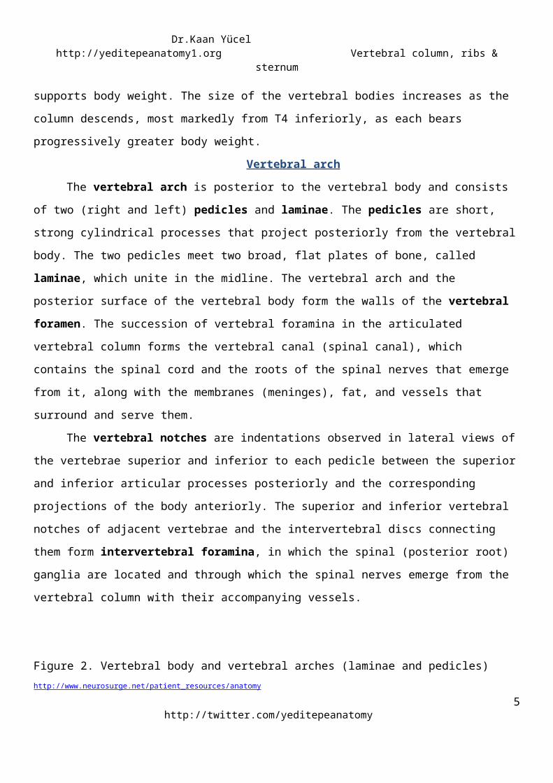
Vertebral archThe vertebral arch is posterior to the vertebral body and consists of two (right and left) pedicles
and laminae. The pedicles are short, strong cylindrical processes that project posteriorly from the vertebral
body. The two pedicles meet two broad, flat plates of bone, called laminae, which unite in the midline. The
vertebral arch and the posterior surface of the vertebral body form the walls of the vertebral foramen. The
succession of vertebral foramina in the articulated vertebral column forms the vertebral canal (spinal
canal), which contains the spinal cord and the roots of the spinal nerves that emerge from it, along with
the membranes (meninges), fat, and vessels that surround and serve them.
The vertebral notches are indentations observed in lateral views of the vertebrae superior and
inferior to each pedicle between the superior and inferior articular processes posteriorly and the
corresponding projections of the body anteriorly. The superior and inferior vertebral notches of adjacent
vertebrae and the intervertebral discs connecting them form intervertebral foramina, in which the spinal
(posterior root) ganglia are located and through which the spinal nerves emerge from the vertebral column
with their accompanying vessels.
Figure 2. Vertebral body and vertebral arches (laminae and pedicles)
http://www.youtube.com/yeditepeanatomy 4
A TYPICAL VERTEBRA
Dr.Kaan Yücel http://yeditepeanatomy1.org Vertebral column, ribs & sternum
http://www.neurosurge.net/patient_resources/anatomy
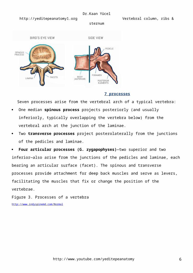
7 processesSeven processes arise from the vertebral arch of a typical vertebra:
One median spinous process projects posteriorly (and usually inferiorly, typically overlapping the
vertebra below) from the vertebral arch at the junction of the laminae.
Two transverse processes project posterolaterally from the junctions of the pedicles and laminae.
Four articular processes (G. zygapophyses)—two superior and two inferior—also arise from the
junctions of the pedicles and laminae, each bearing an articular surface (facet). The spinous and transverse
processes provide attachment for deep back muscles and serve as levers, facilitating the muscles that fix or
change the position of the vertebrae.
Figure 3. Processes of a vertebrahttp://www.indyspinemd.com/Normal
http://twitter.com/yeditepeanatomy5
Dr.Kaan Yücel http://yeditepeanatomy1.org Vertebral column, ribs & sternum
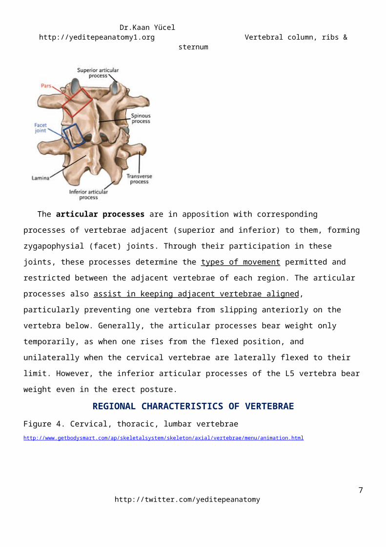
The articular processes are in apposition with corresponding processes of vertebrae adjacent (superior
and inferior) to them, forming zygapophysial (facet) joints. Through their participation in these joints, these
processes determine the types of movement permitted and restricted between the adjacent vertebrae of
each region. The articular processes also assist in keeping adjacent vertebrae aligned, particularly
preventing one vertebra from slipping anteriorly on the vertebra below. Generally, the articular processes
bear weight only temporarily, as when one rises from the flexed position, and unilaterally when the
cervical vertebrae are laterally flexed to their limit. However, the inferior articular processes of the L5
vertebra bear weight even in the erect posture.
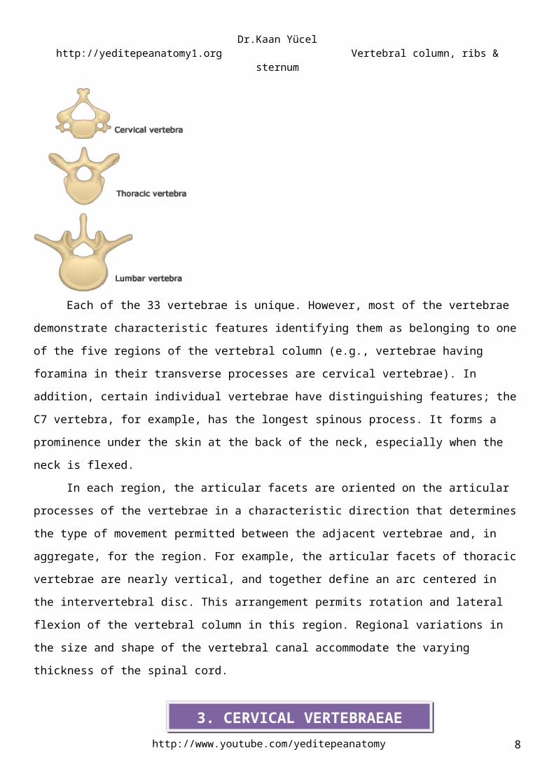
REGIONAL CHARACTERISTICS OF VERTEBRAEFigure 4. Cervical, thoracic, lumbar vertebraehttp://www.getbodysmart.com/ap/skeletalsystem/skeleton/axial/vertebrae/menu/animation.html
Each of the 33 vertebrae is unique. However, most of the vertebrae demonstrate characteristic
features identifying them as belonging to one of the five regions of the vertebral column (e.g., vertebrae
having foramina in their transverse processes are cervical vertebrae). In addition, certain individual
vertebrae have distinguishing features; the C7 vertebra, for example, has the longest spinous process. It
forms a prominence under the skin at the back of the neck, especially when the neck is flexed.
In each region, the articular facets are oriented on the articular processes of the vertebrae in a
characteristic direction that determines the type of movement permitted between the adjacent vertebrae
and, in aggregate, for the region. For example, the articular facets of thoracic vertebrae are nearly vertical,
and together define an arc centered in the intervertebral disc. This arrangement permits rotation and
lateral flexion of the vertebral column in this region. Regional variations in the size and shape of the
vertebral canal accommodate the varying thickness of the spinal cord.
http://www.youtube.com/yeditepeanatomy 6
Dr.Kaan Yücel http://yeditepeanatomy1.org Vertebral column, ribs & sternum
skeleton of the neck, between the cranium & thoracic vertebrae
1. Smallest of the movable vertebrae as they bear less weight; only the cranium.
2. Relatively larger intervertebral discs: the discs are actually thin, but relative to their small size; thick.
3. Greatest range & variety of movement of all the vertebral regions thanks to: a) relative thickness of the
intervertebral discs, b) nearly horizontal orientation of the articular facets, c) small mass of body.
4. The most distinctive feature of each cervical vertebra is the oval foramen transversarium (transverse
foramen) in the transverse process. The vertebral arteries and their accompanying veins pass through the
transverse foramina (except C7).
5. The transverse processes of cervical vertebrae end laterally in two projections: an anterior tubercle and
a posterior tubercle. The tubercles provide attachment for a laterally placed group of cervical muscles. The
anterior tubercles of vertebra C6 are called carotid tubercles (Chassaignac tubercles) because the
common carotid arteries may be compressed here, in the groove between the tubercle and body, to
control bleeding from these vessels.
6. The spinous processes of the C3-C6 vertebrae are short and usually bifid in white people, especially
males, but usually not as commonly in people of African descent or in females (Duray et al., 1999).
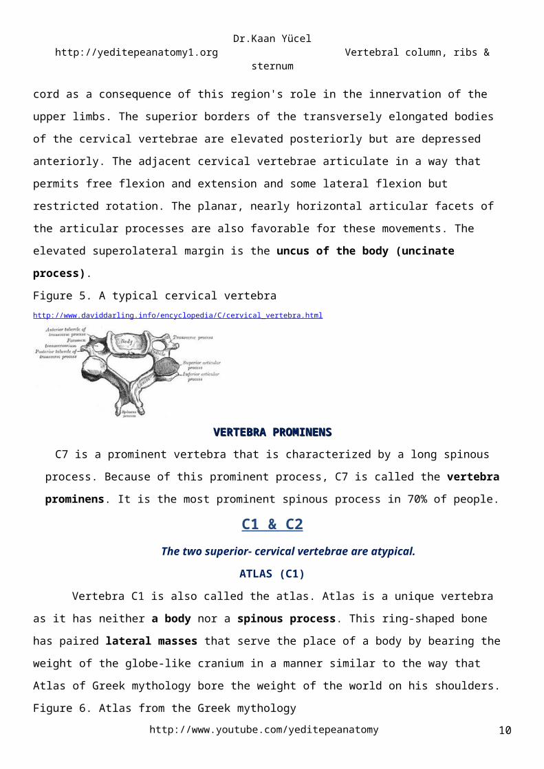
Vertebrae C3-C7 are the typical cervical vertebrae. They have large vertebral foramina to
accommodate the cervical enlargement of the spinal cord as a consequence of this region's role in the
innervation of the upper limbs. The superior borders of the transversely elongated bodies of the cervical
vertebrae are elevated posteriorly but are depressed anteriorly. The adjacent cervical vertebrae articulate
in a way that permits free flexion and extension and some lateral flexion but restricted rotation. The
planar, nearly horizontal articular facets of the articular processes are also favorable for these movements.
The elevated superolateral margin is the uncus of the body (uncinate process).
Figure 5. A typical cervical vertebrahttp://www.daviddarling.info/encyclopedia/C/cervical_vertebra.html
http://twitter.com/yeditepeanatomy7
3. CERVICAL VERTEBRAEAE
FEATURES TYPICAL FOR CERVICAL VERTEBRAEFEATURES TYPICAL FOR CERVICAL VERTEBRAE
Dr.Kaan Yücel http://yeditepeanatomy1.org Vertebral column, ribs & sternum
VERTEBRA PROMINENSVERTEBRA PROMINENSC7 is a prominent vertebra that is characterized by a long spinous process. Because of this prominent
process, C7 is called the vertebra prominens. It is the most prominent spinous process in 70% of people.
C1 & C2The two superior- cervical vertebrae are atypical.
ATLAS (C1) Vertebra C1 is also called the atlas. Atlas is a unique vertebra as it has neither a body nor a spinous
process. This ring-shaped bone has paired lateral masses that serve the place of a body by bearing the
weight of the globe-like cranium in a manner similar to the way that Atlas of Greek mythology bore the
weight of the world on his shoulders.
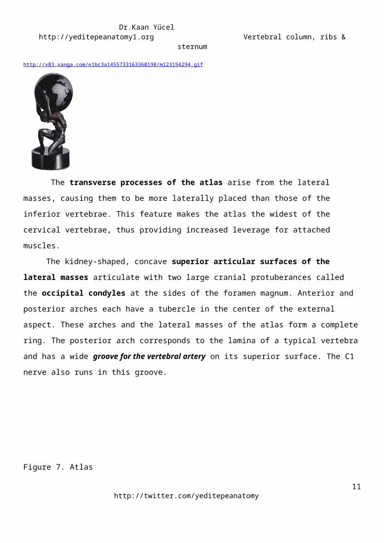
Figure 6. Atlas from the Greek mythologyhttp://x83.xanga.com/e1bc3a1455733163360198/m123194294.gif
The transverse processes of the atlas arise from the lateral masses, causing them to be more
laterally placed than those of the inferior vertebrae. This feature makes the atlas the widest of the cervical
vertebrae, thus providing increased leverage for attached muscles.
The kidney-shaped, concave superior articular surfaces of the lateral masses articulate with two
large cranial protuberances called the occipital condyles at the sides of the foramen magnum. Anterior
and posterior arches each have a tubercle in the center of the external aspect. These arches and the lateral
masses of the atlas form a complete ring. The posterior arch corresponds to the lamina of a typical
vertebra and has a wide groove for the vertebral artery on its superior surface. The C1 nerve also runs in
this groove.
http://www.youtube.com/yeditepeanatomy 8
Dr.Kaan Yücel http://yeditepeanatomy1.org Vertebral column, ribs & sternum
Figure 7. Atlashttp://www.daviddarling.info/encyclopedia/C/cervical_vertebra.htm l
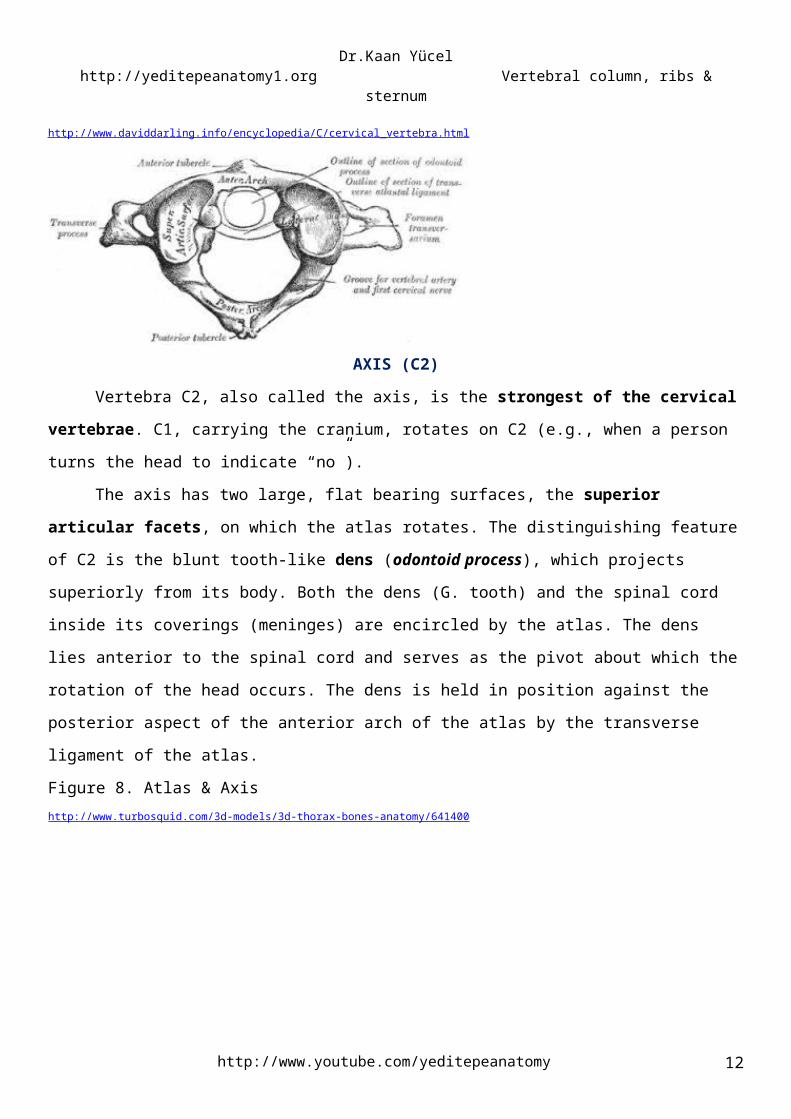
AXIS (C2)Vertebra C2, also called the axis, is the strongest of the cervical vertebrae. C1, carrying the
cranium, rotates on C2 (e.g., when a person turns the head to indicate “no”).
The axis has two large, flat bearing surfaces, the superior articular facets, on which the atlas
rotates. The distinguishing feature of C2 is the blunt tooth-like dens (odontoid process), which projects
superiorly from its body. Both the dens (G. tooth) and the spinal cord inside its coverings (meninges) are
encircled by the atlas. The dens lies anterior to the spinal cord and serves as the pivot about which the
rotation of the head occurs. The dens is held in position against the posterior aspect of the anterior arch of
the atlas by the transverse ligament of the atlas.
Figure 8. Atlas & Axishttp://www.turbosquid.com/3d-models/3d-thorax-bones-anatomy/641400
http://twitter.com/yeditepeanatomy9
Dr.Kaan Yücel http://yeditepeanatomy1.org Vertebral column, ribs & sternum
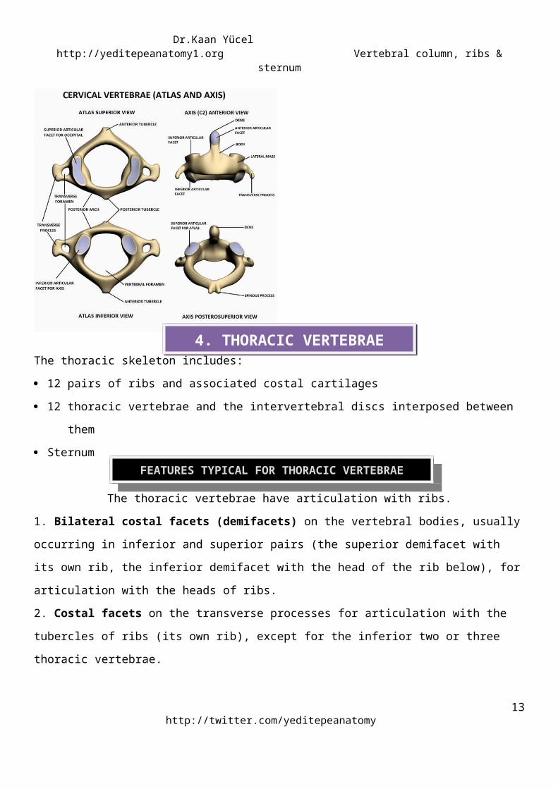
The thoracic skeleton includes:
12 pairs of ribs and associated costal cartilages
12 thoracic vertebrae and the intervertebral discs interposed between them
Sternum
The thoracic vertebrae have articulation with ribs.
1. Bilateral costal facets (demifacets) on the vertebral bodies, usually occurring in inferior and superior
pairs (the superior demifacet with its own rib, the inferior demifacet with the head of the rib below), for
articulation with the heads of ribs.
2. Costal facets on the transverse processes for articulation with the tubercles of ribs (its own rib), except
for the inferior two or three thoracic vertebrae.
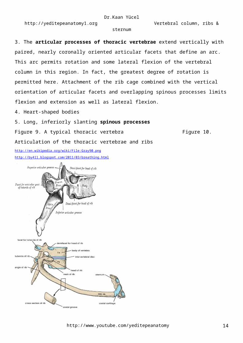
3. The articular processes of thoracic vertebrae extend vertically with paired, nearly coronally oriented
articular facets that define an arc. This arc permits rotation and some lateral flexion of the vertebral
column in this region. In fact, the greatest degree of rotation is permitted here. Attachment of the rib cage
combined with the vertical orientation of articular facets and overlapping spinous processes limits flexion
and extension as well as lateral flexion.
4. Heart-shaped bodies
5. Long, inferiorly slanting spinous processes
Figure 9. A typical thoracic vertebra Figure 10. Articulation of the thoracic vertebrae and ribshttp://en.wikipedia.org/wiki/File:Gray90.png http://by411.blogspot.com/2011/03/breathing.html
The T1-T4 vertebrae share some features of cervical vertebrae. The middle four thoracic vertebrae
(T5-T8) demonstrate all the features typical of thoracic vertebrae.
A typical thoracic vertebra has three sites on each side for articulation with ribs.
http://www.youtube.com/yeditepeanatomy 10
4. THORACIC VERTEBRAE
FEATURES TYPICAL FOR THORACIC VERTEBRAEFEATURES TYPICAL FOR THORACIC VERTEBRAE
Dr.Kaan Yücel http://yeditepeanatomy1.org Vertebral column, ribs & sternum
Two demifacets (i.e., partial facets) are located on the superior and inferior aspects of the body for
articulation with corresponding sites on the heads of adjacent ribs. The superior costal facet articulates
with part of the head of its own rib, and the inferior costal facet articulates with part of the head of the rib
below.
An oval facet (transverse costal facet) at the end of the transverse process articulates with the tubercle
of its own rib.
Atyical articulations of the thoracic vertebrae with the ribs:You should know that not all thoracic vertebrae have this articulation design with ribs. As you will see
in the following section ribs are grouped into two parts: typical and atypical ribs according their articulation
features with the thoracic vertebrae.
Atypical thoracic vertebrae
T1
1. a long, almost horizontal spinous process that may be nearly as prominent as that of
the vertebra prominens.
2. a complete costal facet on the superior edge of its body for the 1st rib; T1 articulates with a single
facet on the head of its own rib-in other words, the head of rib I does not articulate with vertebra
CVII.
3. a demifacet on its inferior edge that contributes to the articular surface for the 2nd rib.
TX
Vertebra TX (and often TIX) articulates only with its own ribs and therefore lacks inferior demifacets on the
body.
TXI & TXII
Vertebrae TXI and TXII articulate only with the heads of their own ribs-they lack transverse costal facets
and have only a single complete facet on each side of their bodies.
The T9-T12 vertebrae have some features of lumbar vertebrae (e.g., tubercles similar to the accessory
processes). Mammillary processes also occur. However, most of the transition in characteristics of
vertebrae from the thoracic to the lumbar region occurs over the length of a single vertebra: vertebra T12.
Generally, its superior half is thoracic in character, having costal facets and articular processes that permit
primarily rotatory movement, whereas its inferior half is lumbar in character, devoid of costal facets and
having articular processes that permit only flexion and extension. Consequently, vertebra T12 is subject to
transitional stresses that cause it to be the most commonly fractured vertebra.
http://twitter.com/yeditepeanatomy11
Dr.Kaan Yücel http://yeditepeanatomy1.org Vertebral column, ribs & sternum
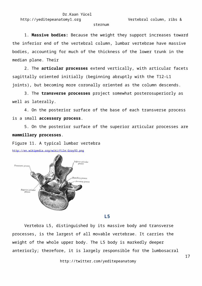
Lumbar vertebrae are in the lower back between the thorax and sacrum.
,1. Massive bodies: Because the weight they support increases toward the inferior end of the
vertebral column, lumbar vertebrae have massive bodies, accounting for much of the thickness of the
lower trunk in the median plane. Their
2. The articular processes extend vertically, with articular facets sagittally oriented initially
(beginning abruptly with the T12-L1 joints), but becoming more coronally oriented as the column
descends.
3. The transverse processes project somewhat posterosuperiorly as well as laterally.
4. On the posterior surface of the base of each transverse process is a small accessory process.
5. On the posterior surface of the superior articular processes are mammillary processes.
Figure 11. A typical lumbar vertebrahttp://en.wikipedia.org/wiki/File:Gray93.png
L5
Vertebra L5, distinguished by its massive body and transverse processes, is the largest of all
movable vertebrae. It carries the weight of the whole upper body. The L5 body is markedly deeper
anteriorly; therefore, it is largely responsible for the lumbosacral angle between the long axis of the lumbar
region of the vertebral column and that of the sacrum. Body weight is transmitted from L5 vertebra to the
base of the sacrum, formed by the superior surface of S1 vertebra.
The L5-S1 facets are distinctly coronal in orientation. In the more sagittally oriented superior
joints, the laterally facing facets of the inferior articular processes of the vertebra above are “gripped” by
http://www.youtube.com/yeditepeanatomy 12
5. LUMBAR VERTEBRAE
FEATURES TYPICAL FOR LUMBAR VERTEBRAEFEATURES TYPICAL FOR LUMBAR VERTEBRAE
Dr.Kaan Yücel http://yeditepeanatomy1.org Vertebral column, ribs & sternum
the medially facing facets of the superior processes of the vertebra below, in a manner that facilitates
flexion and extension and allows lateral flexion, but prohibits rotation.
The wedged-shaped sacrum (L. sacred) is usually composed of five fused sacral vertebrae in adults.
It is located between the hip bones and forms the roof and posterosuperior wall of the posterior half of the
pelvic cavity. The triangular shape of the sacrum results from the rapid decrease in the size of the lateral
masses of the sacral vertebrae during development. The inferior half of the sacrum is not weight-bearing;
therefore, its bulk is diminished considerably. The sacrum supports the vertebral column and forms the
posterior part of the bony pelvis. The sacrum is tilted so that it articulates with the L5 vertebra at the
lumbosacral angle.
The sacrum provides strength and stability to the pelvis and transmits the weight of the body to the
pelvic girdle, the bony ring formed by the hip bones and sacrum, to which the lower limbs are attached.
The sacral canal is the continuation of the vertebral canal in the sacrum. On the pelvic and posterior
surfaces of the sacrum between its vertebral components are typically four pairs of sacral foramina for the
exit of the posterior and anterior rami of the spinal nerves.
The base of the sacrum is formed by the superior surface of the S1 vertebra. Its superior articular
processes articulate with the inferior articular processes of the L5 vertebra. The anterior projecting edge of
the body of the S1 vertebra is the sacral promontory (L. mountain ridge), an important obstetrical
landmark. The apex of the sacrum, its inferior end, has an oval facet for articulation with the coccyx.
The pelvic surface of the sacrum is smooth and concave. Four transverse lines on this surface of
sacra from adults indicate where fusion of the sacral vertebrae occurred. Fusion of the sacral vertebrae
starts after age 20; however, most of the intervertebral discs remain unossified up to or beyond middle
life.
The dorsal surface of the sacrum is rough, convex, and marked by five prominent longitudinal
ridges. The central ridge, the median sacral crest, represents the fused rudimentary spinous processes of
the superior three or four sacral vertebra; S5 has no spinous process. The intermediate sacral crests
represent the fused articular processes, and the lateral sacral crests are the tips of the transverse
processes of the fused sacral vertebrae.
The clinically important features of the dorsal surface of the sacrum are the inverted U-shaped
sacral hiatus and the sacral cornua (L. horns). The sacral hiatus results from the absence of the laminae
http://twitter.com/yeditepeanatomy13
6. SACRUM
Dr.Kaan Yücel http://yeditepeanatomy1.org Vertebral column, ribs & sternum
and spinous process of S5 and sometimes S4. The sacral hiatus leads into the sacral canal. The sacral
cornua, representing the inferior articular processes of S5 vertebra, project inferiorly on each side of the
sacral hiatus and are a helpful guide to its location.
The superior part of the lateral surface of the sacrum looks somewhat like an auricle (L. external
ear); because of its shape, this area is called the auricular surface. During life, the auricular surface is
covered with hyaline cartilage.
Figure 12. Sacrum; anterior view
http://3.bp.blogspot.com/-HQsnxFx0qc0/TqqunS0uSDI/AAAAAAAAAEs/bL8GdNChku0/s1600/sacrum_coccyx_Front.jp g
Figure 13. Sacrum; posterior viewhttp://www.back.com/anatomy-sacral.html
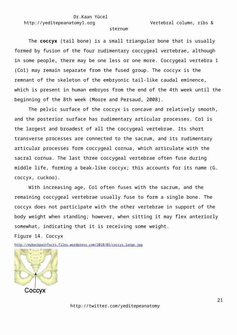
The coccyx (tail bone) is a small triangular bone that is usually formed by fusion of the four
rudimentary coccygeal vertebrae, although in some people, there may be one less or one more. Coccygeal
vertebra 1 (Co1) may remain separate from the fused group. The coccyx is the remnant of the skeleton of
the embryonic tail-like caudal eminence, which is present in human embryos from the end of the 4th week
until the beginning of the 8th week (Moore and Persaud, 2008).
http://www.youtube.com/yeditepeanatomy 14
7. COCCYX
Dr.Kaan Yücel http://yeditepeanatomy1.org Vertebral column, ribs & sternum
The pelvic surface of the coccyx is concave and relatively smooth, and the posterior surface has
rudimentary articular processes. Co1 is the largest and broadest of all the coccygeal vertebrae. Its short
transverse processes are connected to the sacrum, and its rudimentary articular processes form coccygeal
cornua, which articulate with the sacral cornua. The last three coccygeal vertebrae often fuse during
middle life, forming a beak-like coccyx; this accounts for its name (G. coccyx, cuckoo).
With increasing age, Co1 often fuses with the sacrum, and the remaining coccygeal vertebrae
usually fuse to form a single bone. The coccyx does not participate with the other vertebrae in support of
the body weight when standing; however, when sitting it may flex anteriorly somewhat, indicating that it is
receiving some weight.
Figure 14. Coccyxhttp://mybackpainfacts.files.wordpress.com/2010/05/coccyx_large.jpg
OSSIFICATION OF VERTEBRAEVertebrae begin to develop during the embryonic period as mesenchymal condensations around
the notochord. Later, these mesenchymal bone models chondrify and cartilaginous vertebrae form.
Typically, vertebrae begin to ossify toward the end of the embryonic period (8th week), with three primary
ossification centers developing in each cartilaginous vertebra: an endochondral centrum, which will
eventually constitute most of the body of the vertebra, and two perichondral centers, one in each half of
the neural arch.
VARIATIONS IN VERTEBRAEMost people have 33 vertebrae, but developmental errors may result in 32 or 34 vertebrae.
Variations in vertebrae are affected by race, gender, and developmental factors (genetic and
environmental). An increased number of vertebrae occurs more often in males and a reduced number
occurs more frequently in females.
Variations in vertebrae also involve the relationship between the vertebrae and ribs, and the
number of vertebrae that fuse to form the sacrum. The relationship of presacral vertebrae to ribs and/or
sacrum may occur higher (cranial shift) or lower (caudal shift) than normal. A C7 vertebra articulating with
http://twitter.com/yeditepeanatomy15
Dr.Kaan Yücel http://yeditepeanatomy1.org Vertebral column, ribs & sternum
a rudimentary cervical rib(s) is still considered a cervical vertebra. The same is true for lumbar vertebrae
and lumbar ribs. Likewise, an L5 vertebra fused to the sacrum is referred to as a “sacralized 5th lumbar
vertebra”.
There are four natural curves in a healthy spine.
1. The neck or cervical spine, curves gently inward (lordosis)
2. The mid back, or thoracic spine, is curved outward (kyphosis)
3. The low back, or lumbar spine, also curves inward (lordosis)
4. Pelvic (Sacral) curve
The cervical and lumbar curves are considered secondary curves whereas the thoracic and sacral
curves are primary. The spine’s curves work like a coiled spring to absorb shock, maintain balance, and
allow the full range of motion throughout the spinal column. The lumbosacral angle occurs at the junction
of the long axes of the lumbar region of the vertebral column and the sacrum.
Figure 15. Curvartures of the spinehttp://www.spineuniverse.com/anatomy/spinal-curves
DEFORMITIES OF THE VERTEBRAL COLUMNScoliosisScoliosis (from Greek: skoliōsis meaning from skolios, "crooked") is a medical condition in which a person's
spine is curved from side to side. Scoliosis occurs in approximately 2% of women and less than 1/2% of
men. It is a progressive disease whose origin is unknown (or idiopathic) in 80% of the cases, although there
is evidence for a genetic and nutritional component. Females are at 10 times more risk than males.
http://www.youtube.com/yeditepeanatomy 16
CLINICAL ANATOMY
8. CURVATURES IN THE VERTEBRAL COLUMN
Dr.Kaan Yücel http://yeditepeanatomy1.org Vertebral column, ribs & sternum
Scoliosis often includes a twisting of the spine, resulting in distortion of the ribs and entire thorax. It usually
presents in pre-teens and adolescents. Structural scoliosis may require surgical intervention; alternatively
scoliosis may be corrected using orthotics (e.g. braces).
HyperkyphosisKyphosis describes the natural curvatures of the thoracic spine, but hyperkyphosis a pathologically
exaggerated thoracic curvature, commonly called "hunchback." Hyperkyphos is common in aging adults,
usually aided by the vertebral collapse related to osteoporosis. Other common causes may include trauma,
arthritis, and endocrine or other diseases.
HyperlordosisLordosis describes the natural curvature of the lumbar spine, but hyperlordosis is a pathologically
exaggerated lumbar curvature, commonly called "swayback." Hyperlordosis is usually accompanied by the
pelvis tilting abnormally forward, often causing an exaggerated protrusion of the buttocks. Symptoms may
include pain and numbness if the nerve trunks are compromised. Typically, the condition is attributed to
weak back muscles or a habitual hyperextension, such as in pregnant women, men with excessive visceral
fat, and some dance postures. Hyperlordosis is also correlated with puberty.
More @ http://clinicalanatomy.wetpaint.com/page/Vertebral+column
Ribs (L. costae) are curved, flat bones that form most of the thoracic cage. There are twelve pairs of
ribs, each terminating anteriorly in a costal cartilage. There are three types of ribs that can be classified as
typical or atypical. (In Moore’s Clinically Oriented Anatomy and Gray’s Anatomy for Students; the first seven ribs
that articulate with the sternum directly are described as true ribs. In Gray’s anatomy textbook the remaining five ribs
are considered as false ribs, however, in Moore’s textbook the last two, sometimes three ribs are classified as
floating, free ribs, and these ribs are not grouped under the false ribs, but seen as a different third group. Snell’s
anatomy textbook follows the Moore’s). All the ribs articulate with the thoracic vertebrae posteriorly with
different fashion, though. Note that their connection with the sternum is different anteriorly. These three
types below are according to the articulations of the ribs with the sternum anteriorly, and the terms
atypical and typical ribs refer to the articulations with the thoracic vertebrae posteriorly.
Figure 16. True, false, and floating ribshttp://www.daviddarling.info/encyclopedia/R/rib-cage.html
http://twitter.com/yeditepeanatomy17
9. RIBS
True (vertebrocostal) ribs (1st-7th ribs): They attach directly to the sternum through their own costal cartilages.
False (vertebrochondral) ribs (8th, 9th, and usually 10th ribs): Their cartilages are connected to the cartilage of the rib above them; thus their connection with the sternum is indirect.
Floating (vertebral, free) ribs (11th, 12th, and sometimes 10th ribs): The rudimentary cartilages of these ribs do not connect even indirectly with the sternum; instead they end in the posterior abdominal musculature.
Dr.Kaan Yücel http://yeditepeanatomy1.org Vertebral column, ribs & sternum
Typical ribs (3rd-9th) have the following components:
Head: wedge-shaped and has two facets, separated by the crest of the head; one facet for articulation with
the numerically corresponding vertebra and one facet for the vertebra superior to it.
Neck: connects the head of the rib with the body at the level of the tubercle.
Tubercle: located at the junction of the neck and body; a smooth articular part articulates with the
corresponding transverse process of the vertebra, and a rough nonarticular part provides attachment for
the costotransverse ligament.
Body (shaft): thin, flat, and curved, most markedly at the costal angle where the rib turns anterolaterally.
The angle also demarcates the lateral limit of attachment of the deep back muscles to the ribs. The
concave internal surface of the body has a costal groove paralleling the inferior border of the rib, which
provides some protection for the intercostal nerve and vessels.
Figure 17. Parts of a typical ribhttp://www.blobs.org/science/article.php?article=9
Atypical ribs (1st, 2nd, and 10th-12th) are dissimilar:
The 1st rib is the broadest (i.e., its body is widest and nearly horizontal), shortest, and most sharply
curved of the seven true ribs. It has a single facet on its head for articulation with the T1 vertebra only and
two transversely directed grooves crossing its superior surface for the subclavian vessels; the grooves are
separated by a scalene tubercle and ridge, to which the anterior scalene muscle is attached.
http://www.youtube.com/yeditepeanatomy 18
Dr.Kaan Yücel http://yeditepeanatomy1.org Vertebral column, ribs & sternum
The 2nd rib is has a thinner, less curved body and is substantially longer than the 1st rib. Its head
has two facets for articulation with the bodies of the T1 and T2 vertebrae; its main atypical feature is a
rough area on its upper surface, the tuberosity for serratus anterior, from which part of that muscle
originates.
The 10th-12th ribs, like the 1st rib, have only one facet on their heads and articulate with a single
vertebra. The 11th and 12th ribs are short and have no neck or tubercle.
Figure 18. Atypical and typical ribshttp://www.sciencedirect.com/science/article/pii/S154741270600106X
Costal cartilages prolong the ribs anteriorly and contribute to the elasticity of the thoracic wall,
providing a flexible attachment for their anterior ends (tips). The cartilages increase in length through the
first 7 and then gradually decrease. The first 7 costal cartilages attach directly and independently to the
sternum; the 8th, 9th, and 10th articulate with the costal cartilages just superior to them, forming a
continuous, articulated, cartilaginous costal margin. The 11th and 12th costal cartilages form caps on the
anterior ends of the corresponding ribs and do not reach or attach to any other bone or cartilage. The
costal cartilages of ribs 1-10 clearly anchor the anterior end of the rib to the sternum, limiting its overall
movement as the posterior end rotates around the transverse axis of the rib.
Intercostal spaces separate the ribs and their costal cartilages from one another. The spaces are
named according to the rib forming the superior border of the space—for example, the 4th intercostal
http://twitter.com/yeditepeanatomy19
11. INTERCOSTAL SPACES
10. COSTAL CARTILAGES

Dr.Kaan Yücel http://yeditepeanatomy1.org Vertebral column, ribs & sternum
space lies between ribs 4 and 5. There are 11 intercostal spaces and 11 intercostal nerves. Intercostal
spaces are occupied by intercostal muscles and membranes, and two sets (main and collateral) of
intercostal blood vessels and nerves, identified by the same number assigned to the space. The space
below the 12th rib does not lie between ribs and thus is referred to as the subcostal space, and the
anterior ramus (branch) of spinal nerve T12 is the subcostal nerve. The intercostal spaces are widest
anterolaterally, and they widen further with inspiration. They can also be further widened by extension
and/or lateral flexion of the thoracic vertebral column to the contralateral side.
RIB FRACTURES The short, broad 1st rib, posteroinferior to the clavicle, is rarely fractured because of its protected position
(it cannot be palpated). When it is broken, however, structures crossing its superior aspect may be injured,
including the brachial plexus of nerves and subclavian vessels that serve the upper limb. The middle ribs
are most commonly fractured. The weakest part of a rib is just anterior to its angle; however, direct
violence may fracture a rib anywhere, and its broken end may injure internal organs such as a lung and/or
the spleen. Fractures of the lower ribs may tear the diaphragm and result in a diaphragmatic hernia. Rib
fractures are painful because the broken parts move during respiration, coughing, laughing, and sneezing.
SUPERNUMERARY RIBSPeople usually have 12 ribs on each side, but the number is increased by the presence of cervical and/or
lumbar ribs, or decreased by failure of the 12th pair to form. Cervical ribs are relatively common (0.5-2%)
and may interfere with neurovascular structures exiting the superior thoracic aperture. Lumbar ribs are
less common. Supernumerary (extra) ribs also have clinical significance in that they may confuse the
identification of vertebral levels in radiographs and other diagnostic images.
The sternum (G. sternon, chest) is the flat, elongated bone that forms the middle of the anterior
part of the thoracic cage. It directly overlies and affords protection for mediastinal viscera in general and
much of the heart in particular. The sternum consists of three parts: manubrium, body, and xiphoid
process. In adolescents and young adults, the three parts are connected together by cartilaginous joints
(synchondroses) that ossify during middle to late adulthood.
The manubrium (L. handle, as in the handle of a sword, with the sternal body forming the blade) is
a roughly trapezoidal bone. The manubrium is the widest and thickest of the three parts of the sternum.
The easily palpated concave center of the superior border of the manubrium is the jugular notch
http://www.youtube.com/yeditepeanatomy 20
CLINICAL ANATOMY
12. STERNUM
Dr.Kaan Yücel http://yeditepeanatomy1.org Vertebral column, ribs & sternum
(suprasternal notch). The notch is deepened by the medial (sternal) ends of the clavicles, which are much
larger than the relatively small clavicular notches in the manubrium that receive them, forming the
sternoclavicular (SC) joints. Inferolateral to the clavicular notch, the costal cartilage of the 1st rib is tightly
attached to the lateral border of the manubrium—the synchondrosis of the first rib. The manubrium and
body of the sternum lie in slightly different planes superior and inferior to their junction, the
manubriosternal joint; hence, their junction forms a projecting sternal angle (of Louis).
The body of the sternum, is longer, narrower, and thinner than the manubrium, and is located at
the level of the T5-T9 vertebrae. Its width varies because of the scalloping of its lateral borders by the
costal notches. In young people, four sternebrae (primordial segments of the sternum) are obvious. The
sternebrae articulate with each other at primary cartilaginous joints (sternal synchondroses). These joints
begin to fuse from the inferior end between puberty (sexual maturity) and age 25. The nearly flat anterior
surface of the body of the sternum is marked in adults by three variable transverse ridges, which represent
the lines of fusion (synostosis) of its four originally separate sternebrae.
The xiphoid process, the smallest and most variable part of the sternum, is thin and elongated. Its
inferior end lies at the level of T10 vertebra. Although often pointed, the process may be blunt, bifid,
curved, or deflected to one side or anteriorly. It is cartilaginous in young people but more or less ossified in
adults older than age 40. In elderly people, the xiphoid process may fuse with the sternal body.
Figure 19. Sternum and its parts
http://medical-dictionary.thefreedictionary.com/sternum
Surface Anatomy: Key LandmarksJugular (suprasternal) notch:T2 vertebra in male, T4 in female
Sternal angle (of Louis) is at the level of the intervertebral disc between the 4th and 5th thoracal vertebra
and is useful for counting intercostal spaces (2nd ribs articulate here).
The xiphoid process is an important landmark in the median plane because
http://twitter.com/yeditepeanatomy21
Dr.Kaan Yücel http://yeditepeanatomy1.org Vertebral column, ribs & sternum
• Its junction with the sternal body at the xiphisternal joint indicates the inferior limit of the central part
of the thoracic cavity projected onto the anterior body wall;
• This joint is also the site of the infrasternal angle (subcostal angle) formed by the right and left costal
margins.
• It is a midline marker for the superior limit of the liver, the central tendon of the diaphragm, and the
inferior border of the heart.
STERNAL FRACTURESDespite the subcutaneous location of the sternum, sternal fractures are not common. Crush injuries
can occur after traumatic compression of the thoracic wall in automobile accidents when the driver's chest
is forced into the steering column, for example. The installation and use of air bags in vehicles has reduced
the number of sternal fractures. A fracture of the sternal body is usually a comminuted fracture (a break
resulting in several pieces). The most common site of sternal fracture in elderly people is at the sternal
angle, where the manubriosternal joint has fused. The concern in sternal injuries is not primarily for the
fracture itself but for the likelihood of heart injury (myocardial contusion, cardiac rupture, tamponade) or
lung injury.
MEDIAN STERNOTOMYTo gain access to the thoracic cavity for surgical operations in the mediastinum—such as coronary
artery bypass grafting, for example—the sternum is divided (split) in the median plane and retracted. Such
“sternal splitting” also gives good exposure for removal of tumors in the superior lobes of the lungs. After
surgery, the halves of the sternum are joined using wire sutures.
STERNAL ANOMALIESComplete sternal cleft is an uncommon anomaly through which the heart may protrude (ectopia
cordis). Partial clefts involving the manubrium and superior half of the body are V- or U-shaped and can be
repaired during infancy by direct apposition and fixation of the sternal halves. Sometimes only a
perforation (sternal foramen) remains in the sternal body because of the incomplete fusion. It is not
clinically significant; however, one should be aware of its possible presence so that it will not be
misinterpreted on chest X-ray, as a being an unhealed bullet wound for example. A receding (pectus
excavatum, or funnel chest) or projecting (pectus cavinatum, or pigeon breast) sternum are anomalous
variations that may become evident or more pronounced during childhood.
http://www.youtube.com/yeditepeanatomy 22
CLINICAL ANATOMY