Embed Size (px)
Citation preview

بسم الله الرحمن الرحيم
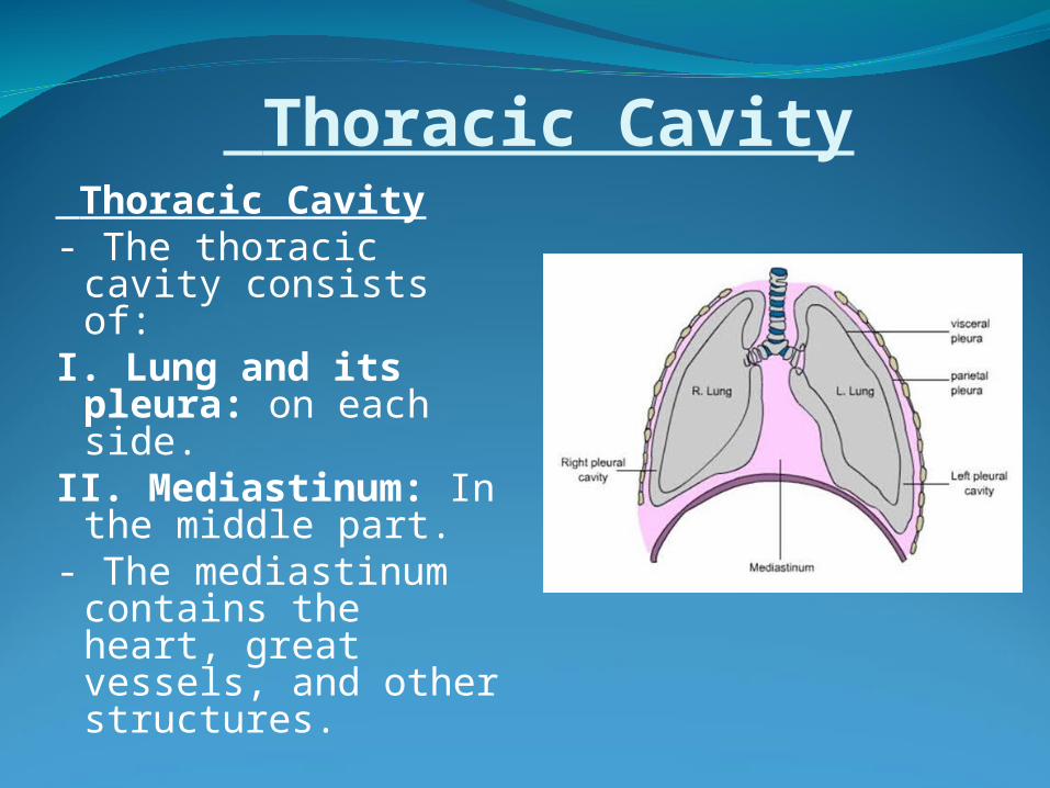
Thoracic Cavity Thoracic Cavity- The thoracic cavity
consists of:I. Lung and its
pleura: on each side.
II. Mediastinum: In the middle part.
- The mediastinum contains the heart, great vessels, and other structures.
PleuraDefinition:- It is a closed serous sac which is invaginated
by the lung from its medial side.
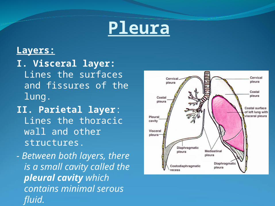
PleuraLayers:I. Visceral layer: Lines
the surfaces and fissures of the lung.
II. Parietal layer: Lines the thoracic wall and other structures.
- Between both layers, there is a small cavity called the pleural cavity which contains minimal serous fluid.
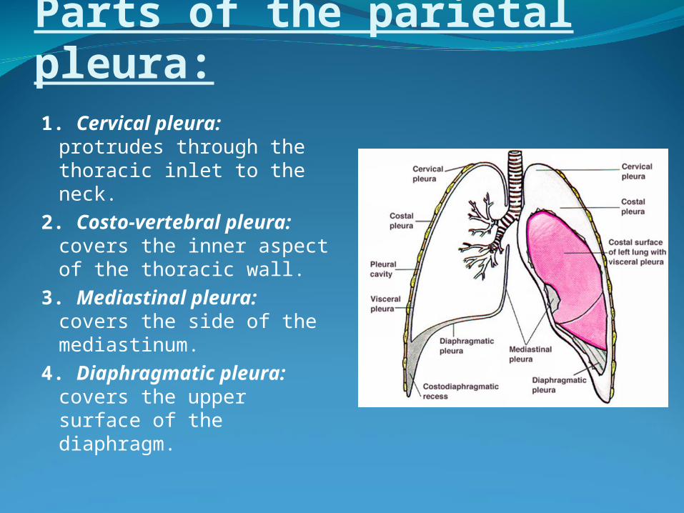
Parts of the parietal pleura:1. Cervical pleura:
protrudes through the thoracic inlet to the neck.
2. Costo-vertebral pleura: covers the inner aspect of the thoracic wall.
3. Mediastinal pleura: covers the side of the mediastinum.
4. Diaphragmatic pleura: covers the upper surface of the diaphragm.
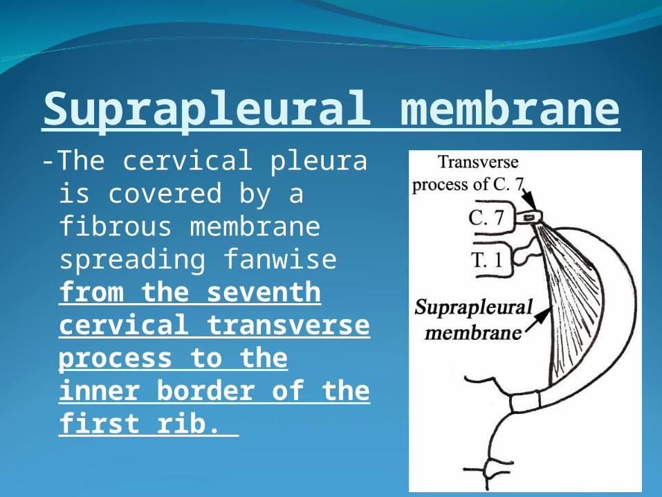
Suprapleural membrane-The cervical pleura is
covered by a fibrous membrane spreading fanwise from the seventh cervical transverse process to the inner border of the first rib.
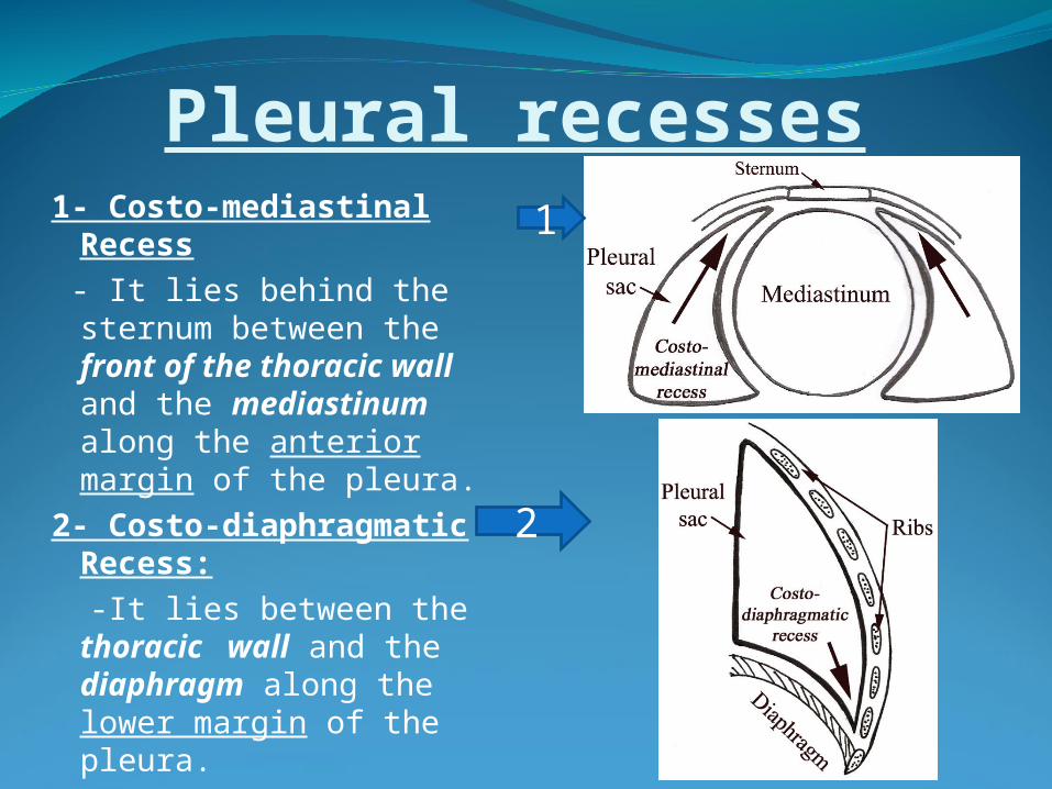
Pleural recesses1- Costo-mediastinal
Recess - It lies behind the sternum
between the front of the thoracic wall and the mediastinum along the anterior margin of the pleura.
2- Costo-diaphragmatic Recess:
-It lies between the thoracic wall and the diaphragm along the lower margin of the pleura.
2
1
Nerve supply of the pleuraParietal pleura:1-Costal pleura and peripheral diaphragmatic pleura:
Intercostal nerves .2-Mediastinal and central diaphragmatic pleura : Phrenic nerves.Viseral pleura: Not sensitive (has autonomic nerve supply).
Arterial supplyPartial Pleura:1. Intercostal arteries. 2. Internal thoracic
artery.3. Musculo-phrenic and pericardiaco-phrenic arteries.Visceral Pleura:- Bronchial arteries.
Applied anatomy- Pain due to irritation of the costal and
peripheral diaphragmatic pleura is referred to the thoracic or abdominal walls along the intercostal nerves.
- Pain due to irritation of the mediastinal and central diaphragmatic pleura is referred to the root of the neck and shoulder, because they are supplied by the same spinal segments through the supra clavicular nerves (C. 3,4).
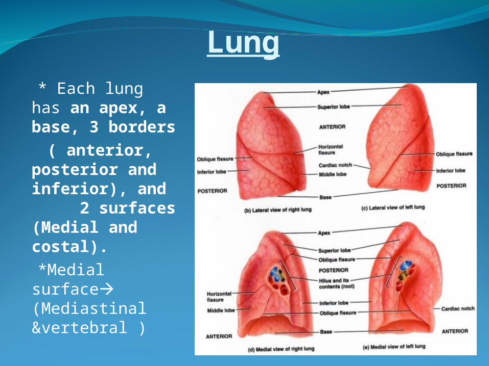
Lung * Each lung has
an apex, a base, 3 borders
( anterior, posterior and inferior), and 2 surfaces (Medial and costal).
*Medial surface (Mediastinal &vertebral )
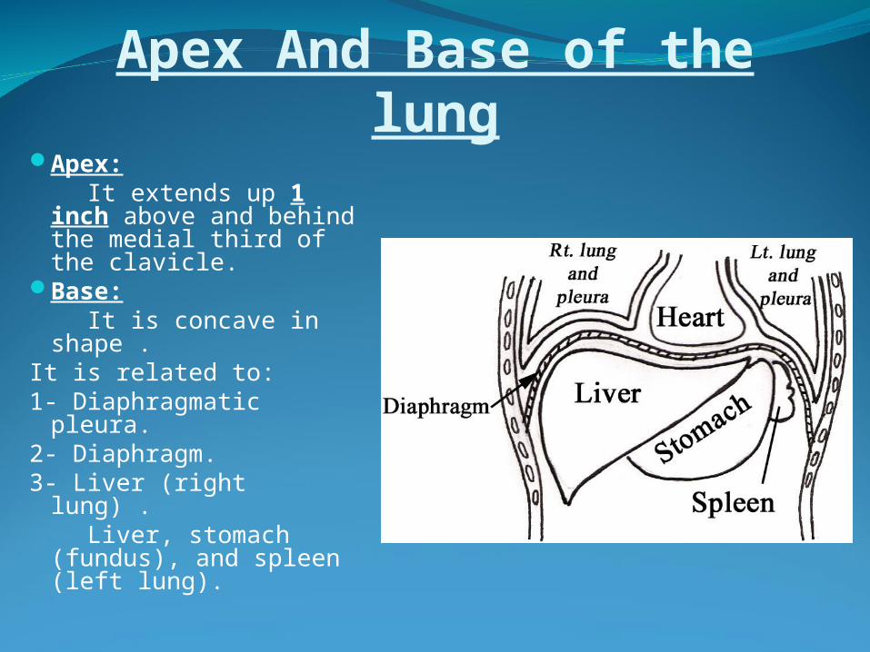
Apex And Base of the lungApex: It extends up 1 inch
above and behind the medial third of the clavicle.
Base: It is concave in shape
.It is related to:1- Diaphragmatic
pleura.2- Diaphragm.3- Liver (right lung) . Liver, stomach
(fundus), and spleen (left lung).
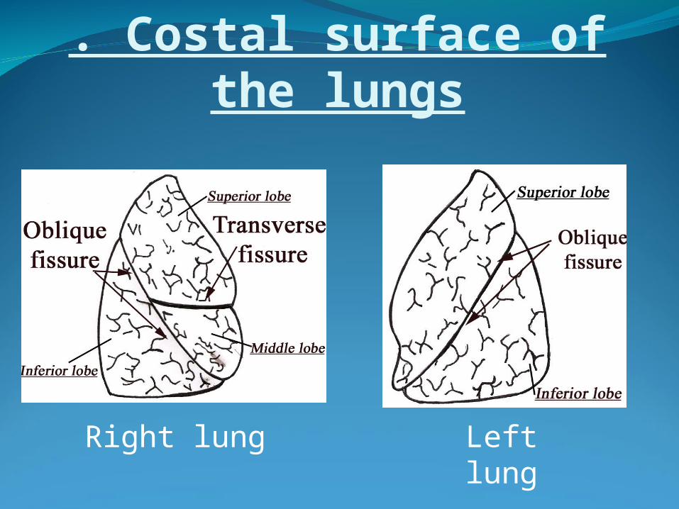
. Costal surface of the lungs
Right lung Left lung
Lobes &Fissures of the lungs
- The right lung is divided into three lobes(superior, middle, and inferior) by :
1-Oblique fissure: It begins at the posterior
border of the lung opposite the third thoracic spine & ends at the inferior border opposite the six costo-chondral junction.
2-Transverse fissure : It begins at the anterior
border opposite the fourth costal cartilage.
- The left lung is divided into two lobes (superior and inferior) by the oblique fissure.
* Oblique fissure (surface anatomy)
- It begins at the posterior border of the lung opposite the third thoracic spine (1 inch lateral to the spine) & ends at the inferior border opposite the six costo-chondral junction
Right lung Left lung
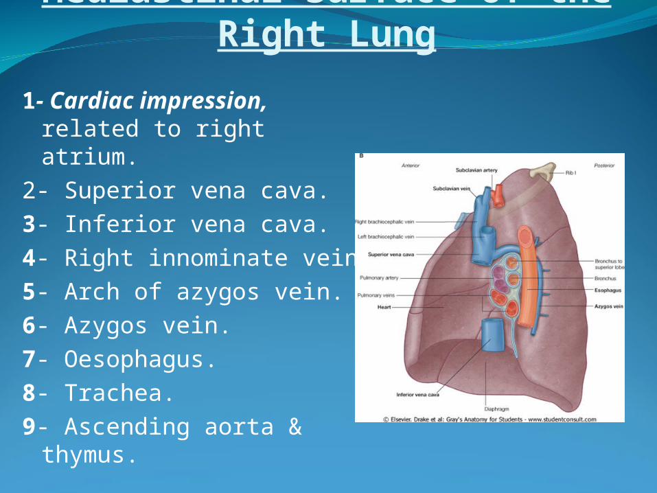
Mediastinal Surface of the Right Lung
1- Cardiac impression, related to right atrium.
2- Superior vena cava.3- Inferior vena cava.4- Right innominate vein.5- Arch of azygos vein.6- Azygos vein.7- Oesophagus.8- Trachea.9- Ascending aorta &
thymus.
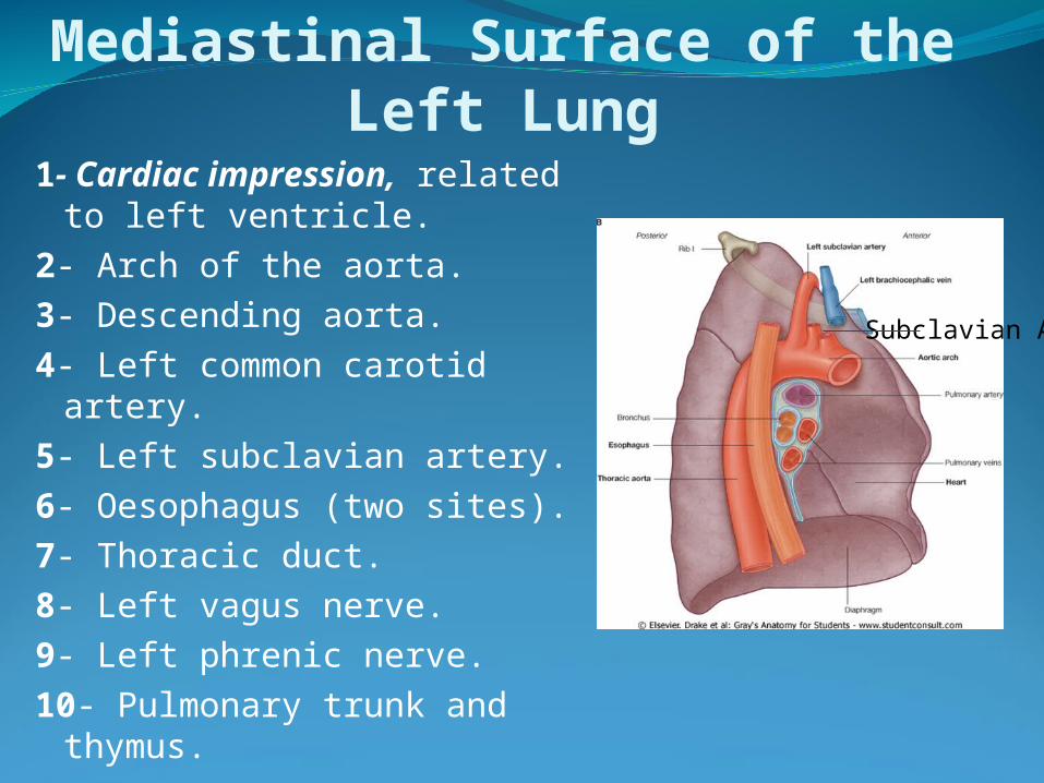
Mediastinal Surface of the Left Lung1- Cardiac impression,
related to left ventricle.2- Arch of the aorta.3- Descending aorta.4- Left common carotid
artery.5- Left subclavian artery.6- Oesophagus (two sites).7- Thoracic duct.8- Left vagus nerve.9- Left phrenic nerve. 10- Pulmonary trunk and
thymus.
Subclavian A
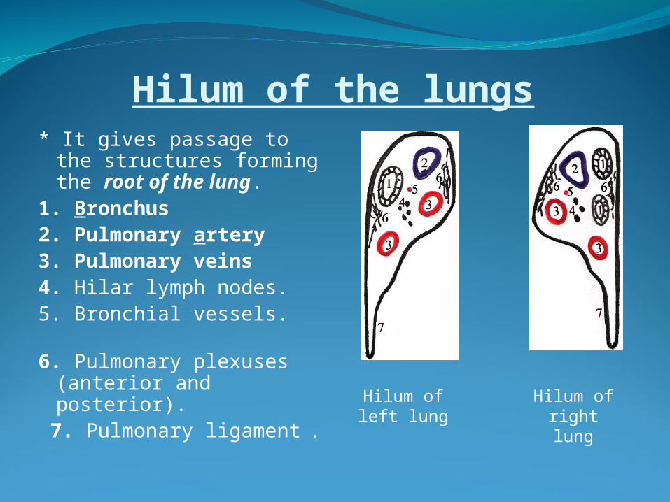
Hilum of the lungs* It gives passage to the
structures forming the root of the lung.
1. Bronchus2. Pulmonary artery3. Pulmonary veins4. Hilar lymph nodes.5. Bronchial vessels. 6. Pulmonary plexuses
(anterior and posterior).
7. Pulmonary ligament .
Hilum of left lung
Hilum of right lung
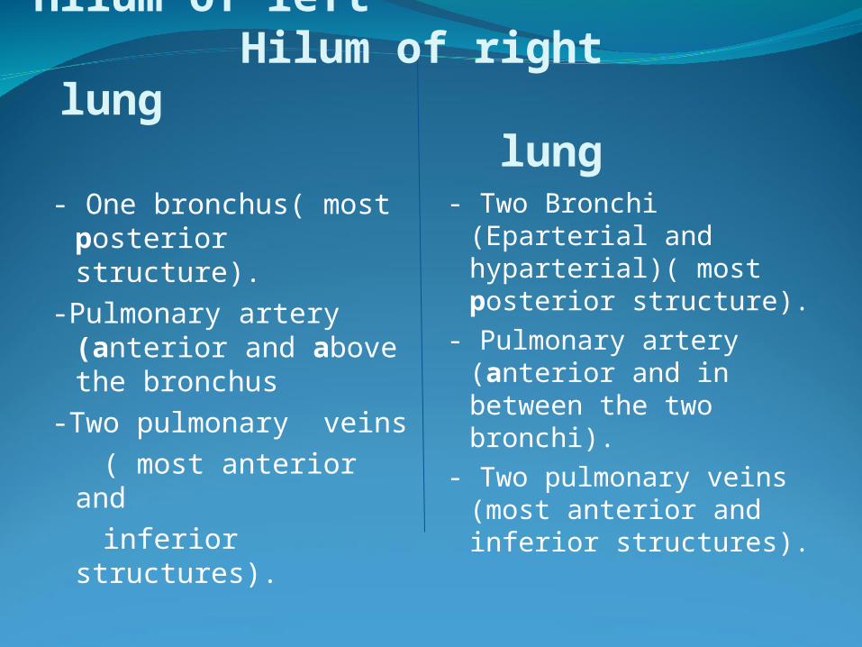
Hilum of left Hilum of right lung lung
- One bronchus( most posterior structure).
-Pulmonary artery (anterior and above the bronchus
-Two pulmonary veins ( most anterior and inferior structures).
- Two Bronchi (Eparterial and hyparterial)( most posterior structure).
- Pulmonary artery (anterior and in between the two bronchi).
- Two pulmonary veins (most anterior and inferior structures).
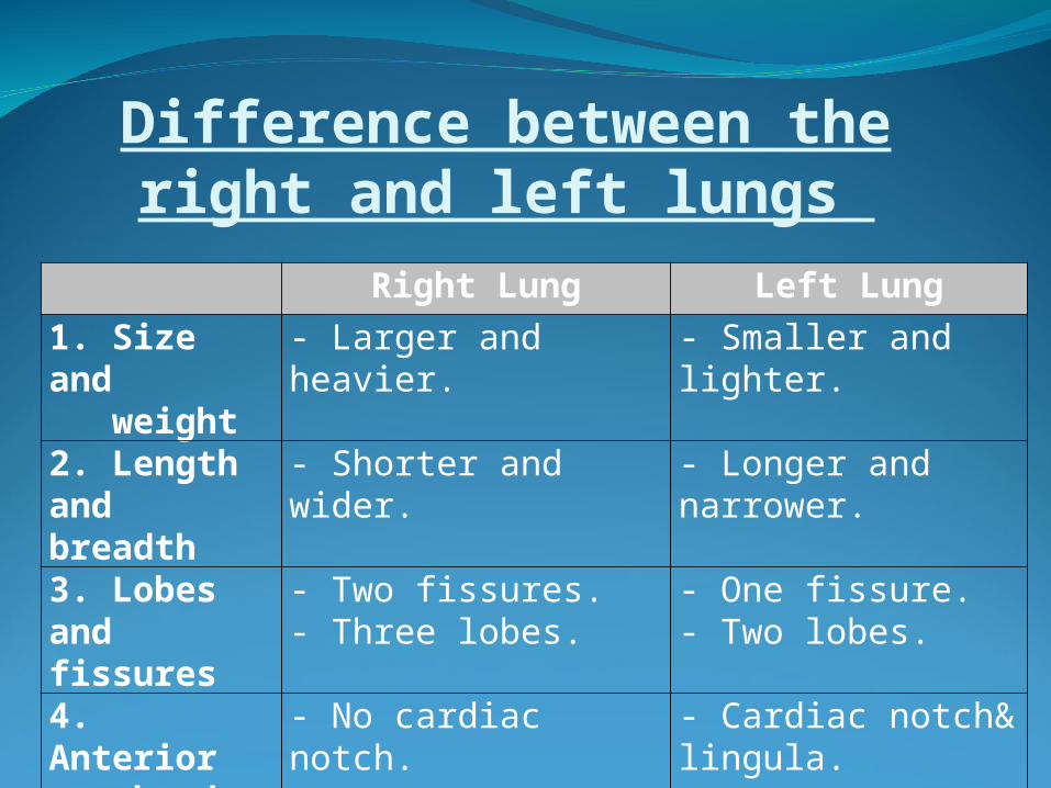
Difference between the right and left lungs
Right Lung Left Lung
1. Size and weight
- Larger and heavier. - Smaller and lighter.
2. Length and breadth
- Shorter and wider. - Longer and narrower.
3. Lobes and fissures
- Two fissures. - Three lobes.
- One fissure. - Two lobes.
4. Anterior border
- No cardiac notch. - Cardiac notch& lingula.
5. Hilum
Broncho-pulmonary segments- The trachea divides into right and left main bronchi, with more division of the main bronchi it gives rise to lobar then segmental bronchi.
- Each segmental bronchus is distributed to a localized part of lung tissue forming what is called the broncho-pulmonary segments.
- The right bronchus divides into: 1- superior lobar bronchus and 2-middle and inferior lobar bronchus.
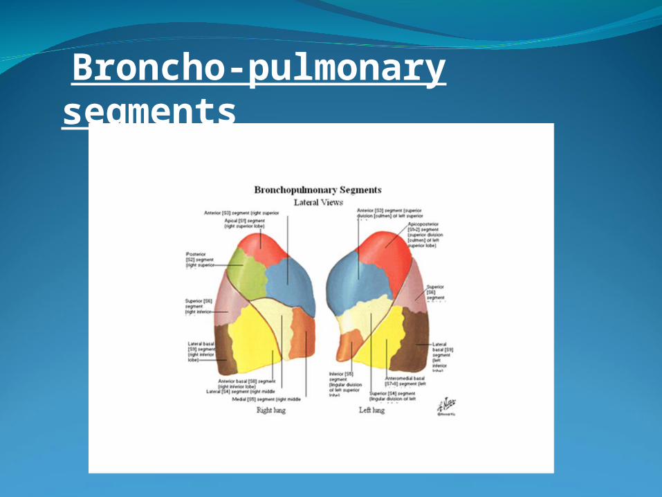
Broncho-pulmonary segments
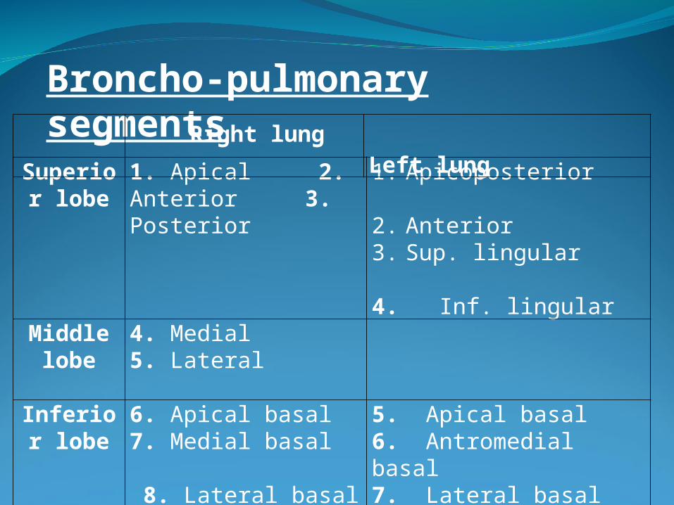
Superior lobe
1. Apical 2. Anterior 3. Posterior
1. Apicoposterior 2. Anterior 3. Sup. lingular 4. Inf. lingular
Middle lobe
4. Medial 5. Lateral
Inferior lobe
6. Apical basal7. Medial basal 8. Lateral basal9. Anterior basal 10. Posterior basal
5. Apical basal 6. Antromedial basal 7. Lateral basal 8. Posterior basal
Broncho-pulmonary segmentsRight lung Left lung
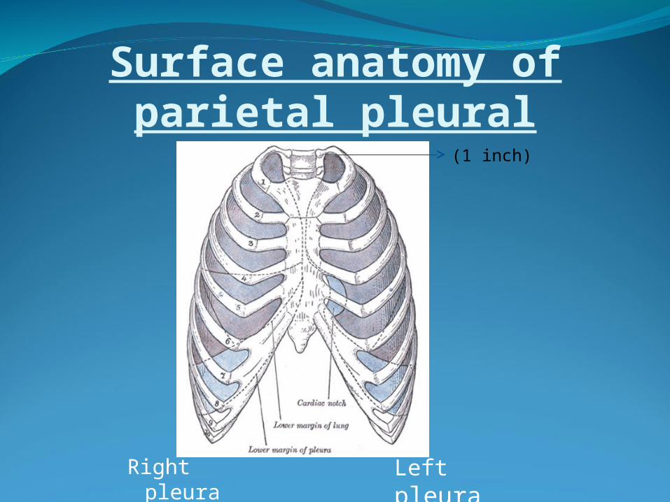
Surface anatomy of parietal pleural
Right pleura
Left pleura
(1 inch) (1 inch)
1-Apex of lung: It lies one inch above the medial end of the clavicle.2-A point behind the sternal angle close to the middle line
at the level of the second costal cartilage.3-A point behind the sternum close to the middle line at
the level of the fourth costal cartilage. 4- On the right side: A point behind the sternum close to the middle line at
the level of the sixth costal cartilage. On the left side: A point behind the sixth costal cartilage 2 cm from the
sternum.5-A point on the eighth rib in the mid-clavicular line.6-A point on the tenth rib in the mid-axillary line.7-A point just below the medial end of the twelfth rib
opposite the twelfth thoracic spine.
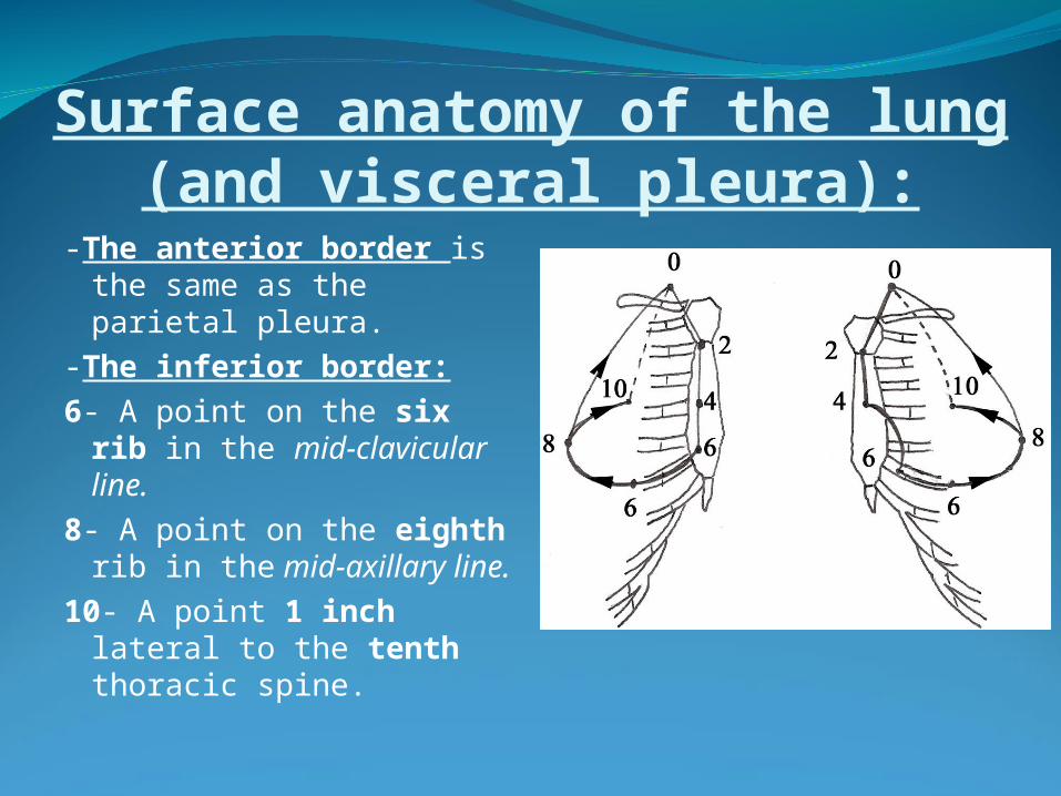
Surface anatomy of the lung (and visceral pleura):
-The anterior border is the same as the parietal pleura.
-The inferior border:6- A point on the six rib in
the mid-clavicular line.8- A point on the eighth
rib in the mid-axillary line.
10- A point 1 inch lateral to the tenth thoracic spine.
Nerve supply- Both lungs are supplied by the anterior and posterior
pulmonary plexuses.a. Sympathetic component (2 - 5 thoracic
sympathetic ganglia) Causes broncho-dilatation.b. Parasympathetic component (vagi) Causes
broncho-constriction + Increases the secretion of the glands.
Blood supply- The lungs are supplied by the bronchial arteries
( Aorta)and drained by the bronchial veins ( Azygos system).
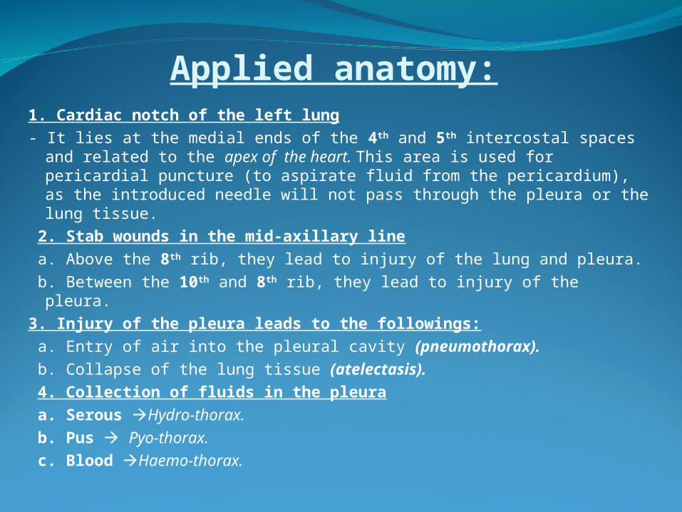
Applied anatomy:1. Cardiac notch of the left lung- It lies at the medial ends of the 4th and 5th intercostal spaces and
related to the apex of the heart. This area is used for pericardial puncture (to aspirate fluid from the pericardium), as the introduced needle will not pass through the pleura or the lung tissue.
2. Stab wounds in the mid-axillary line a. Above the 8th rib, they lead to injury of the lung and pleura. b. Between the 10th and 8th rib, they lead to injury of the pleura.3. Injury of the pleura leads to the followings: a. Entry of air into the pleural cavity (pneumothorax). b. Collapse of the lung tissue (atelectasis). 4. Collection of fluids in the pleura a. Serous Hydro-thorax. b. Pus Pyo-thorax. c. Blood Haemo-thorax.
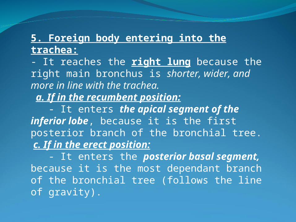
5. Foreign body entering into the trachea: - It reaches the right lung because the right main bronchus is shorter, wider, and more in line with the trachea. a. If in the recumbent position: - It enters the apical segment of the inferior lobe, because it is the first posterior branch of the bronchial tree. c. If in the erect position: - It enters the posterior basal segment, because it is the most dependant branch of the bronchial tree (follows the line of gravity).

Prof.: Dr. Wafaa Abdel-Rahman


![Solitary fi brous tumors of the pleura · tumor of the pleura from other lun g tumors, while the contribution of thoracic CT is rather moderate [4]. Although preoperative dia gnosis](https://img.pdfslide.us/doc/110x75/6081a9dfae78a40b630c556a/solitary-i-brous-tumors-of-the-pleura-tumor-of-the-pleura-from-other-lun-g-tumors.jpg)