Embed Size (px)
Citation preview

THORACIC CAVITYTHORACIC CAVITY
HEARTHEART
Pericardial CavityPericardial Cavity
Heart is located in the middle inferior Heart is located in the middle inferior mediastinum within the pericardial cavity.mediastinum within the pericardial cavity.
Visceral PericardiumVisceral Pericardium
Serous membrane Serous membrane Fig 1.37, p 139)Fig 1.37, p 139)Referred to as epicardium.Referred to as epicardium.Arterial mesocardium.Arterial mesocardium.Venous mesocardium.Venous mesocardium.
Visceral PericardiumVisceral Pericardium
Transverse sinus:Transverse sinus:Space between arterial and venous Space between arterial and venous
mesocardium.mesocardium.Oblique sinus:Oblique sinus:
Space between right and left pulmonary Space between right and left pulmonary veins.veins.
PericardiumPericardium
Pericardial cavity.Pericardial cavity.Parietal (serous):Parietal (serous):
Fused to fibrous pericardium.Fused to fibrous pericardium.Fibrous:Fibrous:
Fused with central tendon of diaphragm:Fused with central tendon of diaphragm:Pericardiacophrenic ligament.Pericardiacophrenic ligament.
Fused with sternum:Fused with sternum:Superior and inferior sternopericardial Superior and inferior sternopericardial ligaments.ligaments.
PericardiumPericardium
Blood supply (Fig. 1.38, p 139):Blood supply (Fig. 1.38, p 139):Pericardial arteries.Pericardial arteries.
Pericardiacophrenic arteries.Pericardiacophrenic arteries.
Musculophrenic arteries.Musculophrenic arteries.Nerve supply:Nerve supply:
CN X.CN X.
Phrenic nerve.Phrenic nerve.
Sympathetic trunk.Sympathetic trunk.

Cardiac ProjectionsCardiac Projections
Apex is down and to left.Apex is down and to left.Base of heart is superior to apex:Base of heart is superior to apex:
Upside down triangle.Upside down triangle.Base projection:Base projection:
Horizontal plane across sternum.Horizontal plane across sternum.
Level of costal cartilage 3.Level of costal cartilage 3.
Ends 2 cm to left of left parasternal line.Ends 2 cm to left of left parasternal line.
Cardiac ProjectionsCardiac Projections
Apex projection:Apex projection:55thth intercostal space. intercostal space.
8 cm (interclavicular line) to left of median 8 cm (interclavicular line) to left of median line of sternum.line of sternum.
Pericardium extends up to sternal angle.Pericardium extends up to sternal angle.
Layers of the HeartLayers of the Heart
External:External:EpicardiumEpicardium
Visceral layer of serous pericardium + Visceral layer of serous pericardium + subserous layer of connective tissue.subserous layer of connective tissue.
Middle:Middle:
Myocardium:Myocardium:ThickestThickest
Cardiac muscle tissue.Cardiac muscle tissue.
Layers of the HeartLayers of the Heart
Inner:Inner:Endocardium:Endocardium:
Endothelial layerEndothelial layer
Cardiac SkeletonCardiac Skeleton
4 interlocking fibrous rings:4 interlocking fibrous rings:Annuli fibrosi.Annuli fibrosi.
Interconnected with membranous portion Interconnected with membranous portion of of interventricular septum.interventricular septum.
Fig. 1.40B, p 143)Fig. 1.40B, p 143)
Cardiac SkeletonCardiac Skeleton
Functions of annuli fibrosi:Functions of annuli fibrosi:Rigid attachment for cardiac muscle Rigid attachment for cardiac muscle
fibers.fibers.
Support for valves.Support for valves.
External HeartExternal Heart
Base:Base:Posterior aspect of heart.Posterior aspect of heart.
Mostly left atrium + small part of right Mostly left atrium + small part of right atrium.atrium.
Apex:Apex:Located at intercostal space 5 medial to Located at intercostal space 5 medial to
nipple.nipple.
Formed from left ventricleFormed from left ventricleFig. 1.42A, p 146.Fig. 1.42A, p 146.
External HeartExternal Heart
Diaphragmatic surface:Diaphragmatic surface:Rests on diaphragm.Rests on diaphragm.
Mostly left ventricle and a small part of Mostly left ventricle and a small part of right.right.
Sternocostal surface:Sternocostal surface:Faces anteriorly.Faces anteriorly.
Mostly right ventricle.Mostly right ventricle.
External HeartExternal Heart
Left margin (border):Left margin (border):Left side of heart formed mostly of left ventricle Left side of heart formed mostly of left ventricle
and and left auricle.left auricle.
Inferior margin (border):Inferior margin (border):Intersection of diaphragmatic and sternocostal Intersection of diaphragmatic and sternocostal
surfaces.surfaces.
Formed mostly from right ventricle.Formed mostly from right ventricle.
Superior border:Superior border:Left and right atria and auricles.Left and right atria and auricles.
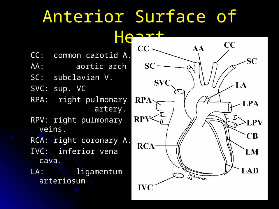
Anterior Surface of HeartAnterior Surface of Heart
CC: common carotid A.CC: common carotid A.
AA:AA: aortic arch aortic arch
SC: subclavian V.SC: subclavian V.
SVC: sup. VCSVC: sup. VC
RPA: right pulmonary RPA: right pulmonary artery.artery.
RPV: right pulmonary RPV: right pulmonary veins.veins.
RCA: right coronary A.RCA: right coronary A.
IVC: inferior vena cava.IVC: inferior vena cava.
LA:LA: ligamentum ligamentum arteriosumarteriosum
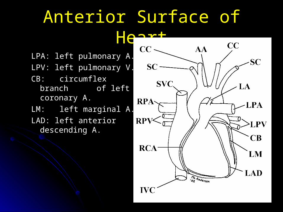
Anterior Surface of HeartAnterior Surface of Heart
LPA: left pulmonary A.LPA: left pulmonary A.
LPV: left pulmonary V.LPV: left pulmonary V.
CB: circumflex branch CB: circumflex branch of of left coronary A.left coronary A.
LM: left marginal A.LM: left marginal A.
LAD: left anterior LAD: left anterior descending A.descending A.
Blood Supply to HeartBlood Supply to Heart
Blood supply to heart is via two Blood supply to heart is via two coronary arteries (Fig. 1.49A&B, 157):coronary arteries (Fig. 1.49A&B, 157):
Coronary arteries are the direct and only Coronary arteries are the direct and only branches off the ascending aorta.branches off the ascending aorta.
Right Coronary ArteryRight Coronary Artery
Passes between pulmonary trunk and right Passes between pulmonary trunk and right auricle.auricle.
To coronary sulcus.To coronary sulcus.Follows coronary sulcus to diaphragmatic Follows coronary sulcus to diaphragmatic
surface.surface.Anastomoses with left coronary artery.Anastomoses with left coronary artery.
Right Coronary Artery Supplies:Right Coronary Artery Supplies:
Right atrium.Right atrium.Right ventricle.Right ventricle.Posterior half of interventricular septum.Posterior half of interventricular septum.
Right Coronary Artery BranchesRight Coronary Artery Branches
Artery to SA node.Artery to SA node.Artery to AV node.Artery to AV node.Right marginal artery.Right marginal artery.Posterior interventricular arteryPosterior interventricular artery
= posterior descending artery (PDA)= posterior descending artery (PDA)
Left Coronary ArteryLeft Coronary Artery
Passes between pulmonary trunk and Passes between pulmonary trunk and left atrium.left atrium.
Supplies:Supplies:Left atrium.Left atrium.
Left ventricle.Left ventricle.
Anterior half of interventricular septum.Anterior half of interventricular septum.
Left Coronary ArteryLeft Coronary Artery
Major branches:Major branches:Anterior interventricular artery (= left Anterior interventricular artery (= left
anterior anterior descending LAD).descending LAD).
Circumflex artery.Circumflex artery.
Left marginal artery.Left marginal artery.
Venous Drainage from HeartVenous Drainage from Heart
Fig. 1.51, page 159Fig. 1.51, page 159Coronary sinus:Coronary sinus:
Located in posterior coronary sulcus.Located in posterior coronary sulcus.
Opens into right atrium.Opens into right atrium.
Direct continuation of great cardiac vein.Direct continuation of great cardiac vein.
Tributaries:Tributaries:Great cardiac vein.Great cardiac vein.
Middle cardiac vein.Middle cardiac vein.
Small cardiac vein.Small cardiac vein.

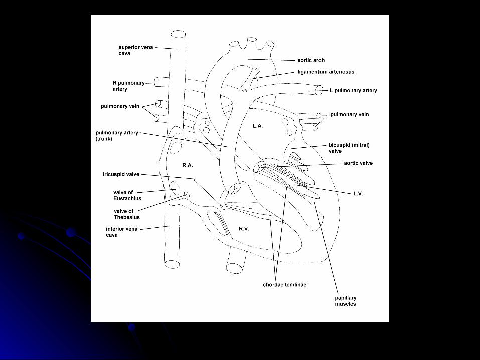
Right AtriumRight Atrium
Fig. 1.43, p 148:Fig. 1.43, p 148:Receives blood from:Receives blood from:
Superior vena cava.Superior vena cava.
Inferior vena cava.Inferior vena cava.
Coronary sinus.Coronary sinus.
Anterior cardiac veins.Anterior cardiac veins.Large, thin-walled chamber.Large, thin-walled chamber.
Right AtriumRight Atrium
Sub-chambers:Sub-chambers:Main posterior cavity:Main posterior cavity:
Sinus venarum.Sinus venarum.
Anterior cavity:Anterior cavity:Auricle.Auricle.
Lined with pectinate muscles.Lined with pectinate muscles.

Right VentricleRight Ventricle
Fig. 1.44, p 149Fig. 1.44, p 149Receives blood from right atrium.Receives blood from right atrium.Thicker walled than right atrium.Thicker walled than right atrium.Trabeculae carnae.Trabeculae carnae.Moderator band:Moderator band:
(septomarginal trabecula)(septomarginal trabecula)
Conveys right branch of atrioventricular Conveys right branch of atrioventricular bundle.bundle.
Right Atrioventricular ValveRight Atrioventricular Valve
Also called the tricuspid valve.Also called the tricuspid valve.Chordae tendinae.Chordae tendinae.Papillary muscles.Papillary muscles.Fig. 1.45 B&C, p 150Fig. 1.45 B&C, p 150
Right VentricleRight Ventricle
Pulmonary valve:Pulmonary valve:Formed from three semilunar cusps.Formed from three semilunar cusps.
Pulmonary trunk:Pulmonary trunk:Divides into left and right pulmonary Divides into left and right pulmonary
arteries.arteries.
Left AtriumLeft Atrium
Left atrium (Fig. 1.46, p 152):Left atrium (Fig. 1.46, p 152):Receives blood from four pulmonary veins.Receives blood from four pulmonary veins.
Smaller and thicker-walled than right Smaller and thicker-walled than right atrium.atrium.
Posterior smooth portion receives Posterior smooth portion receives pulmonary pulmonary veins.veins.
Anterior portion = auricle:Anterior portion = auricle:with pectinate muscles.with pectinate muscles.

Left VentricleLeft Ventricle
Fig. 1.47, p 153Fig. 1.47, p 153Wall = 2-3 x as thick as wall of right Wall = 2-3 x as thick as wall of right
ventricle.ventricle.Trabeculae carnae are less coarse than Trabeculae carnae are less coarse than
those of right ventricle.those of right ventricle.No moderator band.No moderator band.Two large papillary muscles:Two large papillary muscles:
Anterior and posterior.Anterior and posterior.
Left Atrioventricular/Aortic ValvesLeft Atrioventricular/Aortic Valves
Left atrioventricular valve:Left atrioventricular valve:= bicuspid or mitral valve.= bicuspid or mitral valve.
Aortic valve:Aortic valve:Composed of three semilunar cusps.Composed of three semilunar cusps.
Conduction System of the HeartConduction System of the Heart
Composed of modified specialized cardiac Composed of modified specialized cardiac muscle cells.muscle cells.
No nervous tissue in heart.No nervous tissue in heart.Fig. 1.52, p 163)Fig. 1.52, p 163)
Sinoatrial NodeSinoatrial Node
Referred to as pacemaker of heart.Referred to as pacemaker of heart.Located in right atrium near opening of Located in right atrium near opening of
SVC:SVC:Superior end of sulcus terminalis.Superior end of sulcus terminalis.
Receives direct stimulation from:Receives direct stimulation from:Sympathetic cardiac nerves.Sympathetic cardiac nerves.
Parasympathetic vagus nerves.Parasympathetic vagus nerves.

Conduction System of the HeartConduction System of the Heart
Atrioventricular node:Atrioventricular node:Located in interatrial septum near tricuspid Located in interatrial septum near tricuspid valve.valve.
Interventricular bundle:Interventricular bundle:Descends through channel in fibrous Descends through channel in fibrous
skeleton.skeleton.Reaches membranous interventricular Reaches membranous interventricular
septum.septum.Only connection between myocardium of Only connection between myocardium of atria and that of ventricles.atria and that of ventricles.

Conduction System of the HeartConduction System of the Heart
Interventricular bundle:Interventricular bundle:Divides into two bundles in membranous Divides into two bundles in membranous portion:portion:
Right crus (bundle branches) passes Right crus (bundle branches) passes through through moderator band.moderator band.
Left crus (bundle branches)Left crus (bundle branches)
Conduction System of the HeartConduction System of the Heart
Purkinje fibers:Purkinje fibers:Terminal endings of bundle fibers.Terminal endings of bundle fibers.
Embedded in myocardium of ventricle.Embedded in myocardium of ventricle.
Great VesselsGreat Vessels
Ascending aorta:Ascending aorta:Runs behind sternum to sternal angle.Runs behind sternum to sternal angle.
Only branches are the two coronary Only branches are the two coronary arteries.arteries.
Fig. 1.58, p 172Fig. 1.58, p 172
Arch of the AortaArch of the Aorta
Lies within superior mediastinum.Lies within superior mediastinum.Arches to the left over the left pulmonary Arches to the left over the left pulmonary
artery.artery.
Apex of the arch reaches the middle of the Apex of the arch reaches the middle of the manubrium.manubrium.
Three main branches:Three main branches:Brachiocephalic.Brachiocephalic.
Left common carotid.Left common carotid.Left subclavian.Left subclavian.
Arch of the AortaArch of the Aorta
Anterior relationships:Anterior relationships:Left phrenic nerve.Left phrenic nerve.
Left vagus nerve.Left vagus nerve.
Superficial cardiac plexus.Superficial cardiac plexus.

Arch of the AortaArch of the Aorta
Inferior relationships:Inferior relationships:Left recurrent laryngeal nerve.Left recurrent laryngeal nerve.
Ligamentum arteriosum.Ligamentum arteriosum.
Pulmonary trunk.Pulmonary trunk.
Left primary bronchus.Left primary bronchus.
Arch of the AortaArch of the Aorta
Posterior relationships:Posterior relationships:TracheaTrachea
Left recurrent laryngeal nerve.Left recurrent laryngeal nerve.
Descending AortaDescending Aorta
Lies within posterior mediastinum.Lies within posterior mediastinum.Begins at level of sternal angle.Begins at level of sternal angle.Ends in front of thoracic vertebra 12.Ends in front of thoracic vertebra 12.Continuous with abdominal aorta.Continuous with abdominal aorta.

Descending Aorta BranchesDescending Aorta Branches
Paired intercostal arteries.Paired intercostal arteries.Paired subcostal arteries.Paired subcostal arteries.Two or more bronchial arteries.Two or more bronchial arteries.Two to five esophageal arteries.Two to five esophageal arteries.
Other Thoracic VesselsOther Thoracic Vessels
Supreme intercostal:Supreme intercostal:From costocervical trunk of subclavian From costocervical trunk of subclavian
artery.artery.
Supplies IC spaces one and two.Supplies IC spaces one and two. Internal thoracics:Internal thoracics:
Arise within root of neck.Arise within root of neck.
Descend lateral to sternum.Descend lateral to sternum.
Internal Thoracic ArteriesInternal Thoracic Arteries
Branches:Branches:
Musculophrenic (terminal):Musculophrenic (terminal):
To diaphragmTo diaphragm
To intercostal spaces 7-9To intercostal spaces 7-9
Superior epigastric (terminal)Superior epigastric (terminal)
Internal Thoracic ArteriesInternal Thoracic Arteries
Branches:Branches:Pericardioacophrenic arteries:Pericardioacophrenic arteries:
Accompanies phrenic nerve.Accompanies phrenic nerve.
Supplies pericardium, mediastinal Supplies pericardium, mediastinal pleura, diaphragm.pleura, diaphragm.
Perforating branches:Perforating branches:Accompany anterior cutaneous Accompany anterior cutaneous branches of intercostal nerves.branches of intercostal nerves.
Largest in intercostal spaces 2-4 in Largest in intercostal spaces 2-4 in females.females.
Right Brachiocephalic VeinRight Brachiocephalic Vein
From:From:Right internal jugular.Right internal jugular.
Right subclavian.Right subclavian.Tributary:Tributary:
Right internal intercostal vein.Right internal intercostal vein. Fig. 1.64, p 182Fig. 1.64, p 182
Left Brachiocephalic VeinLeft Brachiocephalic Vein
Formed from:Formed from:Left internal jugular vein.Left internal jugular vein.
Left subclavian vein.Left subclavian vein.Tributaries:Tributaries:
Left internal thoracic vein.Left internal thoracic vein.
Left superior intercostal.Left superior intercostal.
Inferior thyroid veins.Inferior thyroid veins.

Superior Vena CavaSuperior Vena Cava
Formed from:Formed from:Right brachiocephalic vein.Right brachiocephalic vein.
Left brachiocephalic vein.Left brachiocephalic vein.Receives:Receives:
Azygos vein.Azygos vein.
Azygos SystemAzygos System
Drains most of blood from thoracic wall.Drains most of blood from thoracic wall.Consists of longitudinal veins lying on Consists of longitudinal veins lying on
either side of thoracic vertebral bodies.either side of thoracic vertebral bodies.Variable.Variable.
Azygos VeinAzygos Vein
Forms in abdomen:Forms in abdomen:From right subcostal and ascending From right subcostal and ascending
lumbar veins.lumbar veins.Drains all right posterior intercostal veins Drains all right posterior intercostal veins
except first.except first.Also receives blood from the bronchial and Also receives blood from the bronchial and
esophageal veins.esophageal veins.
Hemiazygos VeinHemiazygos Vein
Forms in abdomen:Forms in abdomen:From left subcostal and left ascending From left subcostal and left ascending
lumbar veins.lumbar veins.Receives four posterior intercostal veins.Receives four posterior intercostal veins.Crosses over thoracic vertebrae at T8 Crosses over thoracic vertebrae at T8
level.level.Empties into azygos vein.Empties into azygos vein.
Other Thoracic VesselsOther Thoracic Vessels
Accessory hemiazygos vein:Accessory hemiazygos vein:Drains intercostal spaces 4-7(8) on left Drains intercostal spaces 4-7(8) on left
side.side.
Crosses over thoracic vertebrae at level Crosses over thoracic vertebrae at level T7.T7.
Empties into azygos vein.Empties into azygos vein.Note: Intercostal space 1 is drained by the Note: Intercostal space 1 is drained by the
supreme intercostal vein emptying into the supreme intercostal vein emptying into the brachiocephalic vein.brachiocephalic vein.