Embed Size (px)
Citation preview
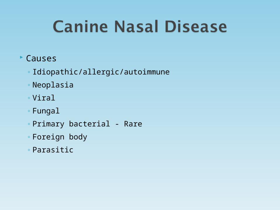
Causes
◦ Idiopathic/allergic/autoimmune
◦ Neoplasia
◦ Viral
◦ Fungal
◦ Primary bacterial - Rare
◦ Foreign body
◦ Parasitic
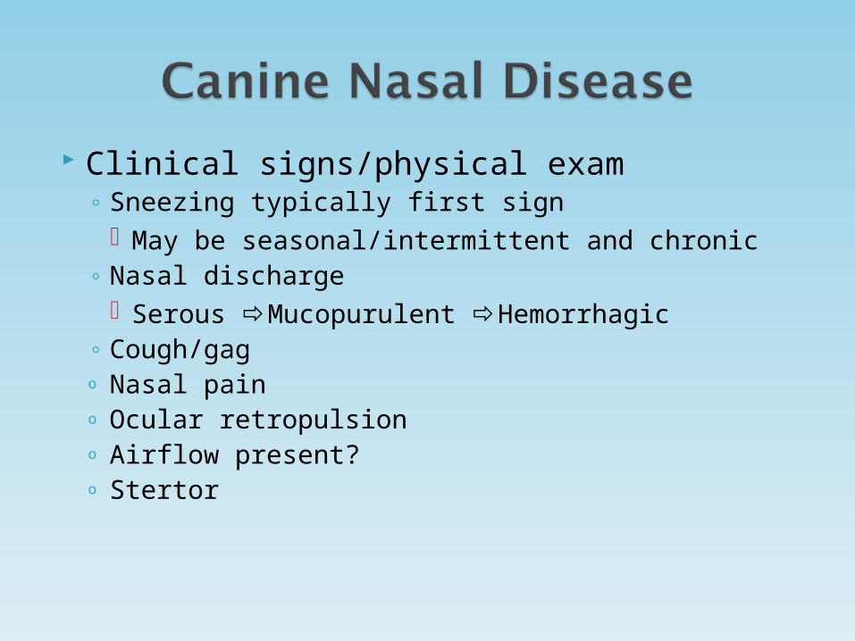
Clinical signs/physical exam◦ Sneezing typically first sign
May be seasonal/intermittent and chronic◦ Nasal discharge
Serous Mucopurulent Hemorrhagic ◦ Cough/gago Nasal paino Ocular retropulsiono Airflow present?o Stertor
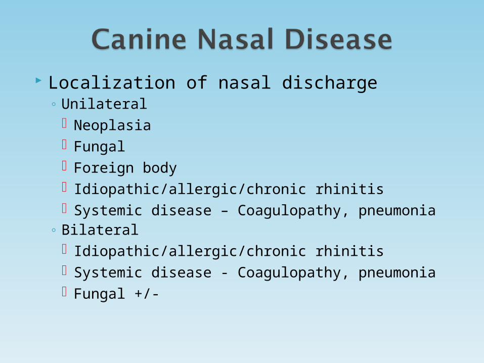
Localization of nasal discharge◦ Unilateral
Neoplasia Fungal Foreign body Idiopathic/allergic/chronic rhinitis Systemic disease – Coagulopathy, pneumonia
◦ Bilateral Idiopathic/allergic/chronic rhinitis Systemic disease - Coagulopathy, pneumonia Fungal +/-

Epistaxis◦ Local disease
Neoplasia Fungal Chronic idiopathic rhinitis
◦ Systemic disease Thrombocytopenia Hypertension Hyperviscosity Vasculitis
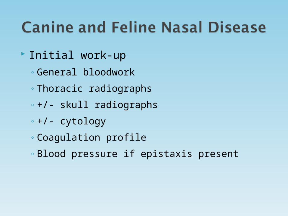
Initial work-up
◦ General bloodwork
◦ Thoracic radiographs
◦ +/- skull radiographs
◦ +/- cytology
◦ Coagulation profile
◦ Blood pressure if epistaxis present

Initial work-up◦ Culture?◦ Sedated oral exam Use spay hook and good light source Deep sedation sometimes necessary Maxillary 3rd incisor and premolars 1, 2, 3
(mesial root) Dental probe indicated in many cases
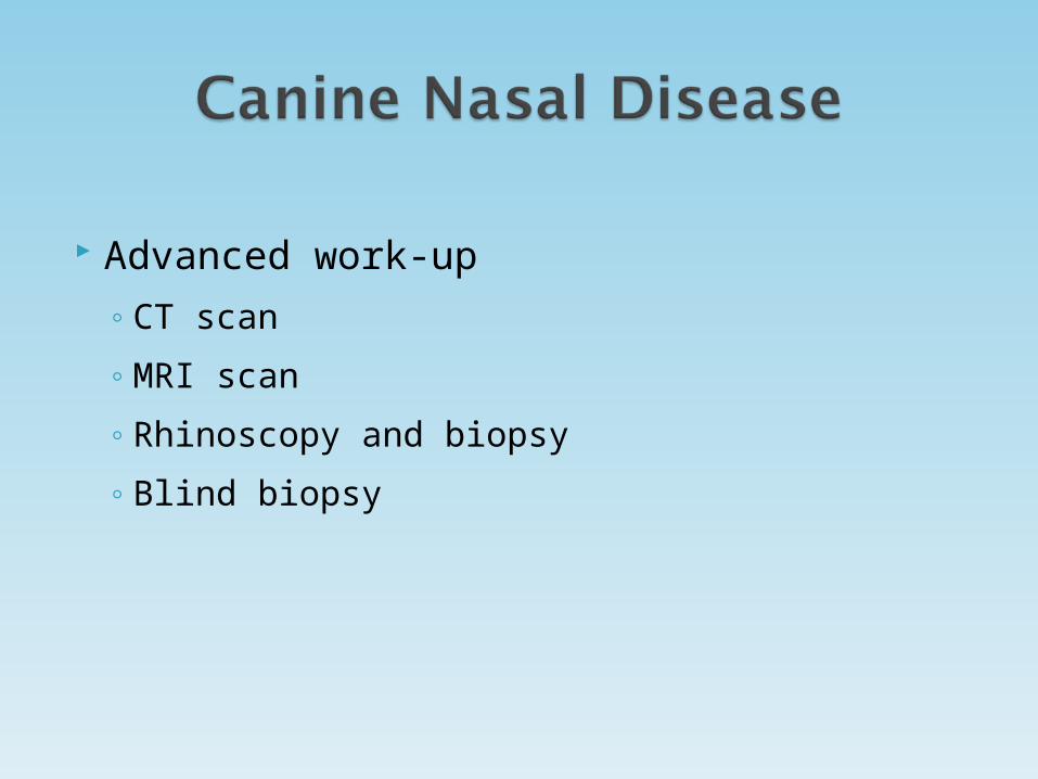
Advanced work-up
◦ CT scan
◦ MRI scan
◦ Rhinoscopy and biopsy
◦ Blind biopsy
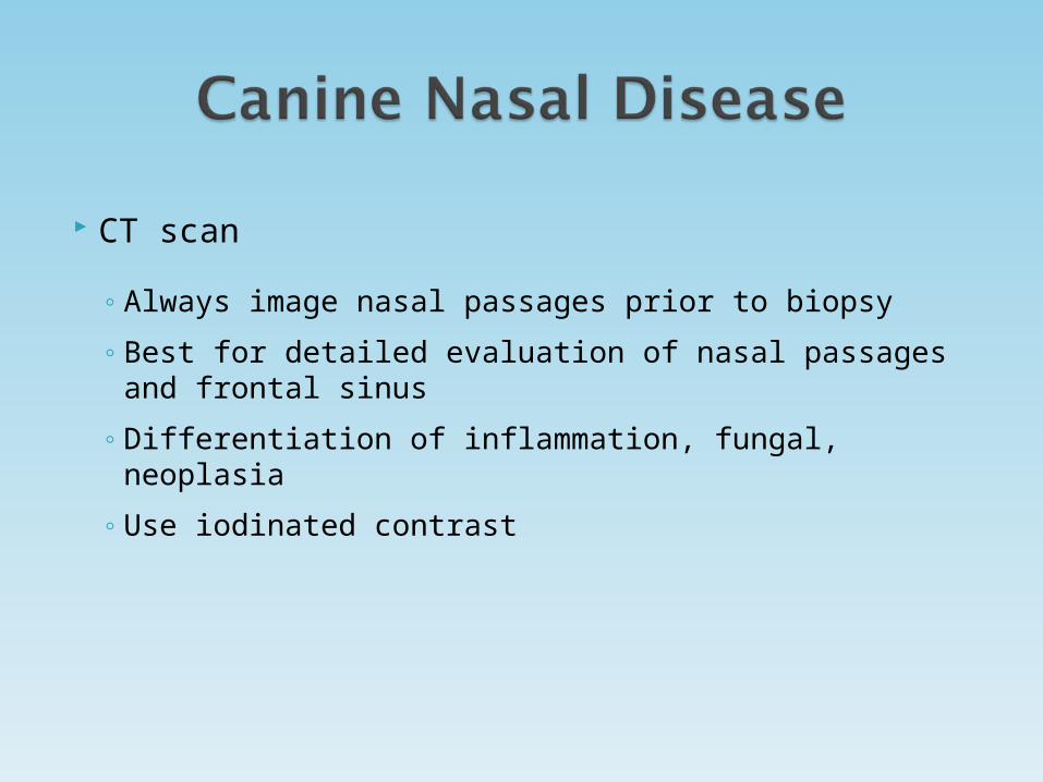
CT scan
◦ Always image nasal passages prior to biopsy
◦ Best for detailed evaluation of nasal passages and frontal sinus
◦ Differentiation of inflammation, fungal, neoplasia
◦ Use iodinated contrast
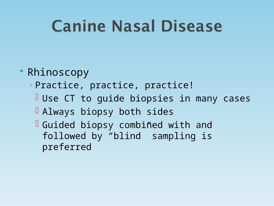
Rhinoscopy◦ Practice, practice, practice!
Use CT to guide biopsies in many cases Always biopsy both sides Guided biopsy combined with and followed by
“blind” sampling is preferred
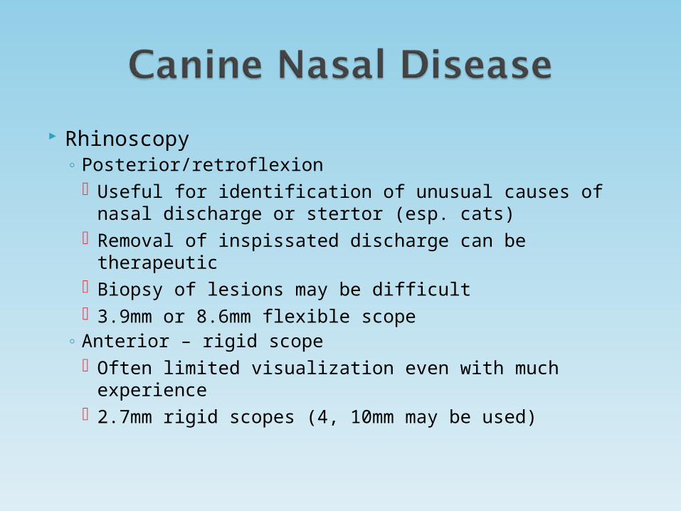
Rhinoscopy◦ Posterior/retroflexion
Useful for identification of unusual causes of nasal discharge or stertor (esp. cats)
Removal of inspissated discharge can be therapeutic Biopsy of lesions may be difficult 3.9mm or 8.6mm flexible scope
◦ Anterior – rigid scope Often limited visualization even with much experience 2.7mm rigid scopes (4, 10mm may be used)
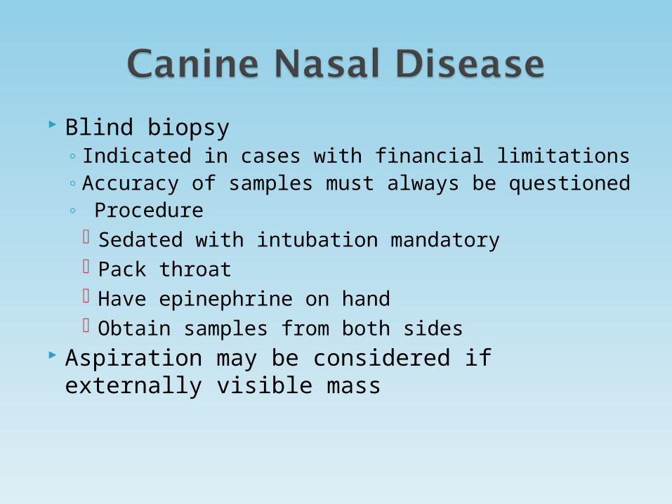
Blind biopsy◦ Indicated in cases with financial limitations◦ Accuracy of samples must always be
questioned◦ Procedure
Sedated with intubation mandatory Pack throat Have epinephrine on hand Obtain samples from both sides
Aspiration may be considered if externally visible mass

Limitations of all nasal biopsies◦ Inflammation surrounding masses◦ Differentiating neoplasia from true/primary ◦ Owners should always be made aware of:
Potential need to repeat scope and biopsy if biopsy results do not coincide with physical exam, imaging findings, or clinical impressions
Rhinoscopy and biopsy procedures are rarely, if ever therapeutic!!

Cytology◦ Indicated for cats with nasal discharge and
clinical suspicion of fungal disease◦ Not useful for diagnosis of neoplasia, idiopathic
rhinitis, fungal rhinitis in dogs, or true bacterial infection
◦ Brush cytology generally does not correlate with biopsy results

Nasal culture◦ Fairly useless in most cases◦ False positive for fungal and bacterial infection◦ False negative often found in dogs with
Aspergillosis◦ Mainly indicated in cats with chronic
rhinitis/nasal discharge and dogs with non-responsive to therapy for “chronic rhinitis”

Fungal rhinitis◦ Potential pathogens
Aspergillosis Rhinosporidium seeberi Penicillium
◦ Differentiating signs Dramatic Depigmentation and nasal pain (tip of nose) Severe turbinate loss on CT or radiographs Fungal plaques seen on rhinoscopy Typically unilateral
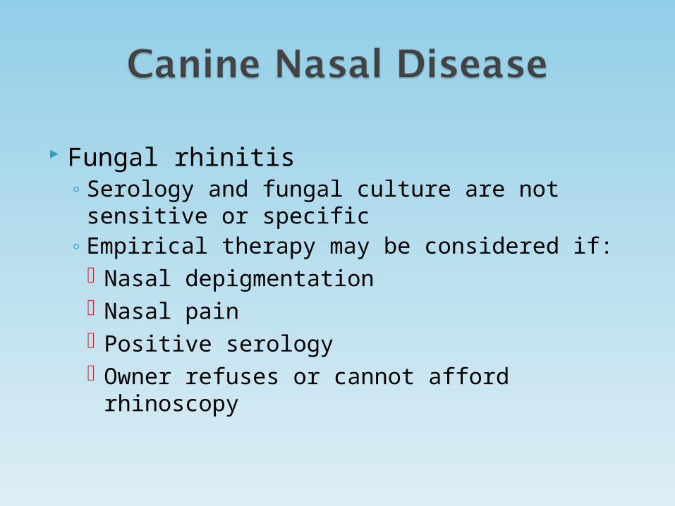
Fungal rhinitis◦ Serology and fungal culture are not sensitive or
specific◦ Empirical therapy may be considered if:
Nasal depigmentation Nasal pain Positive serology Owner refuses or cannot afford rhinoscopy
Fungal rhinitis◦ CT scan/radiographs
Severe turbinate loss Fluid/granuloma opacity in nasal passage and
possibly frontal sinus +/- bone erosion +/- erosion of cribiform plate
◦ Histopathology Generally sensitive for obvious infection, but
can miss in presence of severe inflammation
Fungal rhinitis◦ Rhinoscopy
Severe turbinate loss in most (too much room!) Friable mucosa, erythema, hyperemia, edema White fungal plaques
Seen in 83% of cases within the nasal cavity 17% localized exclusively in sinus(‘) Need ability to reach sinus for this reason as
well as for catheter placement during therapy Very time consuming during therapeutic phase
$$$

Fungal rhinitis◦ Rhinoscopic topical therapy best
Enilconazole 1% (nasal) and 2% (sinus), compared to 1% clotrimazole infusion
May have long term nasal signs following infusion with both treatments Approximately 50% of the time Typically antibiotic responsive
Discouraged, but can be done if cribiform plate is not intact

From Peeters, D. and Clerx C., Update on Canine Sinonasal Aspergillosis. Vet Clin North Am Small Anim Pract 2007; 37 (5): 909.
Fungal rhinitis therapy◦ Meticulous debridement◦ Follow-up rhinoscopy◦ Combine with oral antifungals?◦ Surgery
For inaccessible suspected sinus infection Clotrimazole liquid topical combined with
cream instillation as depot therapy
Oral antifungal therapy◦ Oral therapy alone is not recommended◦ Use if cribiform plate is not intact◦ Reported 50-70% cure rate (best case scenario)◦ Options (best to worst)
Itraconazole 5mg/kg BID X 10 weeks Fluconazole 2.5mg/kg BID X 10 weeks Ketoconazole 5mg/kg BID 12 weeks Thiabendazole 10mg/kg BID X 6-8 weeks Terbinafine 5-10mg/kg BID X 10 weeks
◦ Cost, GI side effects, and hepatotoxicity
Lymphoplasmacytic rhinitis◦ Fairly common disease of dogs◦ Diagnosis may obtained with other underlying
causes Fungal Foreign body Neoplasia Parasitic
Mites True bacterial infection
Lymphoplasmacytic rhinitis◦ Causes
Idiopathic Inhaled allergens Irritants Hypersensitivity to bacteria or fungi? Dust mites? (n=3)

Lymphoplasmacytic rhinitis radiographic findings Turbinate destruction Soft tissue/fluid opacity Obvious bone lysis/remodeling
◦ CT findings May be difficult for differentiation of
inflammation from neoplasia in cats, but fairly good in dogs
Allows clinician to target biopsy collection from areas of interest
Turbinate destruction can mimic fungal rhinitis Fluid in nasal passages and sinuses Suspect fungal disease or neoplasia if bone
destruction noted
Lymphoplasmacytic rhinitis◦ Rhinoscopy
Erythema, hyperemia, edema, normal Not sensitive for detection of turbinate destruction Right and left sides may differ on gross inspection
considerably, but disease present on both sides in most
◦ Histopathology Biopsy results may not correlate with disease
severity or clinical signs Always correlate with imaging findings
Lymphoplasmacytic rhinitis◦ Therapy – General considerations
FRUSTRATING!!!!! Owner preparation is critical if suspected
diagnosis No cure, but hope to decrease signs to
acceptable level Lifelong treatment often required Seasonal or unpredictable relapse is common Allergen avoidance
Smoke, forced air heat, wood burning stoves, fireplace, etc.

Lymphoplasmacytic rhinitis◦ Drug therapy
Antihistamines Many formulations, but none evaluated critically Sometimes effective but durable response rarely
achieved Oral corticosteroids
Prednisone 0.5-1mg/kg BID to start with taper over 2-3 weeks
Use at beginning of combined therapeutic regimen in selected cases
Only in those with serous discharge Generally poor response overall esp. when used
alone

Lymphoplasmacytic rhinitis - Therapy◦ Antibiotic therapy
Combine with oral or topical anti-inflammatory therapy
Doxycycline 3-5mg mg/kg BID X 2 weeks Reduce to once daily if responsive
Azithromycin 10mg/kg daily 5 days Reduce to 2X/week if initially responsive
Use at standard dose intermittently or alternative antibiotic based on C & S if persistent purulent or mucopurulent discharge noted
Lymphoplasmacytic rhinitis - Therapy◦ Oral antiinflammatory therapy
Oral corticosteroids Prednisone 0.5-1mg/kg BID to start with taper
over 2-3 weeks Use at beginning of combined therapeutic regimen
in selected cases Only in those with serous discharge Generally poor response overall esp. when used
alone NSAIDs - Piroxicam 0.3mg/kg daily
Use with misoprostol 3mcg/kg (2-5mcg/kg) BID
◦ Topical antiinflammatory therapy Flovent 110-220mcg/actuation BID to start
May reduce to once daily or every other day if effective
Lower to once daily if significant improvement noted
Less potential side effects Variable responses
Nasal confirmation Presence of severe discharge Compliance

Lymphoplasmacytic rhinitis – Therapy Ideally 2-3X per week antiinflammatory and
intermittent antibiotic courses vs. 2-3X/week of both indefinitely or seasonally May consider pulse therapy with antibiotics
If responsive, most require long term/lifelong therapy
Compliance is a major issue when patients improve Bacterial rhinitis - Canine
◦ Pasteurella multocida, Bordatella bronchiseptica may be primary pathogens - RARE
◦ Last line diagnostic test if no resolution of clinical signs after treatment of rhinitis

Nasal neoplasia – General considerations◦ Seen in approximately 1/3 of dogs with chronic nasal
disease◦ Nasal carcinoma 2/3 of all nasal neoplasms
Adenocarcinoma, undifferentiated, squamous cell◦ Others = 1/3
Lymphoma Fibrosarcoma Neuroendocrine Hemangiosarcoma MCT TVT – extremely rare
◦ Nasal polyps – Rare and typically secondary to inflammation or underlying neoplasia
Neoplasia – General considerations◦ Metastasis
Local lymph nodes Lungs – Rare
◦ Most express COX-2 receptors ◦ Clinical signs
Dramatic Unilateral epistaxis and discharge are common Facial deformity – other considerations?
Sporotrichosis, severe aspergillosis Angiomatous proliferation of nasal cavity - rare
Neurologic signs may be very late Caudal nasal passage
Nasal neoplasia◦ Radiographic findings
Non-specific Loss of turbinates May see bone lysis Fluid in frontal sinus Soft tissue opacity late in course of disease

◦ CT Very good at determining neoplasia vs. non-
neoplastic disease Bone erosion/lysis usually consistent with
neoplasia◦ MRI
Mass effect on MRI not necessarily associated with neoplasia Other factors: cribiform plate erosion, vomer
bone lysis etc. must be present to discriminate Bone erosion/lysis usually consistent with
neoplasia
Nasal neoplasia◦ Rhinoscopy
Sometimes limited by location Difficult in most cases due to presence of
hemorrhage, occlusion of nasal passage, and magnification
Retroflexion will allow diagnostic specimens in some
◦ Blind biopsy Always followed by rhinoscopic assisted biopsies Help improve diagnostic accuracy?

Nasal neoplasia◦ Prognosis - Carcinomas
No therapy = MST 95d (73-113) Epistaxis
Present = 88d Absent = 224d
Nasal neoplasia – Therapy and prognosis◦ Surgery alone
Mixed results, but generally disappointing MST = 3-6 months
◦ Radiation CT planning is best to prevent normal tissue
damage No evidence that CT planning improves
prognosis MST = 8-20 months when used alone
◦ IMRT/Cyberknife
Nasal neoplasia – Therapy and prognosis◦ Radiation followed by surgery
Best outcome to date 54 dogs 4yr MST vs. 2 yr MST with radiation alone in
one study More side effects when compared to either
alone Osteomyelitis Fistula formation Fungal rhinitis
Nasal neoplasia – Therapy and prognosis◦ Chemotherapy
Single agent cisplatin MST = 5 months
Combination adriamycin, carboplatin, piroxicam MST is unknown Clinical response has been favorable in
those in which it has been used 81% of canine nasal tumors expressed COX-2
receptors in one study