Embed Size (px)
Citation preview

NeoplasiaNeoplasia
Dr. Gehan MohamedDr. Gehan Mohamed
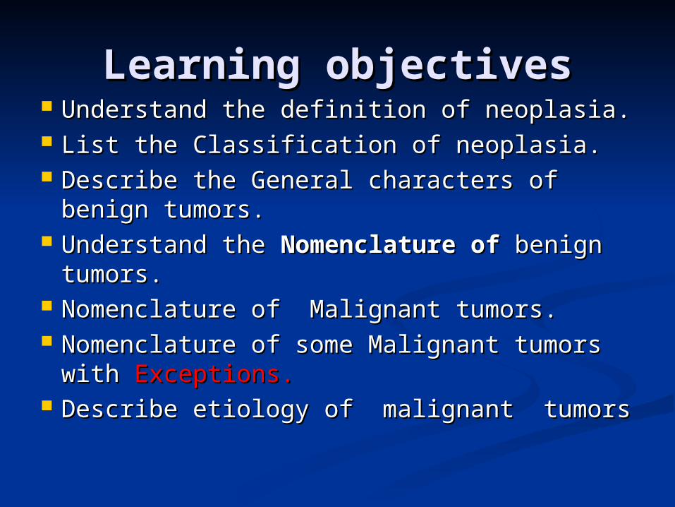
Learning objectivesLearning objectives Understand the definition of neoplasia.Understand the definition of neoplasia. List the Classification of neoplasia.List the Classification of neoplasia. Describe the General characters of Describe the General characters of
benign tumors.benign tumors. Understand the Understand the Nomenclature of Nomenclature of
benign tumors.benign tumors. Nomenclature of Malignant tumors.Nomenclature of Malignant tumors. Nomenclature of some Malignant tumors Nomenclature of some Malignant tumors
with with Exceptions.Exceptions. Describe etiology of malignant tumorsDescribe etiology of malignant tumors
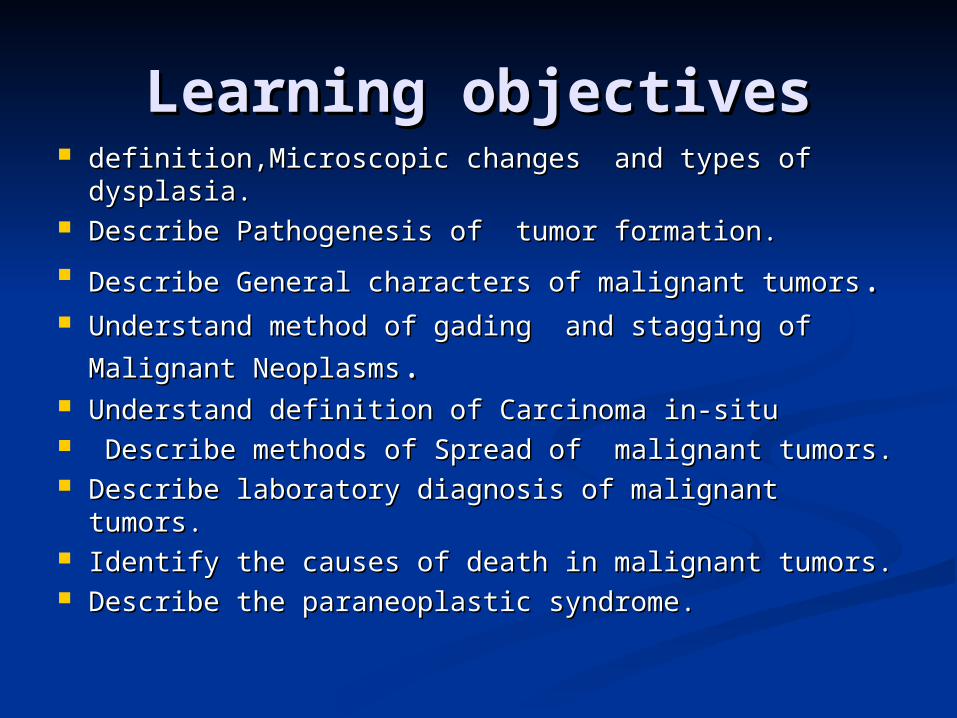
Learning objectivesLearning objectives definition,Microscopic changes and types of definition,Microscopic changes and types of
dysplasia.dysplasia. Describe Pathogenesis of tumor formation.Describe Pathogenesis of tumor formation.
Describe General characters of malignant tumorsDescribe General characters of malignant tumors.. Understand method of gading and stagging of Malignant Understand method of gading and stagging of Malignant
NeoplasmsNeoplasms.. Understand definition of Understand definition of Carcinoma in-situCarcinoma in-situ Describe methods of Describe methods of Spread of malignant tumors.Spread of malignant tumors. Describe laboratory diagnosis of malignant tumors.Describe laboratory diagnosis of malignant tumors. Identify the causes of death in malignant tumors.Identify the causes of death in malignant tumors. Describe the paraneoplastic syndrome.Describe the paraneoplastic syndrome.
NeoplasiaNeoplasia
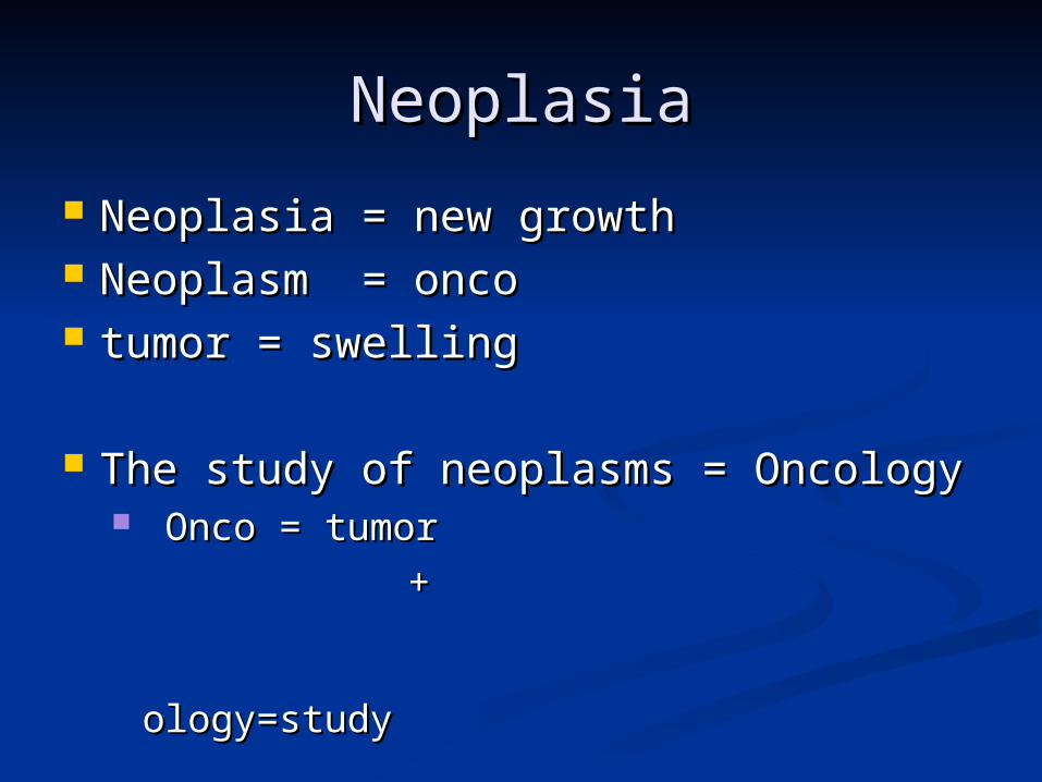
Neoplasia = new growthNeoplasia = new growth Neoplasm = oncoNeoplasm = onco tumor = swellingtumor = swelling
The study of neoplasms = OncologyThe study of neoplasms = Oncology Onco = tumor Onco = tumor
+ + ology=study ology=study
NeoplasiaNeoplasia
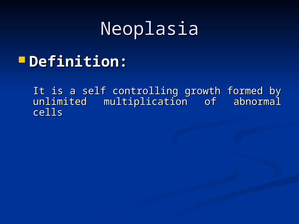
Definition:Definition:
It is a self controlling growth formed by It is a self controlling growth formed by unlimited multiplication of abnormal cellsunlimited multiplication of abnormal cells
NeoplasiaNeoplasia
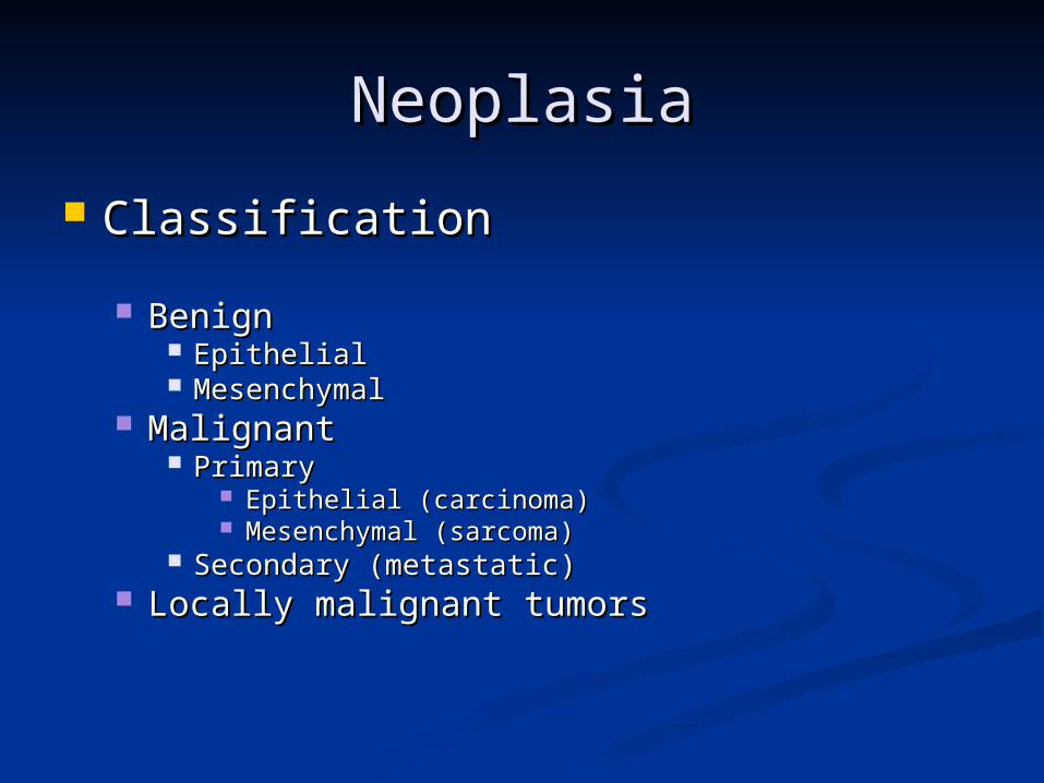
ClassificationClassification
BenignBenign EpithelialEpithelial MesenchymalMesenchymal
MalignantMalignant PrimaryPrimary
Epithelial (carcinoma)Epithelial (carcinoma) Mesenchymal (sarcoma)Mesenchymal (sarcoma)
Secondary (metastatic)Secondary (metastatic) Locally malignant tumors Locally malignant tumors
General characters General characters of benign tumorsof benign tumors
PathologyPathology Gross pathologyGross pathology
Size: Usually small in sizeSize: Usually small in size Shape: usually ovoid or rounded in shapeShape: usually ovoid or rounded in shape Capsule: usually capsulatedCapsule: usually capsulated Cut section solid or cysticCut section solid or cystic Hemorrhage and necrosis: usually absentHemorrhage and necrosis: usually absent
Microscopic pathologyMicroscopic pathology Differentiation: The cells are well differentiated i.e Differentiation: The cells are well differentiated i.e
tumor cells closely similar to the tissue of origin.tumor cells closely similar to the tissue of origin. Nucleocytoplasmic ratio (N/C) ratio: small or normalNucleocytoplasmic ratio (N/C) ratio: small or normal Stroma: is usually well formed with few blood vesselsStroma: is usually well formed with few blood vessels

Behavior of benign tumorsBehavior of benign tumors Rate of growth: usually slowRate of growth: usually slow Mode of growth: by expansionMode of growth: by expansion Localization: usually localizedLocalization: usually localized Effects on the host: usually do not destroy the Effects on the host: usually do not destroy the
surrounding structures and do not kill the surrounding structures and do not kill the patient (except in certain sites as in brain)patient (except in certain sites as in brain)
Recurrence: usually not recurrentRecurrence: usually not recurrent Metastasis: Do not metastatiseMetastasis: Do not metastatise Malignant change: may occurMalignant change: may occur
Benign tumorsBenign tumors
NomenclatureNomenclature Benign tumors: Benign tumors:
prefix + suffixprefix + suffix Type of cell + (-oma)Type of cell + (-oma)

NeoplasiaNeoplasia
Examples:Examples: Benign tumor arising in fibrous tissue:Benign tumor arising in fibrous tissue:
Fibro + oma = FibromaFibro + oma = Fibroma
Benign tumor arising in fatty tissue:Benign tumor arising in fatty tissue:
Lipo + oma = lipomaLipo + oma = lipoma
Benign epithelial tumorsBenign epithelial tumors PapillomaPapilloma AdenomaAdenoma
Benign mesenchymal tumorsBenign mesenchymal tumors CT tumorsCT tumors
FibromaFibroma LipomaLipoma ChondromaChondroma OsteomaOsteoma
Benign tumors of musclesBenign tumors of muscles LeiomyomaLeiomyoma RhabdomyomaRhabdomyoma
Benign tumors of vesselsBenign tumors of vessels HaemangiomaHaemangioma lymphangioma lymphangioma

Lipoma
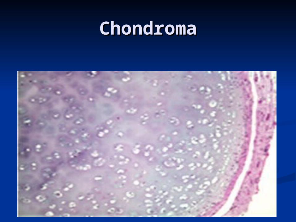
ChondromaChondroma
Adenoma Adenoma : benign epithelial neoplasms : benign epithelial neoplasms producing gland pattern….OR … derived producing gland pattern….OR … derived from glands but not necessarily from glands but not necessarily exhibiting gland pattern exhibiting gland pattern
Examples : Examples : Respiratory airways: Bronchial adenomaRespiratory airways: Bronchial adenoma Renal epithelium: Renal tubular adenomaRenal epithelium: Renal tubular adenoma Liver cell : Liver cell adenomaLiver cell : Liver cell adenoma Papilloma Papilloma : benign epithelial neoplasms : benign epithelial neoplasms
growing on any surface that produce growing on any surface that produce microscopic or macroscopic finger-like microscopic or macroscopic finger-like patternpattern
Squamous epithelium: squamous Squamous epithelium: squamous papillomapapilloma

Adenoma
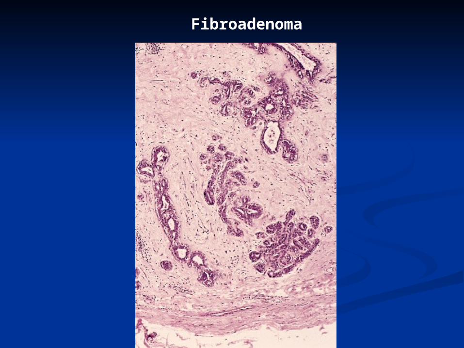
Fibroadenoma
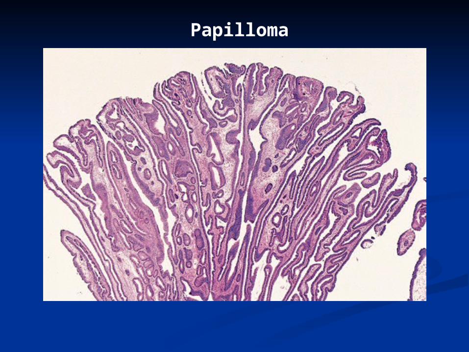
Papilloma
Malignant tumorsMalignant tumors
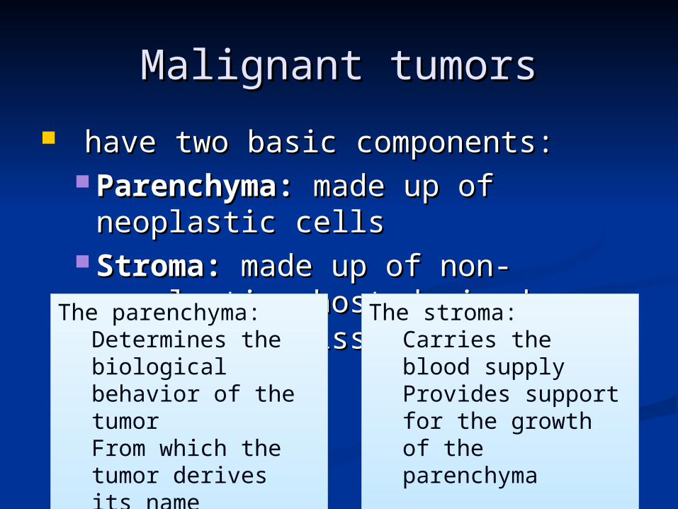
have two basic components:have two basic components: Parenchyma: Parenchyma: made up of made up of
neoplastic cellsneoplastic cells Stroma: Stroma: made up of non-made up of non-
neoplastic, host-derived neoplastic, host-derived connective tissue and blood connective tissue and blood vessels.vessels.
The parenchyma:Determines the biological behavior of the tumorFrom which the tumor derives its name
The parenchyma:Determines the biological behavior of the tumorFrom which the tumor derives its name
The stroma:Carries the blood supplyProvides support for the growth of the parenchyma
The stroma:Carries the blood supplyProvides support for the growth of the parenchyma
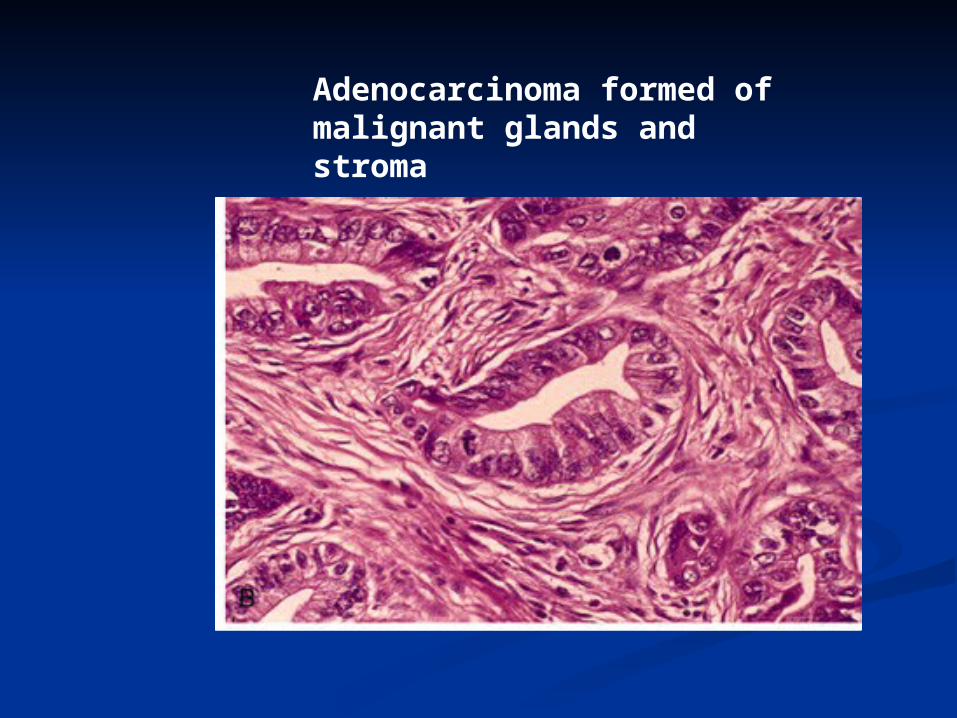
Adenocarcinoma formed of malignant glands and stroma
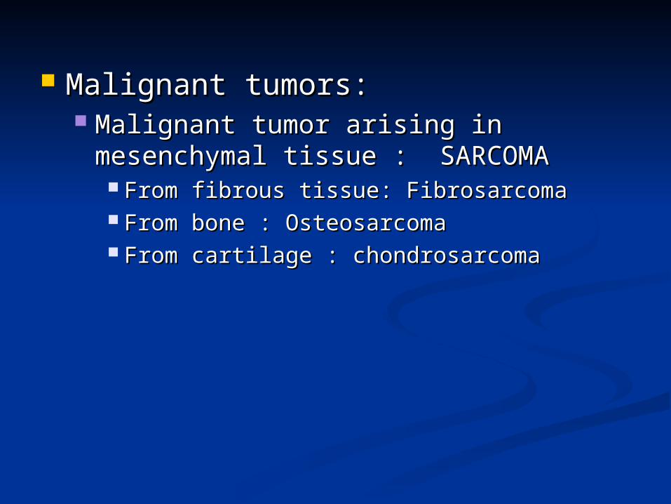
Malignant tumors:Malignant tumors: Malignant tumor arising in Malignant tumor arising in
mesenchymal tissue : SARCOMAmesenchymal tissue : SARCOMA From fibrous tissue: FibrosarcomaFrom fibrous tissue: Fibrosarcoma From bone : OsteosarcomaFrom bone : Osteosarcoma From cartilage : chondrosarcomaFrom cartilage : chondrosarcoma

OsteosarcomaOsteosarcoma
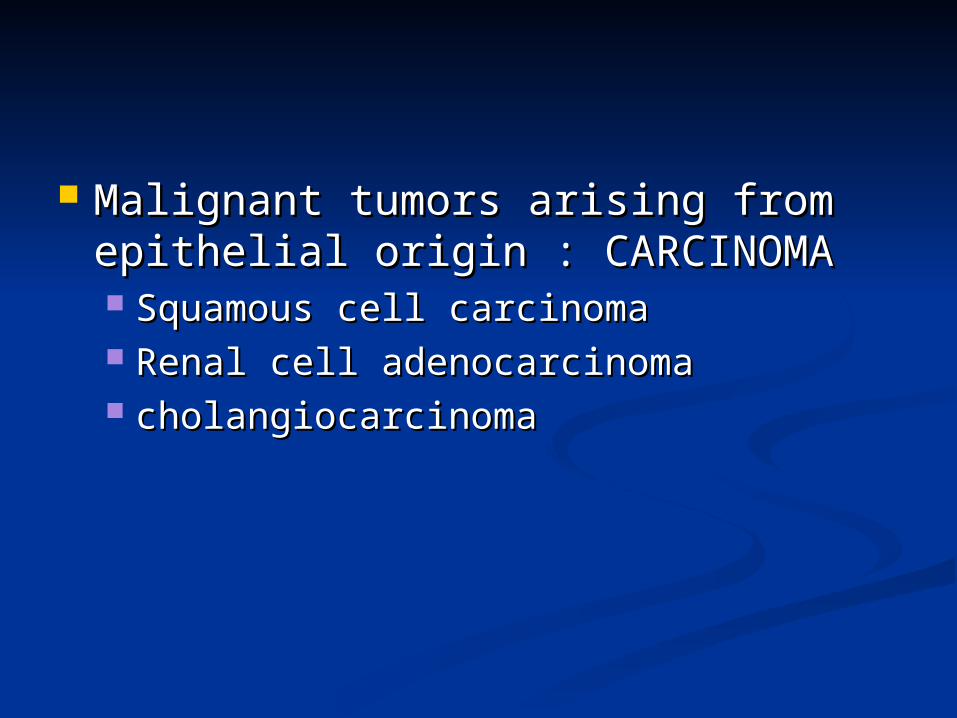
Malignant tumors arising from Malignant tumors arising from epithelial origin : CARCINOMAepithelial origin : CARCINOMA Squamous cell carcinomaSquamous cell carcinoma Renal cell adenocarcinomaRenal cell adenocarcinoma cholangiocarcinomacholangiocarcinoma

Carcinomas arising from any epithelium of the body that exhibit squamous differentiation are termed squamous cell carcinoma.
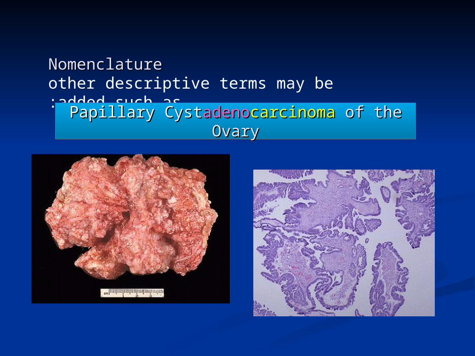
NomenclatureNomenclatureother descriptive terms may be added such as:
PapillaryPapillary CystCystadenoadenocarcinomacarcinoma of the Ovaryof the OvaryPapillaryPapillary CystCystadenoadenocarcinomacarcinoma of the Ovaryof the Ovary

Nomenclature of some Nomenclature of some Malignant tumors with Malignant tumors with
ExceptionsExceptions Melanoma ( skin )Melanoma ( skin ) Mesothelioma (mesothelium )Mesothelioma (mesothelium ) Seminoma ( testis )Seminoma ( testis ) Lymphoma ( lymphoid tissue ) Lymphoma ( lymphoid tissue )
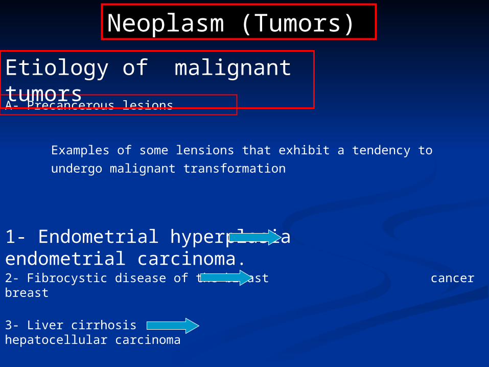
Neoplasm (Tumors)
A- Precancerous lesions
Etiology of malignant tumors
Examples of some lensions that exhibit a tendency to
undergo malignant transformation
1- Endometrial hyperplasia endometrial carcinoma.
2- Fibrocystic disease of the breast cancer breast
3- Liver cirrhosis hepatocellular carcinoma
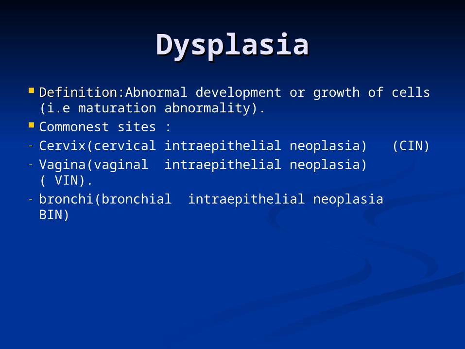
DysplasiaDysplasia Definition:Definition:Abnormal development or growth of
cells (i.e maturation abnormality). Commonest sites :- Cervix(cervical intraepithelial neoplasia) (CIN)- Vagina(vaginal intraepithelial neoplasia) ( VIN).- bronchi(bronchial intraepithelial neoplasia BIN)
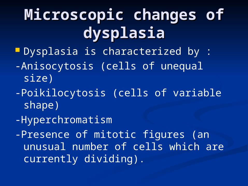
Microscopic changes of Microscopic changes of dysplasiadysplasia
Dysplasia is characterized by :-Anisocytosis (cells of unequal size)-Poikilocytosis (cells of variable shape)-Hyperchromatism-Presence of mitotic figures (an
unusual number of cells which are currently dividing).

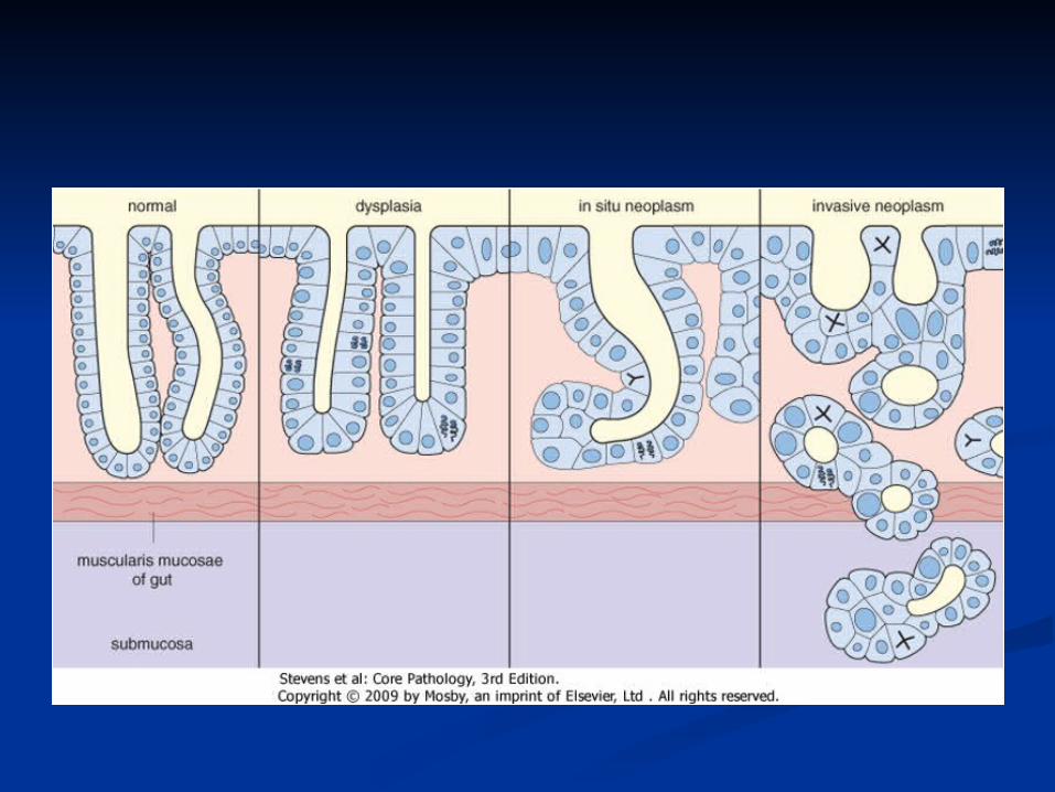
Types of dysplasiaTypes of dysplasia
1- low grade dysplasia: 1- low grade dysplasia:
-not affect the whole thickness of -not affect the whole thickness of epithelium.epithelium.
-it is reversible if the irritant is removed.-it is reversible if the irritant is removed. High grade dysplasia:High grade dysplasia:
- it is precancerous (not reversible)- it is precancerous (not reversible)
-affect the whole thickness of epithelium.-affect the whole thickness of epithelium.
-it is also called carcinoma in situ as the -it is also called carcinoma in situ as the basement membrane not invaded by the basement membrane not invaded by the abnormal cellsabnormal cells
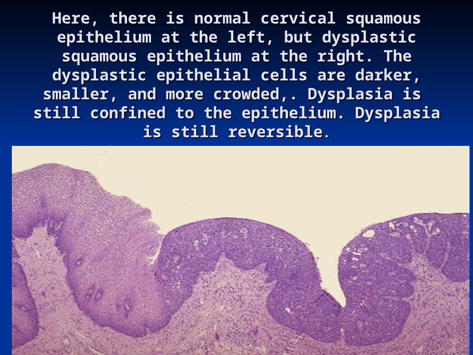
Here, there is normal cervical squamous Here, there is normal cervical squamous epithelium at the left, but dysplastic epithelium at the left, but dysplastic
squamous epithelium at the right. The squamous epithelium at the right. The dysplastic epithelial cells are darker, smaller, dysplastic epithelial cells are darker, smaller, and more crowded,. Dysplasia is still confined and more crowded,. Dysplasia is still confined to the epithelium. Dysplasia is still reversibleto the epithelium. Dysplasia is still reversible..
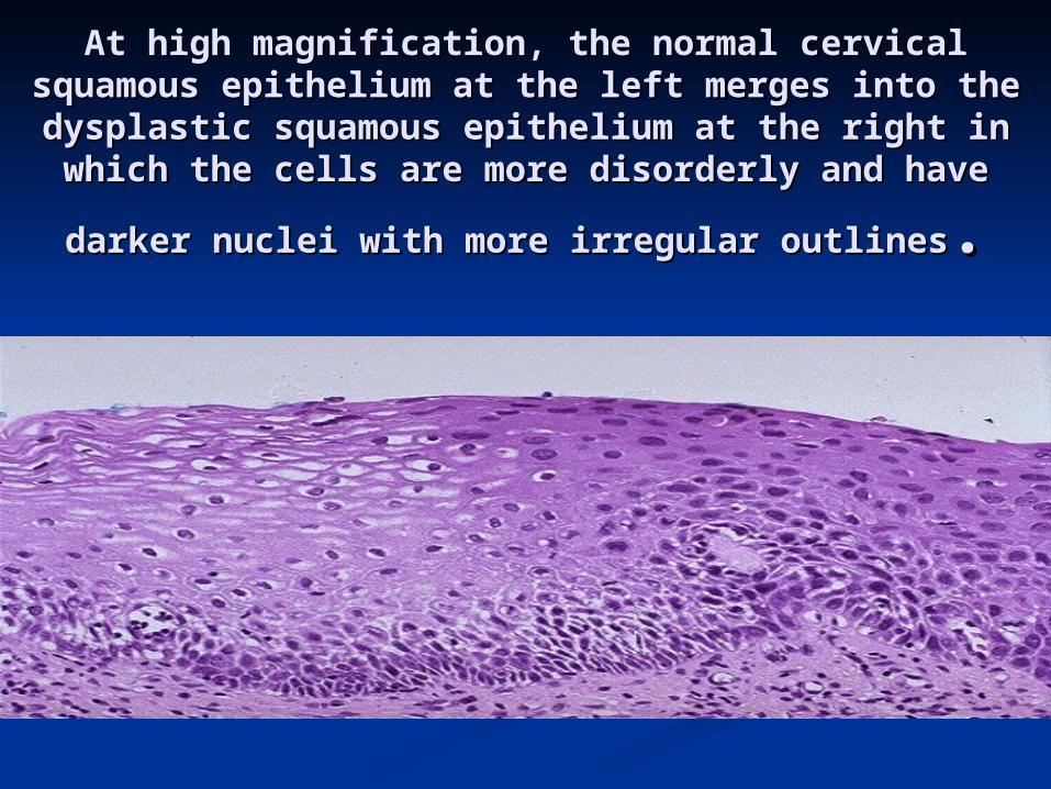
At high magnification, the normal cervical At high magnification, the normal cervical squamous epithelium at the left merges into the squamous epithelium at the left merges into the dysplastic squamous epithelium at the right in dysplastic squamous epithelium at the right in
which the cells are more disorderly and have darker which the cells are more disorderly and have darker
nuclei with more irregular outlinesnuclei with more irregular outlines..
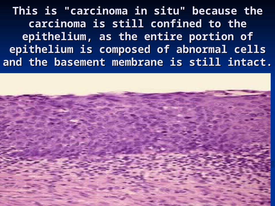
This is "carcinoma in situ" because the This is "carcinoma in situ" because the carcinoma is still confined to the carcinoma is still confined to the
epithelium, as the entire portion of epithelium, as the entire portion of epithelium is composed of abnormal cells epithelium is composed of abnormal cells
and the basement membrane is still intact.and the basement membrane is still intact.
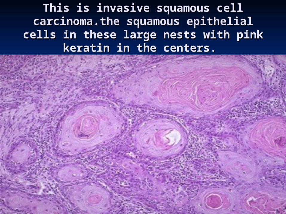
This is invasive squamous cell This is invasive squamous cell carcinoma.the squamous epithelial cells carcinoma.the squamous epithelial cells in these large nests with pink keratin in in these large nests with pink keratin in
the centers. the centers.
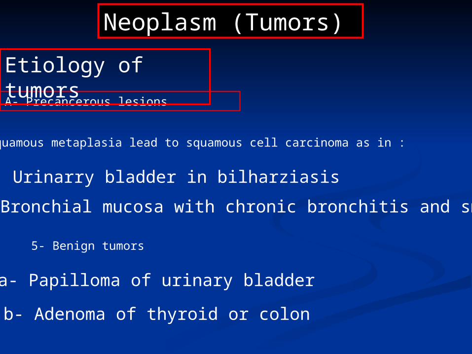
Neoplasm (Tumors)
A- Precancerous lesions
Etiology of tumors
4- Squamous metaplasia lead to squamous cell carcinoma as in :
a- Urinarry bladder in bilharziasis
b- Bronchial mucosa with chronic bronchitis and smoking
5- Benign tumors
a- Papilloma of urinary bladder
b- Adenoma of thyroid or colon
Neoplasm (Tumors)
Etiology of tumors
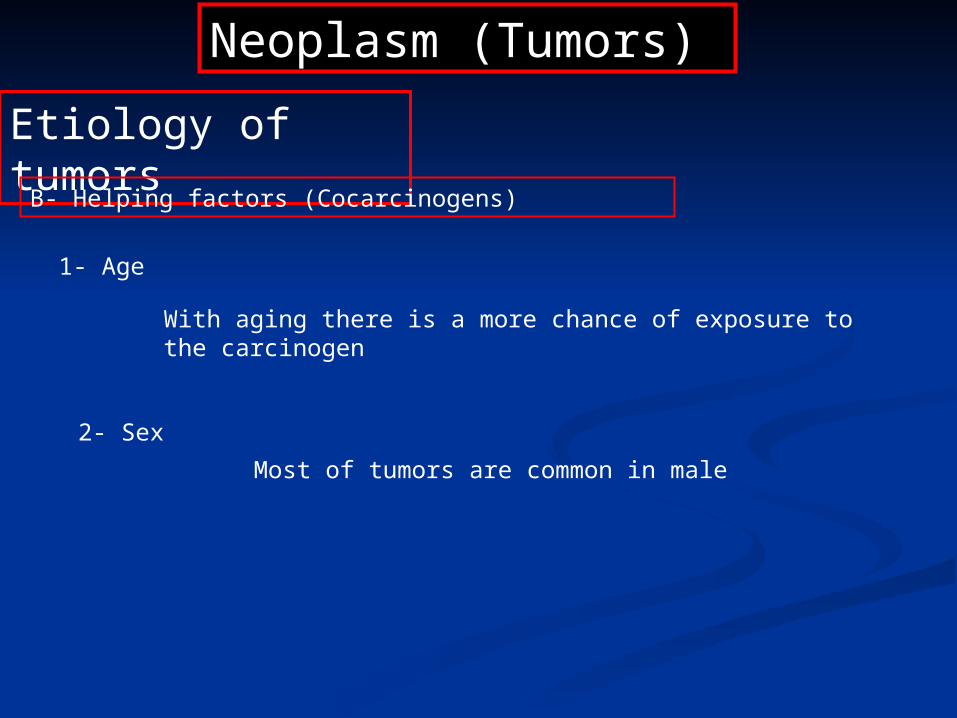
1- Age
B- Helping factors (Cocarcinogens)
With aging there is a more chance of exposure to the carcinogen
2- Sex
Most of tumors are common in male

Neoplasm (Tumors)
Etiology of tumors
B- Helping factors (Cocarcinogens)
4- Smoking
May lead to lung cancer
Some tumors are inherited i.e. retinoblastoma and colonic cancer
5- Heredity
3- Diet
Fat may be related to colonic cancer.
Smoked fish is related to gastric carcinoma.
Excess alcohol is related to liver cancer.

Neoplasm (Tumors)
Etiology of tumors
C- Carcinogens
Types of carcinogens
1- Chemical carcinogens
Methylated hydrocarbons-A 20 dyes bladder cancer
2- Viruses
Aflatoxins produced from Aspergillus fungus liver cancer
Hepatitis B virus liver cancer
Human papilloma virus cancer cervix
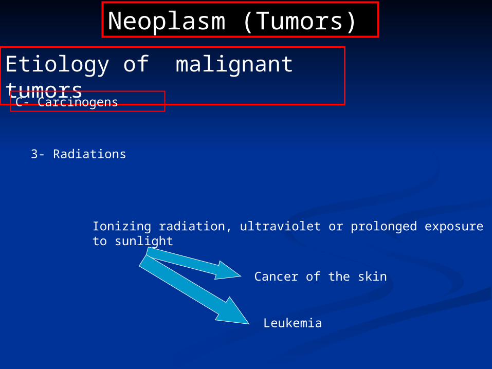
Neoplasm (Tumors)
Etiology of malignant tumors
C- Carcinogens
3- Radiations
Ionizing radiation, ultraviolet or prolonged exposure to sunlight
Cancer of the skin
Leukemia

Neoplasm (Tumors)
Etiology of malignant tumors
C-mechanism of action of Carcinogens
Chemical carcinogens, viruses and radiation result in DNA damage and initiation of cancer by :
1- Activation of oncogenes e.g myc gene,K-ras (genes responsible for abnormal growth and proliferation of cell).
2- Inactivation of cancer suppressor genes e.g RB ,P53 genes.
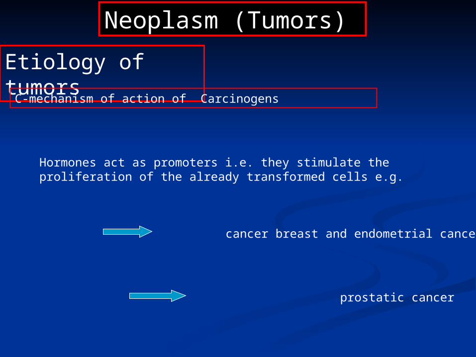
Neoplasm (Tumors)
Etiology of tumors
C-mechanism of action of Carcinogens
Hormones act as promoters i.e. they stimulate the proliferation of the already transformed cells e.g.
Estrogen cancer breast and endometrial cancer
Androgen prostatic cancer

Pathogenesis of tumor Pathogenesis of tumor formationformation
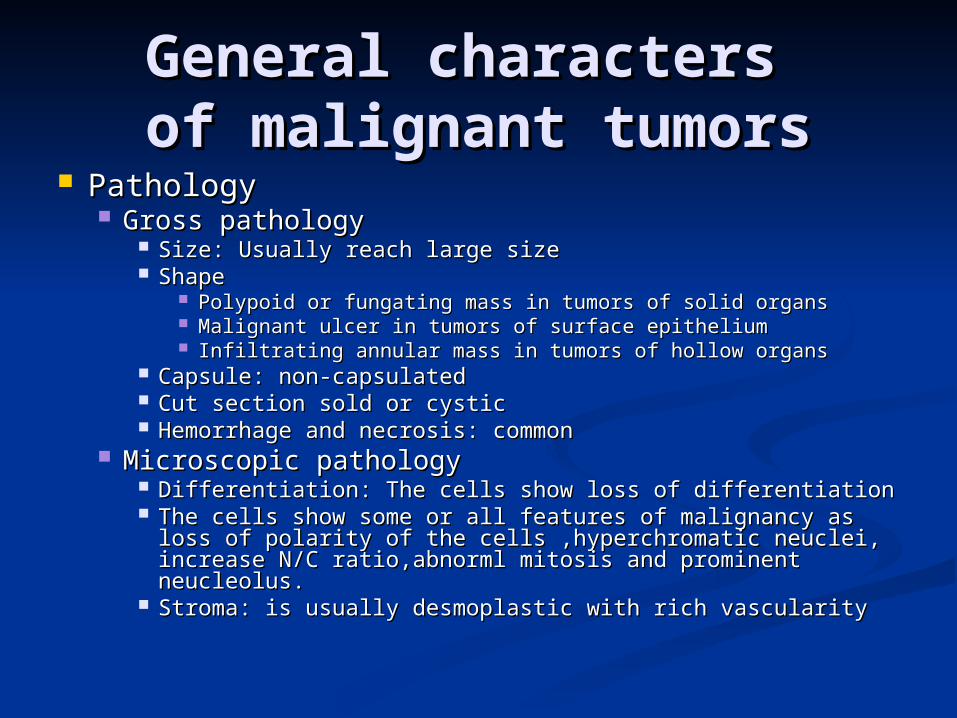
General characters General characters of malignant tumorsof malignant tumors
PathologyPathology Gross pathologyGross pathology
Size: Usually reach large sizeSize: Usually reach large size ShapeShape
Polypoid or fungating mass in tumors of solid organsPolypoid or fungating mass in tumors of solid organs Malignant ulcer in tumors of surface epitheliumMalignant ulcer in tumors of surface epithelium Infiltrating annular mass in tumors of hollow organsInfiltrating annular mass in tumors of hollow organs
Capsule: non-capsulatedCapsule: non-capsulated Cut section sold or cysticCut section sold or cystic Hemorrhage and necrosis: commonHemorrhage and necrosis: common
Microscopic pathologyMicroscopic pathology Differentiation: The cells show loss of differentiationDifferentiation: The cells show loss of differentiation The cells show some or all features of malignancy as loss The cells show some or all features of malignancy as loss
of polarity of the cells ,hyperchromatic neuclei, increase of polarity of the cells ,hyperchromatic neuclei, increase N/C ratio,abnorml mitosis and prominent neucleolus.N/C ratio,abnorml mitosis and prominent neucleolus.
Stroma: is usually desmoplastic with rich vascularityStroma: is usually desmoplastic with rich vascularity
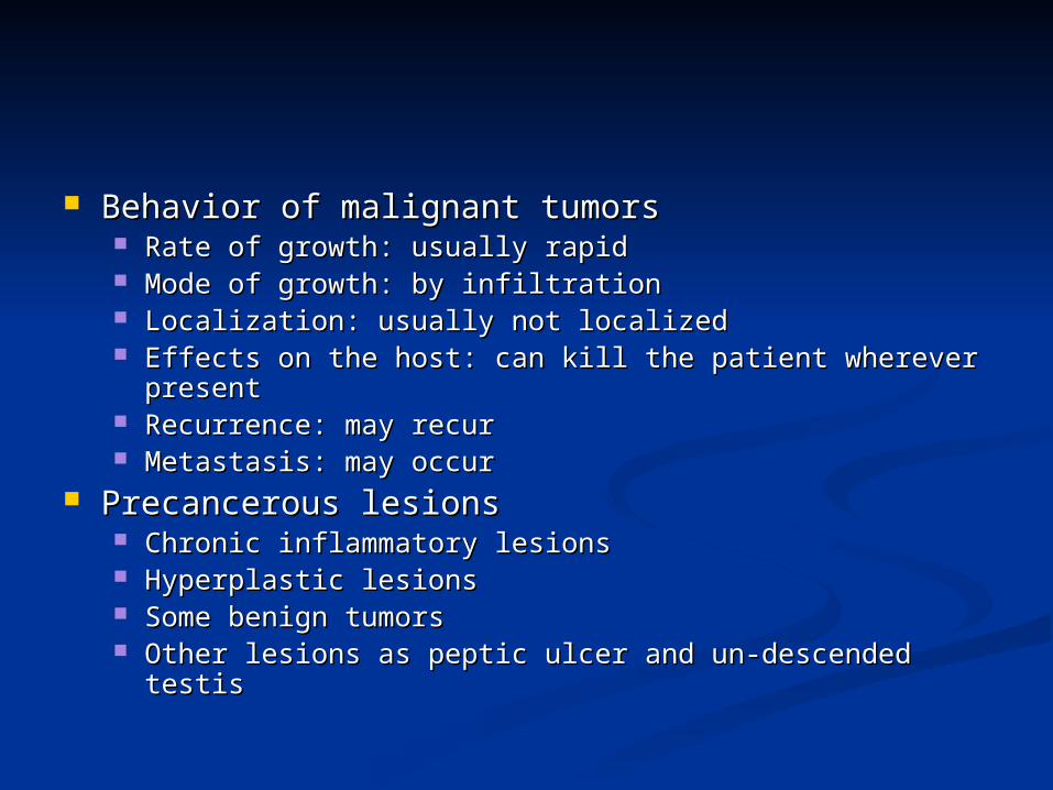
Behavior of malignant tumorsBehavior of malignant tumors Rate of growth: usually rapidRate of growth: usually rapid Mode of growth: by infiltrationMode of growth: by infiltration Localization: usually not localizedLocalization: usually not localized Effects on the host: can kill the patient wherever Effects on the host: can kill the patient wherever
presentpresent Recurrence: may recurRecurrence: may recur Metastasis: may occurMetastasis: may occur
Precancerous lesionsPrecancerous lesions Chronic inflammatory lesionsChronic inflammatory lesions Hyperplastic lesionsHyperplastic lesions Some benign tumorsSome benign tumors Other lesions as peptic ulcer and un-descended testisOther lesions as peptic ulcer and un-descended testis
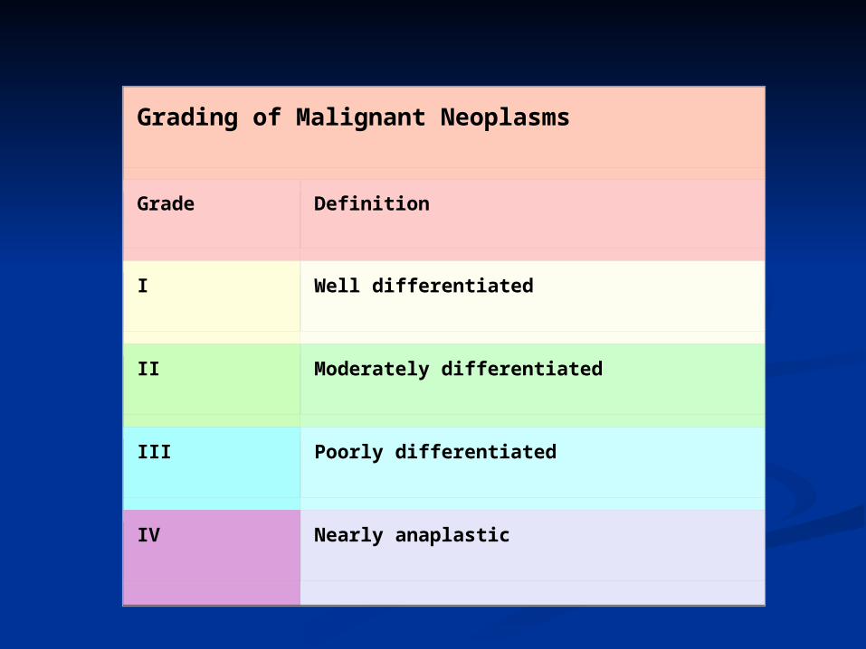
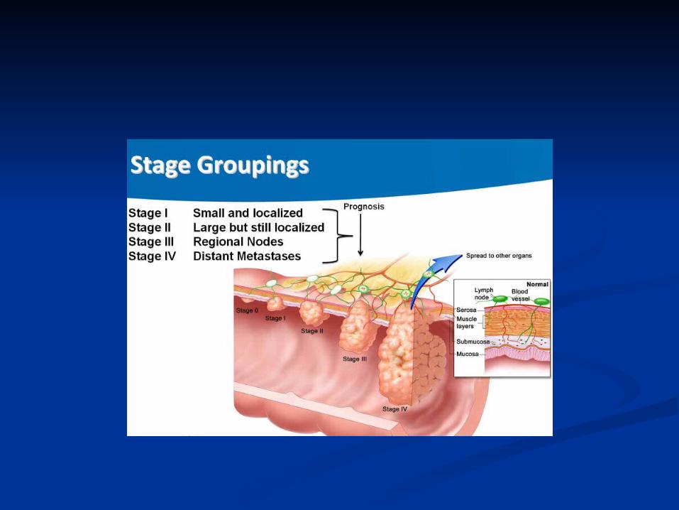
Grading of Malignant Neoplasms
Grade Definition
I Well differentiated
II Moderately differentiated
III Poorly differentiated
IV Nearly anaplastic
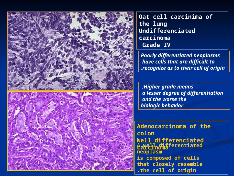
Oat cell carcinima of the lungUndifferenciated carcinomaGrade IV
Adenocarcinoma of the colonWell differenciated carcinoma
Higher grade means : a lesser degree of differentiation
and the worse the biologic behavior
A well differentiated neoplasm is composed of cells that closely resemble
the cell of origin.
Poorly differentiated neoplasms have cells that are difficult to
recognize as to their cell of origin.

Clinical StagingClinical Staging
T (primary tumor): T1, T2, T3, T4T (primary tumor): T1, T2, T3, T4 N (regional lymph nodes): N0, N1, N (regional lymph nodes): N0, N1,
N2, N3N2, N3 M (metastasis): M0, M1M (metastasis): M0, M1
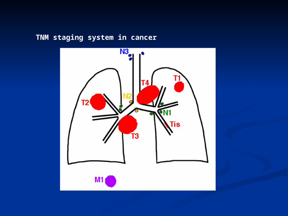
TNM staging system in cancer
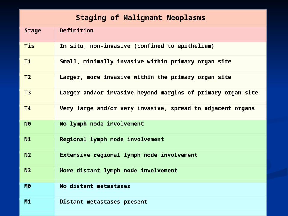
Staging of Malignant Neoplasms
Stage Definition
Tis In situ, non-invasive (confined to epithelium)
T1 Small, minimally invasive within primary organ site
T2 Larger, more invasive within the primary organ site
T3 Larger and/or invasive beyond margins of primary organ site
T4 Very large and/or very invasive, spread to adjacent organs
N0 No lymph node involvement
N1 Regional lymph node involvement
N2 Extensive regional lymph node involvement
N3 More distant lymph node involvement
M0 No distant metastases
M1 Distant metastases present
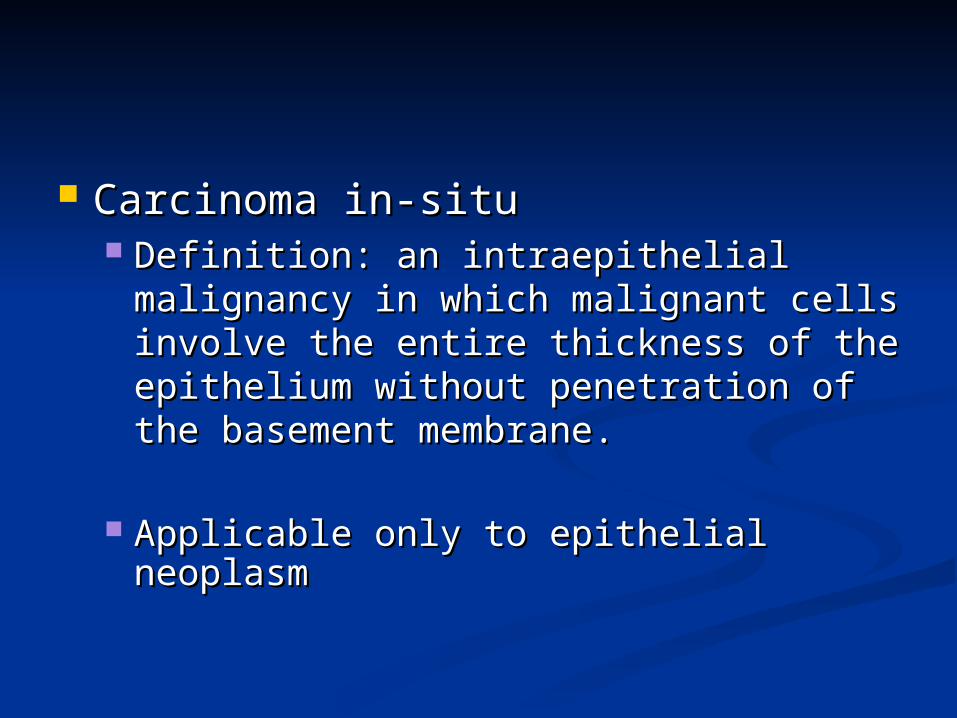
Carcinoma in-situCarcinoma in-situ Definition: an intraepithelial malignancy Definition: an intraepithelial malignancy
in which malignant cells involve the in which malignant cells involve the entire thickness of the epithelium entire thickness of the epithelium without penetration of the basement without penetration of the basement membrane.membrane.
Applicable only to epithelial neoplasmApplicable only to epithelial neoplasm
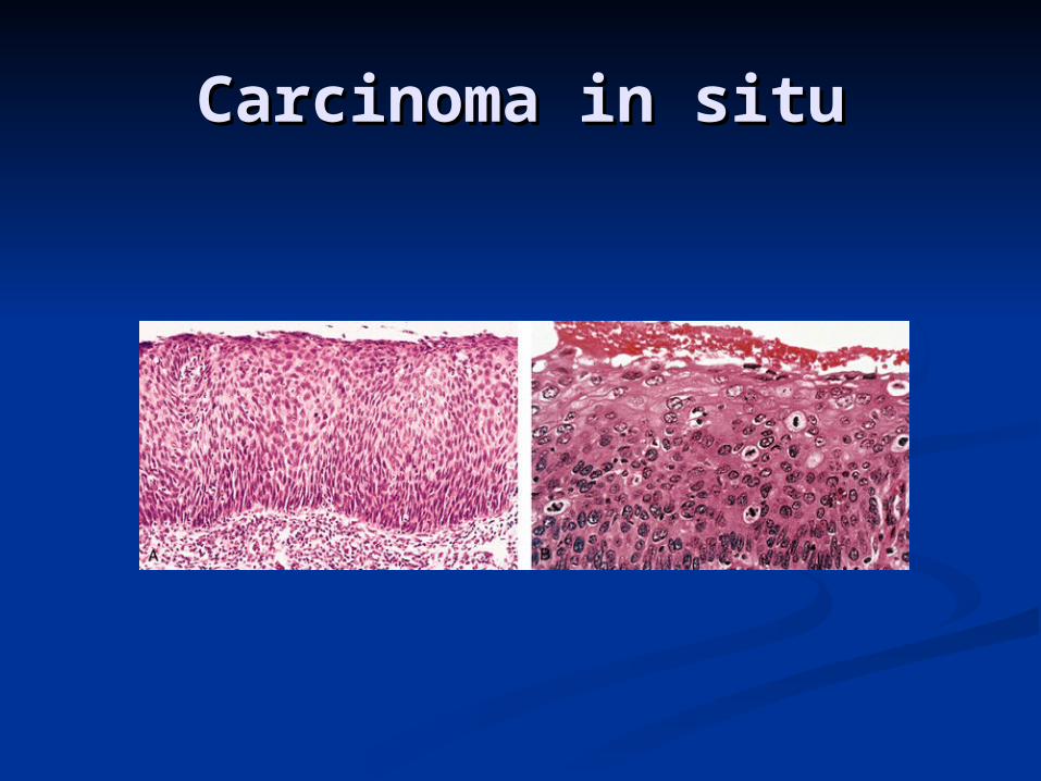
Carcinoma in situCarcinoma in situ
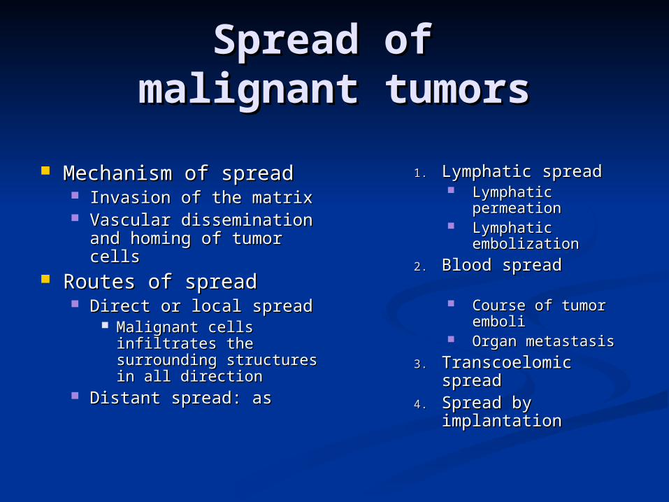
Spread of Spread of malignant tumorsmalignant tumors
Mechanism of spreadMechanism of spread Invasion of the matrixInvasion of the matrix Vascular dissemination Vascular dissemination
and homing of tumor and homing of tumor cellscells
Routes of spreadRoutes of spread Direct or local spreadDirect or local spread
Malignant cells Malignant cells infiltrates the infiltrates the surrounding structures surrounding structures in all directionin all direction
Distant spread: asDistant spread: as
1.1. Lymphatic spreadLymphatic spread Lymphatic Lymphatic
permeationpermeation Lymphatic Lymphatic
embolizationembolization
2.2. Blood spreadBlood spread Course of tumor Course of tumor
emboliemboli Organ metastasisOrgan metastasis
3.3. Transcoelomic spreadTranscoelomic spread4.4. Spread by Spread by
implantationimplantation
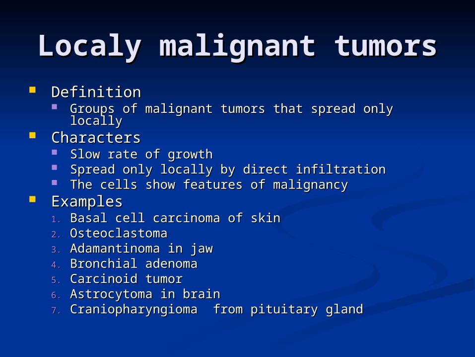
Localy malignant tumorsLocaly malignant tumors DefinitionDefinition
Groups of malignant tumors that spread only locallyGroups of malignant tumors that spread only locally CharactersCharacters
Slow rate of growthSlow rate of growth Spread only locally by direct infiltrationSpread only locally by direct infiltration The cells show features of malignancyThe cells show features of malignancy
ExamplesExamples1.1. Basal cell carcinoma of skinBasal cell carcinoma of skin2.2. OsteoclastomaOsteoclastoma3.3. Adamantinoma in jawAdamantinoma in jaw4.4. Bronchial adenomaBronchial adenoma5.5. Carcinoid tumorCarcinoid tumor6.6. Astrocytoma in brainAstrocytoma in brain7.7. Craniopharyngioma from pituitary glandCraniopharyngioma from pituitary gland
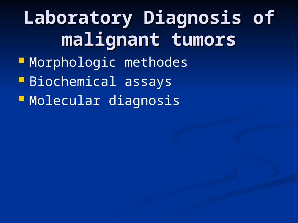
Laboratory Diagnosis of Laboratory Diagnosis of malignant tumorsmalignant tumors
Morphologic methodes Biochemical assays Molecular diagnosis
Laboratory DiagnosisLaboratory Diagnosis
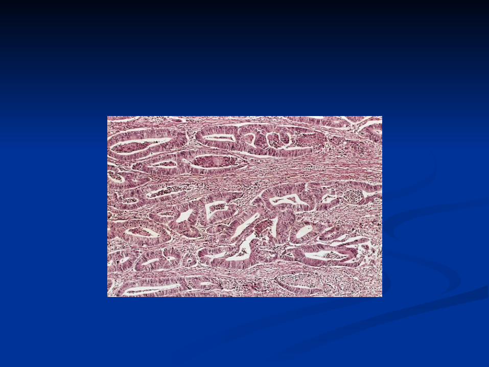
Microscopic Tissue DiagnosisMicroscopic Tissue Diagnosis the gold standard of cancer diagnosis.. Several sampling approaches are available:
Excision or biopsy Frozen section
fine-needle aspiration Cytologic smears
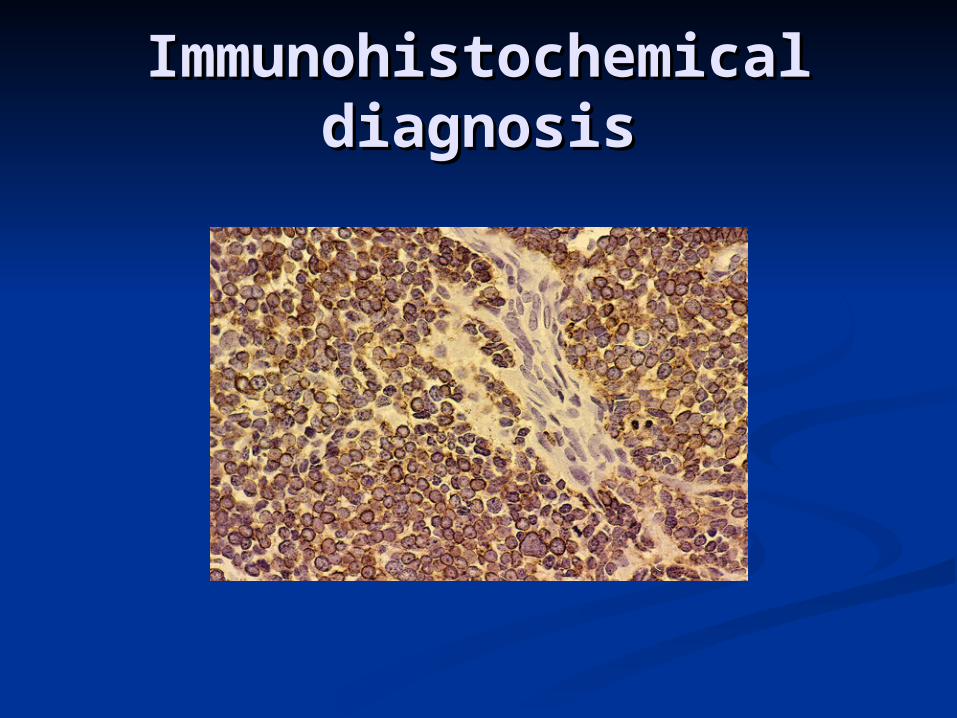
Immunohistochemical Immunohistochemical diagnosisdiagnosis
Laboratory DiagnosisLaboratory Diagnosis
Biochemical assaysBiochemical assays:: Useful for measuring the levels of tumor
associated enzymes, hormones, and tumor markers in serum.
Useful in determining the effectiveness of therapy and detection of recurrences after excision
Only few tumor markers are proved to be clinically useful, example CEA in colonic adenocarcinoma and α- fetoprotein in hepatoma.
Molecular diagnosisMolecular diagnosis
Polymerase chain reaction (PCR) example: detection of BCR-ABL
transcripts in chronic myeloid leukemia. Fluorescent in situ hybridization (fish) it is useful for detecting chromosomes
translocation characteristic of many tumors
Both PCR and Fish can show amplification of oncogenes (HER2 and N-MYC)
Neoplasm (Tumors)
1- Destruction of vital tissues such as brain, liver, kidney
Causes of death in malignant tumors
2- Malnutrition due to interference of food intake, digestion and absorption.
3- Obstructive effects e.g. urinary tract obstruction.
4- Anemia due to
a- Continuous blood loss from bleeding malignant ulcers.
b- Destruction of red bone marrow by metastatic deposites.
5- Malignant cachexia with wasting, loss of weight and muscular weakness.
6- Secondary infection by bacteria, viruses and fungi.

Paraneoplastic syndromes
They are symptoms that occur in cancer patients due to secretion of some hormone like substances and not due to distant metastasis.
They are diverse and are associated with many different tumors.
They appear in 10% to 15% of pateints. They may represent the earliest
manifestation of an occult neoplasm. They may represent significant clinical
problems and may be lethal. They may mimic metastatic disease.
The most common paraneoplastic syndrome are: Hypercalcemia Cushing syndrome Nonbacterial thrombotic endocarditis
The most often neoplasms associated with these syndromes: Lung and breast cancers and
hematologic malignancies