Embed Size (px)
Citation preview

© 2012 Pearson Education, Inc.
An Introduction to Blood and the Cardiovascular System
• The Cardiovascular System consists of:
• A pump (the heart)
• A conducting system (blood vessels)
• A fluid medium (blood)
• Is specialized fluid of connective tissue
• Contains cells suspended in a fluid matrix
© 2012 Pearson Education, Inc.
An Introduction to Blood and the Cardiovascular System
• Blood
• Transports materials to and from cells
• Oxygen and carbon dioxide
• Nutrients
• Hormones
• Immune system components
• Waste products

© 2012 Pearson Education, Inc.
19-1 Physical Characteristics of Blood
• Important Functions of Blood
• Transportation of dissolved substances
• Regulation of pH and ions
• Restriction of fluid losses at injury sites
• Defense against toxins and pathogens
• Stabilization of body temperature
© 2012 Pearson Education, Inc.
19-1 Physical Characteristics of Blood
• Whole Blood
• Plasma
• Fluid consisting of:
• Water
• Dissolved plasma proteins
• Other solutes
• Formed elements
• All cells and solids
© 2012 Pearson Education, Inc.
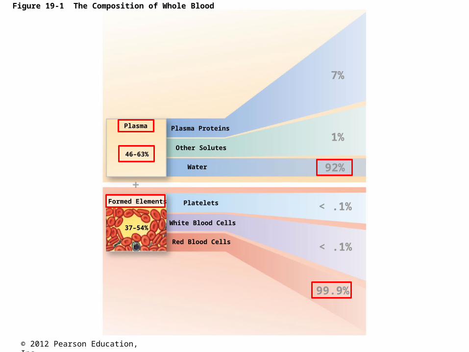
Figure 19-1 The Composition of Whole Blood
Plasma Proteins
Other Solutes
Water
Platelets
White Blood Cells
Red Blood Cells
Plasma
Formed Elements
46–63%
37–54%
7%
1%
92%
< .1%
< .1%
99.9%

© 2012 Pearson Education, Inc.
19-1 Physical Characteristics of Blood
• Three Types of Formed Elements
1. Red blood cells (RBCs) or erythrocytes
• Transport oxygen
2. White blood cells (WBCs) or leukocytes
• Part of the immune system
3. Platelets
• Cell fragments involved in clotting
© 2012 Pearson Education, Inc.
19-1 Physical Characteristics of Blood
• Hemopoiesis
• Process of producing formed elements
• By myeloid and lymphoid stem cells
• Fractionation
• Process of separating whole blood for clinical analysis
• Into plasma and formed elements
© 2012 Pearson Education, Inc.
19-1 Physical Characteristics of Blood
• Three General Characteristics of Blood
1. 38C (100.4F) is normal temperature
2. High viscosity
3. Slightly alkaline pH (7.35–7.45)
© 2012 Pearson Education, Inc.
19-1 Physical Characteristics of Blood
• Characteristics of Blood
• Blood volume (liters) = 7% of body weight (kilograms)
• Adult male 5 to 6 liters
• Adult female 4 to 5 liters
• Hypovolemic = low blood volume.
• Hypervolemic = high blood volume.

© 2012 Pearson Education, Inc.
19-2 Plasma
• The Composition of Plasma
• Makes up 50–60% of blood volume
• More than 90% of plasma is water
• Extracellular fluids
• Interstitial fluid (IF) and plasma
• Materials plasma and IF exchange across capillary walls
• Water
• Ions
• Small solutes

© 2012 Pearson Education, Inc.
19-2 Plasma
• Plasma Proteins
• Albumins (60%)
• Globulins (35%)
• Fibrinogen (4%)

© 2012 Pearson Education, Inc.
19-2 Plasma
• Albumins (60%)
• Transport substances such as fatty acids, thyroid hormones, and steroid hormones
• Globulins (35%)
• Antibodies, also called immunoglobulins
• Transport globulins (small molecules): hormone-binding proteins, metalloproteins, apolipoproteins (lipoproteins), and steroid-binding proteins
• Fibrinogen (4%)
• Molecules that form clots and produce long, insoluble strands of fibrin

© 2012 Pearson Education, Inc.
19-2 Plasma
• Serum
• Liquid part of a blood sample
• In which dissolved fibrinogen has converted to
solid fibrin

© 2012 Pearson Education, Inc.
19-2 Plasma
• Other Plasma Proteins
• 1% of plasma
• Changing quantities of specialized plasma proteins
• Peptide hormones normally present in circulating blood
• Insulin, prolactin (PRL), and the glycoproteins
thyroid-stimulating hormone (TSH), follicle-
stimulating hormone (FSH), and luteinizing
hormone (LH)
© 2012 Pearson Education, Inc.
19-2 Plasma
• Origins of Plasma Proteins
• More than 90% made in liver
• Antibodies made by plasma cells
• Peptide hormones made by endocrine organs

© 2012 Pearson Education, Inc.
19-3 Red Blood Cells
• Red blood cells (RBCs)
• Make up 99.9% of blood’s formed elements
• Hemoglobin
• The red pigment that gives whole blood its color
• Binds and transports oxygen and carbon dioxide

© 2012 Pearson Education, Inc.
19-3 Red Blood Cells
• Abundance of RBCs
• Red blood cell count - the number of RBCs in 1
microliter of whole blood
• Male: 4.5–6.3 million
• Female: 4.2–5.5 million
© 2012 Pearson Education, Inc.
19-3 Red Blood Cells
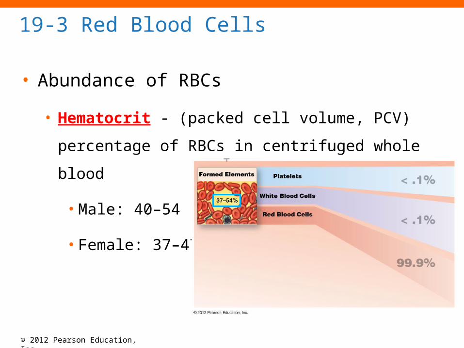
• Abundance of RBCs
• Hematocrit - (packed cell volume, PCV) percentage
of RBCs in centrifuged whole blood
• Male: 40–54
• Female: 37–47

© 2012 Pearson Education, Inc.
19-3 Red Blood Cells
• Structure of RBCs
• Small and highly specialized discs
• Thin in middle and thicker at edge
© 2012 Pearson Education, Inc.
19-3 Red Blood Cells
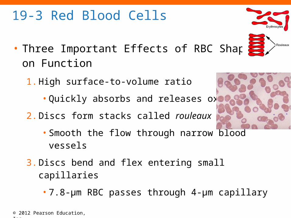
• Three Important Effects of RBC Shape on Function
1. High surface-to-volume ratio
• Quickly absorbs and releases oxygen
2. Discs form stacks called rouleaux
• Smooth the flow through narrow blood vessels
3. Discs bend and flex entering small capillaries
• 7.8-µm RBC passes through 4-µm capillary

© 2012 Pearson Education, Inc.
Figure 19-2ab The Anatomy of Red Blood Cells
Red blood cells
The three-dimensionalshape of RBCs
SEM 1838Blood smear LM 477
When viewed in a standardblood smear, RBCs appear as two-dimensional objects,because they are flattenedagainst the surface of the slide.

© 2012 Pearson Education, Inc.
Figure 19-2c The Anatomy of Red Blood Cells
A sectional view of a mature RBC,showing the normal ranges for itsdimensions
0.45–1.16 μm 2.31–2.85 μm
7.2–8.4 μm
© 2012 Pearson Education, Inc.
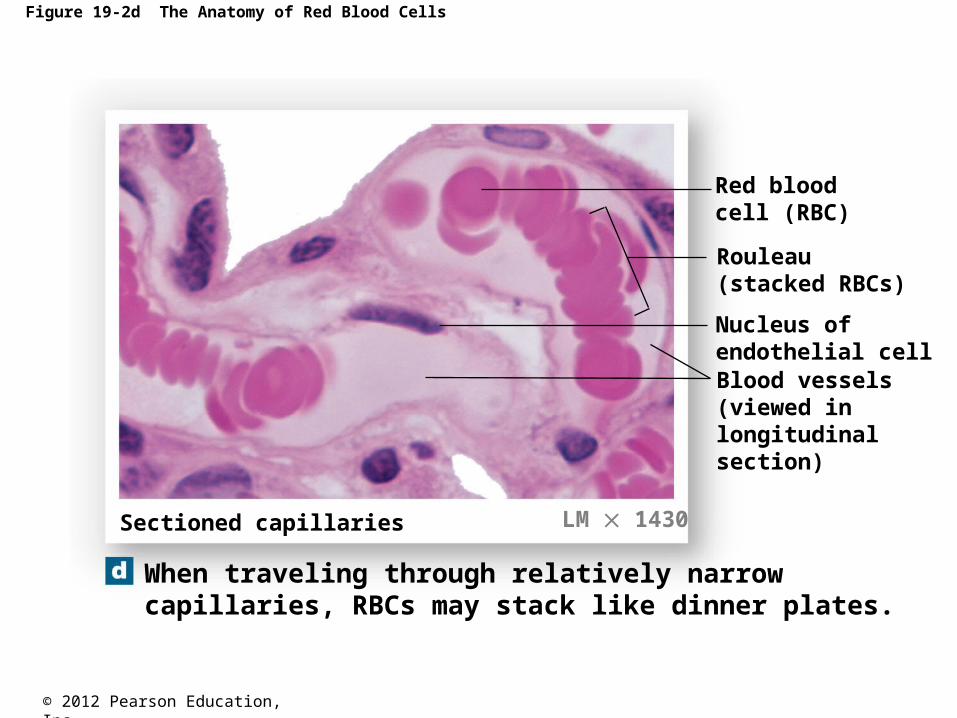
Figure 19-2d The Anatomy of Red Blood Cells
Sectioned capillaries
Red blood cell (RBC)
Rouleau(stacked RBCs)
Nucleus ofendothelial cellBlood vessels(viewed inlongitudinalsection)
When traveling through relatively narrowcapillaries, RBCs may stack like dinner plates.
LM 1430
© 2012 Pearson Education, Inc.
19-3 Red Blood Cells
• Life Span of RBCs
• Lack nuclei, mitochondria, and ribosomes
• Means no repair and anaerobic metabolism
• Live about 120 days

© 2012 Pearson Education, Inc.
19-3 Red Blood Cells
• Hemoglobin (Hb)
• Protein molecule that transports respiratory gases
(oxygen to cells and carbon dioxide away from cells)
• Normal hemoglobin (adult male)
• 14–18 g/dL whole blood
• Normal hemoglobin (adult female)
• 12–16 g/dL whole blood

© 2012 Pearson Education, Inc.
19-3 Red Blood Cells
• Hemoglobin Structure
• Complex quaternary structure
• Four globular protein subunits
• Each with one molecule of heme
• Each heme ring in Hg encloses an atom of iron

© 2012 Pearson Education, Inc.
19-3 Red Blood Cells
• Hemoglobin Structure
• Iron ions
• Associate easily with oxygen (oxyhemoglobin,
HbO2)
• Dissociate easily from oxygen
(deoxyhemoglobin)

© 2012 Pearson Education, Inc.
Figure 19-3 The Structure of Hemoglobin
Hemoglobin molecule
HemeHeme chain 2
chain 2
chain 1
chain 1

© 2012 Pearson Education, Inc.
19-3 Red Blood Cells
• Fetal Hemoglobin
• Strong form of hemoglobin found in embryos
• Takes oxygen from mother’s hemoglobin

© 2012 Pearson Education, Inc.
19-3 Red Blood Cells
• Hemoglobin Function
• Carries oxygen
• With low oxygen (peripheral capillaries)
• Hemoglobin releases oxygen
• Binds carbon dioxide and carries it to lungs
• Forms carbaminohemoglobin

© 2012 Pearson Education, Inc.
Figure 19-4 “Sickling” in Red Blood Cells
Sickle cell anemia:A gene for adult hemoglobin is abnormal.Mutation in the AA sequence of the beta chains of the Hb molecule.
Thalassemia:Inadequate production of alpha or beta chains of Hb resulting in slower RBC production, fragile, and short-lived RBCs.Transfusions help keep adequate numbers of RBCs in the bloodstream.

© 2012 Pearson Education, Inc.
19-3 Red Blood Cells
• RBC Formation and Turnover
• 1% of circulating RBCs wear out per day
• About 3 million RBCs per second
• Hemoglobin Conversion and Recycling
• Macrophages of liver, spleen, and bone marrow
• Monitor RBCs
• Engulf RBCs before membranes rupture (hemolyze)

© 2012 Pearson Education, Inc.
19-3 Red Blood Cells
• Hemoglobin Conversion and Recycling
• Phagocytes break hemoglobin into components
• Globular proteins to amino acids
• Heme to biliverdin
• Iron

© 2012 Pearson Education, Inc.
19-3 Red Blood Cells
• Hemoglobin Conversion and Recycling
• Hemoglobinuria
• Hemoglobin breakdown products in urine due to
excess hemolysis in bloodstream
• Hematuria
• Whole red blood cells in urine due to kidney or
tissue damage

© 2012 Pearson Education, Inc.
19-3 Red Blood Cells
• Breakdown of Biliverdin
• Biliverdin (green) is converted to bilirubin (yellow)
• Bilirubin
• Is excreted by liver (bile)
• Jaundice is caused by bilirubin buildup
• Converted by intestinal bacteria to urobilins and
stercobilins

© 2012 Pearson Education, Inc.
19-3 Red Blood Cells
• Iron Recycling
• Iron removed from heme leaving biliverdin
• To transport proteins (transferrin)
• To storage proteins (ferritin and hemosiderin)
• -ferritin and hemosiderin store excess iron in the liver
and spleen.

© 2012 Pearson Education, Inc.
Figure 19-5 Recycling of Red Blood Cell Components

© 2012 Pearson Education, Inc.
19-3 Red Blood Cells
• RBC Production
• Erythropoiesis
• Occurs only in myeloid tissue (red bone marrow)
in adults
• Stem cells mature to become RBCs
© 2012 Pearson Education, Inc.
19-3 Red Blood Cells
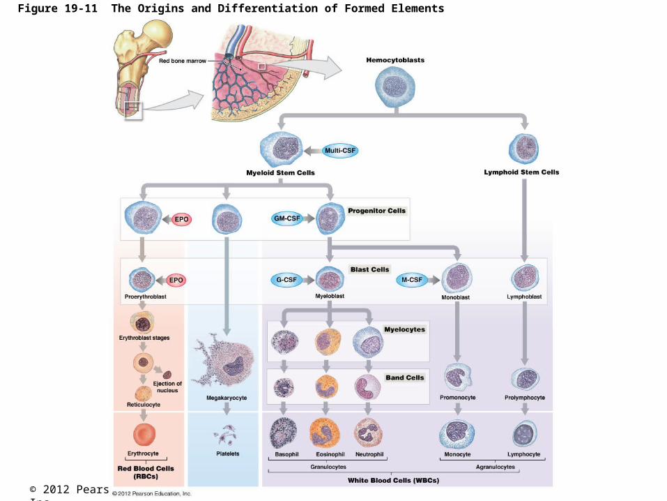
• Hemocytoblasts
• Stem cells in myeloid tissue divide to produce:
1. Myeloid stem cells become RBCs, some WBCs
2. Lymphoid stem cells become lymphocytes

© 2012 Pearson Education, Inc.
19-3 Red Blood Cells
• Stages of RBC Maturation
• Myeloid stem cell
• Proerythroblast
• Erythroblasts
• Reticulocyte
• Mature RBC
© 2012 Pearson Education, Inc.
19-3 Red Blood Cells
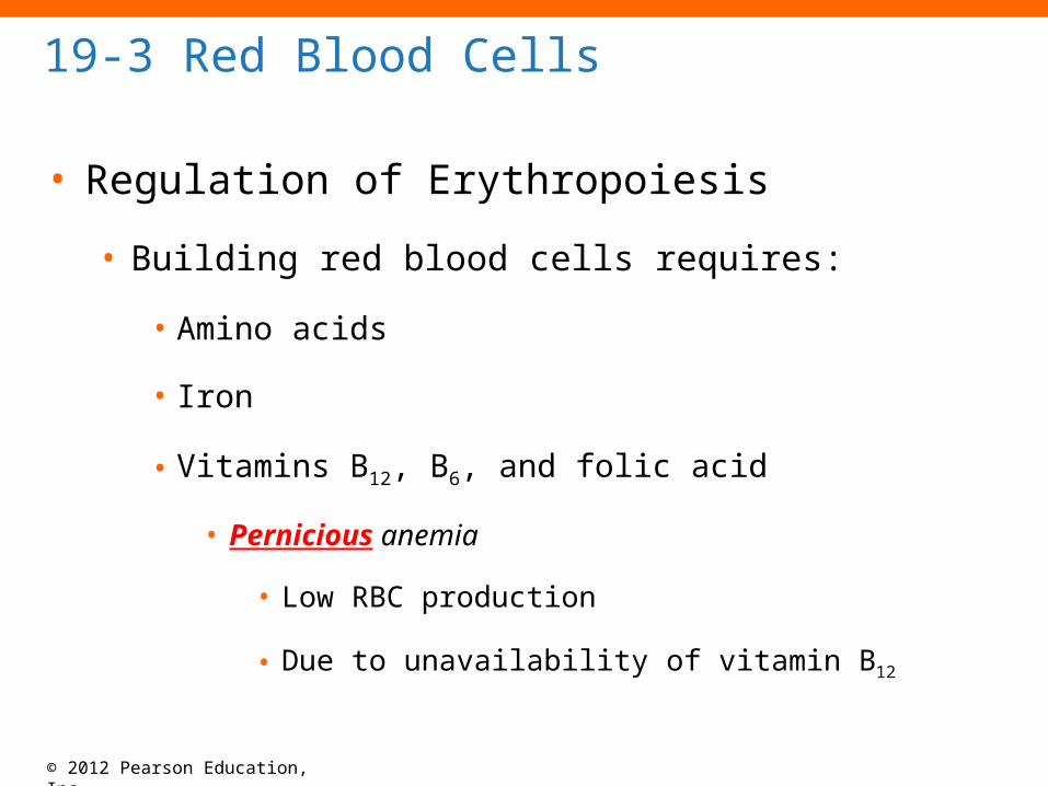
• Regulation of Erythropoiesis
• Building red blood cells requires:
• Amino acids
• Iron
• Vitamins B12, B6, and folic acid
• Pernicious anemia
• Low RBC production
• Due to unavailability of vitamin B12

© 2012 Pearson Education, Inc.
19-3 Red Blood Cells
• Stimulating Hormones
• Erythropoietin (EPO)
• Also called erythropoiesis-stimulating hormone
• Secreted when oxygen in peripheral tissues is low
(hypoxia)
• Due to disease or high altitude

© 2012 Pearson Education, Inc.
Table 19-1 RBC Tests and Related Terminology
Polycythemia: Proportion of blood volume occupied by RBCs increases.Leukocytosis: High leukocyte count normally due to an inflammatory response.Reticulocytosis: Increase in immature RBCs. Common in anemia when the BM is trying to replace RBCs.Macrocytosis: Not a specific disease, but may indicate an underlying problem.Microcytosis: Usually due to iron deficiency, thalassemia, or autoimmune.Hyperchromic: RBC count is low, but cells are larger.
© 2012 Pearson Education, Inc.
If you were to vacation in the Alps, you would expect __.
A.a drop in oxygen levels.B.the release of erythropoietin.C.a rise in hematocrit.D.an increase in RBC production.E.All of the answers are correct.

© 2012 Pearson Education, Inc.
19-4 Blood Typing
• Surface Antigens
• Are cell surface proteins that identify cells to immune
system
• Normal cells are ignored and foreign cells attacked
• Blood Types
• Are genetically determined
• By presence or absence of RBC surface antigens A, B,
Rh (or D)
• (the ABO and Rh blood groups = blood type)

© 2012 Pearson Education, Inc.
19-4 Blood Typing
• Four Basic Blood Types
1. A (surface antigen A)
2. B (surface antigen B)
3. AB (antigens A and B)
4. O (neither A nor B)

© 2012 Pearson Education, Inc.
19-4 Blood Typing
• Blood Plasma Antibodies
• Type A
• Type B antibodies
• Type B
• Type A antibodies
• Type O
• Both A and B antibodies
• Type AB
• Neither A nor B antibodies

© 2012 Pearson Education, Inc.
Figure 19-7a Blood Types and Cross-Reactions
© 2012 Pearson Education, Inc.
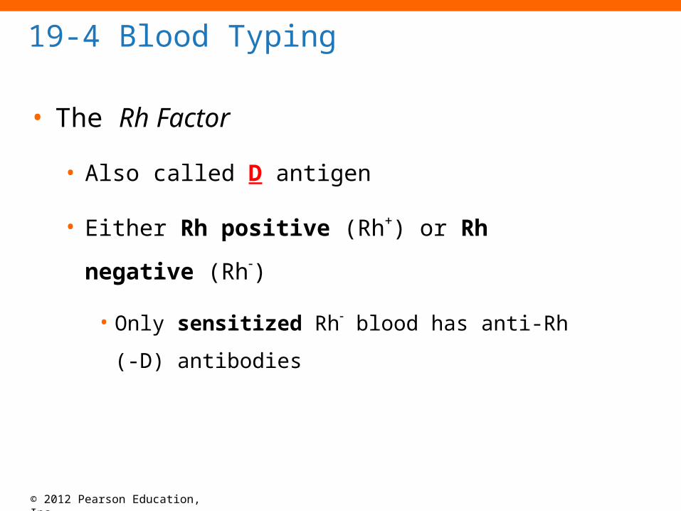
19-4 Blood Typing
• The Rh Factor
• Also called D antigen
• Either Rh positive (Rh) or Rh negative (Rh)
• Only sensitized Rh blood has anti-Rh (-D)
antibodies

© 2012 Pearson Education, Inc.
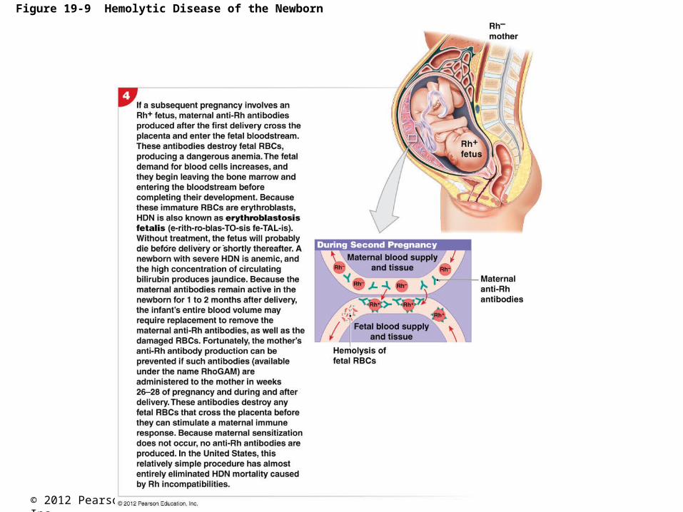
Figure 19-9 Hemolytic Disease of the Newborn

© 2012 Pearson Education, Inc.
Figure 19-9 Hemolytic Disease of the Newborn

© 2012 Pearson Education, Inc.
Figure 19-9 Hemolytic Disease of the Newborn
© 2012 Pearson Education, Inc.
Figure 19-9 Hemolytic Disease of the Newborn
© 2012 Pearson Education, Inc.
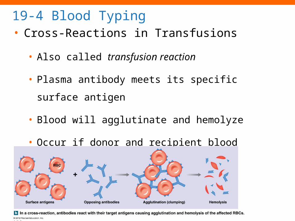
19-4 Blood Typing• Cross-Reactions in Transfusions
• Also called transfusion reaction
• Plasma antibody meets its specific surface antigen
• Blood will agglutinate and hemolyze
• Occur if donor and recipient blood types not
compatible

© 2012 Pearson Education, Inc.
19-4 Blood Typing
• Testing for Transfusion Compatibility
• Performed on donor and recipient blood for
compatibility
• Without cross-match, type O is universal donor

© 2012 Pearson Education, Inc.
Table 19-2 Differences in Blood Group Distribution

© 2012 Pearson Education, Inc.
19-5 White Blood Cells
• White Blood Cells (WBCs)
• Also called leukocytes
• Do not have hemoglobin
• Have nuclei and other organelles
• WBC functions:
• Defend against pathogens
• Remove toxins and wastes
• Attack abnormal cells
© 2012 Pearson Education, Inc.
19-5 White Blood Cells
• WBC Circulation and Movement
• Most WBCs in:
• Connective tissue proper
• Lymphatic system organs
• Small numbers in blood
• 5000 to 10,000 per microliter

© 2012 Pearson Education, Inc.
19-5 White Blood Cells
• WBC Circulation and Movement
• Four Characteristics of Circulating WBCs
1. Can migrate out of bloodstream
2. Have amoeboid movement
3. Attracted to chemical stimuli (positive chemotaxis)
4. Some are phagocytic
• Neutrophils, eosinophils, and monocytes
© 2012 Pearson Education, Inc.
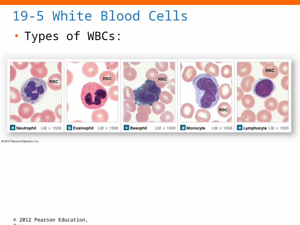
19-5 White Blood Cells
• Types of WBCs:

© 2012 Pearson Education, Inc.
19-5 White Blood Cells
• Neutrophils
• Also called polymorphonuclear
leukocytes
• 50–70% of circulating WBCs
• Pale cytoplasm granules with:
• Lysosomal enzymes
• Bactericides (hydrogen peroxide and
superoxide)
© 2012 Pearson Education, Inc.
19-5 White Blood Cells• Neutrophil Action
• Very active, first to attack bacteria
• Engulf and digest pathogens
• Degranulation
• Removing granules from cytoplasm
• Defensins (peptides from lysosomes) attack pathogen
membranes
• Release prostaglandins and leukotrienes
• Live about 10 hours (30 min when engulfing debris).
• Form pus

© 2012 Pearson Education, Inc.
19-5 White Blood Cells
• Eosinophils (Acidophils)
• 2–4% of circulating WBCs
• Attack large parasites
• Excrete toxic compounds
• Nitric oxide
• Cytotoxic enzymes
• Increase in response to allergens
• Control inflammation with enzymes that counteract inflammatory effects of neutrophils and mast cells
• Destroy antibody-labeled antigens.

© 2012 Pearson Education, Inc.
19-5 White Blood Cells
• Basophils
• Are less than 1% of circulating
WBCs
• Accumulate in damaged tissue
• Release histamine
• Dilates blood vessels
• Release heparin
• Prevents blood clotting
© 2012 Pearson Education, Inc.
19-5 White Blood Cells
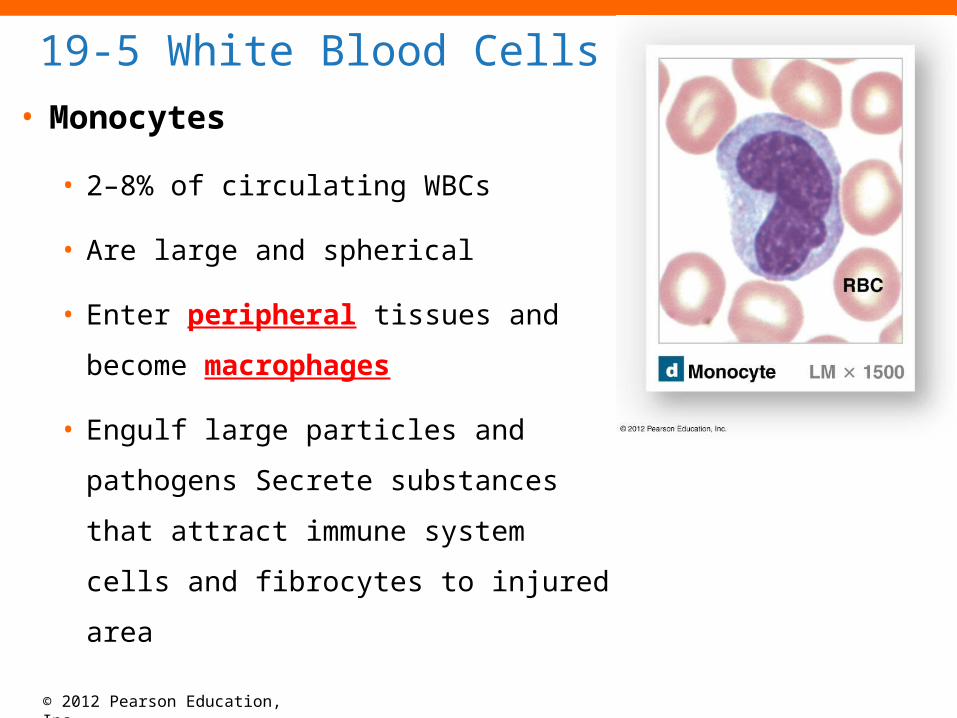
• Monocytes
• 2–8% of circulating WBCs
• Are large and spherical
• Enter peripheral tissues and
become macrophages
• Engulf large particles and pathogens
Secrete substances that attract
immune system cells and fibrocytes
to injured area
© 2012 Pearson Education, Inc.
19-5 White Blood Cells
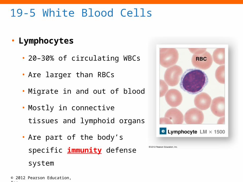
• Lymphocytes
• 20–30% of circulating WBCs
• Are larger than RBCs
• Migrate in and out of blood
• Mostly in connective tissues
and lymphoid organs
• Are part of the body’s specific
immunity defense system

© 2012 Pearson Education, Inc.
19-5 White Blood Cells
• Three Classes of Lymphocytes
1. T cells
• Cell-mediated immunity
• Attack foreign cells directly

© 2012 Pearson Education, Inc.
19-5 White Blood Cells
• Three Classes of Lymphocytes
2. B cells
• Humoral immunity
• Differentiate into plasma cells
• Synthesize antibodies
3. Natural killer (NK) cells
• Detect and destroy abnormal tissue cells (cancers)

© 2012 Pearson Education, Inc.
19-5 White Blood Cells
• The Differential Count and Changes in WBC
Profiles
• Detects changes in WBC populations
• Infections, inflammation, and allergic reactions

© 2012 Pearson Education, Inc.
19-5 White Blood Cells
• WBC Disorders
• Leukopenia
• Abnormally low WBC count
• Leukocytosis
• Abnormally high WBC count
• Leukemia
• Extremely high WBC count
© 2012 Pearson Education, Inc.
19-5 White Blood Cells
• WBC Production
• All blood cells originate from hemocytoblasts
• Which produce progenitor cells called myeloid
stem cells and lymphoid stem cells

© 2012 Pearson Education, Inc.
19-5 White Blood Cells
• WBC Production
• Myeloid Stem Cells
• Produce all WBCs except lymphocytes
• Lymphoid Stem Cells
• Lymphopoiesis - the production of lymphocytes
© 2012 Pearson Education, Inc.
19-5 White Blood Cells
• WBC Development
• WBCs, except monocytes
• Develop in bone marrow
• Monocytes
• Develop into macrophages in peripheral tissues

© 2012 Pearson Education, Inc.
19-5 White Blood Cells
• Regulation of WBC Production
• Colony-stimulating factors (CSFs)
• Hormones that regulate blood cell populations
1. M-CSF stimulates monocyte production
2. G-CSF (Neupogen) stimulates production of granulocytes (neutrophils, eosinophils, and basophils)
3. GM-CSF stimulates granulocyte and monocyte production
4. Multi-CSF accelerates production of granulocytes, monocytes, platelets, and RBCs

© 2012 Pearson Education, Inc.
Table 19-3 Formed Elements of the Blood
© 2012 Pearson Education, Inc.
Figure 19-11 The Origins and Differentiation of Formed Elements

© 2012 Pearson Education, Inc.
All of the following are true of neutrophils, except that they are _____.
A.granular leukocytes.B.phagocytic.C.also known as polymorphonuclear leukocytes.D.important in coagulation.E.active in fighting bacterial infections

© 2012 Pearson Education, Inc.
19-6 Platelets
• Platelets
• Cell fragments involved in human clotting system
• Nonmammalian vertebrates have thrombocytes
(nucleated cells)
• Circulate for 9–12 days
• Are removed by spleen
• 2/3 are reserved for emergencies in the spleen

© 2012 Pearson Education, Inc.
19-6 Platelets
• Platelet Counts
• 150,000 to 500,000 per microliter
• Thrombocytopenia
• Abnormally low platelet count
• Thrombocytosis
• Abnormally high platelet count

© 2012 Pearson Education, Inc.
19-6 Platelets
• Three Functions of Platelets
1. Release important clotting chemicals
2. Temporarily patch damaged vessel walls
3. Reduce size of a break in vessel wall

© 2012 Pearson Education, Inc.
19-6 Platelets
• Platelet Production
• Also called thrombocytopoiesis
• Occurs in bone marrow
• Megakaryocytes
• Giant cells in bone marrow
• Manufacture platelets from cytoplasm

© 2012 Pearson Education, Inc.
19-6 Platelets
• Platelet Production
• Hormonal controls
1. Thrombopoietin (TPO)
2. Interleukin-6 (IL-6)
3. Multi-CSF
© 2012 Pearson Education, Inc.
19-7 Hemostasis
• Hemostasis
• Is the cessation of bleeding
• Consists of three phases
1. Vascular phase
2. Platelet phase
3. Coagulation phase
© 2012 Pearson Education, Inc.
19-7 Hemostasis
• The Vascular Phase
• A cut triggers vascular spasm that lasts 30 minutes
• Three Steps of the Vascular Phase
1. Endothelial cells contract and expose basement
membrane to bloodstream

© 2012 Pearson Education, Inc.
19-7 Hemostasis
• Three Steps of the Vascular Phase
2. Endothelial cells
• Release chemical factors ADP, tissue factor, and
prostacyclin
• Release local hormones, endothelins
• Stimulate smooth muscle contraction and cell division
3. Endothelial plasma membranes become “sticky”
• Seal off blood flow

© 2012 Pearson Education, Inc.
Figure 19-12 The Vascular, Platelet, and Coagulation Phases of Hemostasis and Clot Retraction (Step 1)

© 2012 Pearson Education, Inc.
19-7 Hemostasis
• The Platelet Phase
• Begins within 15 seconds after injury
• Platelet adhesion (attachment)
• To sticky endothelial surfaces
• To basement membranes
• To exposed collagen fibers beneath endothelium
• Platelet aggregation (stick together)
• Forms platelet plug which closes small breaks

© 2012 Pearson Education, Inc.
19-7 Hemostasis
• Platelet Phase
• Activated platelets release clotting compounds
1. Adenosine diphosphate (ADP)
2. Thromboxane A2 and serotonin
3. Clotting factors
4. Platelet-derived growth factor (PDGF)
5. Calcium ions

© 2012 Pearson Education, Inc.
19-7 Hemostasis
• Factors That Limit the Growth of the Platelet Plug
1. Prostacyclin, released by endothelial cells, inhibits
platelet aggregation
2. Inhibitory compounds released by other WBCs
3. Circulating enzymes break down ADP
4. Negative (inhibitory) feedback from serotonin
5. Development of blood clot isolates area

© 2012 Pearson Education, Inc.
Figure 19-12 The Vascular, Platelet, and Coagulation Phases of Hemostasis and Clot Retraction (Step 2)
© 2012 Pearson Education, Inc.
19-7 Hemostasis
• The Coagulation Phase
• Begins 30 seconds or more after the injury
• Blood clotting (coagulation)
• Cascade reactions
• Chain reactions of enzymes and proenzymes
• Form three pathways
• Convert circulating fibrinogen into insoluble fibrin

© 2012 Pearson Education, Inc.
19-7 Hemostasis
• Clotting Factors
• Also called procoagulants
• Proteins or ions in plasma
• Required for normal clotting
• Most protein factor required for clotting are
synthesized by the liver.

© 2012 Pearson Education, Inc.
Table 19-4 Clotting Factors

© 2012 Pearson Education, Inc.
19-7 Hemostasis
• Three Coagulation Pathways
1. Extrinsic pathway
2. Intrinsic pathway
3. Common pathway

© 2012 Pearson Education, Inc.
19-7 Hemostasis
• The Extrinsic Pathway
• Begins in the vessel wall
• Outside bloodstream
• Damaged endothelium releases tissue factor
(Factor III)
• TF + other compounds = enzyme complex
• Activates Factor X

© 2012 Pearson Education, Inc.
19-7 Hemostasis
• The Intrinsic Pathway
• Begins with circulating proenzymes
• Within bloodstream
• Activation of Factor XII exposed to collagen
• Platelets release factors (e.g., PF-3)
• Series of reactions activates Factor X

© 2012 Pearson Education, Inc.
19-7 Hemostasis
• The Common Pathway
• Where intrinsic and extrinsic pathways converge
• Conversion of Factor X to prothrombinase
• Converts prothrombin to thrombin
• Thrombin converts fibrinogen to fibrin

© 2012 Pearson Education, Inc.
Figure 19-12 The Vascular, Platelet, and Coagulation Phases of Hemostasis and Clot Retraction (Step 3)

© 2012 Pearson Education, Inc.
19-7 Hemostasis
• Feedback Control of Blood Clotting
1. Stimulates formation of tissue factor
2. Stimulates release of PF-3
• Forms positive feedback loop (intrinsic and
extrinsic)
• Accelerates clotting

© 2012 Pearson Education, Inc.
19-7 Hemostasis
• Feedback Control of Blood Clotting
• Anticoagulants (plasma proteins)
• Antithrombin-III
• Alpha-2-macroglobulin
• Heparin
• Aspirin
• Protein C (activated by thrombomodulin)
• Prostacyclin

© 2012 Pearson Education, Inc.
19-7 Hemostasis
• Calcium Ions, Vitamin K, and Blood Clotting
• Calcium ions (Ca2+) and vitamin K are both
essential to the clotting process

© 2012 Pearson Education, Inc.
19-7 Hemostasis
• Clot Retraction
1. Pulls torn edges of vessel closer
together
• Reducing residual bleeding and
stabilizing injury site
2. Reduces size of damaged area
• Making it easier for fibrocytes,
smooth muscle cells, and
endothelial cells to complete
repairs

© 2012 Pearson Education, Inc.
19-7 Hemostasis
• Fibrinolysis
• Slow process of dissolving clot
• Thrombin and tissue plasminogen activator (t-PA)
• Activate plasminogen
• Plasminogen produces plasmin
• Digests fibrin strands

© 2012 Pearson Education, Inc.
A digestive disorder that impairs a person’s ability to absorb vitamin K will result in _____.
A.low levels of prothrombinB.low levels of Factor XC.low levels of thromboplastinD.prolonged bleedingE.All of the above