Embed Size (px)
Citation preview

ELAINE N. MARIEB
EIGHTH EDITION
10
Copyright © 2006 Pearson Education, Inc., publishing as Benjamin Cummings
PowerPoint® Lecture Slide Presentation by Jerry L. Cook, Sam Houston University
ESSENTIALSOF HUMANANATOMY
& PHYSIOLOGY
Blood
Copyright © 2006 Pearson Education, Inc., publishing as Benjamin Cummings
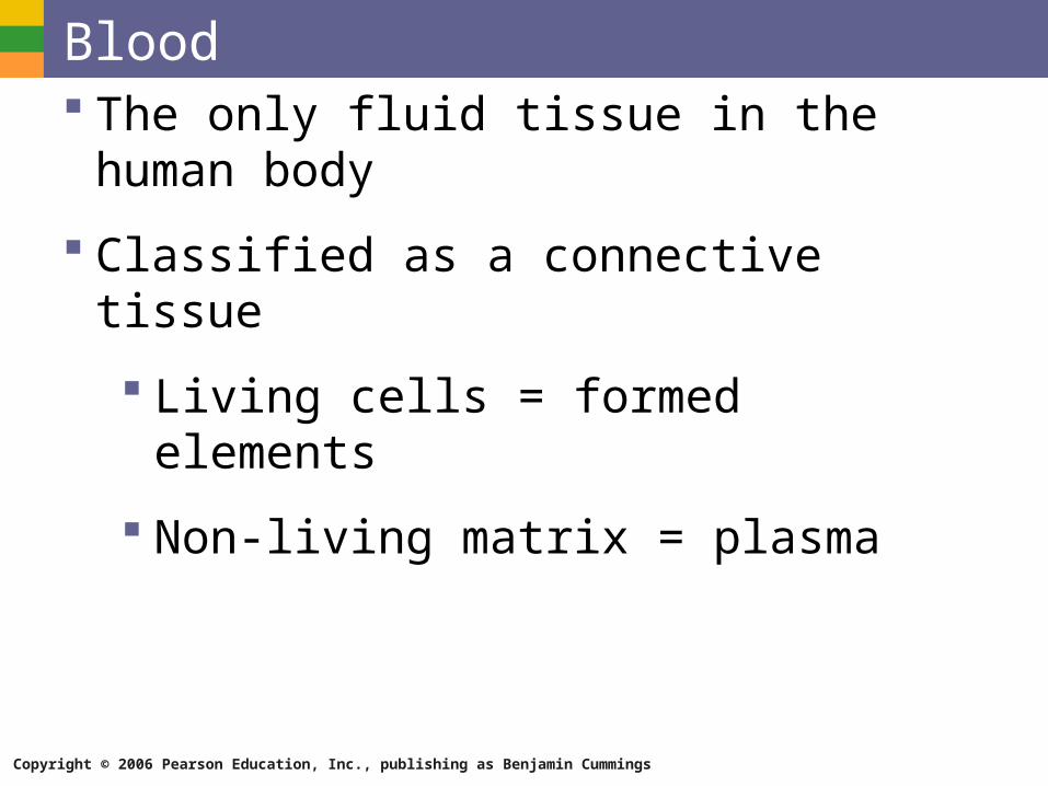
Blood The only fluid tissue in the human body
Classified as a connective tissue
Living cells = formed elements
Non-living matrix = plasma

Copyright © 2006 Pearson Education, Inc., publishing as Benjamin Cummings
Blood
Figure 10.1
Copyright © 2006 Pearson Education, Inc., publishing as Benjamin Cummings
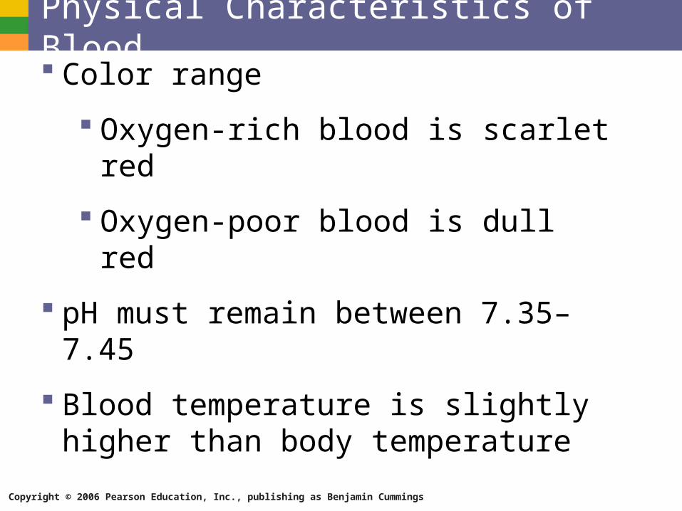
Physical Characteristics of Blood Color range
Oxygen-rich blood is scarlet red
Oxygen-poor blood is dull red
pH must remain between 7.35–7.45
Blood temperature is slightly higher than body temperature
Copyright © 2006 Pearson Education, Inc., publishing as Benjamin Cummings
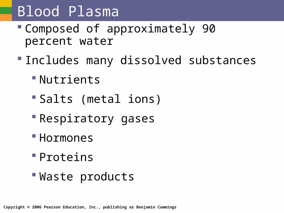
Blood Plasma Composed of approximately 90 percent water
Includes many dissolved substances
Nutrients
Salts (metal ions)
Respiratory gases
Hormones
Proteins
Waste products
Copyright © 2006 Pearson Education, Inc., publishing as Benjamin Cummings
Plasma Proteins Albumin – regulates osmotic pressure
Clotting proteins – help to stem blood loss when a blood vessel is injured
Antibodies – help protect the body from antigens
Copyright © 2006 Pearson Education, Inc., publishing as Benjamin Cummings
Formed Elements Erythrocytes = red blood cells
Leukocytes = white blood cells
Platelets = cell fragments
Copyright © 2006 Pearson Education, Inc., publishing as Benjamin Cummings
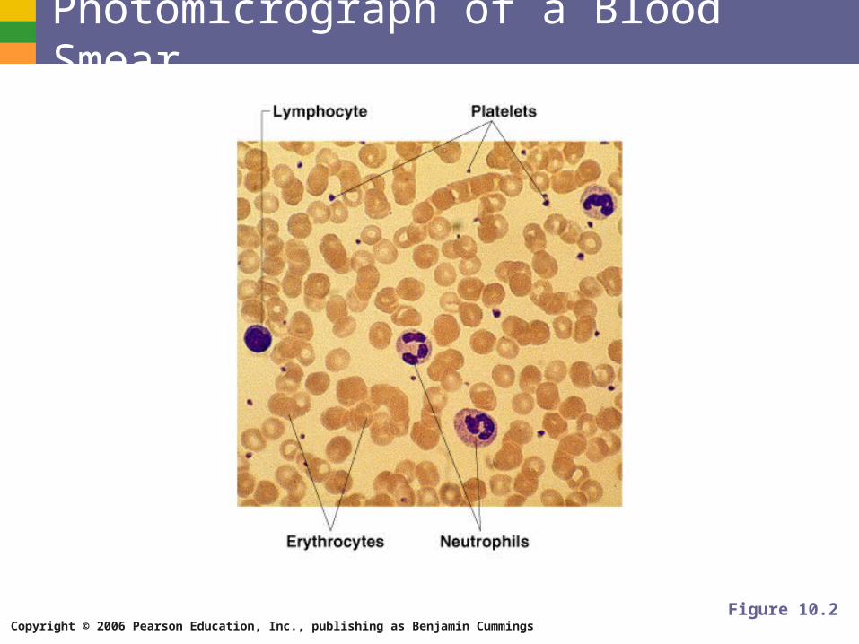
Photomicrograph of a Blood Smear
Figure 10.2
Copyright © 2006 Pearson Education, Inc., publishing as Benjamin CummingsTable 10.2
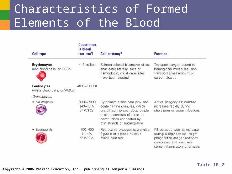
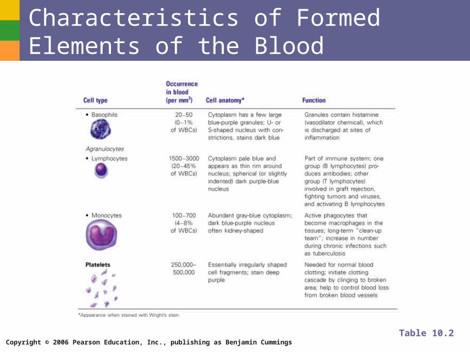
Characteristics of Formed Elements of the Blood
Copyright © 2006 Pearson Education, Inc., publishing as Benjamin Cummings
Characteristics of Formed Elements of the Blood
Table 10.2
Copyright © 2006 Pearson Education, Inc., publishing as Benjamin Cummings
Erythrocytes (Red Blood Cells) The main function is to carry oxygen
Anatomy of circulating erythrocytes
Biconcave disks
Essentially bags of hemoglobin
Anucleate (no nucleus)
Contain very few organelles
Outnumber white blood cells 1000:1

Copyright © 2006 Pearson Education, Inc., publishing as Benjamin Cummings
Hemoglobin Iron-containing protein
Binds strongly, but reversibly, to oxygen
Each hemoglobin molecule has four oxygen binding sites
Each erythrocyte has 250 million hemoglobin molecules
Copyright © 2006 Pearson Education, Inc., publishing as Benjamin Cummings
Leukocytes (White Blood Cells) Crucial in the body’s defense against disease
These are complete cells, with a nucleus and organelles
Able to move into and out of blood vessels (diapedesis)
Can move by ameboid motion
Can respond to chemicals released by damaged tissues
Copyright © 2006 Pearson Education, Inc., publishing as Benjamin Cummings
Leukocyte Levels in the Blood Normal levels are between 4,000 and 11,000 cells
per millimeter
Abnormal leukocyte levels
Leukocytosis
Above 11,000 leukocytes/ml
Generally indicates an infection
Leukopenia
Abnormally low leukocyte level
Commonly caused by certain drugs
Copyright © 2006 Pearson Education, Inc., publishing as Benjamin Cummings
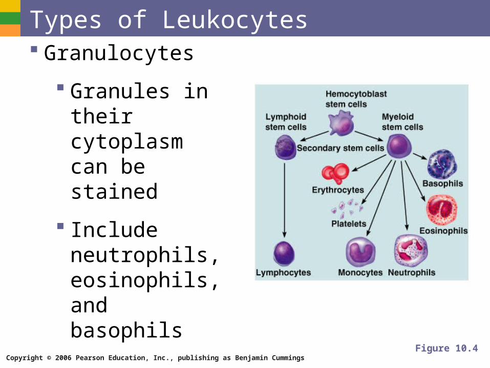
Types of Leukocytes Granulocytes
Granules in their cytoplasm can be stained
Include neutrophils, eosinophils, and basophils
Figure 10.4
Copyright © 2006 Pearson Education, Inc., publishing as Benjamin Cummings
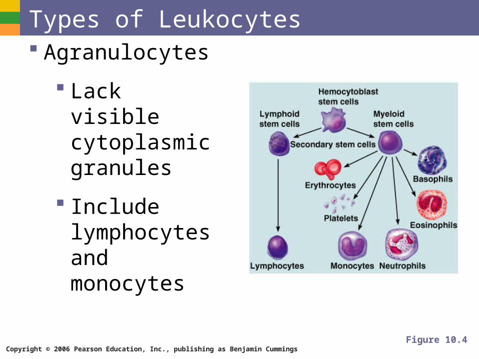
Types of Leukocytes Agranulocytes
Lack visible cytoplasmic granules
Include lymphocytes and monocytes
Figure 10.4
Copyright © 2006 Pearson Education, Inc., publishing as Benjamin Cummings
Granulocytes Neutrophils
Multilobed nucleus with fine granules
Act as phagocytes at active sites of infection
Eosinophils
Large brick-red cytoplasmic granules
Found in repsonse to allergies and parasitic worms
Copyright © 2006 Pearson Education, Inc., publishing as Benjamin Cummings
Granulocytes Basophils
Have histamine-containing granules
Initiate inflammation
Copyright © 2006 Pearson Education, Inc., publishing as Benjamin Cummings
Agranulocytes Lymphocytes
Nucleus fills most of the cell
Play an important role in the immune response
Monocytes
Largest of the white blood cells
Function as macrophages
Important in fighting chronic infection
Copyright © 2006 Pearson Education, Inc., publishing as Benjamin Cummings
Platelets Derived from ruptured multinucleate cells
(megakaryocytes)
Needed for the clotting process
Normal platelet count = 300,000/mm3
Copyright © 2006 Pearson Education, Inc., publishing as Benjamin Cummings
Hematopoiesis Blood cell formation
Occurs in red bone marrow
All blood cells are derived from a common stem cell (hemocytoblast)
Hemocytoblast differentiation
Lymphoid stem cell produces lymphocytes
Myeloid stem cell produces other formed elements
Copyright © 2006 Pearson Education, Inc., publishing as Benjamin Cummings
Fate of Erythrocytes Unable to divide, grow, or synthesize proteins
Wear out in 100 to 120 days
When worn out, are eliminated by phagocytes in the spleen or liver
Lost cells are replaced by division of hemocytoblasts
Copyright © 2006 Pearson Education, Inc., publishing as Benjamin Cummings
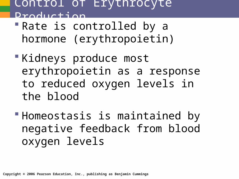
Control of Erythrocyte Production Rate is controlled by a hormone
(erythropoietin)
Kidneys produce most erythropoietin as a response to reduced oxygen levels in the blood
Homeostasis is maintained by negative feedback from blood oxygen levels
Copyright © 2006 Pearson Education, Inc., publishing as Benjamin Cummings
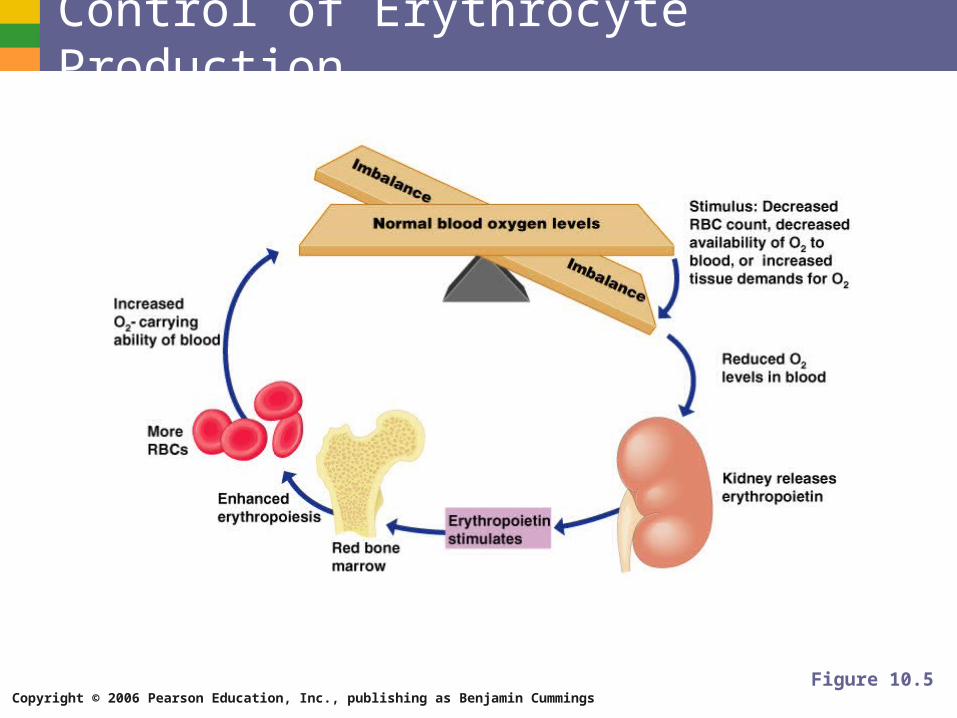
Control of Erythrocyte Production
Figure 10.5
Copyright © 2006 Pearson Education, Inc., publishing as Benjamin Cummings
Hemostasis Stoppage of blood flow
Result of a break in a blood vessel
Hemostasis involves three phases
Platelet plug formation
Vascular spasms
Coagulation
Copyright © 2006 Pearson Education, Inc., publishing as Benjamin Cummings
Platelet Plug Formation Collagen fibers are exposed by a break in a
blood vessel
Platelets become “sticky” and cling to fibers
Anchored platelets release chemicals to attract more platelets
Platelets pile up to form a platelet plug
Copyright © 2006 Pearson Education, Inc., publishing as Benjamin Cummings
Vascular Spasms Anchored platelets release serotonin
Serotonin causes blood vessel muscles to spasm
Spasms narrow the blood vessel, decreasing blood loss
Copyright © 2006 Pearson Education, Inc., publishing as Benjamin Cummings
Coagulation Injured tissues release thromboplastin
PF3 (a phospholipid) interacts with thromboplastin, blood protein clotting factors, and calcium ions to trigger a clotting cascade
Prothrombin activator converts prothrombin to thrombin (an enzyme)
Copyright © 2006 Pearson Education, Inc., publishing as Benjamin Cummings
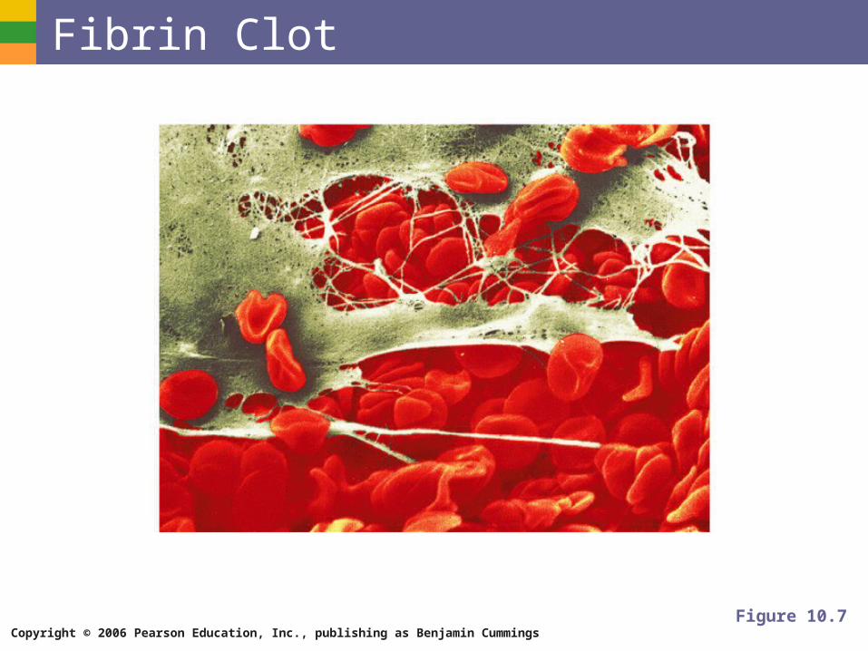
Coagulation Thrombin joins fibrinogen proteins into hair-
like fibrin
Fibrin forms a meshwork (the basis for a clot)
Copyright © 2006 Pearson Education, Inc., publishing as Benjamin Cummings
Blood Clotting Blood usually clots within 3 to 6 minutes
The clot remains as endothelium regenerates
The clot is broken down after tissue repair
Copyright © 2006 Pearson Education, Inc., publishing as Benjamin Cummings
Fibrin Clot
Figure 10.7
Copyright © 2006 Pearson Education, Inc., publishing as Benjamin Cummings
Undesirable Clotting Thrombus
A clot in an unbroken blood vessel
Can be deadly in areas like the heart
Embolus
A thrombus that breaks away and floats freely in the bloodstream
Can later clog vessels in critical areas such as the brain
Copyright © 2006 Pearson Education, Inc., publishing as Benjamin Cummings
Bleeding Disorders Thrombocytopenia
Platelet deficiency
Even normal movements can cause bleeding from small blood vessels that require platelets for clotting
Hemophilia
Hereditary bleeding disorder
Normal clotting factors are missing
Copyright © 2006 Pearson Education, Inc., publishing as Benjamin Cummings
Blood Groups and Transfusions Large losses of blood have serious
consequences
Loss of 15 to 30 percent causes weakness
Loss of over 30 percent causes shock, which can be fatal
Transfusions are the only way to replace blood quickly
Transfused blood must be of the same blood group
Copyright © 2006 Pearson Education, Inc., publishing as Benjamin Cummings
Human Blood Groups Blood contains genetically determined
proteins
A foreign protein (antigen) may be attacked by the immune system
Blood is “typed” by using antibodies that will cause blood with certain proteins to clump (agglutination)
Copyright © 2006 Pearson Education, Inc., publishing as Benjamin Cummings
Human Blood Groups There are over 30 common red blood cell
antigens
The most vigorous transfusion reactions are caused by ABO and Rh blood group antigens
Copyright © 2006 Pearson Education, Inc., publishing as Benjamin Cummings
ABO Blood Groups Based on the presence or absence of two
antigens
Type A
Type B
The lack of these antigens is called type O
Copyright © 2006 Pearson Education, Inc., publishing as Benjamin Cummings
ABO Blood Groups The presence of both A and B is called type
AB
The presence of either A or B is called types A and B, respectively
Copyright © 2006 Pearson Education, Inc., publishing as Benjamin Cummings
Rh Blood Groups Named because of the presence or absence of
one of eight Rh antigens (agglutinogen D)
Most Americans are Rh+
Problems can occur in mixing Rh+ blood into a body with Rh– blood
Copyright © 2006 Pearson Education, Inc., publishing as Benjamin Cummings
Rh Dangers During Pregnancy Danger is only when the mother is Rh– and
the father is Rh+, and the child inherits the Rh+ factor
Copyright © 2006 Pearson Education, Inc., publishing as Benjamin Cummings
Rh Dangers During Pregnancy The mismatch of an Rh– mother carrying an Rh+
baby can cause problems for the unborn child
The first pregnancy usually proceeds without problems
The immune system is sensitized after the first pregnancy
In a second pregnancy, the mother’s immune system produces antibodies to attack the Rh+ blood (hemolytic disease of the newborn)
Copyright © 2006 Pearson Education, Inc., publishing as Benjamin Cummings
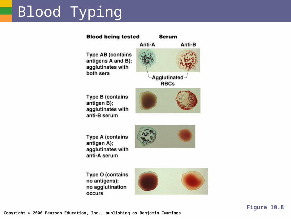
Blood Typing Blood samples are mixed with anti-A and
anti-B serum
Coagulation or no coagulation leads to determining blood type
Typing for ABO and Rh factors is done in the same manner
Cross matching – testing for agglutination of donor RBCs by the recipient’s serum, and vice versa
Copyright © 2006 Pearson Education, Inc., publishing as Benjamin Cummings
Blood Typing
Figure 10.8
Copyright © 2006 Pearson Education, Inc., publishing as Benjamin Cummings
Developmental Aspects of Blood Sites of blood cell formation
The fetal liver and spleen are early sites of blood cell formation
Bone marrow takes over hematopoiesis by the seventh month
Fetal hemoglobin differs from hemoglobin produced after birth