Embed Size (px)
Citation preview
M.Sc. II BiotechnologyAnimal Biotechnology
Dariyus Z Kabraji
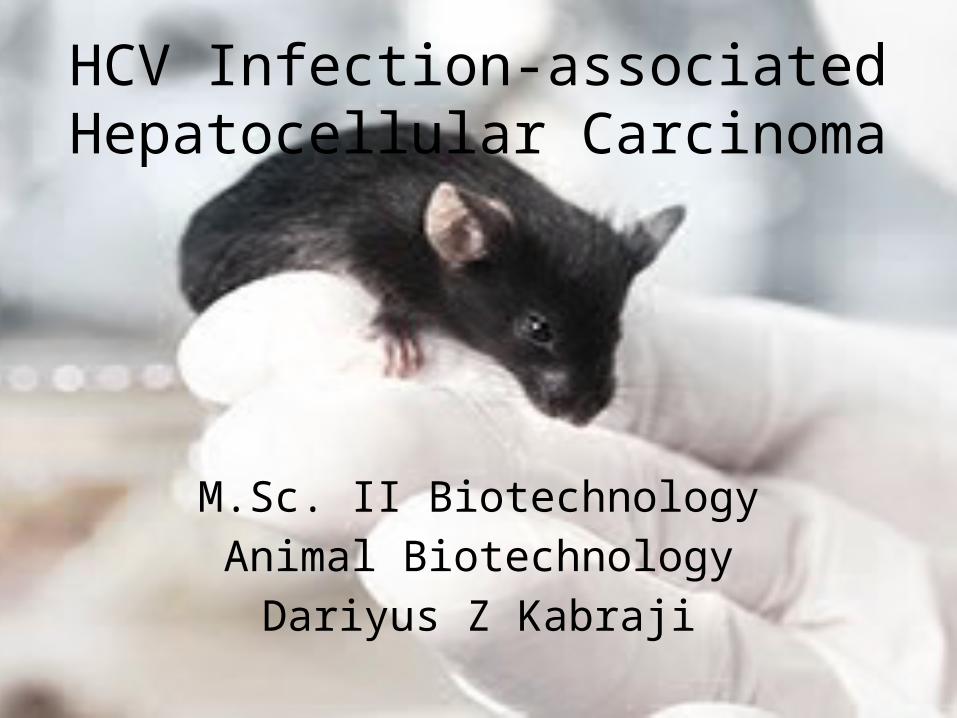
HCV Infection-associated Hepatocellular Carcinoma
M.Sc. II BiotechnologyAnimal Biotechnology
Dariyus Z Kabraji
Introduction• Hepatocellular carcinoma (HCC) is the fifth most common
cancer worldwide; its incidence is increasing because of the prevalence of chronic hepatitis C and B viral infections
• Hepatitis C virus (HCV) infection is a major risk factor for chronic hepatitis, cirrhosis and hepatocellular carcinoma (HCC)
• It would thus be beneficial to explore molecular changes that underlie HCV infection-associated HCC in a humanized mouse model, in order to identify markers of HCC progression and to gain an understanding of the oncogenic changes that underlie it
Generation Of Humanized Mice
• MUP-uPA mice were crossed with SCID/Beige background Balb/c mice • Transgenic mice offspring were identified by PCR, using forward primers
specific for uPA• MUP-uPA/SCID/Bg mice that had been engrafted with human
hepatocytes were inoculated intravenously (i.v.) through the tail vein with 100 uL of diluted plasma from a HCV-infected chimpanzee
• The resultant HCV infections were detected using immunohistochemistry• Sera from the MUP-uPA/SCID/Bg mice were collected and RNA
prepared for measuring HCV RNA. • HCV genome copy number was quantified by one-step real-time qRT-
PCR reaction using Taqman EZ rTth polymerase kit • Mice were bled every 15 days for 4-8 weeks and HBV DNA extracted
(Qiagen DNA extraction kit) from mouse serum. Briefly, 10 uL of DNA was subjected to HBV-specific TaqMan PCR in a 50-ul reaction mixture to obtain HBV genome equivalent copies
Protocols• Protein extraction, subcellular fractionation and
immunoblotting:Liver tissues used for Western blot analysis were from human hepatocyte-engrafted MUP-uPA/SCID/Bg uninfected control, or HCV-infected but HCC-negative or HCV infection-induced liver tumors (HCC positive) mice.
• Real-time quantitative PCR of RNA samples from chimeric mouse liver:1ug of total RNA was added to QuantiMir RT Kit Small RNA Quantification System (System Biosciences) following the manufacturer’s protocol. The reverse-transcribed product was then diluted 40 fold. Real time quantitative PCR was performed
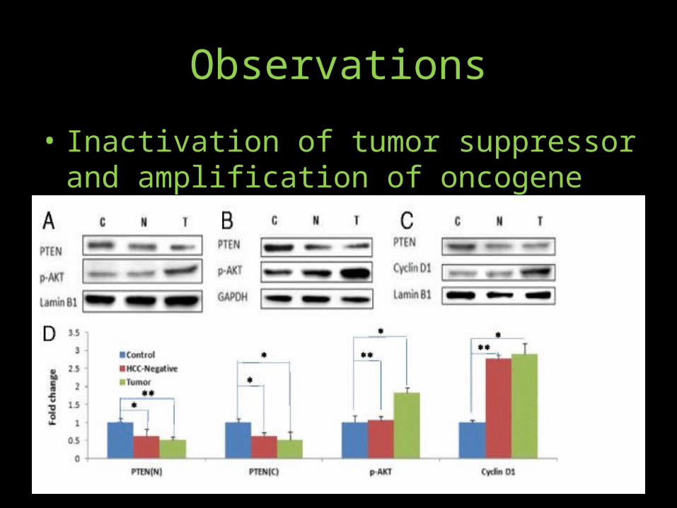
Observations• Inactivation of tumor suppressor and
amplification of oncogene• Induction of c-Myc oncoprotein• Down-regulation of DLC-1 tumor suppressor• P21• Inflammatory response• MicroRNA markers of HCC
Observations• Liver tissues from control animals• HCC-negative mice (engrafted and HCV infected mice that did not develop
HCC), and HCC positive mice (engrafted and HCV infected) were examined by Western blotting
• Panels (a) and (c) show representative Western blots of nuclear protein fraction (with Lamin B1 as loading control); and panel (b) is representative Western blot of corresponding cytoplasmic fraction (with GAPDH as loading control) observed a consistent decline of both nuclear and cytoplasmic PTEN protein in HCV-infected HCC. Interestingly, PTEN protein in HCV-infected but HCC-negative liver (N) also declined to similar extent , suggesting that loss of PTEN may be necessary but insufficient to promote HCC.
• Oncoproteins: Western blots of the control, HCC-negative and liver tumor tissues were probed with antibodies against c-Myc, DLC-1 or p21 proteins (panels a, b and
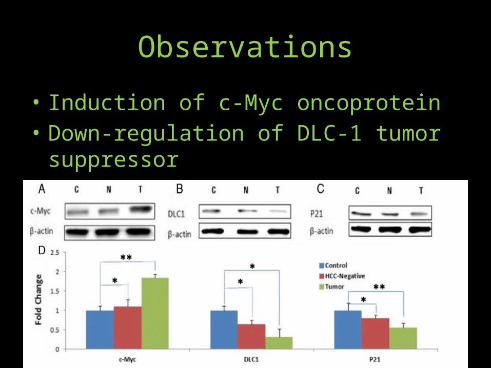
Observations• Induction of c-Myc oncoprotein• Down-regulation of DLC-1 tumor suppressor• P21• Inflammatory response• MicroRNA markers of HCC
Observations• Western blots of the control, HCC-negative and liver
tumor tissues were probed with antibodies against c-Myc, DLC-1 or p21 proteins (panels a, b and b). Panel (d) represents quantitative analysis (based on the loading controls) of liver tissues from uninfected control, HCC negative and HCC positive mice observed increased c-Myc protein levels in HCV-infected liver tumors compared to the control. By contrast, induction of c-Myc in HCC-negative liver was modest, suggesting that induction of c-Myc oncoprotein is a relatively late event in the development of HCV-infection associated HCC.
Observations
• We observed the loss of DLC1 protein in HCV-infected liver tumors and less so in HCV-infected but HCC-negative liver tissue
• it was important to determine if HCV infection of humanized mice modulated p53 to promote HCC. We assessed the modulation of p53 function in HCV-infected chimeric mice on the basis of p21 expression, a direct target of p53 transcriptional regulatory function. We observed a marked decline of p21 protein in HCV-infected liver tumor, and less so in HCC-negative mice, suggesting that HCC progression is correlated with the loss of function of p53 tumor suppressor.
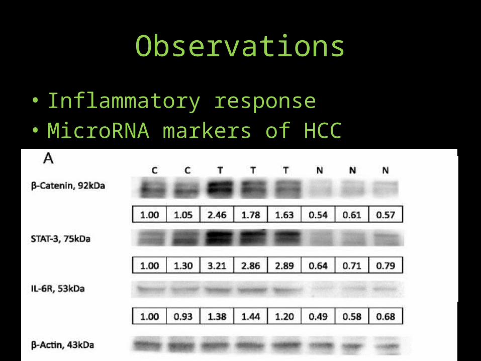
Observations• Persistent viral infection is an underlying cause of inflammation-induced
cancer, including HCC. More than 90 % of HCCs arise in the context of hepatic injury and inflammation. Inflammation-associated oncogenic response is mediated by STAT proteins; in particular, activated STAT3. To ascertain if HCV infection-associated HCC in humanized mice mimics the natural inflammatory response, we assayed activated STAT3 levels in the liver tumors and in HCC-negative as compared to the uninfected control mice. As shown, there is a marked induction of activated (phosphorylated) STAT3 in HCV infection-associated liver tumors as compared to HCV-infected but HCC-negative liver.
• Quantitative assessment of β-Catenin, STAT-3 and IL-6R (from 7 uninfected control, 8 HCC negative and 7 HCC positive mice) was based on B-Actin internal controls analyzed by three independent SDS-PAGE runs
Observations• MicroRNAs can function as tumor suppressors or oncogenes
(oncomiRs). Altered expression levels of miRNAs have been reported in a number of human cancers. In this study we sought to identify miRNAs that would serve as distinguishing markers of HCV infection-associated HCC.
• MicroRNA 141 (miR-141) is induced in HCV-infected human primary hepatocytes. Importantly, miR-141 directly targets DLC1 tumor suppressor protein expression, attesting to its role as bona fide oncomiR. Here we compared expression levels of miR-141 along with other known oncomiRs (miR-21 and miR-221) in HCV infection-induced HCC (Fig. 4). Results suggest that expression of miR-141 and oncomiRs miR-21 and miR-221 that target cell cycle inhibitors [34, 35] is coordinately induced in HCV infection-associated HCC.
Observations• Inflammatory response• MicroRNA markers of HCC
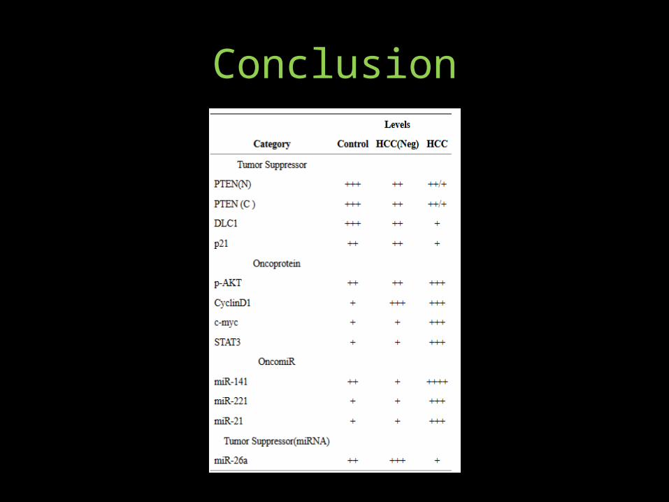
Conclusion• Liver tumor progression requires induction of oncoproteins and
oncomiRs. • HCV utilizes a novel mechanism to induce PTEN insufficiency,
involving viral non-coding RNA directed post-trasncriptional silencing of Transportin-2 that restricts translocation of PTEN protein to the nucleus
• Comparison of PTEN protein levels of HCV-infected but HCC-negative liver with the HCV-infected HCC suggest that loss of PTEN is an early, precancerous event, although PTEN insufficiency by itself does not promote HCC.
• Similarly, loss of DLC-1 tumor suppressor protein appears to be an early indicator of HCV infection-associated HCC. By contrast, induction of oncogenic modulators such as cMyc, miR-21, miR221 and miR-141 appear to be effective in promoting HCC progression.
Conclusion
Conclusion• MicroRNAs (miRNAs) represent a substantial fraction of
tissue-specific small non-protein coding RNA modulators of gene expression.
• MicroRNAs can function as gene silencers by blocking mRNA translation and destabilizing the target mRNA.
• Phenotypic consequences of miRNA-regulated genes evoke essential features of tumor biology, including modulation of apoptosis, cell proliferation, signal transduction and stress response.
• Dysregulated expression of miRNAs has proven valuable in tumor classification and prognosis
References• HCV infection-associated hepatocellular
carcinoma in humanized mice; Zhao Wang, Ningbin Wu, Abeba Tesfaye, Stephen Feinstone, and Ajit Kumar; Infect Agent Cancer. 2015; 10: 24.
• Chimeric Mouse Model for the Infection of Hepatitis B and C Viruses; Abeba Tesfaye, Judith Stift, Dragan Maric, Qingwen Cui, Hans-Peter Dienes, and Stephen M. Feinstone; PLoS One. 2013; 8(10): e77298.