Embed Size (px)
Citation preview

Karnataka Law Society’s
KLS Public School
Science Group
ActivityFA-2
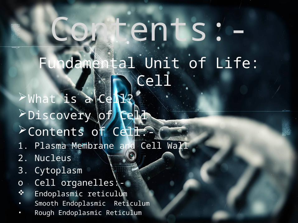
Contents:-Fundamental Unit of Life: Cell
What is a Cell?Discovery of CellContents of Cell:-1. Plasma Membrane and Cell Wall2. Nucleus3. Cytoplasmo Cell organelles:- Endoplasmic reticulum• Smooth Endoplasmic Reticulum• Rough Endoplasmic Reticulum
Ribosomes Golgi Apparatus Lysosomes Mitochondria Plastids (plants Only) Vacuoles Centrioles (animals only)
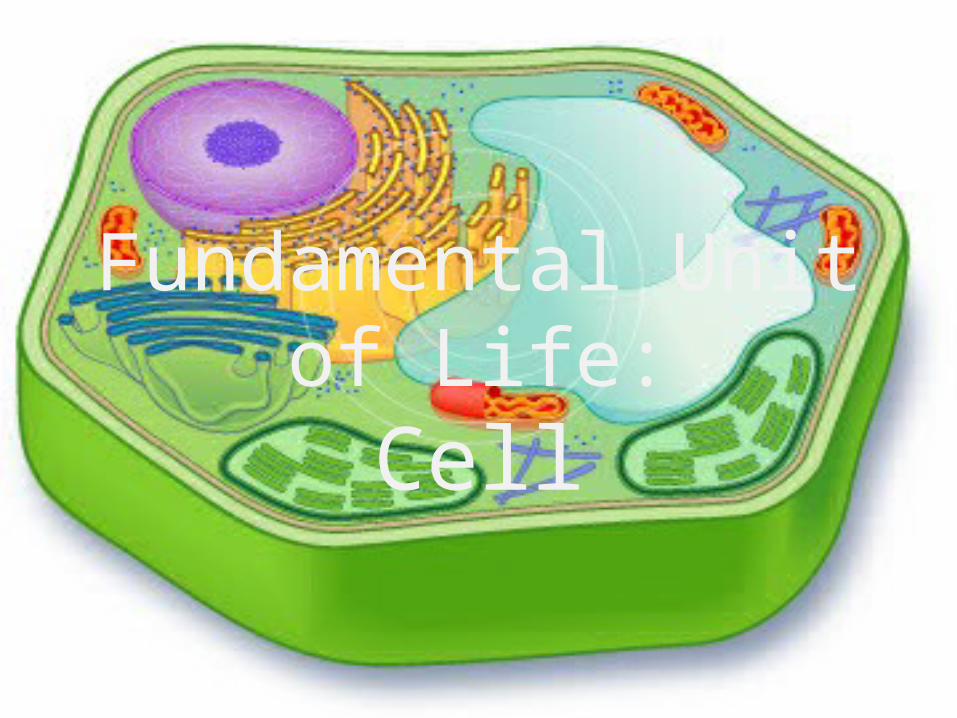
Fundamental Unit of Life:Cell
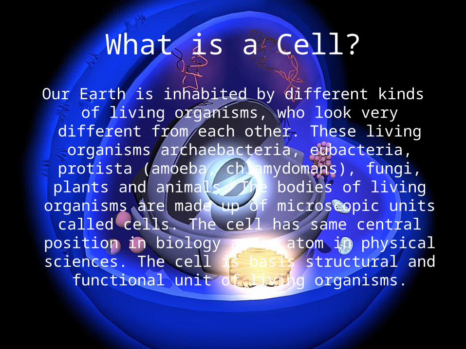
What is a Cell?
Our Earth is inhabited by different kinds of living organisms, who look very different from each other. These living organisms archaebacteria,
eubacteria, protista (amoeba, chlamydomans), fungi, plants and animals. The bodies of living
organisms are made up of microscopic units called cells. The cell has same central position in biology
as an atom in physical sciences. The cell is basis structural and functional unit of living organisms.
Discovery of Cell
While studying a thin slice of cork, Robert Hookesaw that the cork resembled the structure of
honeycomb consisting of many little compartments. Cork is a substance which is obtained from the bark of a tree. This was in the year 1665 when Hooke made this chance discovery through a self designed microscope. Robert Hooke called these boxes-cells. Cell in
Latin means “little room”. Robert Hooke’s discovery was important, because it indicated
for the first time that living organisms consisted of a number of smaller structures or units.
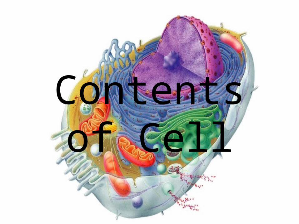
Contents of Cell
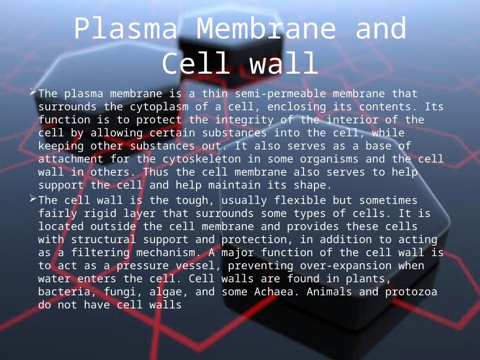
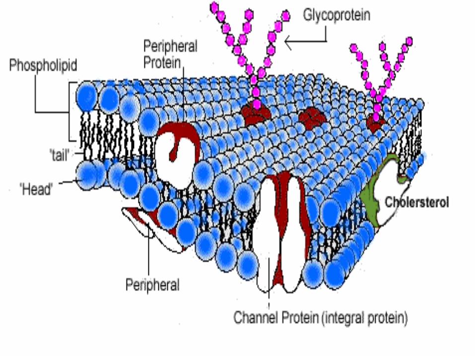
Plasma Membrane and Cell wall The plasma membrane is a thin semi-permeable membrane that
surrounds the cytoplasm of a cell, enclosing its contents. Its function is to protect the integrity of the interior of the cell by allowing certain substances into the cell, while keeping other substances out. It also serves as a base of attachment for the cytoskeleton in some organisms and the cell wall in others. Thus the cell membrane also serves to help support the cell and help maintain its shape.
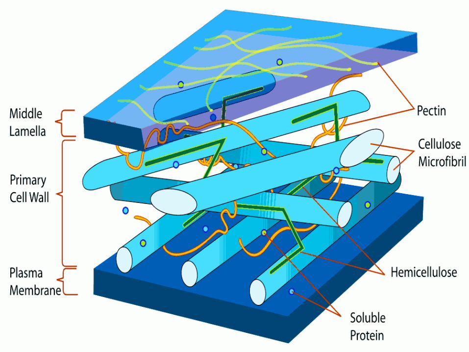
The cell wall is the tough, usually flexible but sometimes fairly rigid layer that surrounds some types of cells. It is located outside the cell membrane and provides these cells with structural support and protection, in addition to acting as a filtering mechanism. A major function of the cell wall is to act as a pressure vessel, preventing over-expansion when water enters the cell. Cell walls are found in plants, bacteria, fungi, algae, and some Achaea. Animals and protozoa do not have cell walls
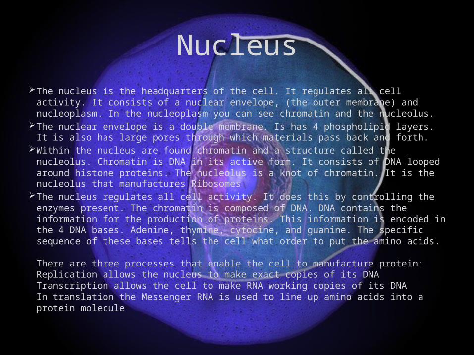
Nucleus The nucleus is the headquarters of the cell. It regulates all cell activity. It consists of a
nuclear envelope, (the outer membrane) and nucleoplasm. In the nucleoplasm you can see chromatin and the nucleolus.
The nuclear envelope is a double membrane. Is has 4 phospholipid layers. It is also has large pores through which materials pass back and forth.
Within the nucleus are found chromatin and a structure called the nucleolus. Chromatin is DNA in its active form. It consists of DNA looped around histone proteins. The nucleolus is a knot of chromatin. It is the nucleolus that manufactures Ribosomes
The nucleus regulates all cell activity. It does this by controlling the enzymes present. The chromatin is composed of DNA. DNA contains the information for the production of proteins. This information is encoded in the 4 DNA bases. Adenine, thymine, cytocine, and guanine. The specific sequence of these bases tells the cell what order to put the amino acids. There are three processes that enable the cell to manufacture protein:Replication allows the nucleus to make exact copies of its DNATranscription allows the cell to make RNA working copies of its DNAIn translation the Messenger RNA is used to line up amino acids into a protein molecule
Cytoplasm Cytoplasm is the inner content of the cell membrane which separates the cell
membrane from the nucleus. Some important features of cytoplasm are as follows: It is composed of Cytosol, organelles and inclusions. Cytosol is the soft, sticky and semi-transparent fluid in which various cell organelles
are suspended. Cytoplasm is not a simple clear fluid. Rather, it is a complex viscous fluid that contains
70% water. The remaining portion is made up of proteins, carbohydrates and lipids. Cytoplasm is one of the most active parts of a cell. While it does not take part in the
cellular processes, it does host most of the metabolic reactions. It helps a cell to perform several vital functions by transporting essential nutrients to
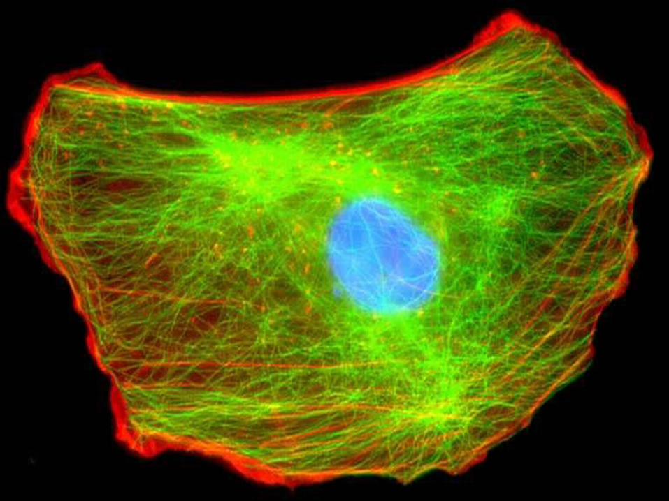
the required destinations. One of the important components of the cytoplasm is the cytoskeleton. Cytoskeleton
is a network of proteins (microtubules and microfilaments) which together form the skeleton of the cytoplasm. The cytoskeleton is responsible for the shape and movement of a cell.
Cell Organelles
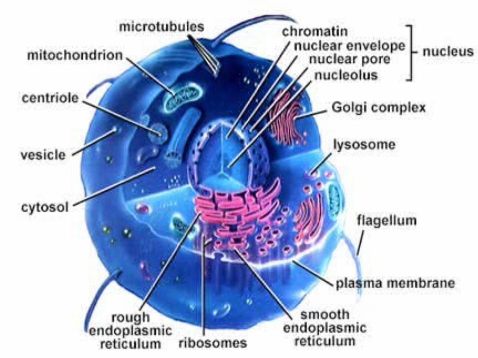
All eukaryotic and prokaryotic cells have within them a variety of different structures called organelles. Organelles are small and function much like organs function in a large organism. Some organelles are responsible for gathering cell energy, others for controlling cell activities. Plant cells have different organelles than animal cells but also have many similar organelles. They all have a large variety of sizes and functions and make life possible.
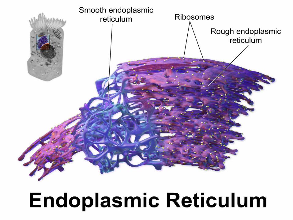
1.Endoplasmic Reticulum Endoplasmic reticulum , or ER , is an interconnected network of membranous structures
like tubules, vesicles, and cisternae . Cisternae are the flattened disk-like membranous structures. Tubules are tubular in shape, while vesicles are sac-like structures.
There are two types of endoplasmic reticulum, namely smooth endoplasmic reticulum (SER) and rough endoplasmic reticulum (RER) . When Ribosomes get attached to the surface of smooth endoplasmic reticulum, it becomes rough endoplasmic reticulum.
Functions of smooth endoplasmic reticulum • Smooth ER synthesizes fats and lipids. • It also takes part in the metabolism of carbohydrates. • It actively participates in drug detoxification. • It maintains the calcium ion concentration in the cytosol. Functions of rough endoplasmic reticulum • Most of the lysosomal proteins are produced in the rough ER. • It transports proteins to various destinations like the plasma membrane. • This is the major site of glycosylation (addition of carbohydrates in proteins).
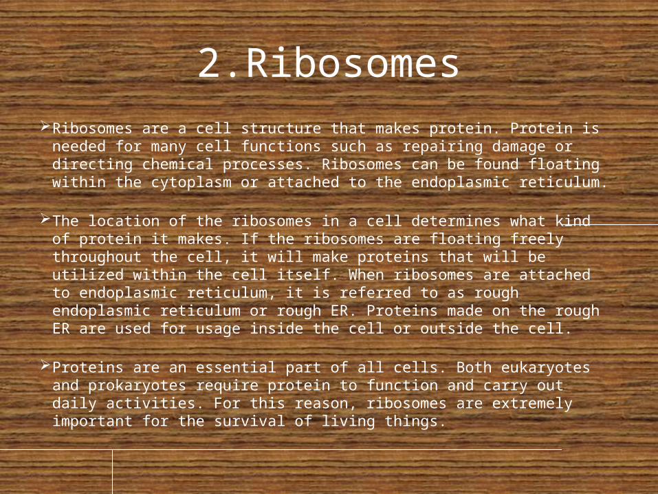
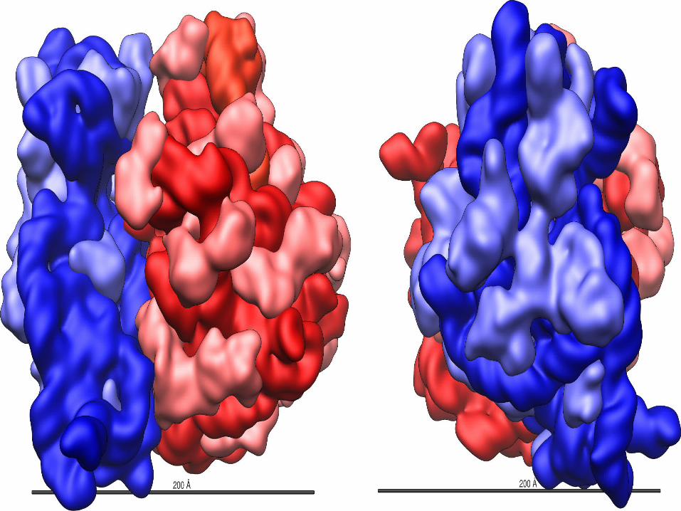
2.Ribosomes Ribosomes are a cell structure that makes protein. Protein is needed for many
cell functions such as repairing damage or directing chemical processes. Ribosomes can be found floating within the cytoplasm or attached to the endoplasmic reticulum.
The location of the ribosomes in a cell determines what kind of protein it makes. If the ribosomes are floating freely throughout the cell, it will make proteins that will be utilized within the cell itself. When ribosomes are attached to endoplasmic reticulum, it is referred to as rough endoplasmic reticulum or rough ER. Proteins made on the rough ER are used for usage inside the cell or outside the cell.
Proteins are an essential part of all cells. Both eukaryotes and prokaryotes require protein to function and carry out daily activities. For this reason, ribosomes are extremely important for the survival of living things.
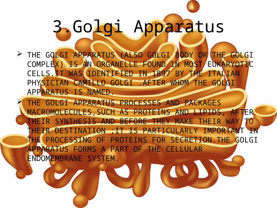
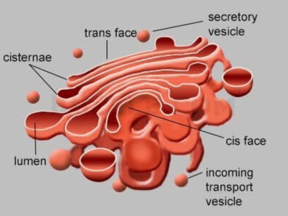
3.Golgi Apparatus THE GOLGI APPARATUS (ALSO GOLGI BODY OR THE GOLGI COMPLEX) IS
AN ORGANELLE FOUND IN MOST EUKARYOTIC CELLS.IT WAS IDENTIFIED IN 1897 BY THE ITALIAN PHYSICIAN CAMILLO GOLGI .AFTER WHOM THE GOLGI APPARATUS IS NAMED.
THE GOLGI APPARATUS PROCESSES AND PACKAGES MACROMOLECULES,SUCH AS PROTEINS AND LIPIDS, AFTER THEIR SYNTHESIS AND BEFORE THEY MAKE THEIR WAY TO THEIR DESTINATION ;IT IS PARTICULARLY IMPORTANT IN THE PROCESSING OF PROTEINS FOR SECRETION.THE GOLGI APPARATUS FORMS A PART OF THE CELLULAR ENDOMEMBRANE SYSTEM.
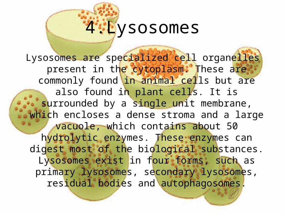
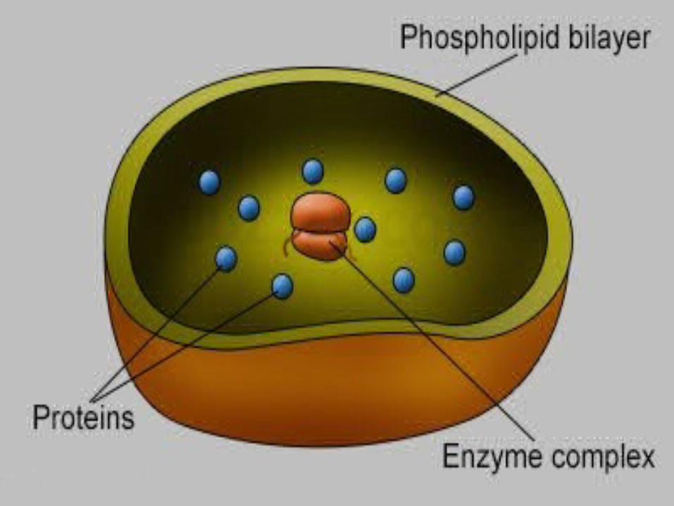
4.Lysosomes
Lysosomes are specialized cell organelles present in the cytoplasm. These are commonly found in
animal cells but are also found in plant cells. It is surrounded by a single unit membrane, which
encloses a dense stroma and a large vacuole, which contains about 50 hydrolytic enzymes. These
enzymes can digest most of the biological substances. Lysosomes exist in four forms, such as primary lysosomes, secondary lysosomes, residual
bodies and autophagosomes.
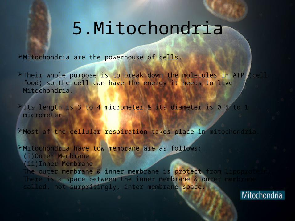
5.Mitochondria Mitochondria are the powerhouse of cells.
Their whole purpose is to break down the molecules in ATP (cell food) so the cell can have the energy it needs to live Mitochondria.
Its length is 3 to 4 micrometer & its diameter is 0.5 to 1 micrometer.
Most of the cellular respiration takes place in mitochondria.
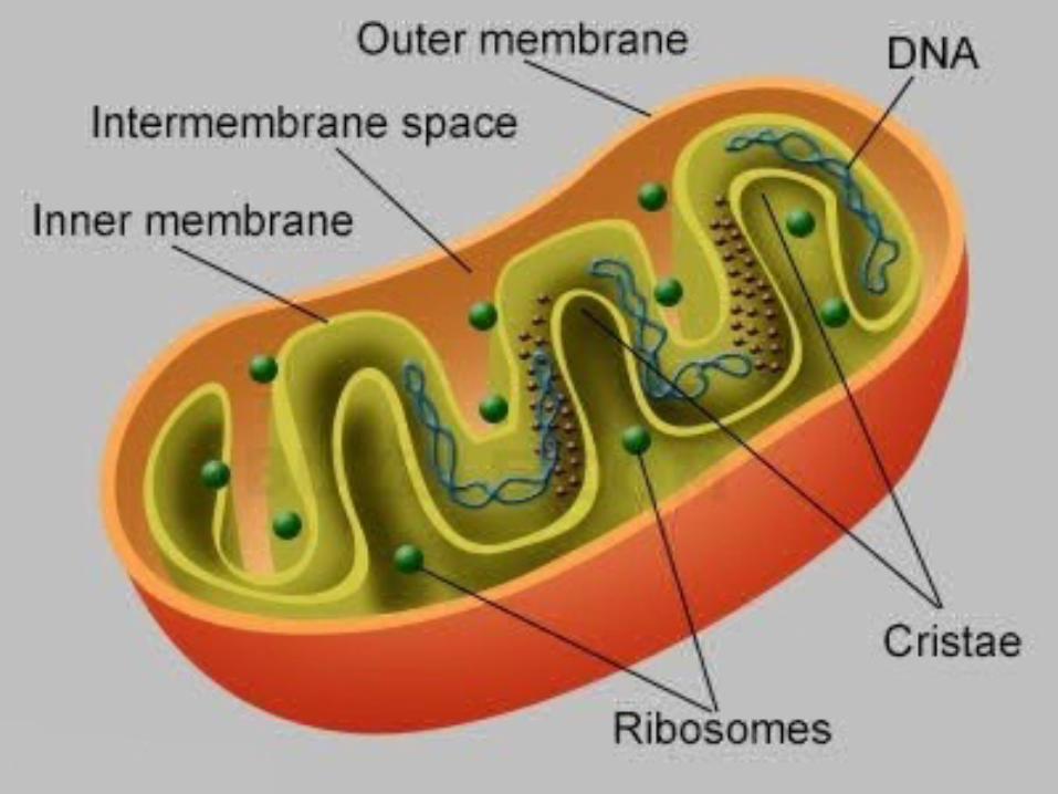
Mitochondria have tow membrane are as follows: (i)Outer Membrane (ii)Inner Membrane The outer membrane & inner membrane is protect from Lipoprotein. There is a space between the inner membrane & outer membrane called, not surprisingly, inter membrane space.
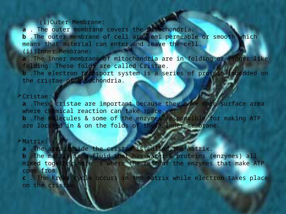
(i)Outer Membrane: a . The outer membrane covers the mitochondria. b .The outer membrane of cell are semi permeable or smooth which means that material can enter and leave the cell. (ii)Inner Membrane: a .The inner membrane of mitochondria are in folding or finger like folding. These folds are called Cristae. b .The electron transport system is a series of protein imbedded on the cristae of mitochondria.
Cristae: a .These cristae are important because they make more surface area where chemical reaction can take space. b .The molecules & some of the enzymes responsible for making ATP are located in & on the folds of these inner membrane.
Matrix: a .The area inside the cristae is called the matrix. b .The matrix is a fluid that has water & proteins (enzymes) all mixed together here 's where the rest of the enzymes that make ATP come from. c . The Krebs cycle occurs in the matrix while electron takes place on the cristae.
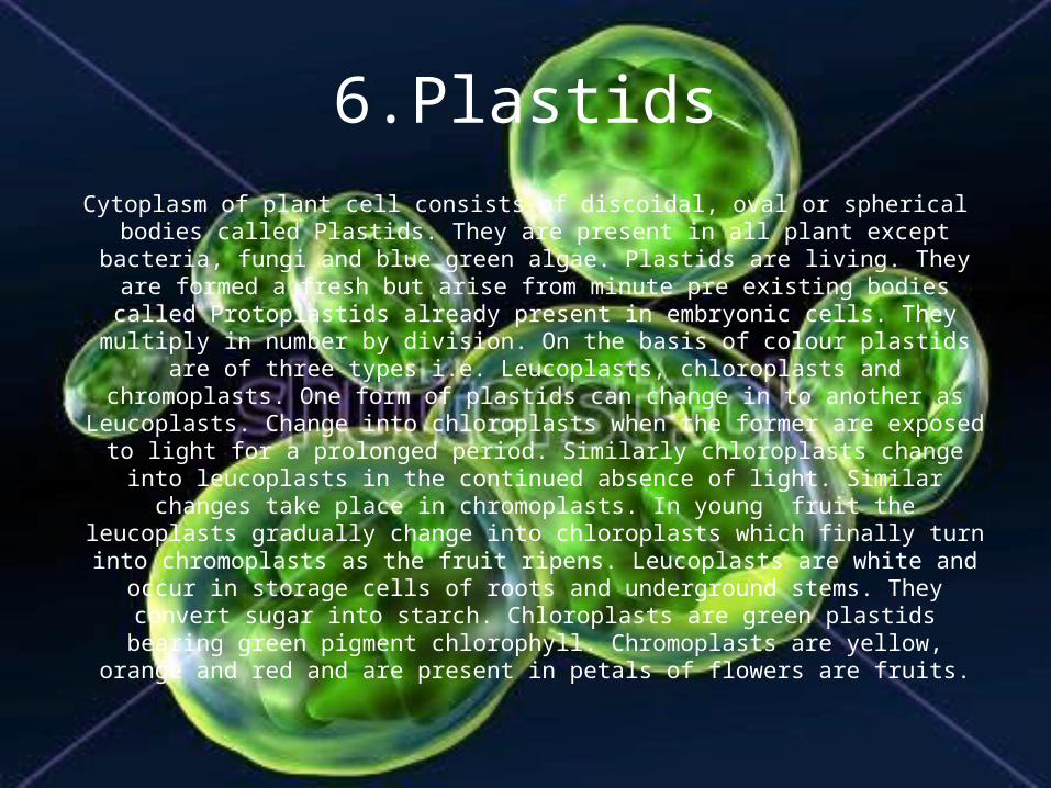
6.PlastidsCytoplasm of plant cell consists of discoidal, oval or spherical bodies called Plastids.
They are present in all plant except bacteria, fungi and blue green algae. Plastids are living. They are formed a fresh but arise from minute pre existing
bodies called Protoplastids already present in embryonic cells. They multiply in number by division. On the basis of colour plastids are of three types i.e.
Leucoplasts, chloroplasts and chromoplasts. One form of plastids can change in to another as Leucoplasts. Change into chloroplasts when the former are exposed to light for a prolonged period. Similarly chloroplasts change into leucoplasts in the continued absence of light. Similar changes take place in
chromoplasts. In young fruit the leucoplasts gradually change into chloroplasts which finally turn into chromoplasts as the fruit ripens. Leucoplasts are white and occur in storage cells of roots and underground stems. They convert sugar into starch. Chloroplasts are green plastids bearing green pigment chlorophyll. Chromoplasts are yellow, orange and red and are present in petals of flowers
are fruits.
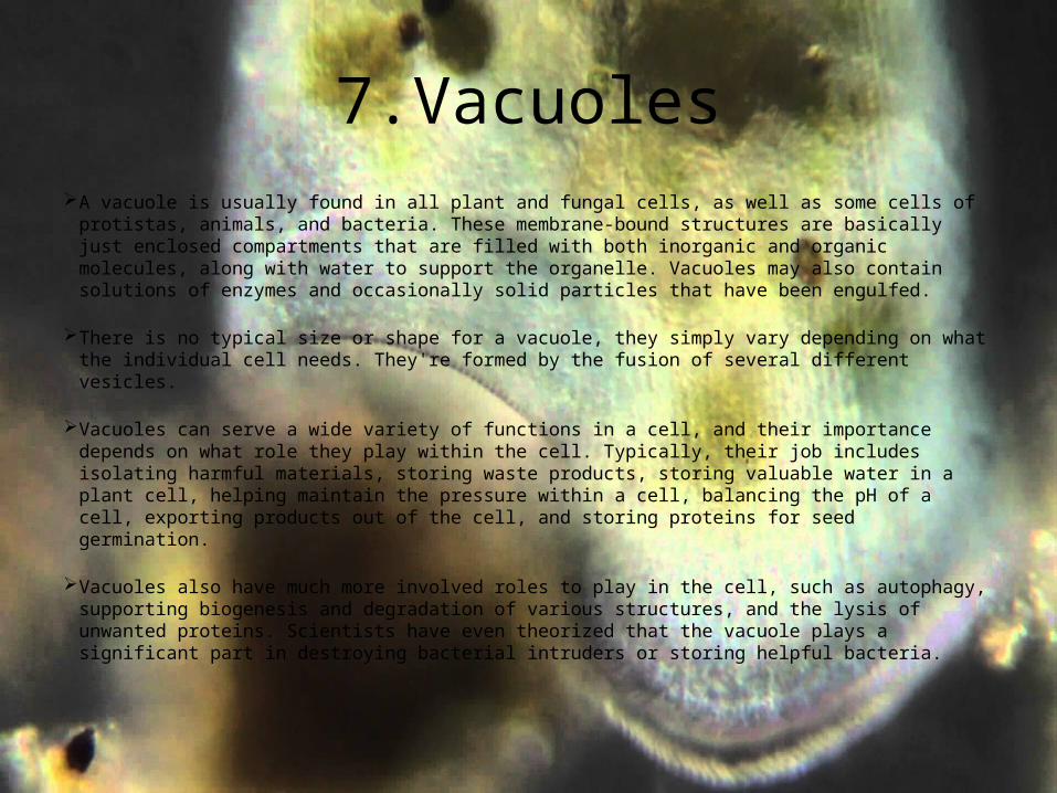
7.Vacuoles A vacuole is usually found in all plant and fungal cells, as well as some cells of protistas, animals, and
bacteria. These membrane-bound structures are basically just enclosed compartments that are filled with both inorganic and organic molecules, along with water to support the organelle. Vacuoles may also contain solutions of enzymes and occasionally solid particles that have been engulfed.
There is no typical size or shape for a vacuole, they simply vary depending on what the individual cell needs. They're formed by the fusion of several different vesicles.
Vacuoles can serve a wide variety of functions in a cell, and their importance depends on what role they play within the cell. Typically, their job includes isolating harmful materials, storing waste products, storing valuable water in a plant cell, helping maintain the pressure within a cell, balancing the pH of a cell, exporting products out of the cell, and storing proteins for seed germination.
Vacuoles also have much more involved roles to play in the cell, such as autophagy, supporting biogenesis and degradation of various structures, and the lysis of unwanted proteins. Scientists have even theorized that the vacuole plays a significant part in destroying bacterial intruders or storing helpful bacteria.
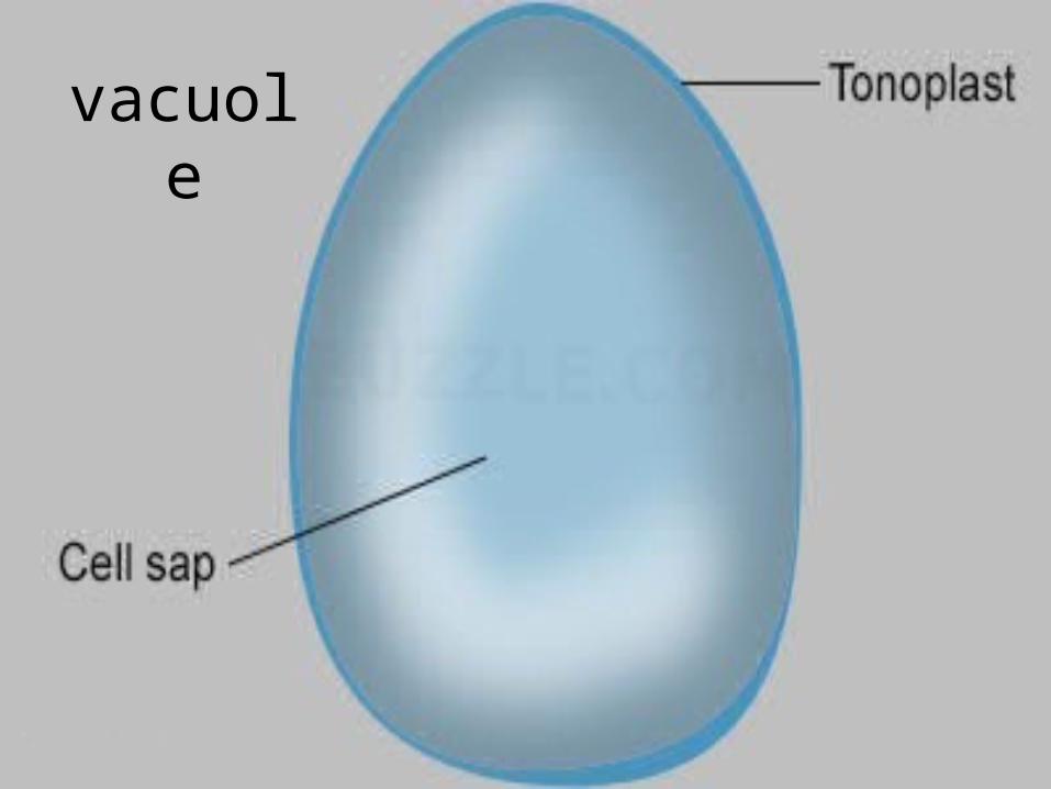
vacuole
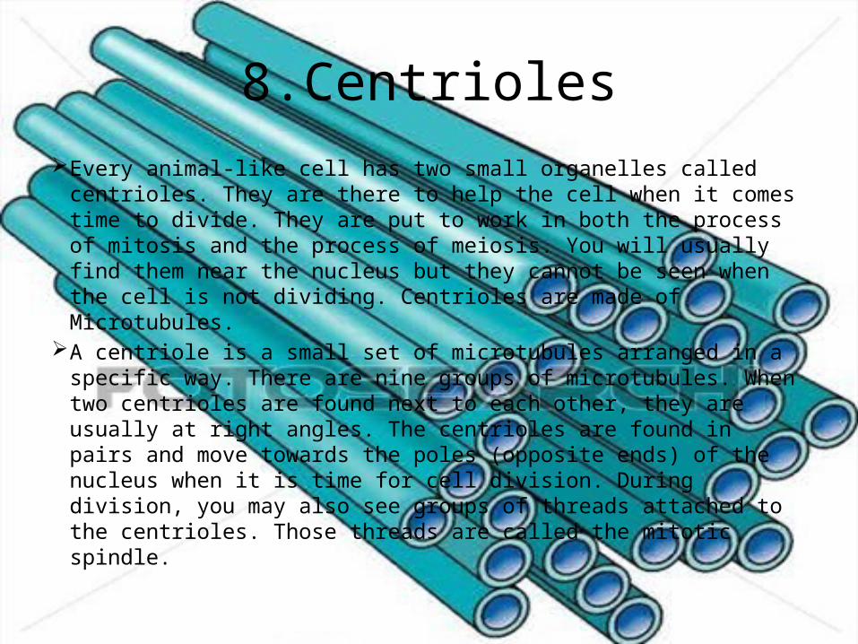
8.Centrioles Every animal-like cell has two small organelles called centrioles.
They are there to help the cell when it comes time to divide. They are put to work in both the process of mitosis and the process of meiosis. You will usually find them near the nucleus but they cannot be seen when the cell is not dividing. Centrioles are made of Microtubules.
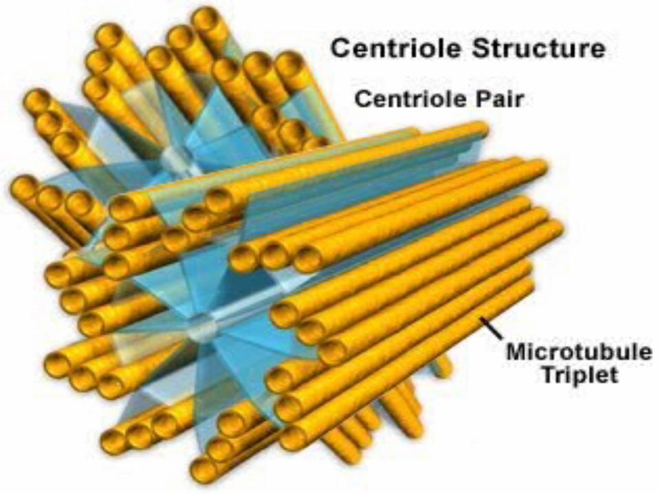
A centriole is a small set of microtubules arranged in a specific way. There are nine groups of microtubules. When two centrioles are found next to each other, they are usually at right angles. The centrioles are found in pairs and move towards the poles (opposite ends) of the nucleus when it is time for cell division. During division, you may also see groups of threads attached to the centrioles. Those threads are called the mitotic spindle.
By:-Varun V. SatputePavan MajalikarAfnaan SayedVinay MansabdarPranit Patil