Embed Size (px)
Citation preview
ORBITDR. REEMA THOMAS
DEPARTMENT OF OPHTHALMOLOGY
TOPICS
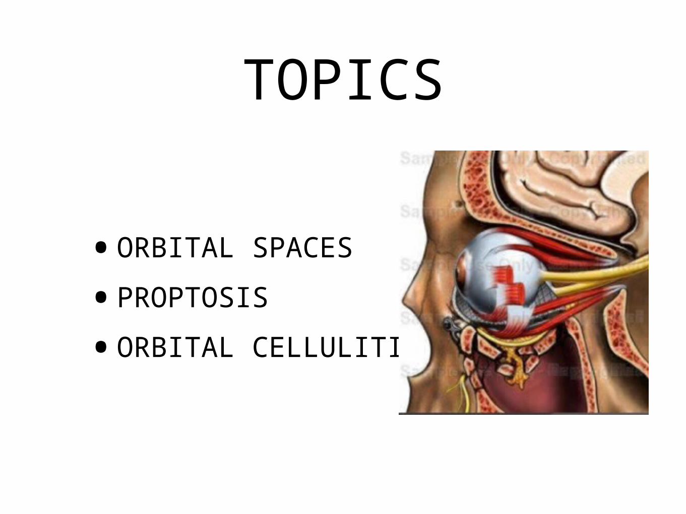
•ORBITAL SPACES•PROPTOSIS•ORBITAL CELLULITIS
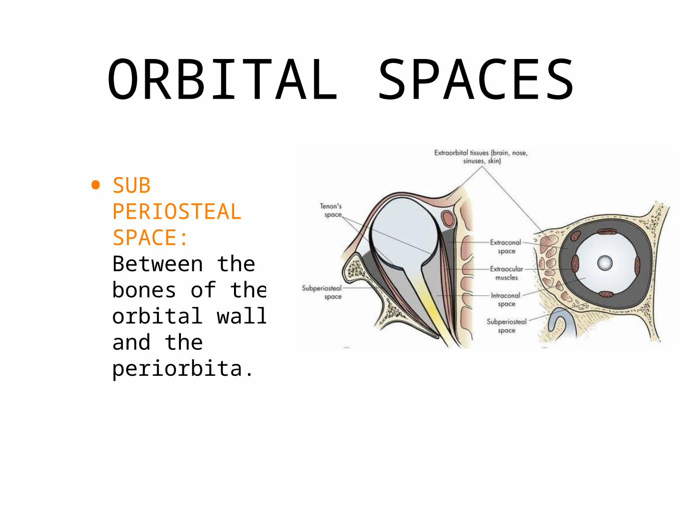
ORBITAL SPACES• SUB
PERIOSTEAL SPACE: Between the bones of the orbital wall and the periorbita.
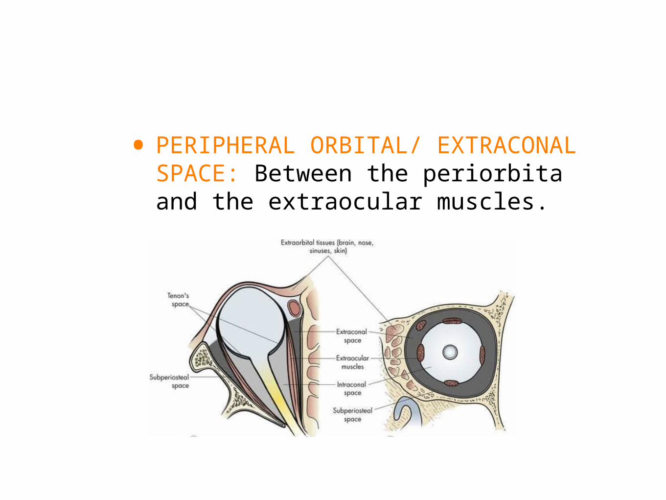
• PERIPHERAL ORBITAL/ EXTRACONAL SPACE: Between the periorbita and the extraocular muscles.
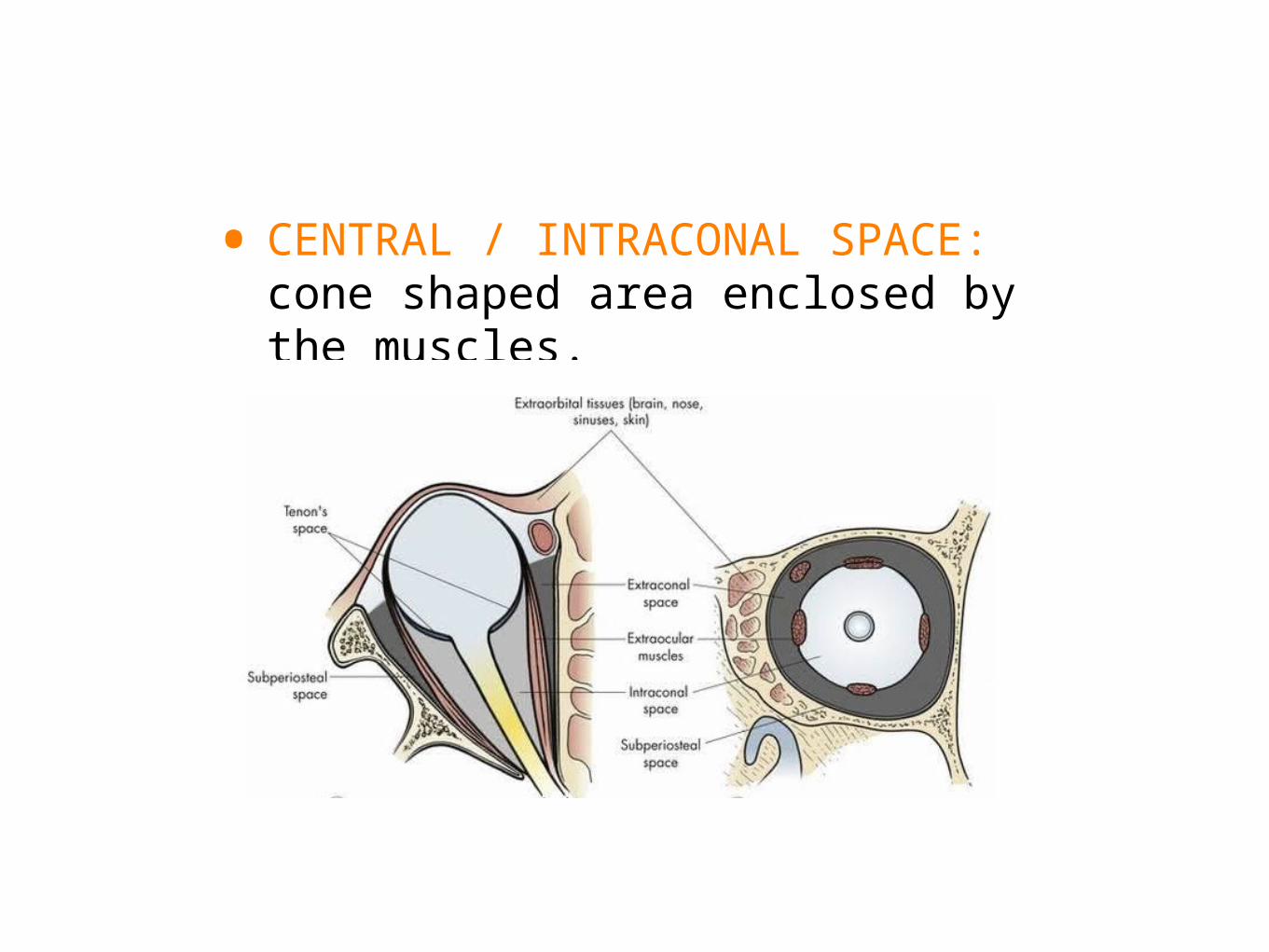
• CENTRAL / INTRACONAL SPACE: cone shaped area enclosed by the muscles.
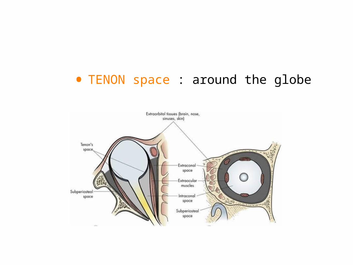
• TENON space : around the globe
PROPTOSIS
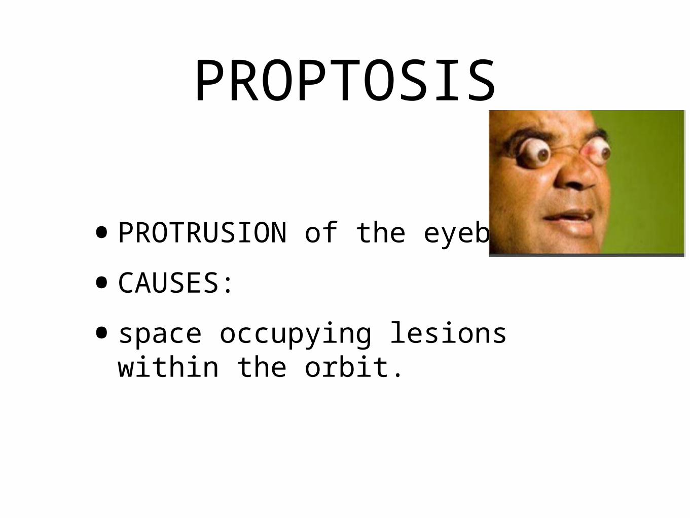
•PROTRUSION of the eyeball.•CAUSES: •space occupying lesions within the
orbit.
UNILATERAL PROPTOSIS
•tumours of the orbit•orbital haemorrhage•orbital cellulitis•pseudotumor
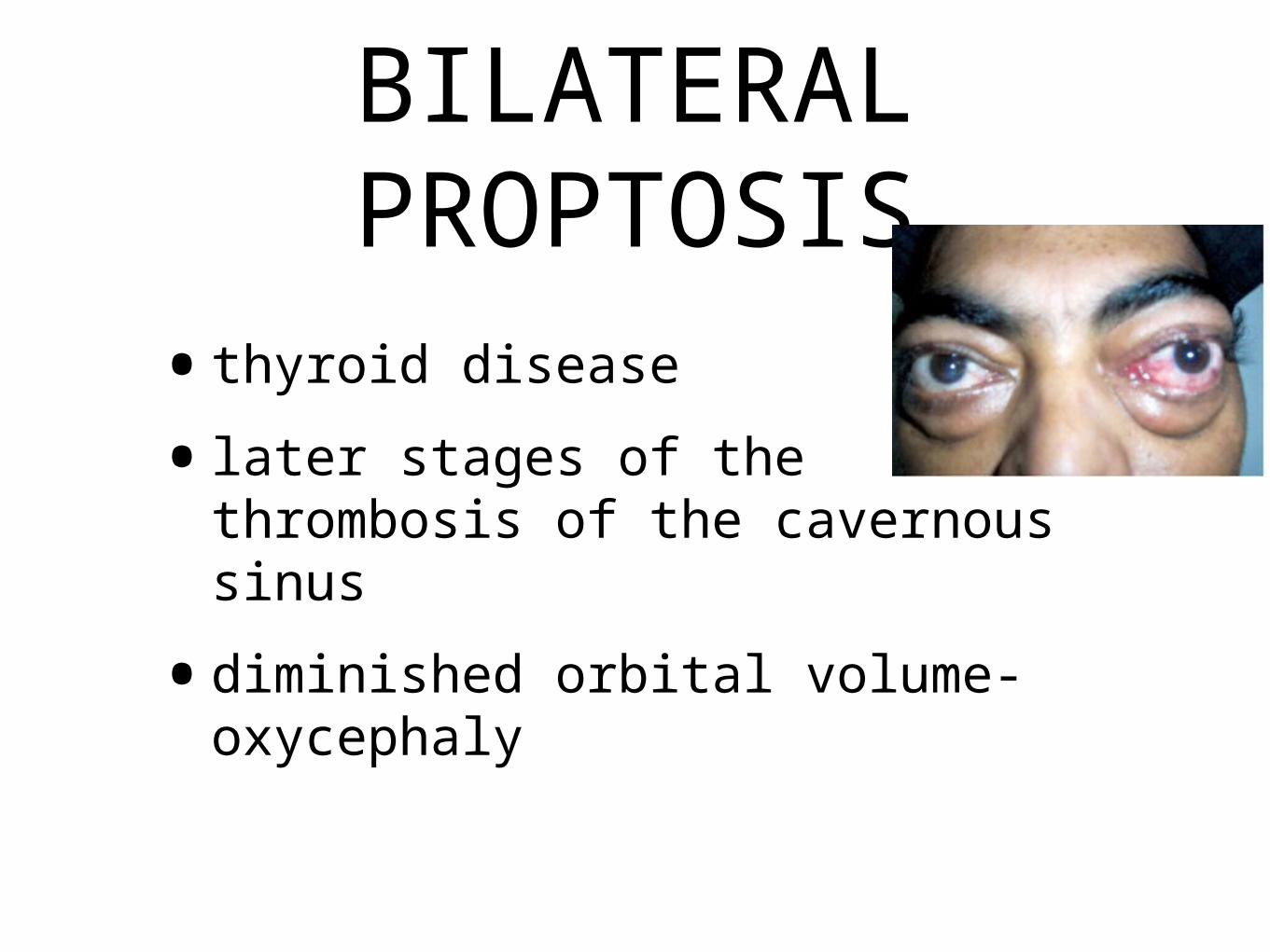
BILATERAL PROPTOSIS
•thyroid disease•later stages of the thrombosis of
the cavernous sinus•diminished orbital volume-
oxycephaly
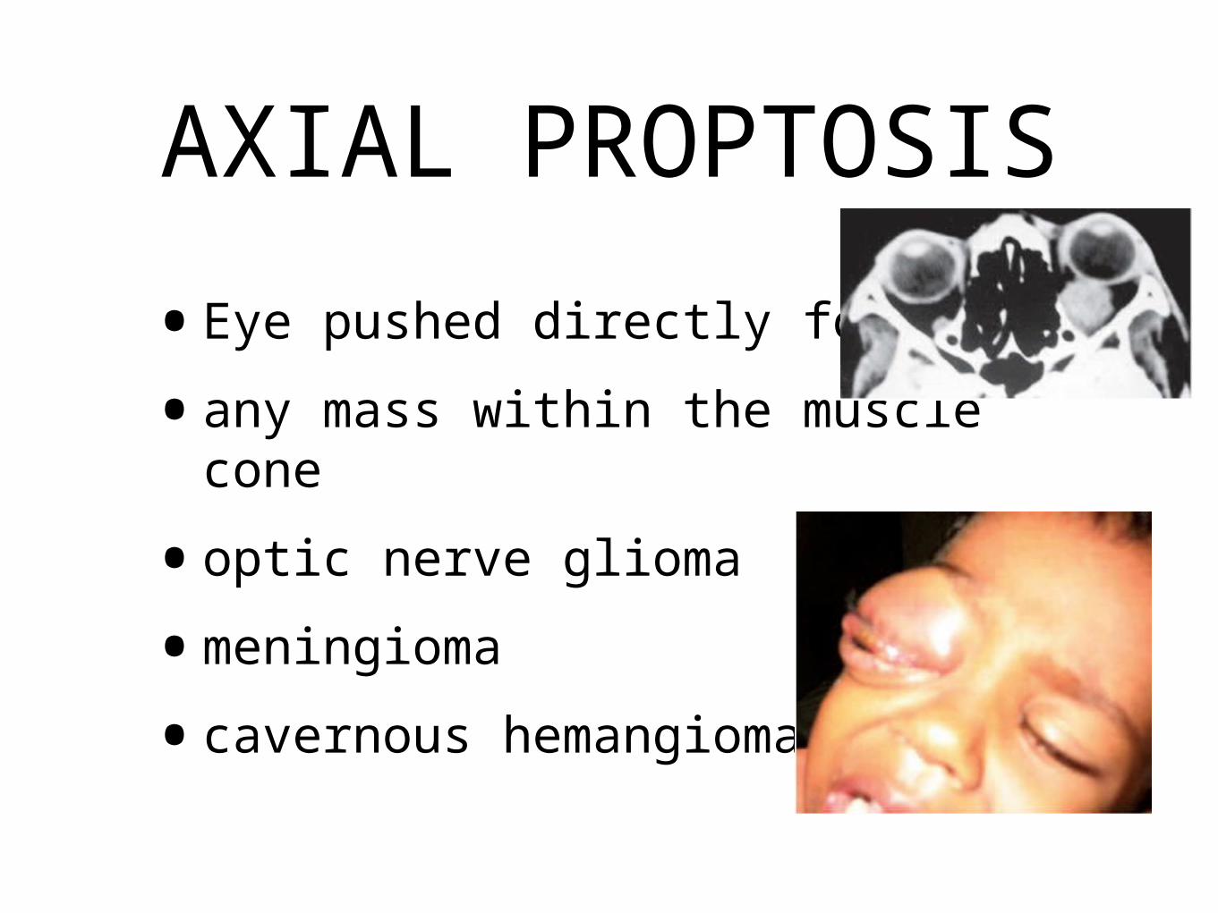
AXIAL PROPTOSIS•Eye pushed directly forwards•any mass within the muscle cone•optic nerve glioma•meningioma•cavernous hemangioma
NON AXIAL DISPLACEMENT•Downwards and Inwards:
•dermoids• lacrimal gland tumours•Superiorly:•maxillary sinus tumour•Laterally:• frontoethmoidal mucocele
PULSATILE PROPTOSIS
•Carotid cavernous fistula•neurofibromatosis
INTERMITTENT PROPTOSIS
• on valsalva manoeuvre/ bending forwards:
• varicosities of the orbital veins
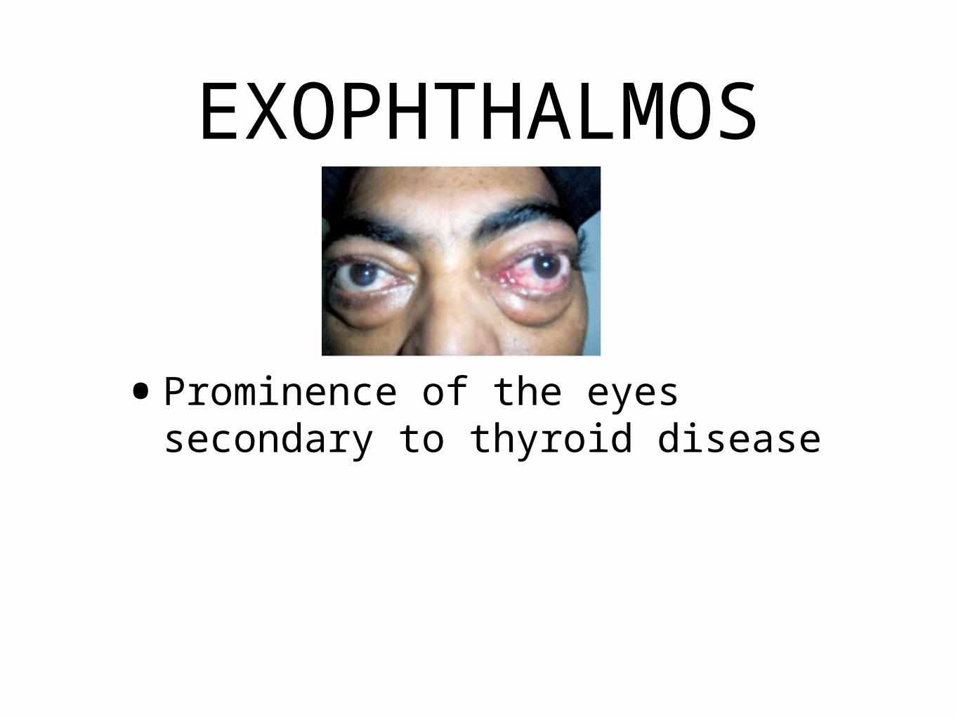
EXOPHTHALMOS
•Prominence of the eyes secondary to thyroid disease
PSEUDOPROPTOSIS
•High myopia•paralysis of the extrinsic muscles•contralateral enophthalmos
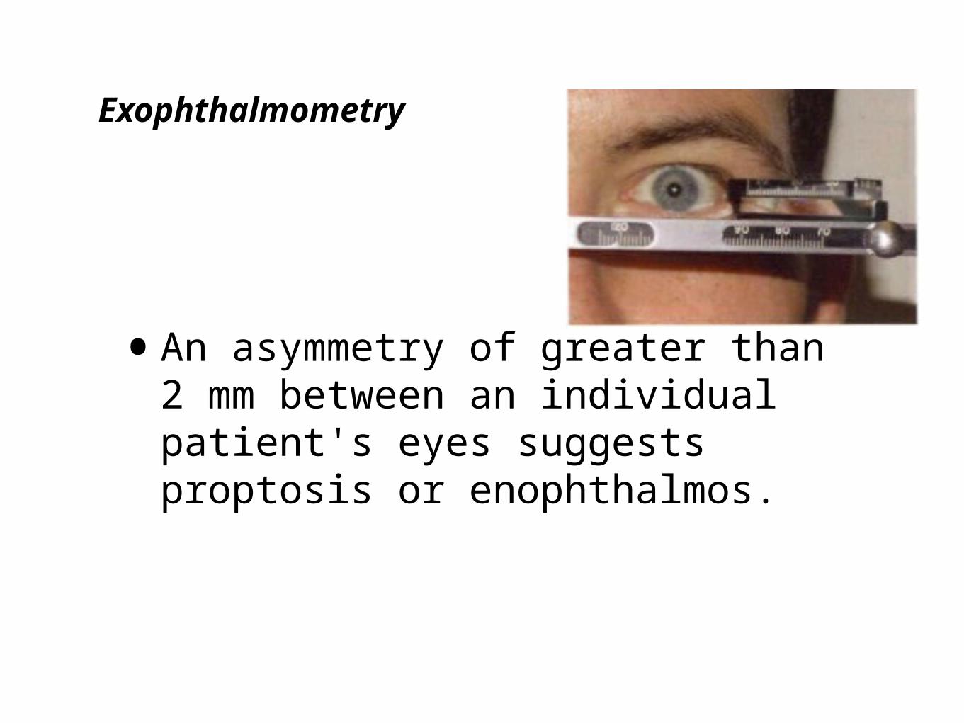
Exophthalmometry
•An asymmetry of greater than 2 mm between an individual patient's eyes suggests proptosis or enophthalmos.
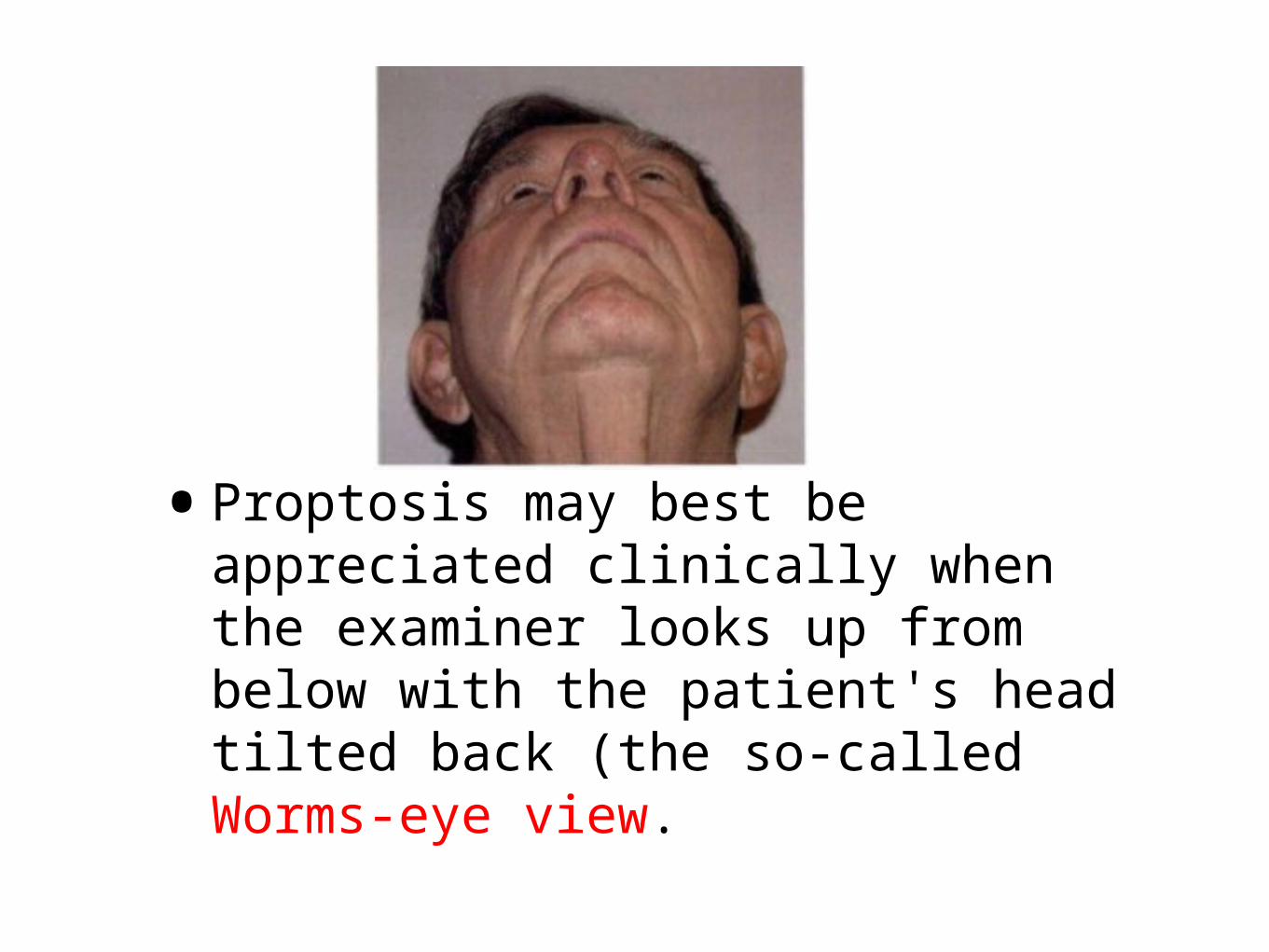
•Proptosis may best be appreciated clinically when the examiner looks up from below with the patient's head tilted back (the so-called Worms-eye view.
ENOPHTHALMOS
• Retrodisplacement of the eye into the orbit
•Fracture of the floor of orbit
THYROID EYE DISEASE
•an autoimmune inflammatory disorder
•eyelid retraction, lid lag, proptosis, restrictive extraocular myopathy, and compressive optic neuropathy
•may also occur with Hashimoto thyroiditis (immune-induced hy- pothyroidism)
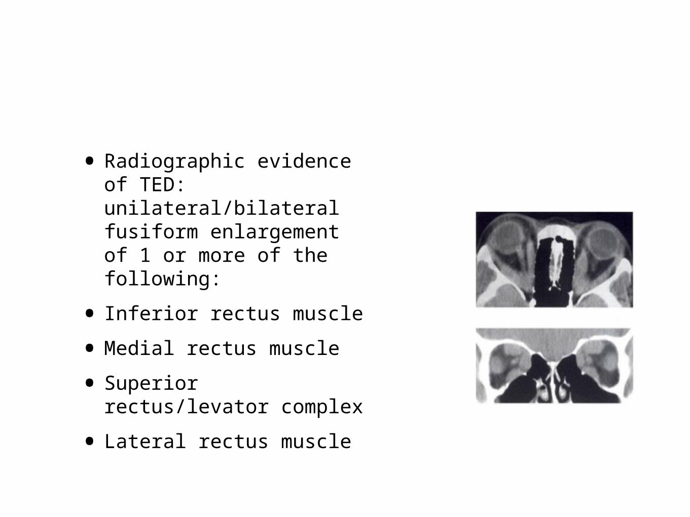
• Radiographic evidence of TED: unilateral/bilateral fusiform enlargement of 1 or more of the following:
• Inferior rectus muscle• Medial rectus muscle• Superior rectus/levator
complex • Lateral rectus muscle
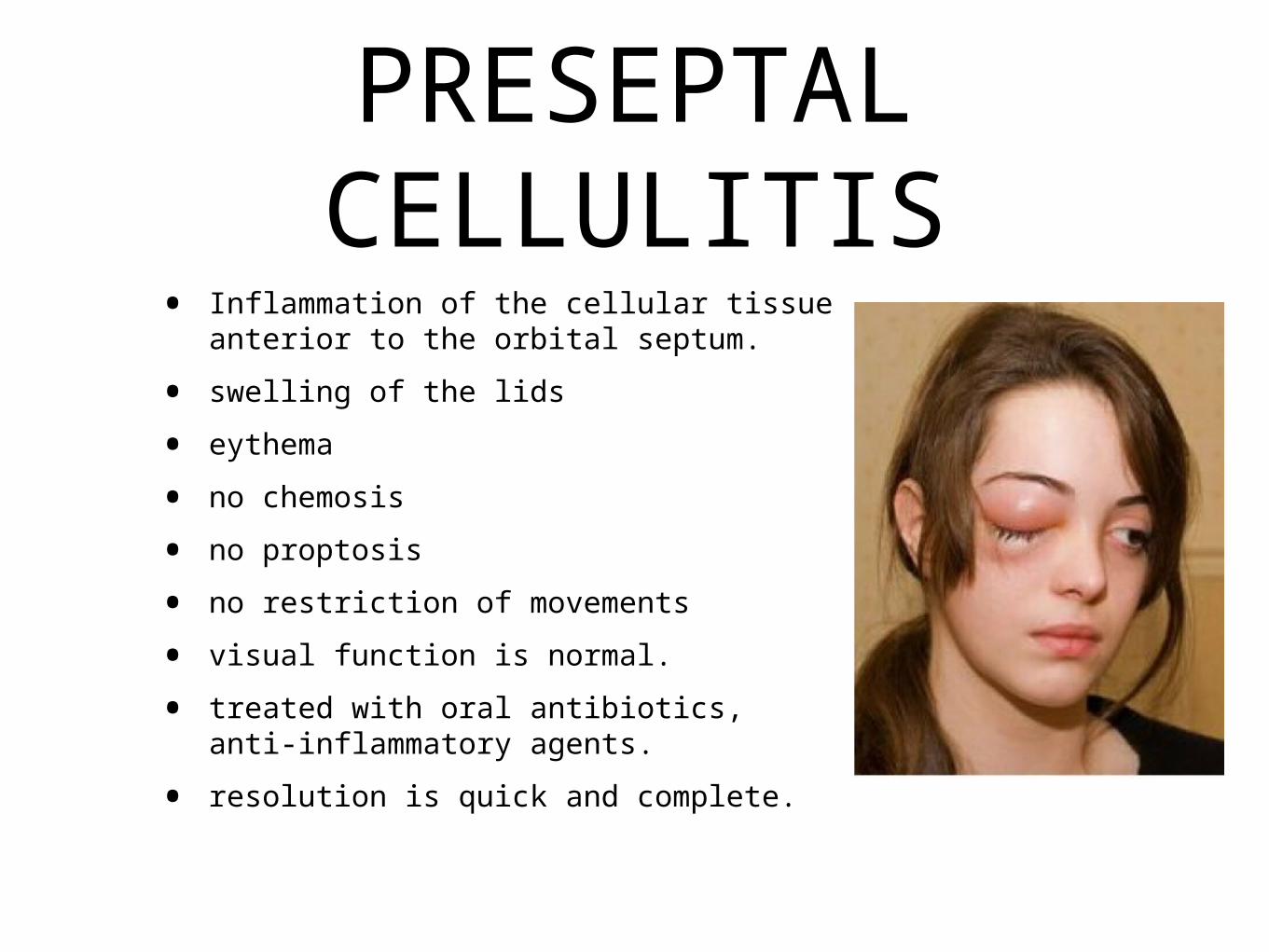
PRESEPTAL CELLULITIS
• Inflammation of the cellular tissue anterior to the orbital septum.
• swelling of the lids• eythema• no chemosis• no proptosis• no restriction of movements• visual function is normal.• treated with oral antibiotics, anti-
inflammatory agents.• resolution is quick and complete.
•causes for preseptal cellulitis:•cutaneous trauma•dacryocystitis•sinusitis
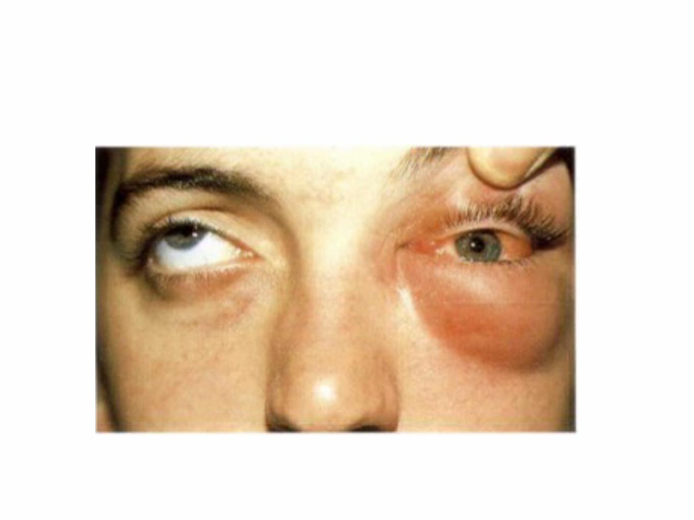
ORBITAL CELLULITIS
•Inflammation of the cellular tissue of the orbit behind the orbital septum.
•most commonly due to the extension of the inflammation from the neighbouring parts, like the nasal sinuses, posterior extensions of suppurative infections of the eyelids, panophthalmitis...
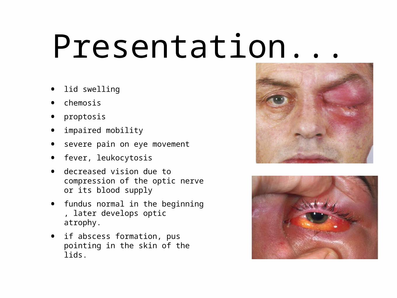
Presentation...• lid swelling• chemosis• proptosis• impaired mobility• severe pain on eye movement• fever, leukocytosis• decreased vision due to
compression of the optic nerve or its blood supply
• fundus normal in the beginning , later develops optic atrophy.
• if abscess formation, pus pointing in the skin of the lids.
•Delay in treatment may result in blindness, cavernous sinus thrombosis, cranial neuropathy, brain abscess, and death.

Treatment....
•blood samples for culture.•intravenous antibiotics•anti-inflammatory drugs.•if abscess, drainage of pus.

•THANK YOU
![World Horse Welfare Presentation - 21.07.2016 [Read-Only] · Title: Microsoft PowerPoint - World Horse Welfare Presentation - 21.07.2016 [Read-Only] Author: RolyO Created Date: 2/14/2017](https://img.pdfslide.us/doc/110x75/5f5bc9d606ad7f5a764f1f26/world-horse-welfare-presentation-21072016-read-only-title-microsoft-powerpoint.jpg)