Embed Size (px)
Citation preview

White Paper | CARESTREAM DRX Plus Detectors
DRX Plus Detectors: Going from Good to Great Authors: Karin Töpfer, Tim Wojcik
Introduction
Carestream’s introduction in 2009 of the world’s first portable, wireless, cassette-sized detector – the CARESTREAM DRX-1 Detector – has fundamentally changed the digital radiography market with a versatile and cost-effective imaging solution. Now available, the third-generation design: the CARESTREAM DRX Plus Detector takes DR to a new level.
This paper describes many of the features and benefits of the DRX Plus at a detailed level to demonstrate that the DRX Plus takes digital radiography with portable wireless detectors from good to great.
Consistently Great Imaging Performance
Keeping in mind that the basic purpose of this product is to consistently and reliably deliver excellent images at the lowest possible patient dose, the DRX Plus incorporates several improvements in fundamental design over the DRX-1 product, delivering a 60% reduction in dark noise along with a 25% increase in sensitivity, for a noticeable improvement in Detective Quantum Efficiency (DQE). The benefit of this improvement is better signal-to-noise ratio (SNR) in the images over previous offerings, or the opportunity to reduce patient exposure while preserving the same SNR in the presented image. Figure 1 illustrates the image noise improvement for a fixed exposure, resulting in an IEC Exposure Index of 135.
Figure 1: 800-speed exposure image comparison: DRX Plus 3543 Detector vs. DRX-1 hand-phantom image at 2x magnification

White Paper | CARESTREAM DRX Plus Detectors
2
Several competitors quote “relative DQE” or “20% improvement in DQE” with their sales promotions, but do not indicate what this is relative to, making it a meaningless metric. Potential buyers should compare absolute DQE numbers in compliance with the recognized international standard IEC 62220 series “Medical Electrical Equipment – Characteristics of digital x-ray imaging devices.” Further, it must be appreciated that DQE is not a single number, as represented in many sales documents – it is a multi-variant response that reflects x-ray beam condition, exposure level, and spatial frequency content. The IEC 62220 standard requires reporting of performance in a manner that covers this response space, so customers should insist on seeing a complete data set when comparing products.
The performance of the DRX Plus is highly competitive for portable wireless detectors and buyers should make this comparison.
Figure 2 illustrates the DQE of the DRX Plus 3543 Detector at RQA-5 beam conditions against a competitive product that claims a similar DQE in its sales brochure.
Note that while the performance of the two devices is similar at the higher exposure level and low spatial frequency, the performance at higher spatial frequencies and lower exposures is substantially better for the DRX Plus.
Of course, DQE characterization reports performance under ideal lab conditions, rather than the extreme conditions of the customer environment. Image uniformity, in particular, can be greatly impacted by conditions such as time from initial power-up to capture, prep hold time, operating temperature differences from calibration temperature, x-ray beam condition as compared to calibration beam condition, and image-to-image capture interval.
Figure 2: DQE comparison at RQA-5 beam of DRX Plus 3543 and Competitor A
Performance similar at lower frequency and high exposure
DRX Plus substantially better at higher frequency and lower exposure

White Paper | CARESTREAM DRX Plus Detectors
3
Figure 3 shows an example of the DRX Plus image uniformity, as compared to a competitor’s detector, at an operating
temperature that is 10 °C different from the calibration temperature. Both were calibrated at a temperature of 25 °C:
Figure 3: Flat-field image comparison: DRX Plus 3543 vs. Competitor A at 35 °C, calibrated at 25 °C, window width is 5 % of level
Finally, the nature of amorphous silicon-based image sensors requires careful attention to panel operating waveforms, including power-up, refresh, signal integration and capture. Carestream has mastered this aspect of detector design as demonstrated in the excellent image uniformity and stability from the first image to the last in a patient study. Figure 4 illustrates the uniformity of the first image taken immediately
after power-up with the DRX Plus detectors as compared to a competitive product. This first-image stability problem is actually addressed in the user manual of one competitor’s product with the statement that “there is no guarantee that the image taken can be used for diagnostic purposes.” Workflow is impacted by the need to discard the first image.

White Paper | CARESTREAM DRX Plus Detectors
4
Figure 4: First image after power-up: DRX Plus vs. Competitor B, window of 30 ADC at 14-bit resolution
Improved Capture Speed
Single-projection imaging is limited by the anatomical noise from overlying structures in the image projection. Advanced applications such as Tomosynthesis and Dual Energy capture techniques enable improved visualization of buried pathology. The DRX Plus detectors are being designed to support these advanced applications in the future with frame rates up to five full-resolution captures per second. (INVESTIGATIONAL: Not commercially available.) This improved speed will also result in faster image access and cycle time with standard projection imaging procedures.
Lighter and More Durable Housing
The weight of the DRX Plus has been reduced, while at the same time improving durability. This set of improvements results in higher weight-bearing capacity, drop tolerance and fluid resistance. In fact, the DRX Plus detectors have achieved IPX Level 7, which means they can tolerate submersion in one meter of water for 30 minutes without failure per the IEC standard 60529. While this helps to illustrate the durability of
the device, it is recommended that good clinical practices such as bagging the detector are followed.
One point of caution regarding load tolerance for detectors – weight-bearing exams must result in acceptable images. Most competitor detectors on the market disclose a specific weight tolerance, but the level provided is the limit before breakage, rather than the limit for which imaging performance is certified. An important design element is to ensure that the critical imaging components perform well under load, for example when a patient is laying on the detector in a bedside procedure.
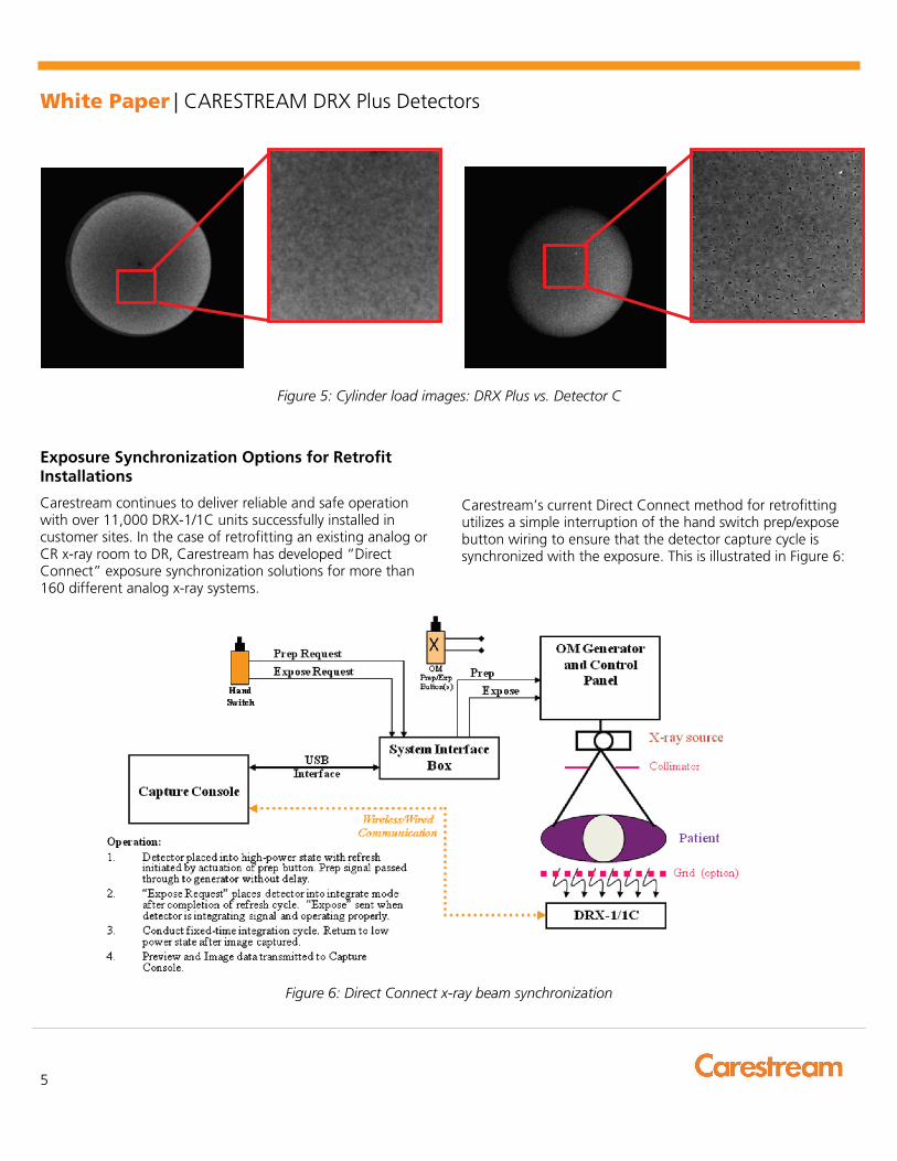
Figure 5 illustrates the image uniformity of the DRX Plus detector with proper scintillator-to-sensor coupling under a 150 lb (68 kg) load on a 4-cm diameter cylinder, as compared to a poorly designed detector under only 50 lb (23 kg) of load. Both devices specify a 150 lb load weight tolerance, but note the points of significant non-uniformity on the device with poor scintillator-to-sensor coupling. Detector C may be able to withstand 150 lb load before breakage, but image quality is severely compromised with a mere 50 lb load.
White Paper | CARESTREAM DRX Plus Detectors
5
Figure 5: Cylinder load images: DRX Plus vs. Detector C
Exposure Synchronization Options for Retrofit Installations
Carestream continues to deliver reliable and safe operation with over 11,000 DRX-1/1C units successfully installed in customer sites. In the case of retrofitting an existing analog or CR x-ray room to DR, Carestream has developed “Direct Connect” exposure synchronization solutions for more than 160 different analog x-ray systems.
Carestream’s current Direct Connect method for retrofitting utilizes a simple interruption of the hand switch prep/expose button wiring to ensure that the detector capture cycle is synchronized with the exposure. This is illustrated in Figure 6:
Figure 6: Direct Connect x-ray beam synchronization

White Paper | CARESTREAM DRX Plus Detectors
carestream.com
©Carestream Health, Inc., 2015. CARESTREAM is a trademark of Carestream Health. CAT 200 0113 07/15
The benefits of this method include:
• Ensuring that the detector is ready to capture before allowing patient exposure
• Keeping the detector in a low power-consuming state as long as possible to maximize battery charge life
• Delivering the highest possible image quality with no loss of exposure
However, it is recognized that certain situations preclude the possibility of connecting to the prep/expose signals on an existing x-ray system. Thus the concept of “Beam Sensitive Triggering” or “Beam Detect” has been implemented by several companies. There are a number of concepts for delivering this functionality, each with their own inherent strengths and weaknesses. Limitations of competitive systems include:
• Requirement to expose a specific portion (usually the center) of the detector, since a dedicated sensor(s) is used to detect the beam. In this case, a collimated beam that is positioned near the perimeter of the image area will not trigger the detector capture cycle.
• Minimum exposure thresholds that can be higher than standard techniques: e.g. collimation must be opened up beyond the object size to ensure adequate exposure impinges onto the detector array, or exposure times must be at least 5 msec. In some cases, the user must decide what detector sensitivity should be used before exposing the patient – if the chosen sensitivity is too low, the image may not get captured, while choosing too high a sensitivity causes false captures from external electromagnetic noise, temperature changes or even gentle bumping of the device.
• Limited “ready-to-capture” time in which only a few seconds are allowed from the user requesting that the detector “get ready” to capture until the beam actually arrives – requires frequent “get ready” prompts by the user.
The introduction of the DRX Plus Detector brings a new level of performance to this capture mode. Carestream’s design utilizes the entire image capture area for signal sensing, and executes an adaptive triggering that ensures a responsiveness that is exceptional for portable detectors. The DRX Plus implementation conducts continuous signal integration so that there is no lost exposure, while some competitive offerings require several milliseconds of signal detection time with the associated loss of exposure. Reliable captures have been demonstrated on Carestream’s DRX Plus detector over a complete set of exposure techniques, with collimation down to extremity (4 x 10 cm) image-capture sizes placed anywhere on the detector surface, and over broad environmental conditions. Carestream’s intelligent beam trigger algorithm rejects false triggers from bumps and external noise while retaining extremely high x-ray sensitivity. Once activated, the DRX Plus detector can await x-ray beam arrival for up to several minutes at the user’s discretion, using a programmable time-out.
Carestream strongly recommends the use of Direct Connect synchronization, as it delivers the best image quality, longest battery charge life, and safest operation through positive prevention of exposure when the detector is not ready. However, if the site situation requires Beam Detect synchronization, the method implemented in the DRX Plus detectors will deliver excellent reliability and performance.
X-Factor Smart
Carestream supports interchangeability and sharing of DRX-1/1C detectors and the new DRX Plus detectors across a broad set of portable, in-room and retrofit systems. This affords the user extreme flexibility in workflow optimization (put the detectors where they are most needed), inherent redundancy for maximum up-time, and obsolescence avoidance. The DRX Plus has capabilities that will support future advanced imaging modes while retaining backward compatibility with the DRX-1/1C environment: “Right for Today, Ready for Tomorrow.”
DRX Plus – from Good to Great
Carestream pioneered the concept of portable, wireless, cassette-sized DR detectors and this leadership position allowed the company to witness the benefits of the original DRX-1 while learning about opportunities to improve. The DRX Plus represents Carestream’s 3rd generation detector design, reflecting the integration of its knowledge and experience in the market.
Karin Töpfer is an Imaging Physicist in Carestream's Research and Innovation Laboratories. She is an expert in medical image quality and performance modeling of digital x-ray detectors. In addition, she designs robust operating cycles and image calibrations and corrections for portable digital X-ray detectors. Tim Wojcik is the Program Leader for Radiographic Image Capture in Carestream's Research and Innovation Laboratories. He has more than 38 years of experience in product development, research and manufacturing and has led projects in digital radiography, computed radiography and medical image printing.