Embed Size (px)
Citation preview
ENHANCED ORAL ASSESSMENT
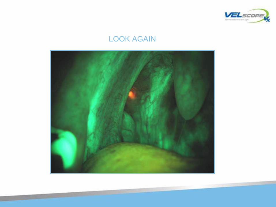
LOOK AGAIN
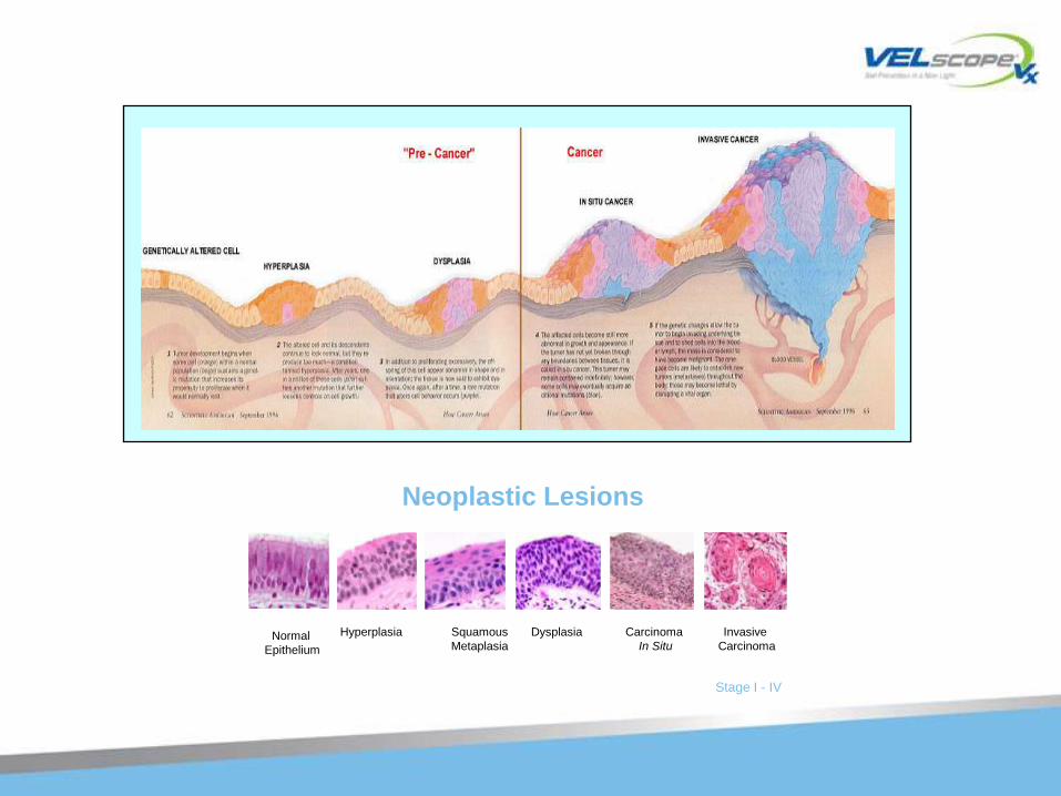
Neoplastic Lesions
Normal
Epithelium
Hyperplasia Dysplasia Carcinoma
In Situ
Invasive
Carcinoma
Squamous
Metaplasia
Stage I - IV
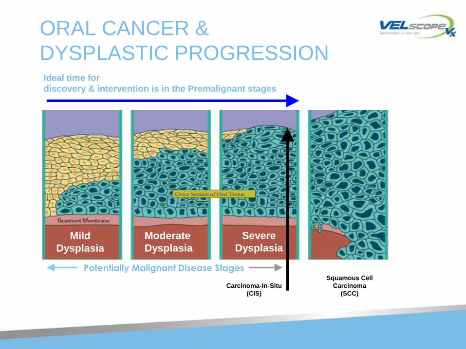
ORAL CANCER &
DYSPLASTIC PROGRESSION
Mild
Dysplasia
Moderate
Dysplasia
Severe
Dysplasia
Potentially Malignant Disease Stages
Ideal time for
discovery & intervention is in the Premalignant stages
Squamous Cell
Carcinoma
(SCC)
Carcinoma-In-Situ
(CIS)
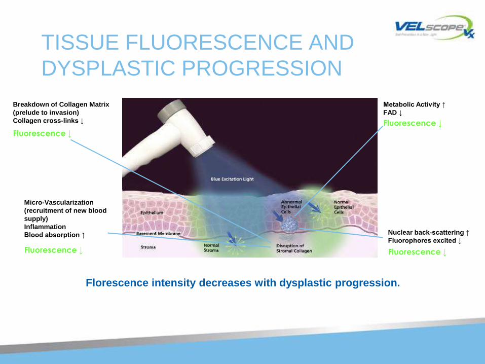
TISSUE FLUORESCENCE AND
DYSPLASTIC PROGRESSION
Florescence intensity decreases with dysplastic progression.
Breakdown of Collagen Matrix
(prelude to invasion)
Collagen cross-links ↓
Micro-Vascularization
(recruitment of new blood
supply)
Inflammation
Blood absorption ↑ Nuclear back-scattering ↑
Fluorophores excited ↓
Metabolic Activity ↑
FAD ↓
Fluorescence ↓
Fluorescence ↓
Fluorescence ↓
Fluorescence ↓
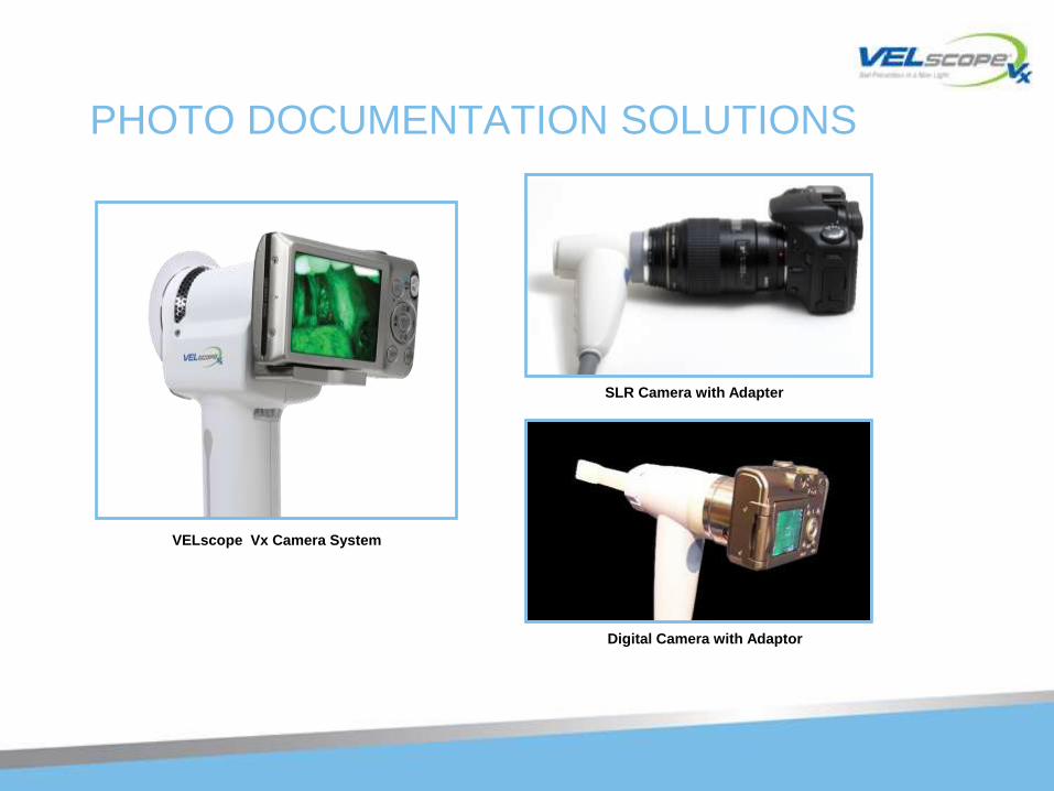
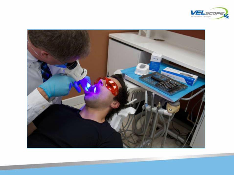
PHOTO DOCUMENTATION SOLUTIONS
Digital Camera with Adaptor
SLR Camera with Adapter
VELscope Vx Camera System
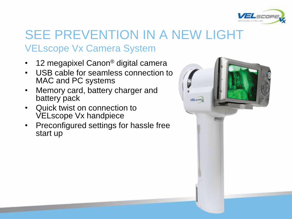
SEE PREVENTION IN A NEW LIGHTVELscope Vx Camera System
• 12 megapixel Canon® digital camera
• USB cable for seamless connection to MAC and PC systems
• Memory card, battery charger and battery pack
• Quick twist on connection to VELscope Vx handpiece
• Preconfigured settings for hassle free start up
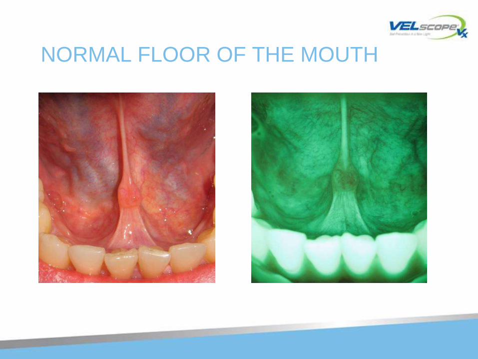
NORMAL FLOOR OF THE MOUTH
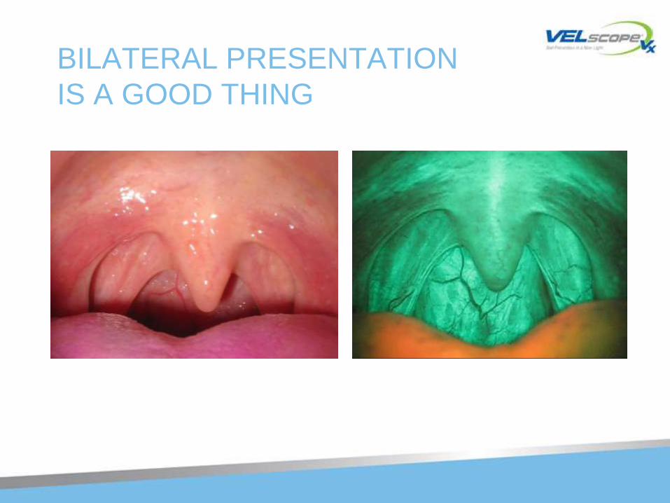
BILATERAL PRESENTATION
IS A GOOD THING
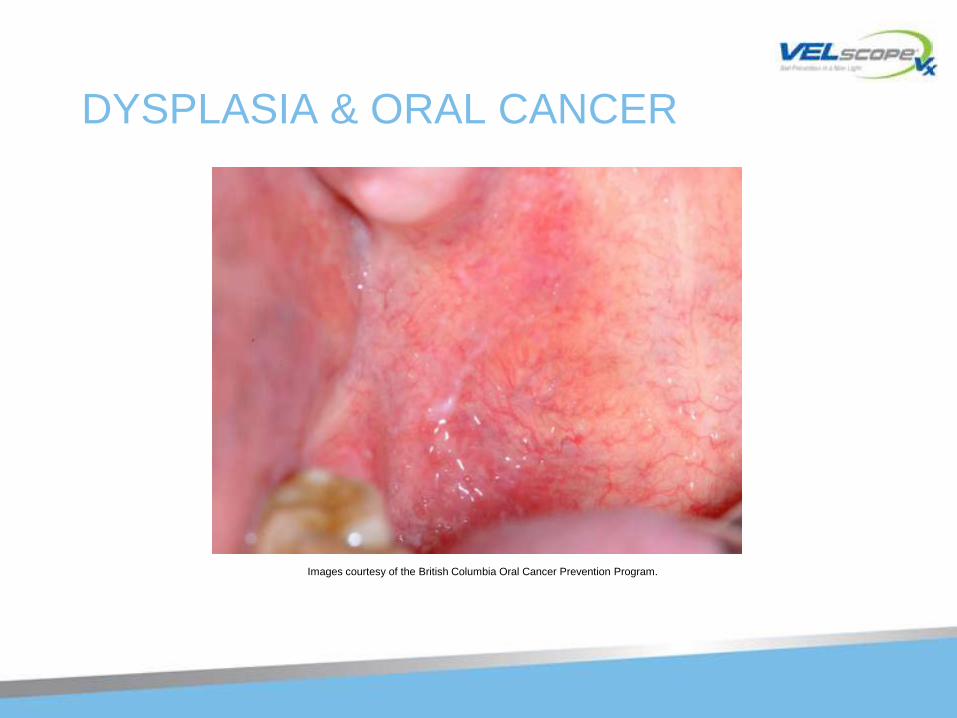
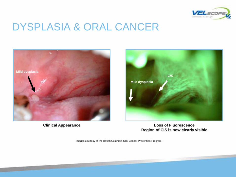
DYSPLASIA & ORAL CANCER
Images courtesy of the British Columbia Oral Cancer Prevention Program.
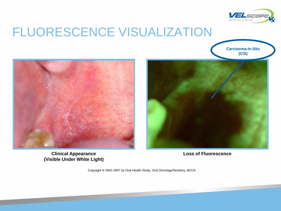
FLUORESCENCE VISUALIZATION
Copyright ® 2002-2007 by Oral Health Study, Oral Oncology/Dentistry, BCCA
Clinical Appearance
(Visible Under White Light)
Loss of Fluorescence
Carcinoma-In-Situ
(CIS)
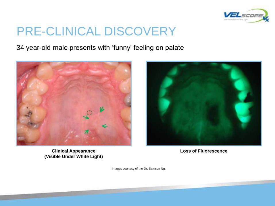
PRE-CLINICAL DISCOVERY
Images courtesy of the Dr. Samson Ng.
Clinical Appearance
(Visible Under White Light)
Loss of Fluorescence
34 year-old male presents with ‗funny‘ feeling on palate
DYSPLASIA & ORAL CANCER
Images courtesy of the British Columbia Oral Cancer Prevention Program.
Clinical Appearance Loss of Fluorescence
Region of CIS is now clearly visible
Mild dysplasia
CIS
Mild dysplasia
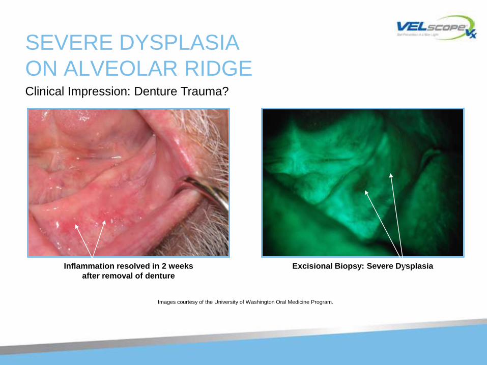
SEVERE DYSPLASIA
ON ALVEOLAR RIDGE
Images courtesy of the University of Washington Oral Medicine Program.
Clinical Impression: Denture Trauma?
Inflammation resolved in 2 weeks
after removal of denture
Excisional Biopsy: Severe Dysplasia
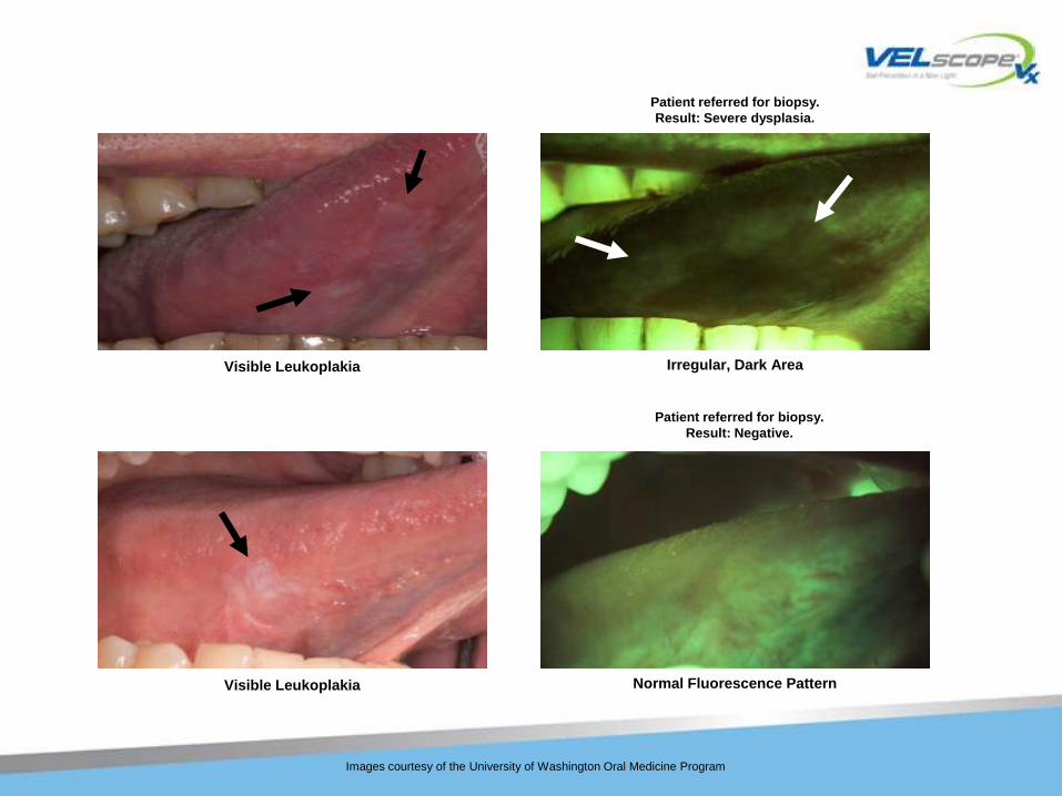
Visible Leukoplakia Irregular, Dark Area
Images courtesy of the University of Washington Oral Medicine Program
Visible Leukoplakia Normal Fluorescence Pattern
Patient referred for biopsy.
Result: Negative.
Patient referred for biopsy.
Result: Severe dysplasia.
VELscope CAN ALSO
DISCOVER THESE CONDITIONS: • Lichen Planus
• Lichenoid mucositis
• Squamous Papillomas
• Candidiasis
• Viral and bacterial infections
• Inflammation from a variety of causes (e.g. trauma)
• Salivary gland tumors
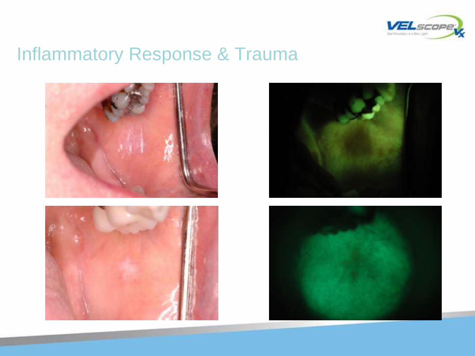
Inflammatory Response & Trauma
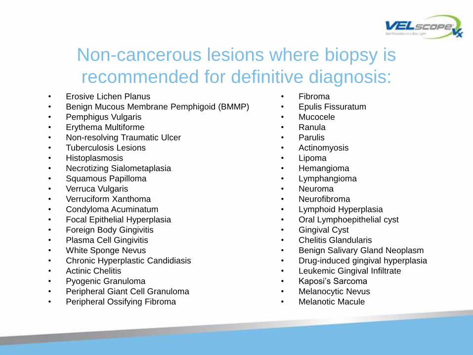
Non-cancerous lesions where biopsy is
recommended for definitive diagnosis:• Erosive Lichen Planus
• Benign Mucous Membrane Pemphigoid (BMMP)
• Pemphigus Vulgaris
• Erythema Multiforme
• Non-resolving Traumatic Ulcer
• Tuberculosis Lesions
• Histoplasmosis
• Necrotizing Sialometaplasia
• Squamous Papilloma
• Verruca Vulgaris
• Verruciform Xanthoma
• Condyloma Acuminatum
• Focal Epithelial Hyperplasia
• Foreign Body Gingivitis
• Plasma Cell Gingivitis
• White Sponge Nevus
• Chronic Hyperplastic Candidiasis
• Actinic Chelitis
• Pyogenic Granuloma
• Peripheral Giant Cell Granuloma
• Peripheral Ossifying Fibroma
• Fibroma
• Epulis Fissuratum
• Mucocele
• Ranula
• Parulis
• Actinomyosis
• Lipoma
• Hemangioma
• Lymphangioma
• Neuroma
• Neurofibroma
• Lymphoid Hyperplasia
• Oral Lymphoepithelial cyst
• Gingival Cyst
• Chelitis Glandularis
• Benign Salivary Gland Neoplasm
• Drug-induced gingival hyperplasia
• Leukemic Gingival Infiltrate
• Kaposi‘s Sarcoma
• Melanocytic Nevus
• Melanotic Macule
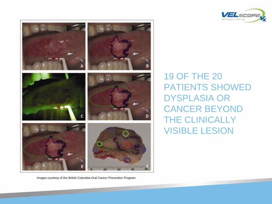
19 OF THE 20
PATIENTS SHOWED
DYSPLASIA OR
CANCER BEYOND
THE CLINICALLY
VISIBLE LESION
Images courtesy of the British Columbia Oral Cancer Prevention Program.
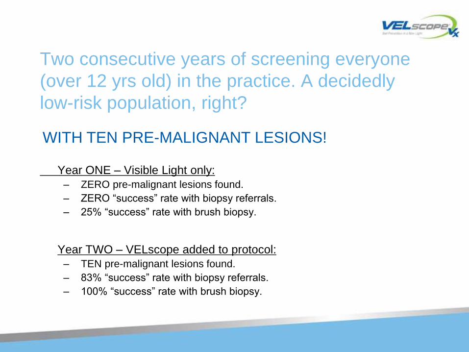
Two consecutive years of screening everyone
(over 12 yrs old) in the practice. A decidedly
low-risk population, right?
Year ONE – Visible Light only:
– ZERO pre-malignant lesions found.
– ZERO ―success‖ rate with biopsy referrals.
– 25% ―success‖ rate with brush biopsy.
Year TWO – VELscope added to protocol:
– TEN pre-malignant lesions found.
– 83% ―success‖ rate with biopsy referrals.
– 100% ―success‖ rate with brush biopsy.
WITH TEN PRE-MALIGNANT LESIONS!
Study at TATA Memorial Hospital, Mumbai
In a recent study conducted at Tata Memorial Hospital (Mumbai), Velscope® was
used to obtain fluorescence images from 261 sites in the oral cavity from 76 patients
and 90 sites in the oral cavity from 33 normal volunteers. From the results of the
study, it was concluded that the performance of Velscope® which is a
simple, objective low-cost system has the potential to improve oral screening
efforts, especially in low resource settings.
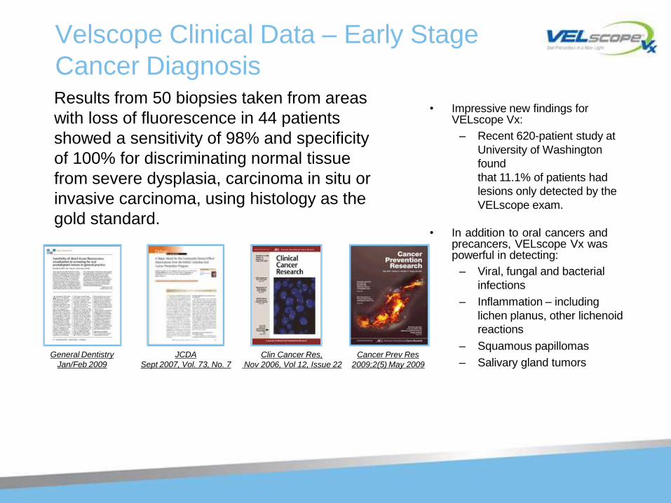
Velscope Clinical Data – Early Stage
Cancer Diagnosis
JCDA
Sept 2007, Vol. 73, No. 7
Clin Cancer Res, Cancer Prev Res
Nov 2006, Vol 12, Issue 22 2009;2(5) May 2009
General Dentistry
Jan/Feb 2009
• Impressive new findings for VELscope Vx:
– Recent 620-patient study at
University of Washington
found
that 11.1% of patients had
lesions only detected by the
VELscope exam.
• In addition to oral cancers andprecancers, VELscope Vx waspowerful in detecting:
– Viral, fungal and bacterial
infections
– Inflammation – including
lichen planus, other lichenoid
reactions
– Squamous papillomas
– Salivary gland tumors
Results from 50 biopsies taken from areas
with loss of fluorescence in 44 patients
showed a sensitivity of 98% and specificity
of 100% for discriminating normal tissue
from severe dysplasia, carcinoma in situ or
invasive carcinoma, using histology as the
gold standard.
NEW CLINICAL STUDIES
• Impressive new findings for VELscope Vx:
– Recent 620-patient study at University of Washington found
that 11.1% of patients had lesions only detected by the
VELscope exam.
• In addition to oral cancers and precancers, VELscope Vx was powerful in detecting:
– Viral, fungal and bacterial infections
– Inflammation – including lichen planus, other lichenoid reactions
– Squamous papillomas
– Salivary gland tumors
VELscope ENDORSEMENTS
• June 30, 2010 – World Health Organization recognizes VELscope Vx as ―an innovative device‖ that addresses global health concerns.
– 1 of 8 commercial devices to be so honored.
– The only dental device to be honored.
• AGD – partnership with LED Dental / VELscope
– 12 oral cancer screening courses with CDE.
– Includes advertising for VELscope Vx.
• University of Washington study led by Dr. Ed Truelove
– Impressive new findings with use of VELscope Vx by GPs.
– Clear case for expanded/routine usage in clinical practice.
• Many NA teaching hospitals and over 100 thought leaders are ―VELscope lovers.‖
VELscope Vx
• Supporting Preventive Health Care
• Preserving Quality Of Life
• Improving Clinical Decisions
• Helping Build Dental Practices
• Maybe Even Saving A Life
A “Win-Win” For The Patient And The Practice
VELscope Vx
• There are many reasons to feel good about giving your patients regular enhanced oral soft tissue exams (featuring both white light and VELscope Vx screenings)
– You‘re taking actions that most practices aren‘t.
– You‘re giving your patients—and yourself—peace of mind.
– You‘re letting patients know that you care about their health.
– It won‘t disrupt your practice.
• The two exams take 5 minutes combined.
• The VELscope Vx exam is free of rinses, dyes and discomfort.
– You‘re using a proven adjunctive technology trusted for millions of
exams
– You‘ll significantly increase your bottom line.
– You‘ll reduce the risk of lawsuit.
– YOU JUST MIGHT SAVE A LIFE OR TWO!