Embed Size (px)
Citation preview
Treatment and outcome of
anatomical factors for
abortions
Dr Rajesh Gajbhiye
Consultant Gynecologist & Laparoscopic Surgeon

Anatomical Factors
15% of women evaluated for RPL
Congenital or acquired.
Infertility, preterm labor, and abnormal
presentation.
Amenable to surgical correction.
Mullerian anomalies
Septate uterus
Absent or incomplete resorption of the
intervening uterovaginal septum
following fusion of the müllerian ducts.
Most common congenital anomaly of
the uterus, comprising approximately
55% of all anomalies.
A septum is partial or complete.
Septate Uterus
Spontaneous abortion rate is high,
averaging approximately 65% .
Raga et al reported a 25.5% incidence
of early miscarriage (< 13 weeks) and
a 6.2% incidence of late miscarriage
(14 to 22 weeks) in women with
septate uterus
Transabdominal metroplasty has been
abandoned because of the higher risk
of complications, including
postoperative reduction of intrauterine
volume, formation of intrauterine and
pelvic adhesions, and tubal occlusion.
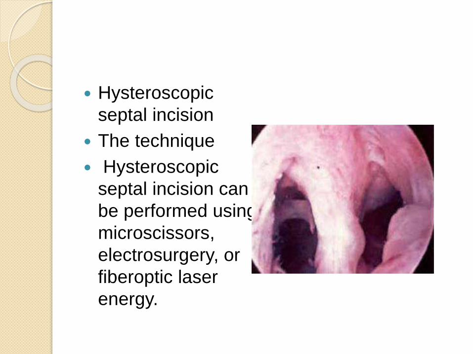
Hysteroscopic
septal incision
The technique
Hysteroscopic
septal incision can
be performed using
microscissors,
electrosurgery, or
fiberoptic laser
energy.

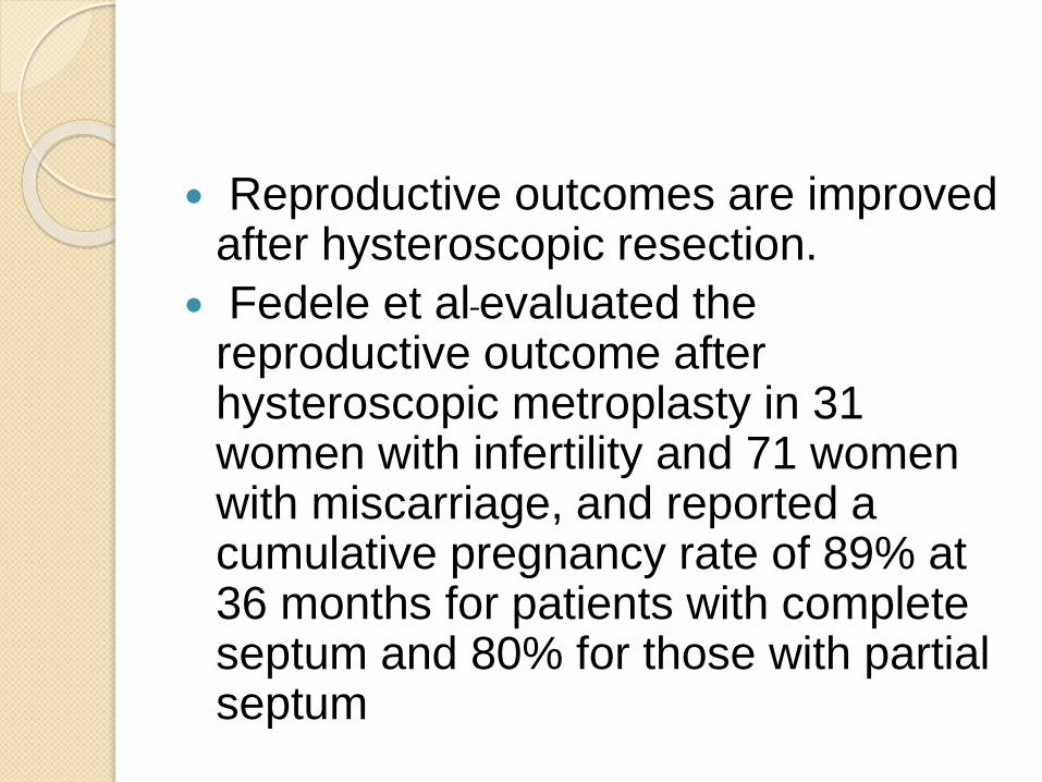
Reproductive outcomes are improved after hysteroscopic resection.
Fedele et al evaluated the reproductive outcome after hysteroscopic metroplasty in 31 women with infertility and 71 women with miscarriage, and reported a cumulative pregnancy rate of 89% at 36 months for patients with complete septum and 80% for those with partial septum
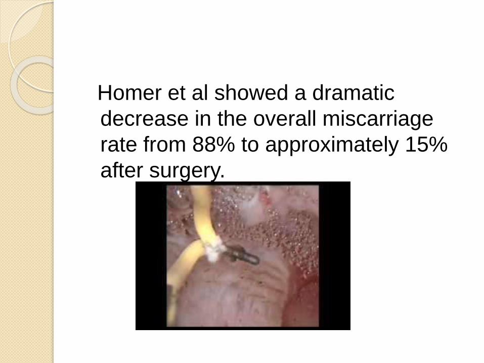
Homer et al showed a dramatic
decrease in the overall miscarriage
rate from 88% to approximately 15%
after surgery.
Laparoscopic guidance frequently is
used during hysteroscopic metroplasty
to reduce the risk of uterine
perforation.
IUD insertion for 3 months with
estrogenisation is only recommended
for complete or wide septa.
Bicornuate uterus
This anomaly is a result of incomplete
fusion of the uterine horns at the level
of the fundus. The distinguishing
aspect of this anomaly is the presence
of two separate but communicating
endometrial cavities and
a single cervix.
Overall, the spontaneous abortion rate
is approximately 32%,
The premature birth rate is
approximately 21%,
The fetal survival rate is approximately
60%.
Strassman metroplasty is most often
reserved for selected patients with
RPL or premature births.
Despite controversy about its
role, there is good evidence that live
birth rates following abdominal
metroplasty improved from 3.7% to
80%.
Unicornuate ut
Spontaneous abortion rates in these
women approach 31%, premature
birth rates approach 15%, and fetal
survival is estimated at 39%.
Other pregnancy complications
include malpresentation, IUGR,
uterine rupture, and ectopic
pregnancies
Current available evidence, women
with a unicornuate uterus and no
previous history of second-trimester
loss or premature birth should be
managed expectantly with frequent
assessment of cervical length and
anatomy.
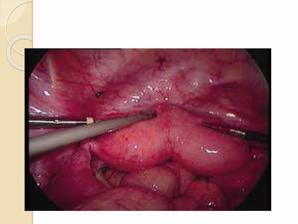
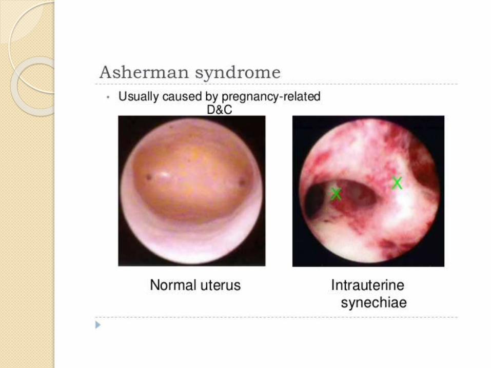
Acquired Uterine anomalies
Intrauterine Adhesions
Intrauterine trauma resulting from
vigorous endometrial curettage
After multiple myomectomy,septum
resection.
Associated with RPL.
The severity of adhesions may range
from minimal to complete
The reproductive outcomes
of women with Asherman syndrome.
are generally poor. In the absence of
treatment, approximately 40% of
pregnancies in these women appear
to end in spontaneous abortion and
another 23% result in preterm
deliveries.
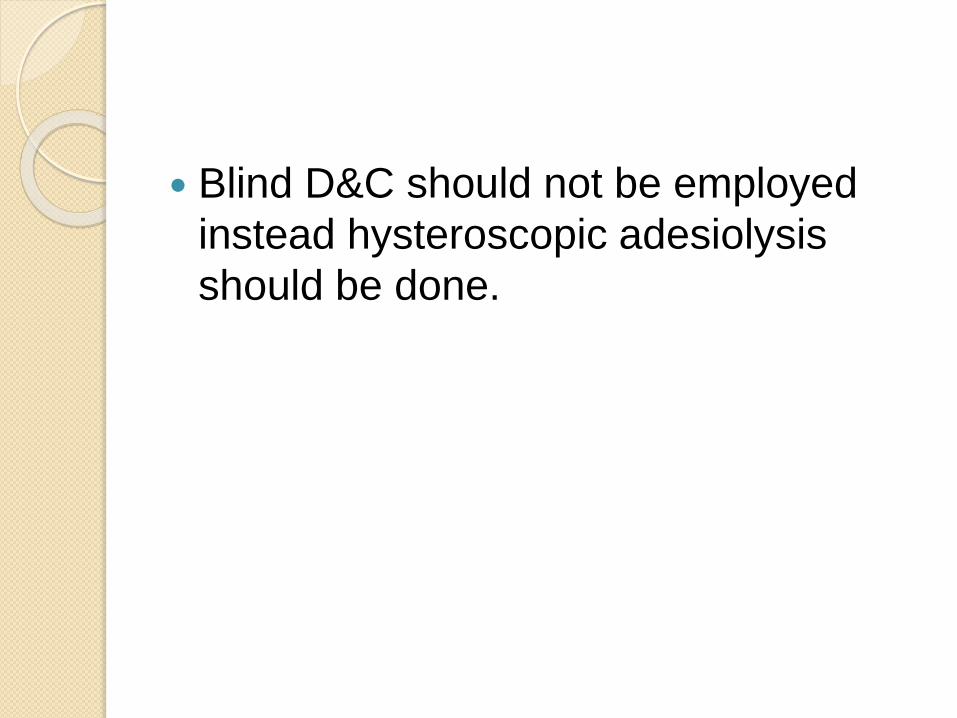
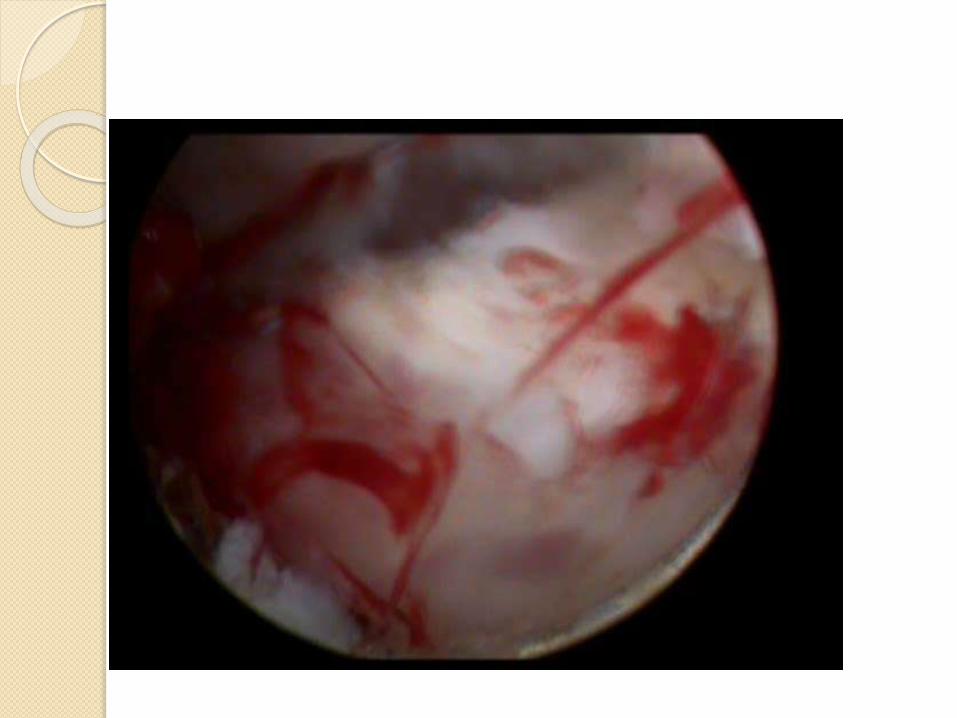
Blind D&C should not be employed
instead hysteroscopic adesiolysis
should be done.
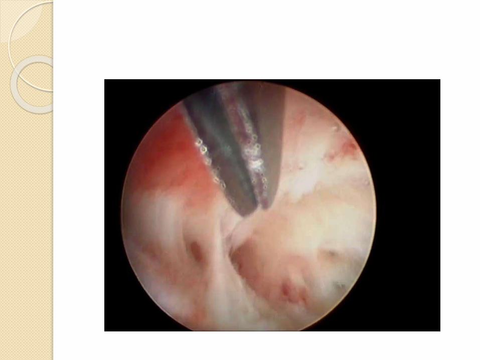
ESGE Grade 3&4 require
electrosurgical adhesiolysis and
pregnancy rates are 20-40%
Post op IUD and estrogen is
adminsitered after electrosurgical
adhesiolysis.
Complication rates are also high
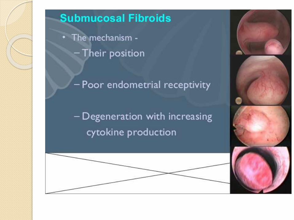
Hysteroscopic myomectomy has been
used to treat women with submucous
fibroids, infertility, and RPL.
Surgery should be adviced in patients
of RPL in which abortuses were
phenotypically normal with viability
upto 9 wks
Women with repetitive second TM
The pregnancy rates after -17-77%
with mean of 45%.
Cervical insufficiency
Cervical Incompetance is now
correctly termed as Cervical
Insufficiency.
It is primarily a clinical diagnosis by
recurrent painless dilatation and
spontaneous midtrimester loss.
It is a component of larger and more
complex preterm birth syndrome.
Diagnosis of Cervical
insufficiency during any
pregnancy.
Cervical insufficiency is defined by TVU cervical length <25 mm and/or advanced cervical changes on physical examination before 24 weeks of gestation in women with either:
One or morepriorpregnancy losses/births at 14 to 36 weeks, and/or
•Other significant risk factors for cervical insufficiency.
History-indicated cerclage
For women with two or more
consecutive prior second trimester
pregnancy losses or three or more
early preterm births
Who have risk factors for cervical
insufficiency and in whom other
causes of preterm birth have been
excluded.
USG indicated cerclage
For women with suspected cervical
insufficiency and prior early preterm
birth who do not meet criteria for
history-indicated cerclage,
sonographic surveillance should be
started early in pregnancy (eg, 14 to
16 weeks). cerclage for women who
develop a short cervix (<25 mm)
before 24 weeks
Physical exam-indicated
cerclage Also called “rescue cerclage” or
“emergency cerclage.
Placement of a physical exam-
indicated cerclage when a dilated
cervix and visible membranes are
detected on digital examination at <24
wks.
Small RCT have shown prolongation
of pregnancy by 4 wks
Even upto 4cm dilatation.
Macdonald and Shirodkars claiming
success rate 80-90%.
It reinforces the internal os with non
absorbable tape or suture.
In proven case prophylactic cerclage
to be done at 14 wks or 2 weeks
before the prior loss as early as 10
weeks
Contraindications
Intrauterine infections
Ruptured membranes
H/O vaginal bleeding
Uterine irritability
Cervical dilatation >4cm
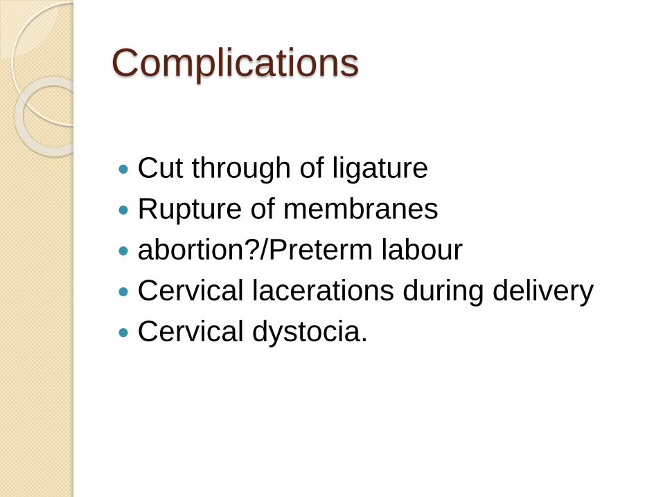
Complications
Cut through of ligature
Rupture of membranes
abortion?/Preterm labour
Cervical lacerations during delivery
Cervical dystocia.
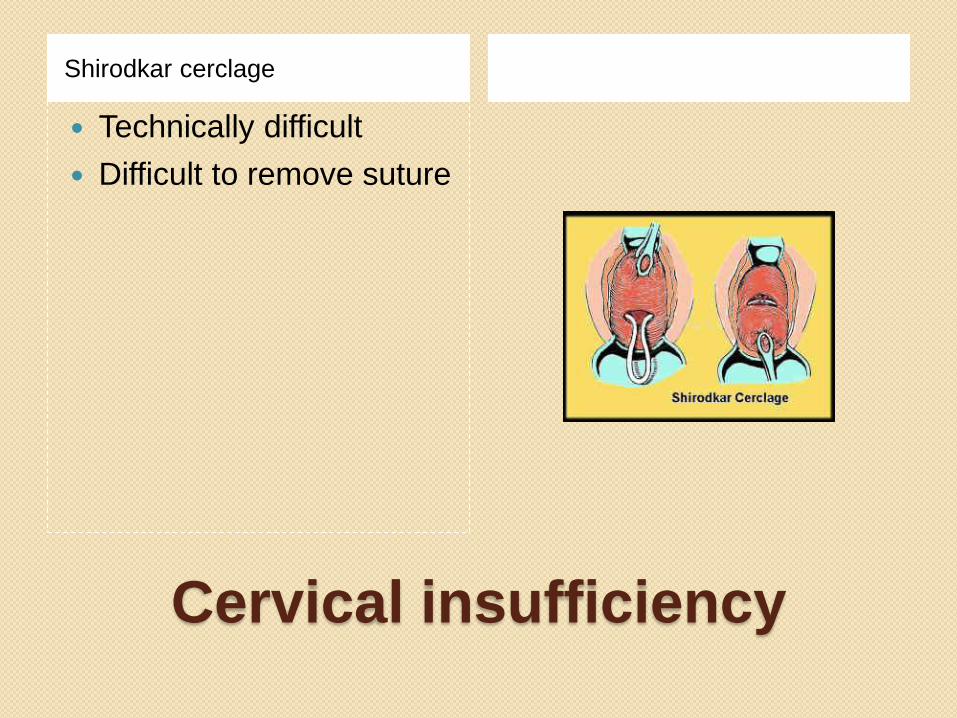
Cervical insufficiency
Shirodkar cerclage
Technically difficult
Difficult to remove suture
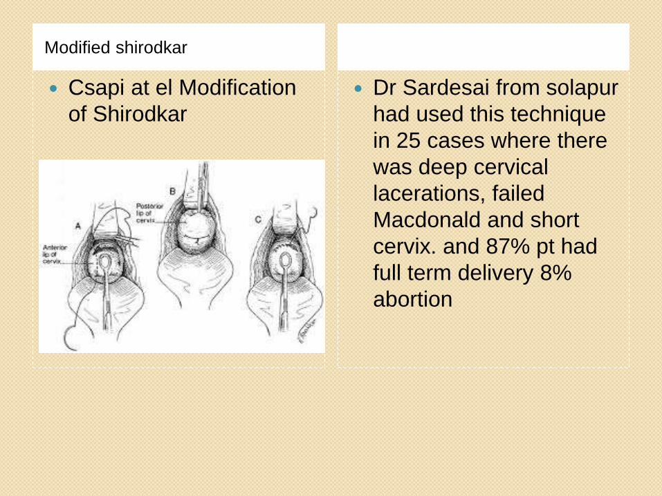
Modified shirodkar
Csapi at el Modification
of Shirodkar
Dr Sardesai from solapur
had used this technique
in 25 cases where there
was deep cervical
lacerations, failed
Macdonald and short
cervix. and 87% pt had
full term delivery 8%
abortion

4-5 Bites as high in the
cervix
Mersiline
Tape,Silk,Prolene
Knot tied anteriorly
Removal at 37 weeks or
if goes in labour.
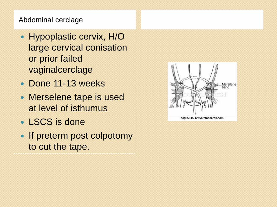
Abdominal cerclage
Hypoplastic cervix, H/O
large cervical conisation
or prior failed
vaginalcerclage
Done 11-13 weeks
Merselene tape is used
at level of isthumus
LSCS is done
If preterm post colpotomy
to cut the tape.
Ludmir
Can be done in non
pregnant state
Disadvantage is inability
to conceive.
Vaginally with USG
guidence
And putting suture in
criss cross fashion.

Alternative treatment include bed rest,
Pharmacological treatment and
Pessary.
Randomised studies are needed for
their evaluation.
Conclusion
Abortion occuring after USG
confirmation of a viable pregnancy at
8-9 weeks may be more attributable to
uterine fusion defects.
Women with second trimester abortion
could benefit from uterine
reconstruction but it is not advised if
losses are restricted to first trimester.
Operative hysteroscopy like septal
resection,myoma
resection,adhesiolysis
Improves the pregnancy outcome
Women with history of previous
painless and spontaneous
midtrimester losses,or previous
preterm birth who then develop short
midtrimester cervical length have a
treatable component of cervical
insufficiency and surgical intervention
in the form of cerclage to be done.

Candidates for ultrasound surveillance and possible ultrasound indicated cerclage —The majority of women with suspected cervical insufficiency do not meet the above criteria for history-indicated cerclage. For these women, we usually initiate TVU cervical length screening (table 2), administer 17-alpha-hydroxyprogesterone caproate prophylaxis, and apply a cerclage if cervical length decreases to <25 mm [27]. The rationale for this approach is:
●Women with a short cervix on transvaginal ultrasound examination are at increased risk of spontaneous preterm birth [28].
•In women with a history of spontaneous preterm birth, a systematic review of controlled studies showed that measurement of cervical length in the second trimester, especially before 24 weeks, predicted the risk of recurrent preterm birth [28]. The use of a TVU cervical length <25 mm at <24 weeks to predict preterm birth at <35 weeks yielded sensitivity of 65.4 percent, specificity of 75.5 percent, positive predictive value of 33.0 percent, and negative predictive value of 92.0 percent. The shorter the cervical length, the higher the positive likelihood ratio for spontaneous preterm birth <35 weeks.
●In randomized trials, progesterone prophylaxis with 17 alpha hydroxy-progesterone caproatestarting at 16 to 20 weeks in women with a history of spontaneous preterm birth and continuing until 36 weeks reduced the risk of recurrent preterm birth [24,25]. (See "Progesterone supplementation to reduce the risk of spontaneous preterm birth".)
●Placement of cerclage upon identification of a short cervix (“ultrasound-indicated cerclage”) is effective in reducing preterm birth [29], results in pregnancy outcomes comparable to those with history-indicated cerclage [30], and avoids cerclage in about 60 percent of patients with a suggestive history [30]. The benefit of ultrasound-indicated cerclage may derive from bolstering cervical strength [31], preventing membranes from being exposed, and retention of the mucus plug.
We usually initiate cervical length
screening at 14 weeks, but may screen as
early as 12 weeks in women with early
second trimester losses, recurrent second
trimester losses, or prior large cold knife
conization (table 2) [32]. In women with
prior preterm birth at 28 to 36 weeks, we
initiate screening at 16 weeks. Ultrasound
examination is generally repeated every
two weeks until 24 weeks as long as the
cervical length is ≥30 mm, and increased
to weekly if cervical length is 25 to 29 mm,
with the expectation that preterm cervical
changes will precede overt preterm labor
or membrane rupture symptoms by three
to six weeks [33]. Transvaginal ultrasound
screening is usually discontinued at 24
weeks of gestation, as cerclage is not
usually performed after this time.
Overall, the prevalence of major
congenital anomalies appears to be
three-fold higher in women with RPL
compared with women without a
history of recurrent miscarriage.
Reduced intraluminal volume and/or inadequate vascular supply to the developing fetus and placenta.
There are no surgical procedures to enlarge the uterus.
The higher prevalence of cervical incompetence in uterine anomalies, however, has led some authors to recommend that cervical cerclage be placed to improve obstetrical outcome.
Methods of hysteroscopic
myomectomy
Unipolar resection with loop electrode
Bipolar resectoscope technique using
NS
Vaporising electrodes
Hysteroscopic Morcellators.


![Visual outcome evaluation of complicated perforating ... · infections [7], and traumatic lens cap sule rupture [8]. This immediate treatment aims to preserve the anatomical integrity](https://img.pdfslide.us/doc/110x75/607478a9d0543615c47c4757/visual-outcome-evaluation-of-complicated-perforating-infections-7-and-traumatic.jpg)