Embed Size (px)
Citation preview

BY – Dr. KANWALPREET KAUR
MODERATOR-Dr. KARUNA GUPTA
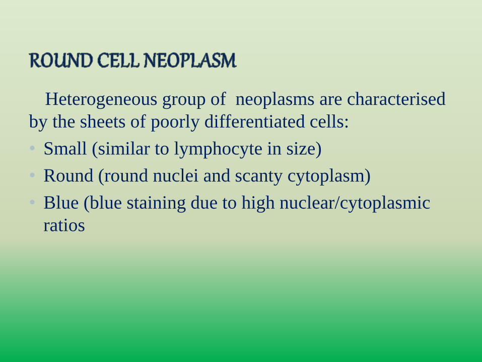
Heterogeneous group of neoplasms are characterised
by the sheets of poorly differentiated cells:
• Small (similar to lymphocyte in size)
• Round (round nuclei and scanty cytoplasm)
• Blue (blue staining due to high nuclear/cytoplasmic
ratios
• Ewings sarcoma/Primitive neuroectodermal tumour(ES/PNET
• Hematopoietic malignancies
• Plasma cell neoplasms
• Small cell osteosarcoma
• Mesenchymal chondrosarcoma
• Poorly differentiated small cell synovial sarcoma
• Rhabdomyosarcoma(embryonal and alveolar subtypes)
• Histological grading of bone sarcomas is an attempt to
predict the biological behaviour of a malignant tumour
• Criteria used—
1. Cellularity i.e., the relative amount of cells compared to
matrix
2.Nuclear features of the tumour cells
• Tumours which are monomorphic, such as small cell
malignancies (Ewing sarcoma, malignant lymphoma and
myeloma), do not lend themselves to histological grading.
• Mesenchymal chondrosarcomas and dedifferentiated
chondrosarcomas are always high grade
• Round cell sarcomas that show varying degrees of
neuroectodermal differentiation
Ewing’s sarcoma - limited neural differentiation
PNET - more neural features
• AGE- 5-20 years (commonly)
• infancy or adulthood rarely
• SEX PREDILITION- males:females=1.4:1
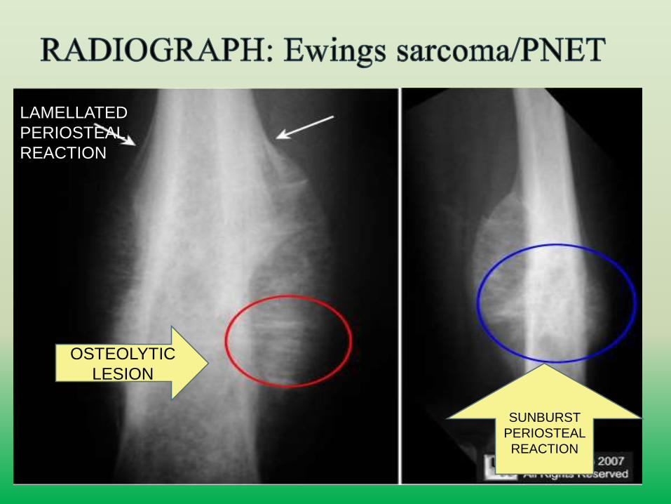
OSTEOLYTIC
LESION
LAMELLATED
PERIOSTEAL
REACTION
SUNBURST
PERIOSTEAL
REACTION
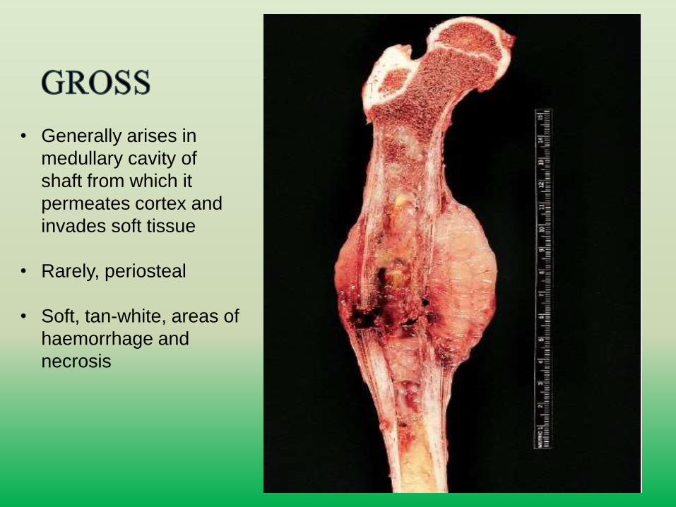
• Generally arises in
medullary cavity of
shaft from which it
permeates cortex and
invades soft tissue
• Rarely, periosteal
• Soft, tan-white, areas of
haemorrhage and
necrosis
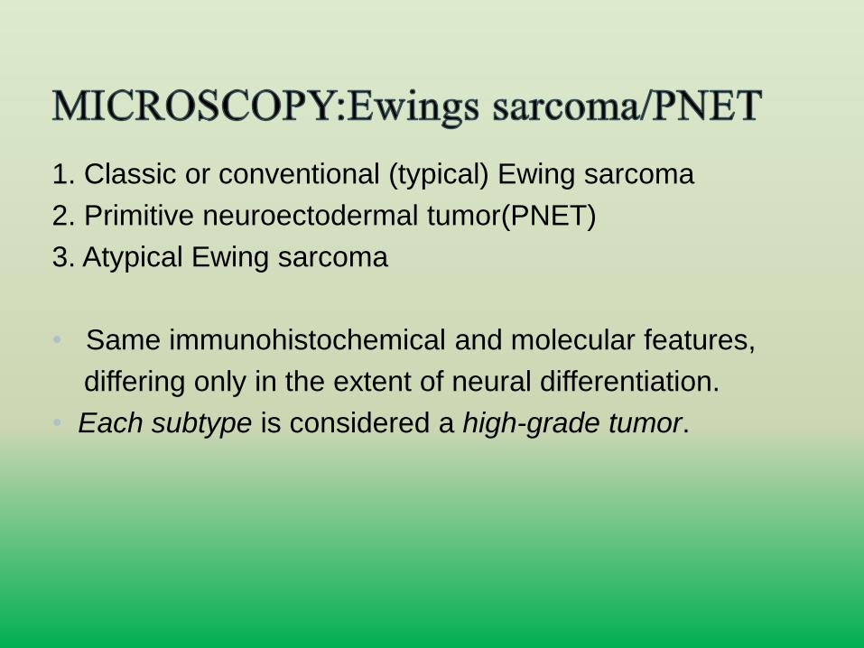
1. Classic or conventional (typical) Ewing sarcoma
2. Primitive neuroectodermal tumor(PNET)
3. Atypical Ewing sarcoma
• Same immunohistochemical and molecular features,
differing only in the extent of neural differentiation.
• Each subtype is considered a high-grade tumor.
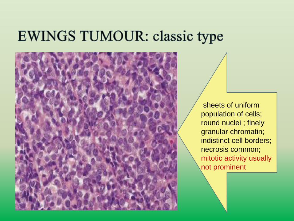
sheets of uniform
population of cells;
round nuclei ; finely
granular chromatin;
indistinct cell borders;
necrosis common;
mitotic activity usually
not prominent
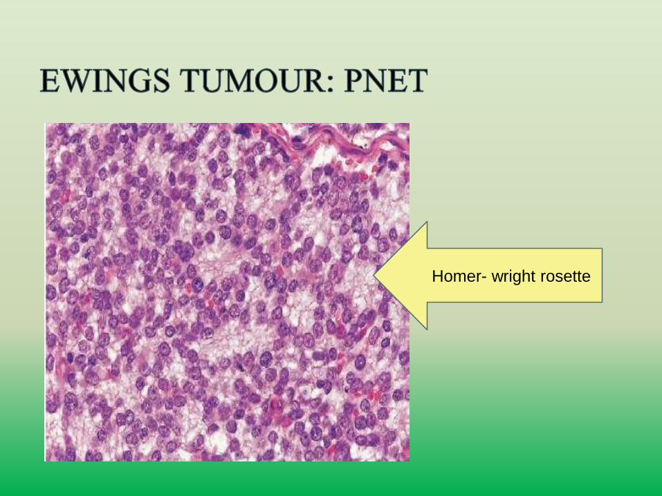
Homer- wright rosette
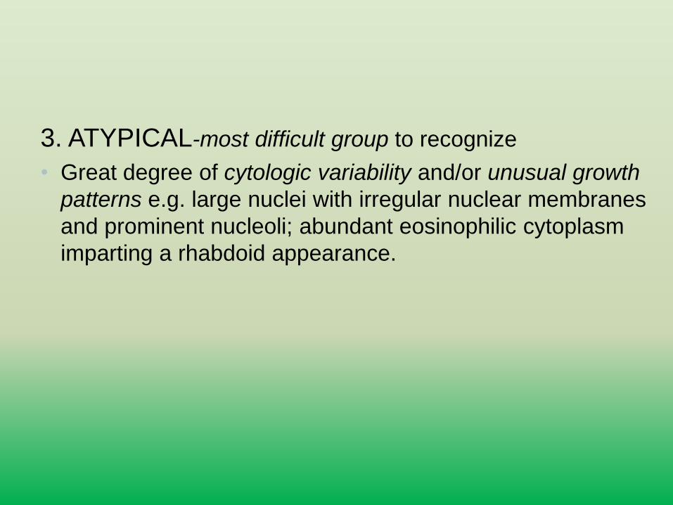
3. ATYPICAL-most difficult group to recognize
• Great degree of cytologic variability and/or unusual growth
patterns e.g. large nuclei with irregular nuclear membranes
and prominent nucleoli; abundant eosinophilic cytoplasm
imparting a rhabdoid appearance.
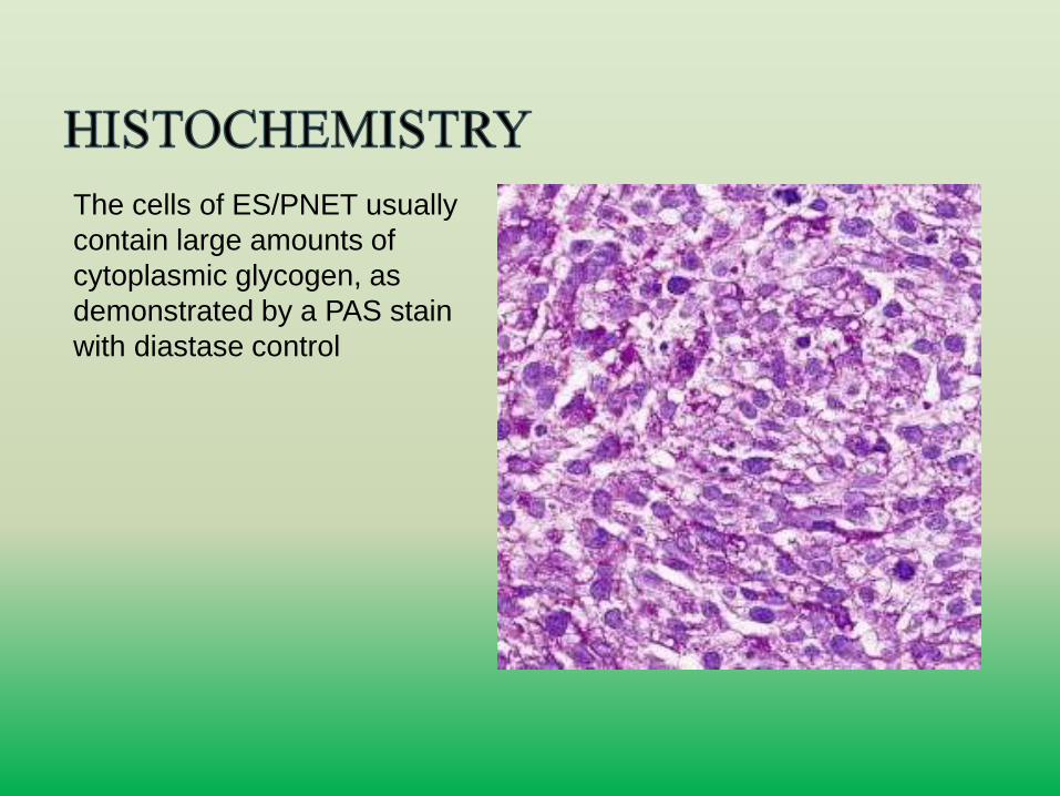
The cells of ES/PNET usually
contain large amounts of
cytoplasmic glycogen, as
demonstrated by a PAS stain
with diastase control
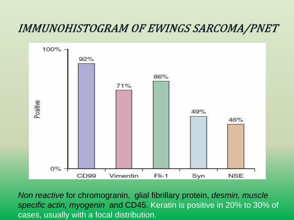
Non reactive for chromogranin, glial fibrillary protein, desmin, muscle
specific actin, myogenin and CD45. Keratin is positive in 20% to 30% of
cases, usually with a focal distribution.
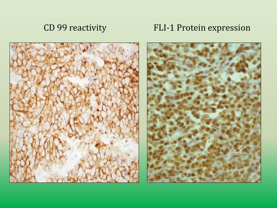
CD 99 reactivity FLI-1 Protein expression
• CD99- strong, diffuse membranous staining pattern
• 84-100% sensitive. If a tumor is negative for
CD99, it is highly unlikely to be Ewing sarcoma
• NOT A SPECIFIC MARKER
• Can be seen in RMS, glial tumours, neuroendocrine
tumours, lymphoblastic lymphoma, WT, clear cell
sarcoma of kidney, teratoma, synovial sarcoma,
osteosarcoma and mesenchymal chondrosarcoma
• CD99 is important for distinction between Ewings
sarcoma/PNET and metastatic neuroblastoma
• FLI1- Only nuclear staining is considered positive
NOT SPECIFIC MARKER
But also positive in lymphoblastic lymphoma,
myeloid neoplasms, DSRCT, Malignant melanoma, merkel
cell carcinoma,synovial sarcoma, and some vascular
neoplasms.
• Reciprocal translocation t(11;22)(q24;q12)
• Fusion of EWSR1 gene(encodes for RNA binding protein) at 22q12 with FLI1 gene(member of ETS family of transcription factors)
• The t(21;22)(q12;q12) translocation involves the gene ERG, which is located on chromosome 21
• t(7;22)(p22;q12) translocation involves a gene known as ETV1 at 7p22.
• Recently a translocation involving chromosomes 4 and 9 with CIC and DUX4 gene has been identified
• Rearrangements of EWSR1 with non–ETS-family
genes—including NFATC2,POU5F1, SMARCA5, ZSG,
and SP3—are also rarely identified
• FISH for EWSR1 genomic rearrangements
is highly sensitive (>95%) but nonspecific because
other tumours may show rearrangement of this locus.
• RT-PCR for EWSR1 fusion genes - highly sensitive
(>95%) and specific (100%).
• Primary Lymphoma- Originates in bone with no evidence of extraskeletal disease or disseminated bone marrow
involvement. Rare
• Secondary skeletal involvement by a primary extraosseouslymphoma is much more common than primary bone lymphoma
• AGE-usually affects 40-60 years
• Lymphoma however can involve children; though it is much less common than Ewing sarcoma in this age group
• SITE- meta-diaphyses of long bones (femur, humerus, and tibia) and axial skeleton (pelvis and vertebrae)
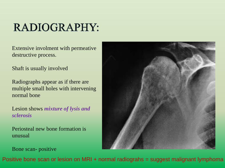
Extensive involment with permeative
destructive process.
Shaft is usually involved
Radiographs appear as if there are
multiple small holes with intervening
normal bone
Lesion shows mixture of lysis and
sclerosis
Periosteal new bone formation is
unusual
Bone scan- positive
Positive bone scan or lesion on MRI + normal radiograhs = suggest malignant lymphoma
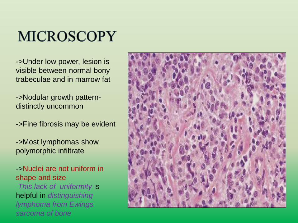
->Under low power, lesion is
visible between normal bony
trabeculae and in marrow fat
->Nodular growth pattern-
distinctly uncommon
->Fine fibrosis may be evident
->Most lymphomas show
polymorphic infiltrate
->Nuclei are not uniform in
shape and size
This lack of uniformity is
helpful in distinguishing
lymphoma from Ewings
sarcoma of bone
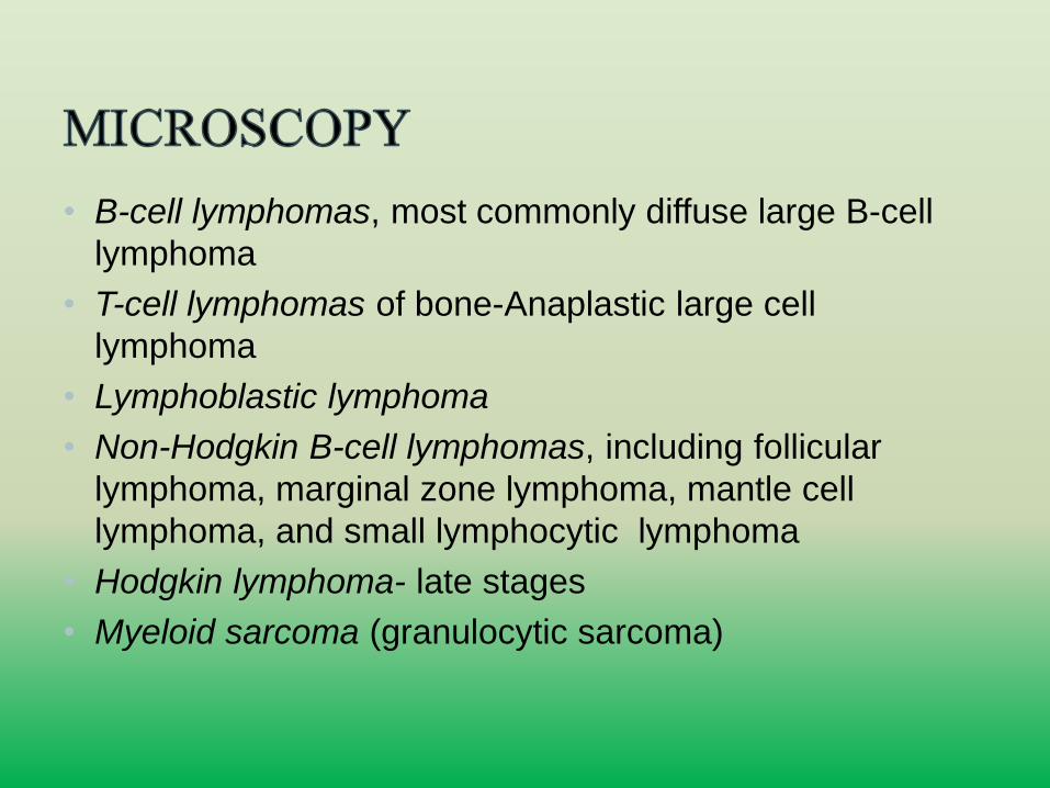
• B-cell lymphomas, most commonly diffuse large B-cell
lymphoma
• T-cell lymphomas of bone-Anaplastic large cell
lymphoma
• Lymphoblastic lymphoma
• Non-Hodgkin B-cell lymphomas, including follicular
lymphoma, marginal zone lymphoma, mantle cell
lymphoma, and small lymphocytic lymphoma
• Hodgkin lymphoma- late stages
• Myeloid sarcoma (granulocytic sarcoma)
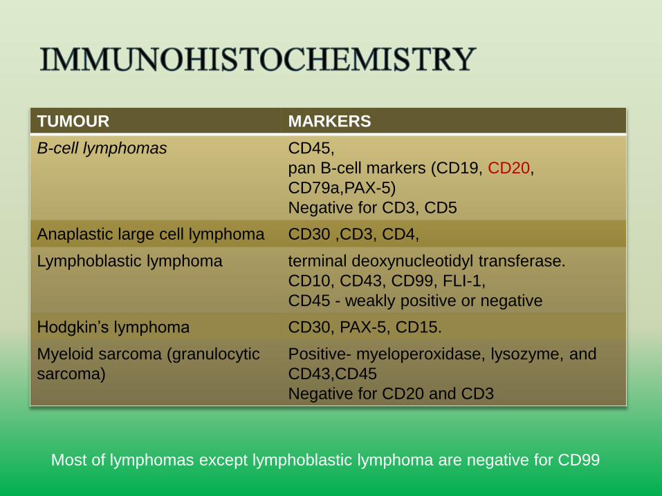
TUMOUR MARKERS
B-cell lymphomas CD45,
pan B-cell markers (CD19, CD20,
CD79a,PAX-5)
Negative for CD3, CD5
Anaplastic large cell lymphoma CD30 ,CD3, CD4,
Lymphoblastic lymphoma terminal deoxynucleotidyl transferase.
CD10, CD43, CD99, FLI-1,
CD45 - weakly positive or negative
Hodgkin’s lymphoma CD30, PAX-5, CD15.
Myeloid sarcoma (granulocytic
sarcoma)
Positive- myeloperoxidase, lysozyme, and
CD43,CD45
Negative for CD20 and CD3
Most of lymphomas except lymphoblastic lymphoma are negative for CD99
• Lymphoblastic lymphoma may be positive for CD99 and
negative for CD45, an immunoprofile that overlaps with
that of Ewing sarcoma
• TdT, CD34,CD43,CD10, CD79a and genetic
rearrangement studies- distinguish lymphoblastic
lymphoma from EWS/PNET
• Specific studies on primary lymphomas of bone are
lacking.
• malignant proliferation of monoclonal plasma cells
• can present as a solitary lesions (solitary plasmacytoma)
or more commonly as part of widespread disease
(multiple myeloma)
• AGE-50-70years
• rare below 40 years
• SITE-: vertebrae, ribs, skull, pelvis, femur, clavicle and
scapula
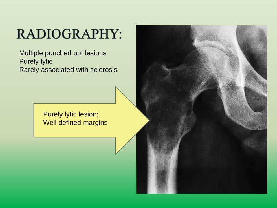
Multiple punched out lesions
Purely lytic
Rarely associated with sclerosis
Purely lytic lesion;
Well defined margins
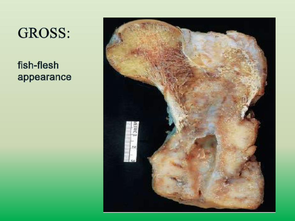
• Usually has a soft red-brown appearance.
• However, some myelomas show the fish-flesh
appearance typical of malignant lymphoma.
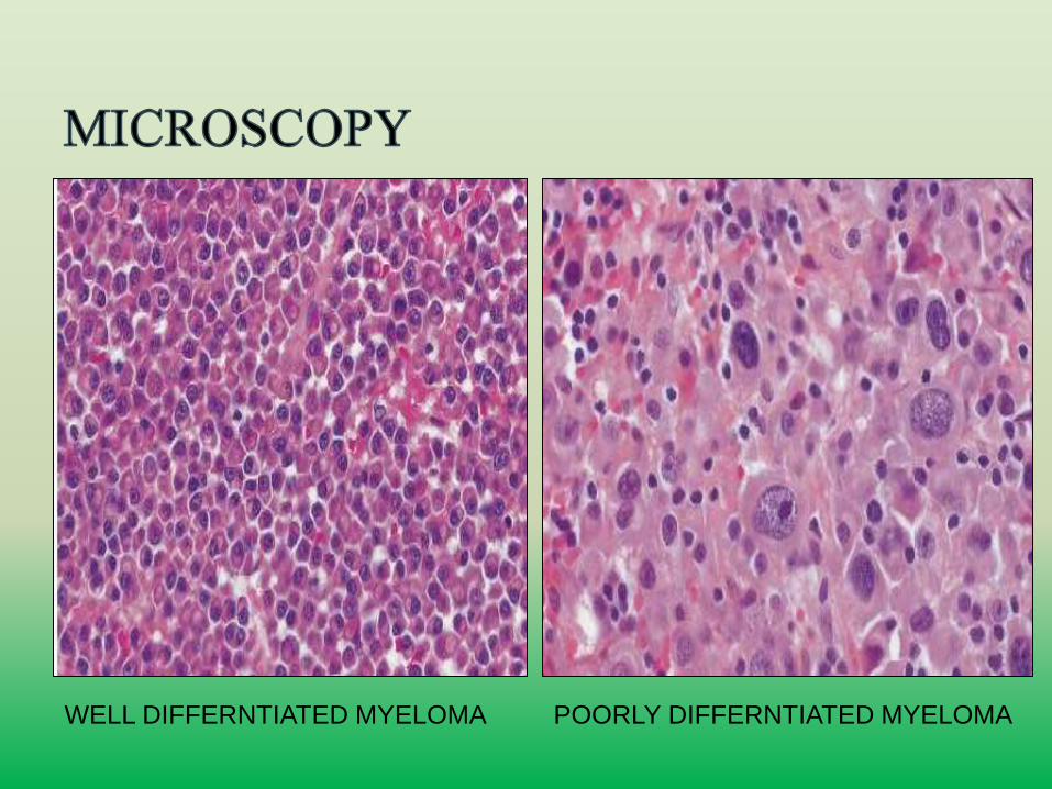
WELL DIFFERNTIATED MYELOMA
.
POORLY DIFFERNTIATED MYELOMA
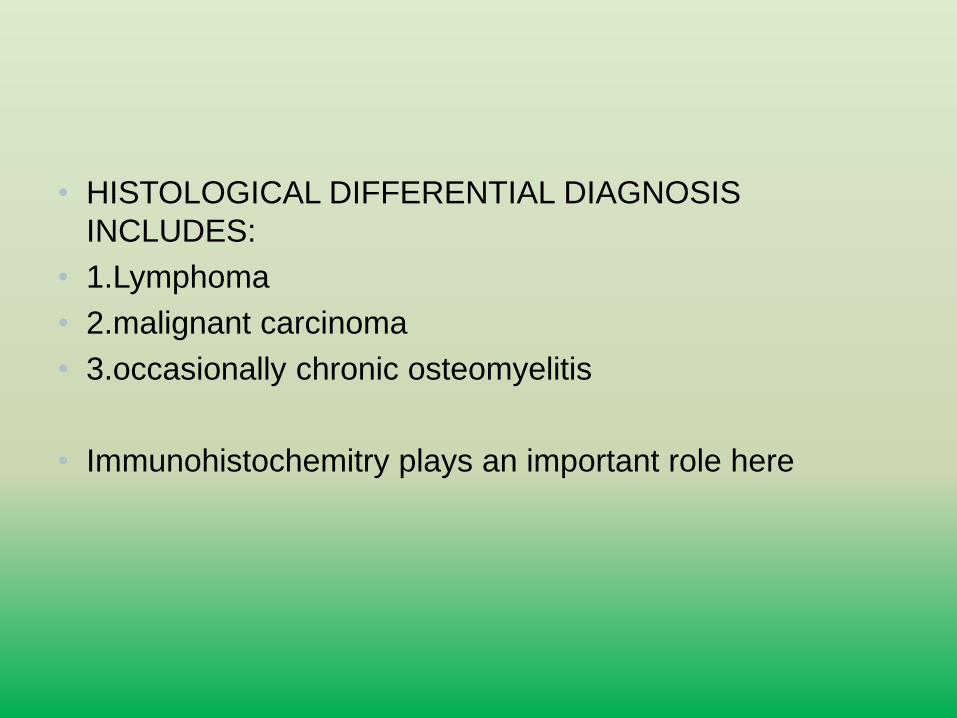
• HISTOLOGICAL DIFFERENTIAL DIAGNOSIS
INCLUDES:
• 1.Lymphoma
• 2.malignant carcinoma
• 3.occasionally chronic osteomyelitis
• Immunohistochemitry plays an important role here
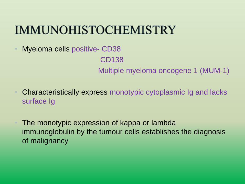
• Myeloma cells positive- CD38
CD138
Multiple myeloma oncogene 1 (MUM-1)
• Characteristically express monotypic cytoplasmic Ig and lacks
surface Ig
• The monotypic expression of kappa or lambda
immunoglobulin by the tumour cells establishes the diagnosis
of malignancy
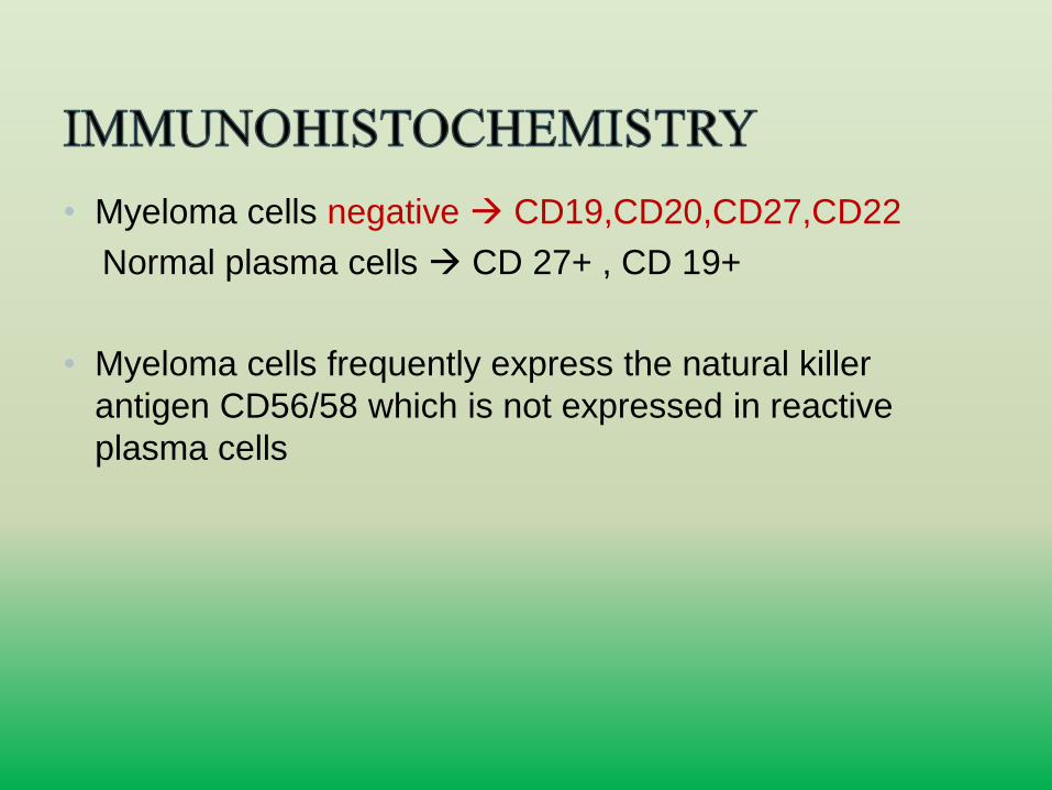
• Myeloma cells negative CD19,CD20,CD27,CD22
Normal plasma cells CD 27+ , CD 19+
• Myeloma cells frequently express the natural killer
antigen CD56/58 which is not expressed in reactive
plasma cells
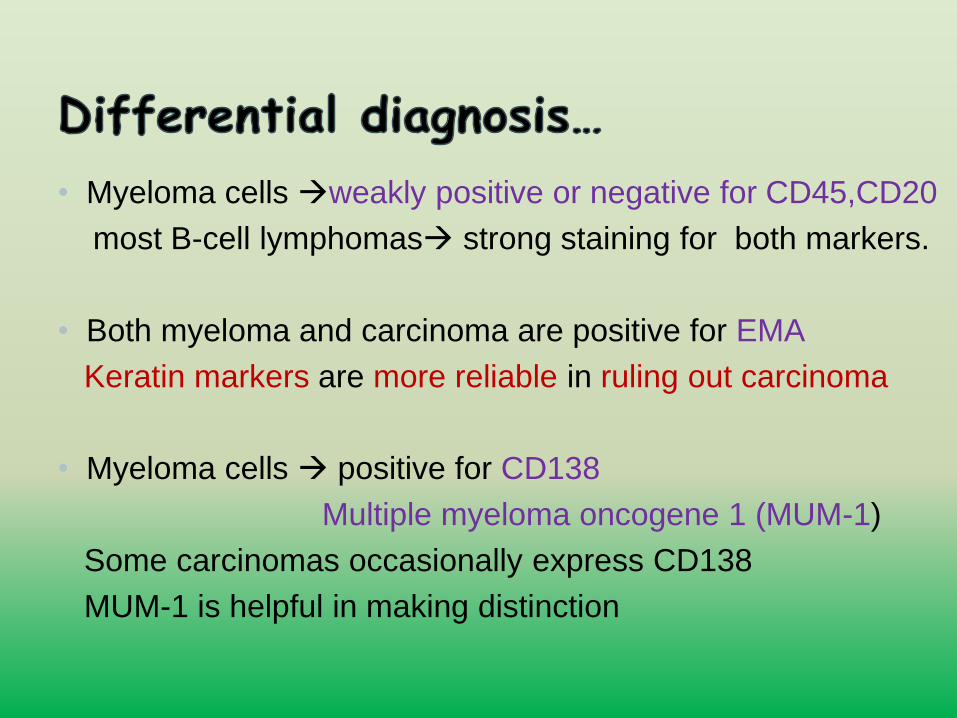
• Myeloma cells weakly positive or negative for CD45,CD20
most B-cell lymphomas strong staining for both markers.
• Both myeloma and carcinoma are positive for EMA
Keratin markers are more reliable in ruling out carcinoma
• Myeloma cells positive for CD138
Multiple myeloma oncogene 1 (MUM-1)
Some carcinomas occasionally express CD138
MUM-1 is helpful in making distinction
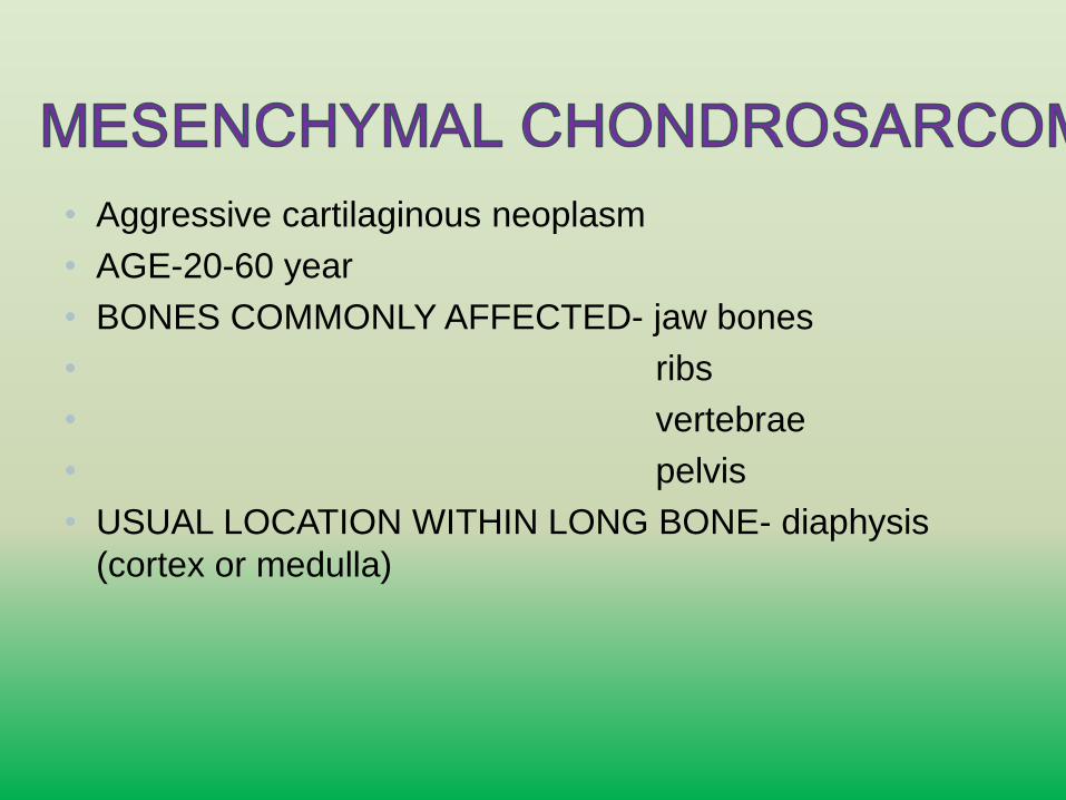
• Aggressive cartilaginous neoplasm
• AGE-20-60 year
• BONES COMMONLY AFFECTED- jaw bones
• ribs
• vertebrae
• pelvis
• USUAL LOCATION WITHIN LONG BONE- diaphysis
(cortex or medulla)
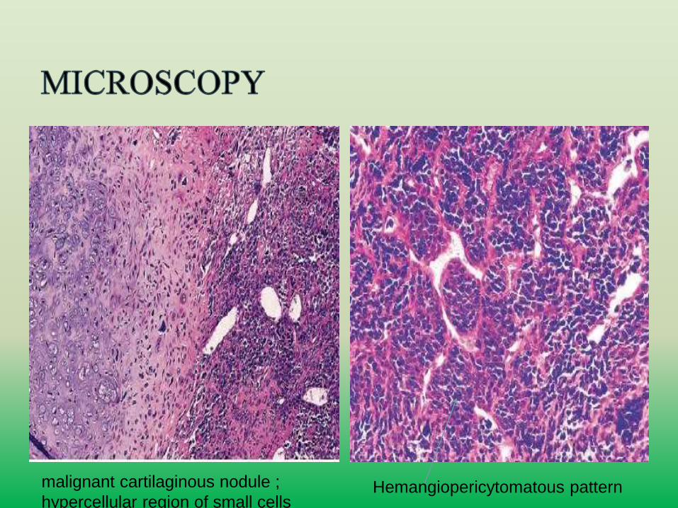
Hemangiopericytomatous patternmalignant cartilaginous nodule ;
hypercellular region of small cells
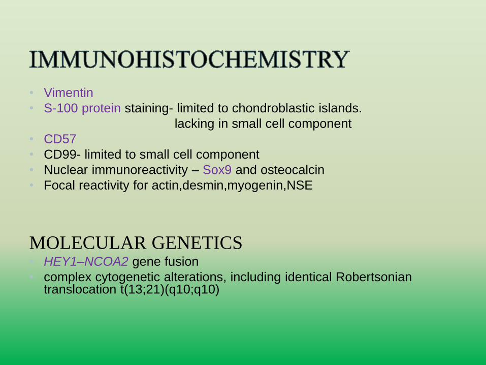
• Vimentin
• S-100 protein staining- limited to chondroblastic islands.
lacking in small cell component
• CD57
• CD99- limited to small cell component
• Nuclear immunoreactivity – Sox9 and osteocalcin
• Focal reactivity for actin,desmin,myogenin,NSE
MOLECULAR GENETICS• HEY1–NCOA2 gene fusion
• complex cytogenetic alterations, including identical Robertsoniantranslocation t(13;21)(q10;q10)
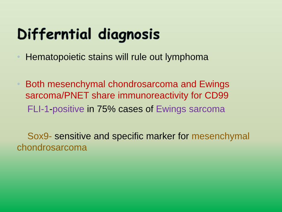
• Hematopoietic stains will rule out lymphoma
• Both mesenchymal chondrosarcoma and Ewings
sarcoma/PNET share immunoreactivity for CD99
FLI-1-positive in 75% cases of Ewings sarcoma
Sox9- sensitive and specific marker for mesenchymal
chondrosarcoma
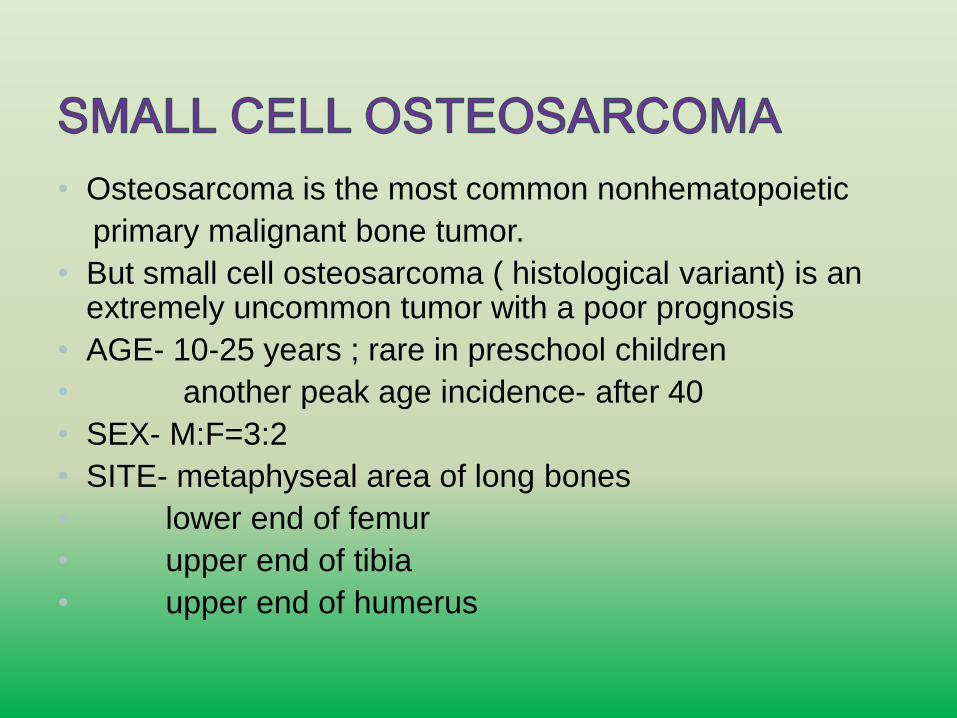
• Osteosarcoma is the most common nonhematopoietic
primary malignant bone tumor.
• But small cell osteosarcoma ( histological variant) is an extremely uncommon tumor with a poor prognosis
• AGE- 10-25 years ; rare in preschool children
• another peak age incidence- after 40
• SEX- M:F=3:2
• SITE- metaphyseal area of long bones
• lower end of femur
• upper end of tibia
• upper end of humerus
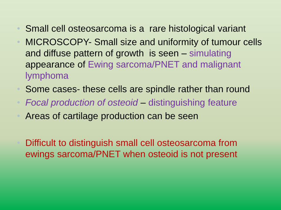
• Small cell osteosarcoma is a rare histological variant
• MICROSCOPY- Small size and uniformity of tumour cells
and diffuse pattern of growth is seen – simulating
appearance of Ewing sarcoma/PNET and malignant
lymphoma
• Some cases- these cells are spindle rather than round
• Focal production of osteoid – distinguishing feature
• Areas of cartilage production can be seen
• Difficult to distinguish small cell osteosarcoma from
ewings sarcoma/PNET when osteoid is not present
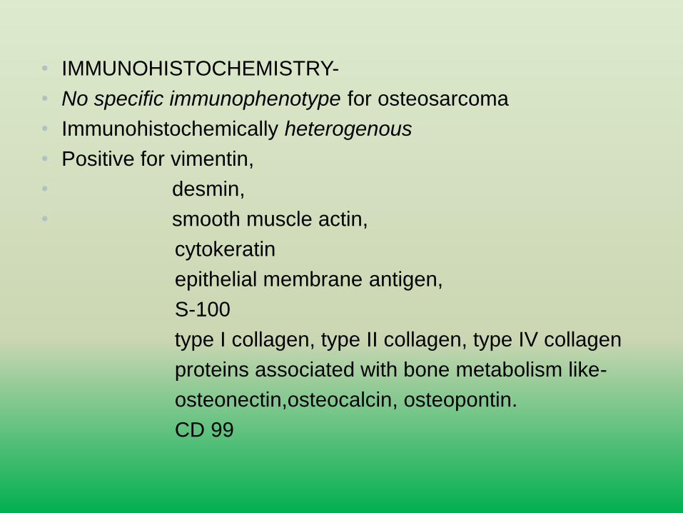
• IMMUNOHISTOCHEMISTRY-
• No specific immunophenotype for osteosarcoma
• Immunohistochemically heterogenous
• Positive for vimentin,
• desmin,
• smooth muscle actin,
cytokeratin
epithelial membrane antigen,
S-100
type I collagen, type II collagen, type IV collagen
proteins associated with bone metabolism like-
osteonectin,osteocalcin, osteopontin.
CD 99
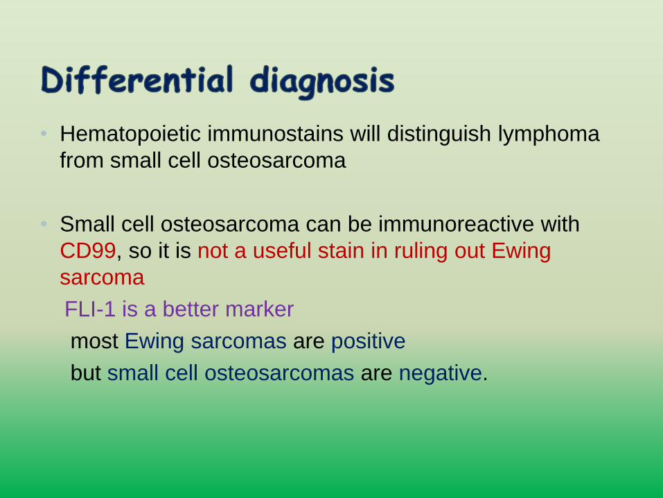
• Hematopoietic immunostains will distinguish lymphoma
from small cell osteosarcoma
• Small cell osteosarcoma can be immunoreactive with
CD99, so it is not a useful stain in ruling out Ewing
sarcoma
FLI-1 is a better marker
most Ewing sarcomas are positive
but small cell osteosarcomas are negative.
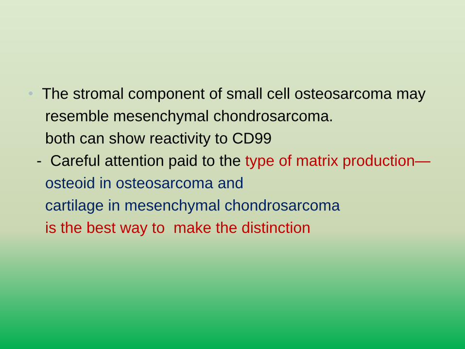
• The stromal component of small cell osteosarcoma may
resemble mesenchymal chondrosarcoma.
both can show reactivity to CD99
- Careful attention paid to the type of matrix production—
osteoid in osteosarcoma and
cartilage in mesenchymal chondrosarcoma
is the best way to make the distinction
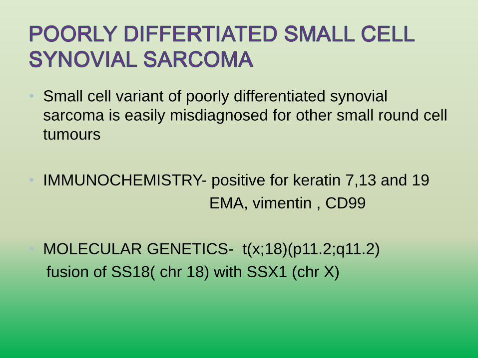
• Small cell variant of poorly differentiated synovial
sarcoma is easily misdiagnosed for other small round cell
tumours
• IMMUNOCHEMISTRY- positive for keratin 7,13 and 19
EMA, vimentin , CD99
• MOLECULAR GENETICS- t(x;18)(p11.2;q11.2)
fusion of SS18( chr 18) with SSX1 (chr X)
• Only two categories show small round cell picture on
histology:
1.Embryonal rhabdomyosarcoma
2.Alveolar rhabdomyosarcoma
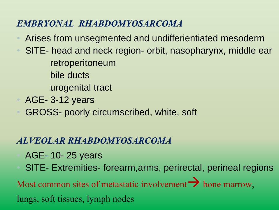
EMBRYONAL RHABDOMYOSARCOMA • Arises from unsegmented and undifferientiated mesoderm
• SITE- head and neck region- orbit, nasopharynx, middle ear
retroperitoneum
bile ducts
urogenital tract
• AGE- 3-12 years
• GROSS- poorly circumscribed, white, soft
ALVEOLAR RHABDOMYOSARCOMA• AGE- 10- 25 years
• SITE- Extremities- forearm,arms, perirectal, perineal regions
Most common sites of metastatic involvement bone marrow, lungs, soft tissues, lymph nodes
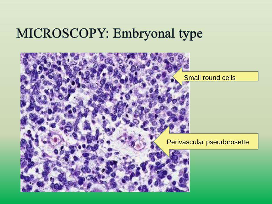
Perivascular pseudorosette
Small round cells
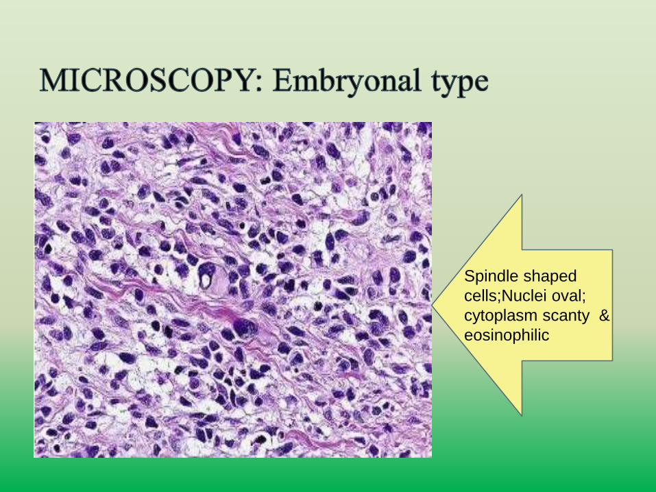
Spindle shaped
cells;Nuclei oval;
cytoplasm scanty &
eosinophilic
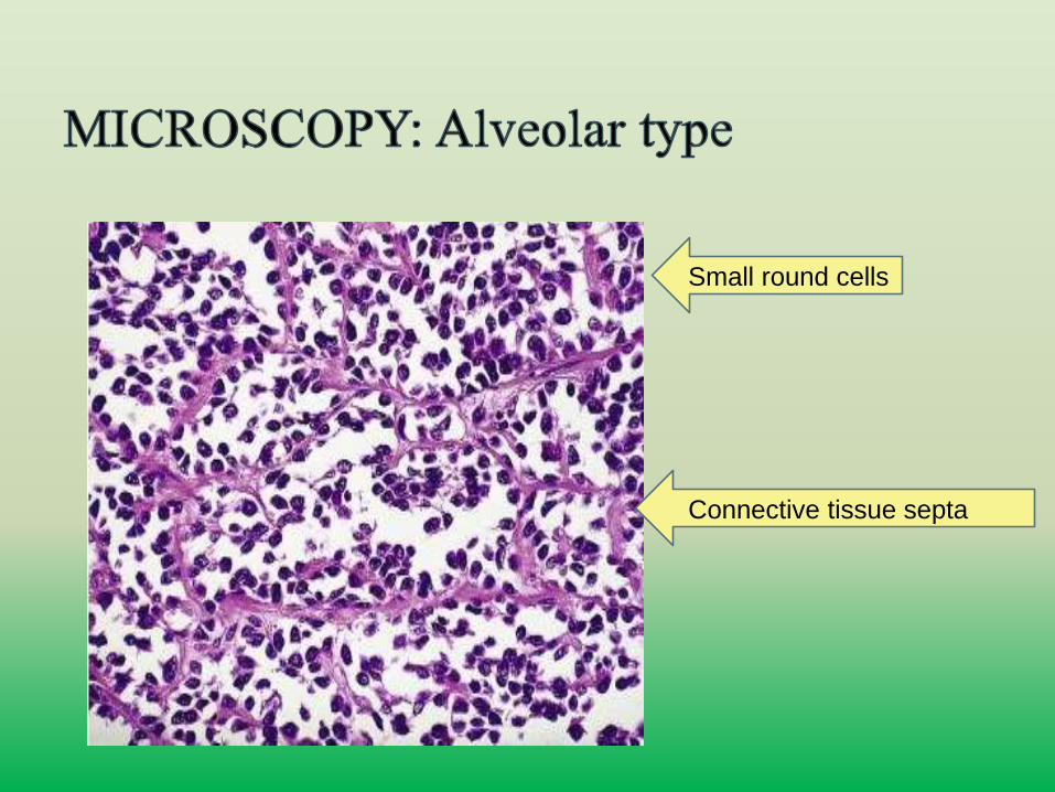
Small round cells
Connective tissue septa
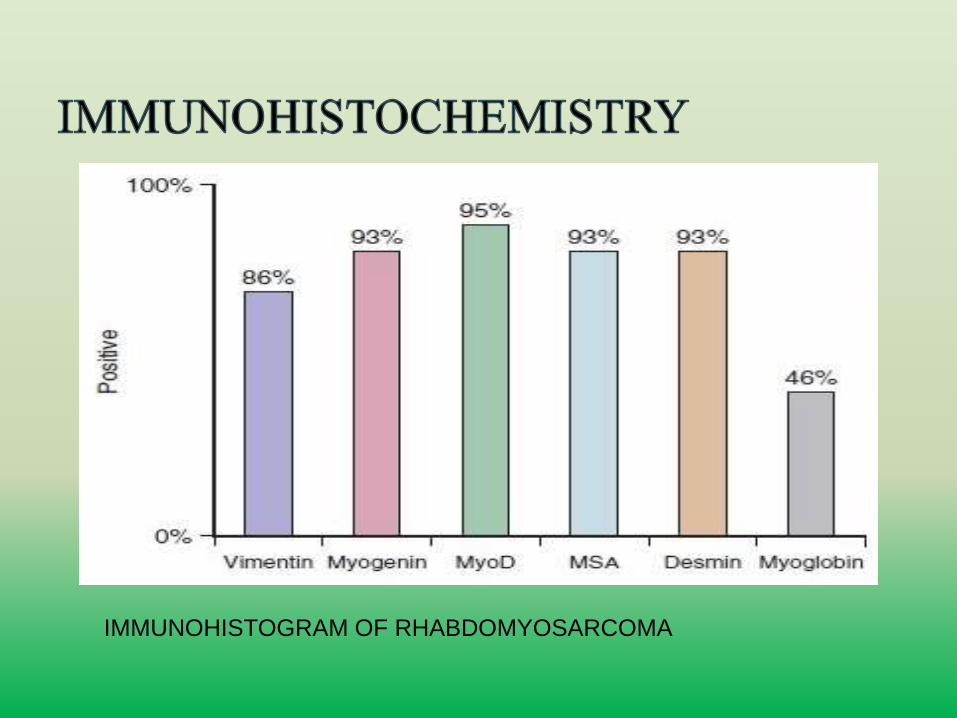
IMMUNOHISTOGRAM OF RHABDOMYOSARCOMA
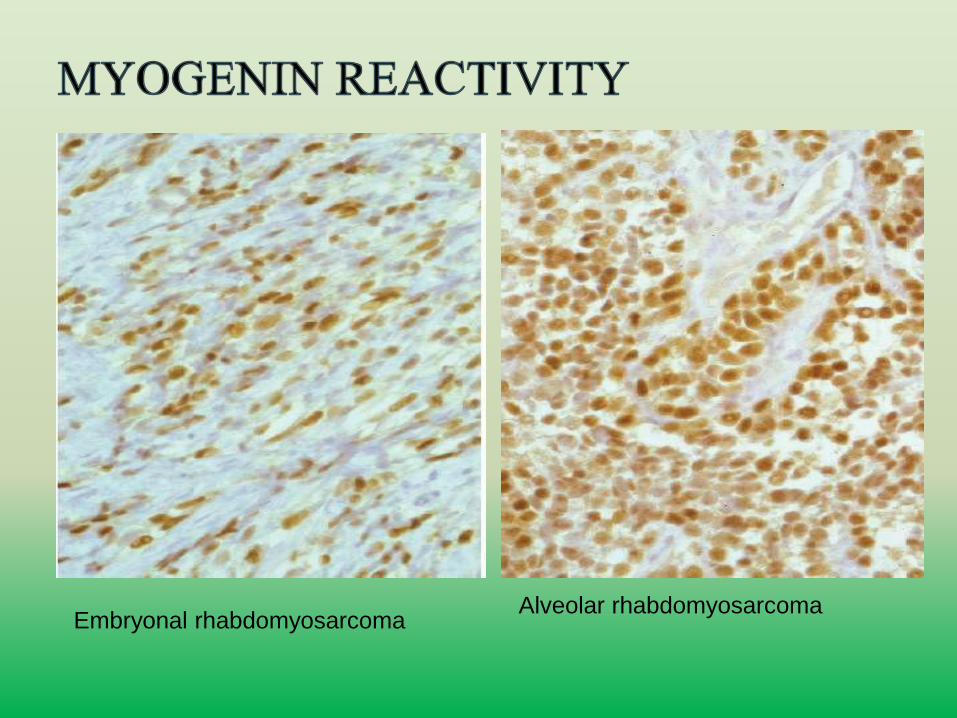
Embryonal rhabdomyosarcomaAlveolar rhabdomyosarcoma
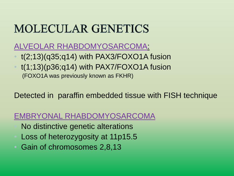
ALVEOLAR RHABDOMYOSARCOMA:
• t(2;13)(q35;q14) with PAX3/FOXO1A fusion
• t(1;13)(p36;q14) with PAX7/FOXO1A fusion(FOXO1A was previously known as FKHR)
Detected in paraffin embedded tissue with FISH technique
EMBRYONAL RHABDOMYOSARCOMA
• No distinctive genetic alterations
• Loss of heterozygosity at 11p15.5
• Gain of chromosomes 2,8,13

Thank you…