Embed Size (px)
Citation preview

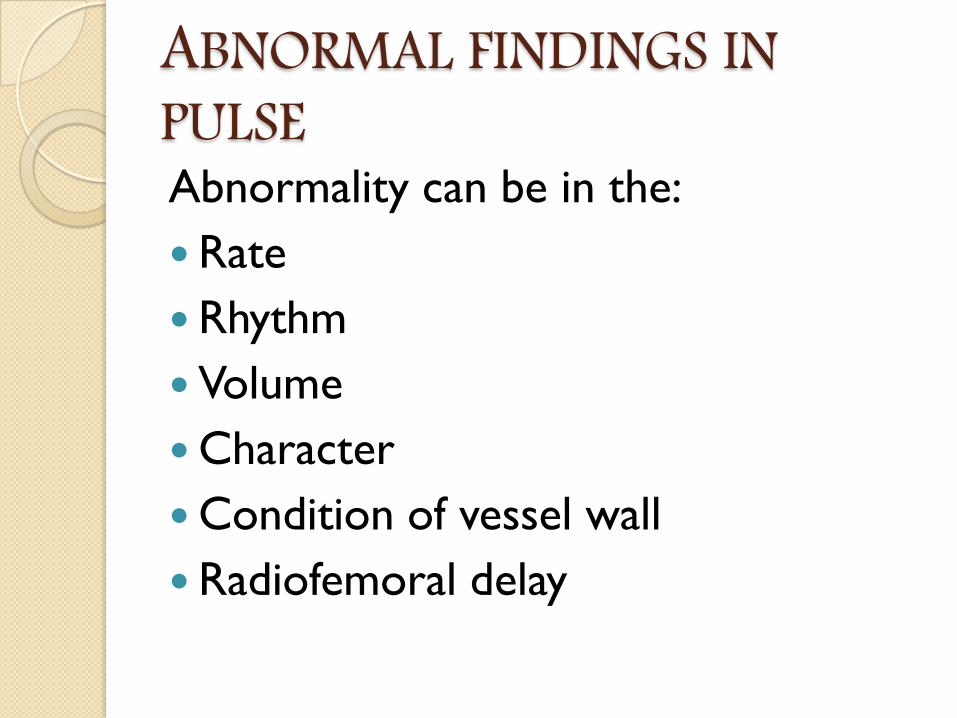
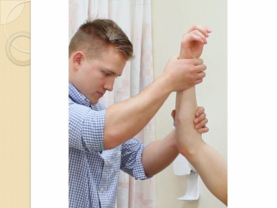
ABNORMAL FINDINGS IN PULSEAbnormality can be in the:
Rate
Rhythm
Volume
Character
Condition of vessel wall
Radiofemoral delay
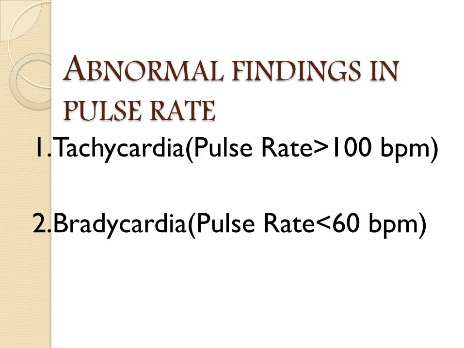
ABNORMAL FINDINGS IN PULSE RATE
1.Tachycardia(Pulse Rate>100 bpm)
2.Bradycardia(Pulse Rate<60 bpm)
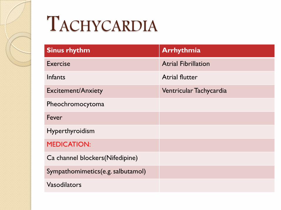
TACHYCARDIASinus rhythm Arrhythmia
Exercise Atrial Fibrillation
Infants Atrial flutter
Excitement/Anxiety Ventricular Tachycardia
Pheochromocytoma
Fever
Hyperthyroidism
MEDICATION:
Ca channel blockers(Nifedipine)
Sympathomimetics(e.g. salbutamol)
Vasodilators
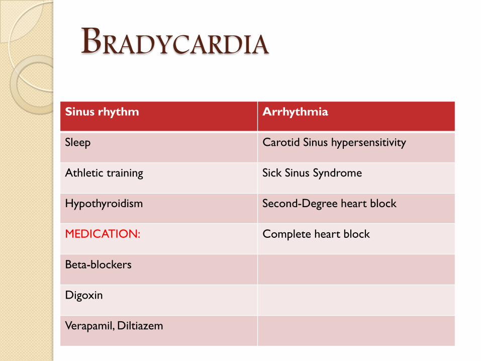
BRADYCARDIA
Sinus rhythm Arrhythmia
Sleep Carotid Sinus hypersensitivity
Athletic training Sick Sinus Syndrome
Hypothyroidism Second-Degree heart block
MEDICATION: Complete heart block
Beta-blockers
Digoxin
Verapamil, Diltiazem

ABNORMAL FINDINGS IN RHYTHM
If Irregular:
Occasionally irregular
Regularly Irregular
Irregularly Irregular
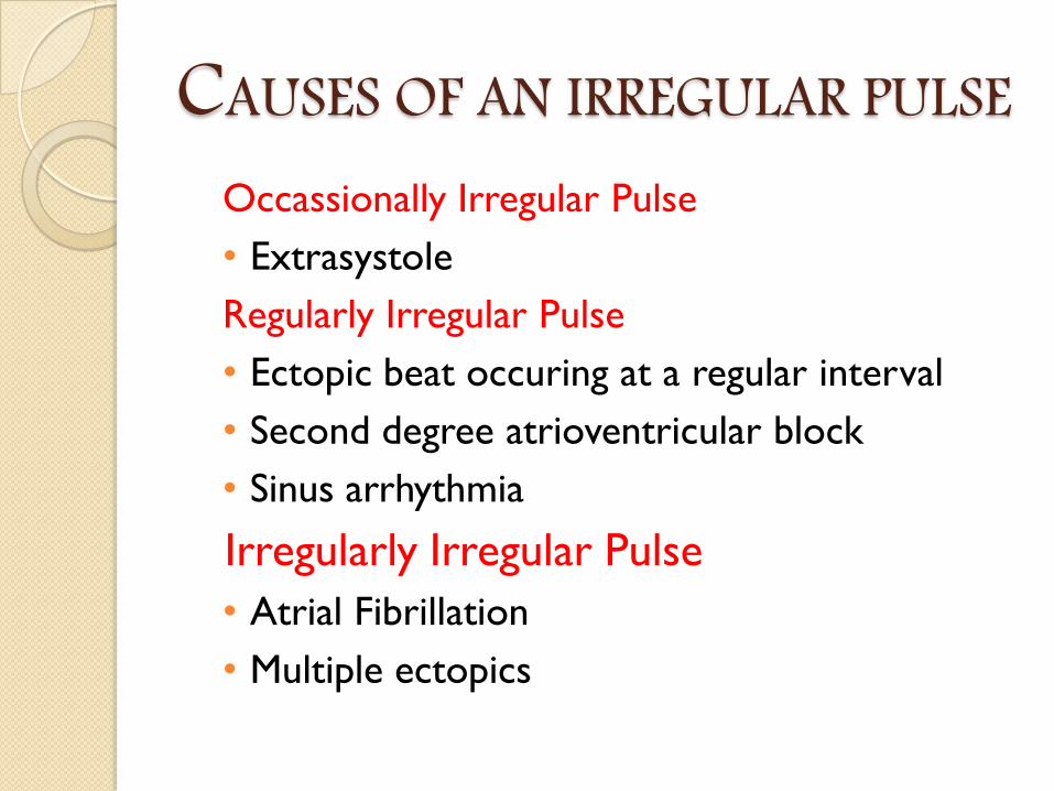
CAUSES OF AN IRREGULAR PULSEOccassionally Irregular Pulse
• Extrasystole
Regularly Irregular Pulse
• Ectopic beat occuring at a regular interval
• Second degree atrioventricular block
• Sinus arrhythmia
Irregularly Irregular Pulse
• Atrial Fibrillation
• Multiple ectopics
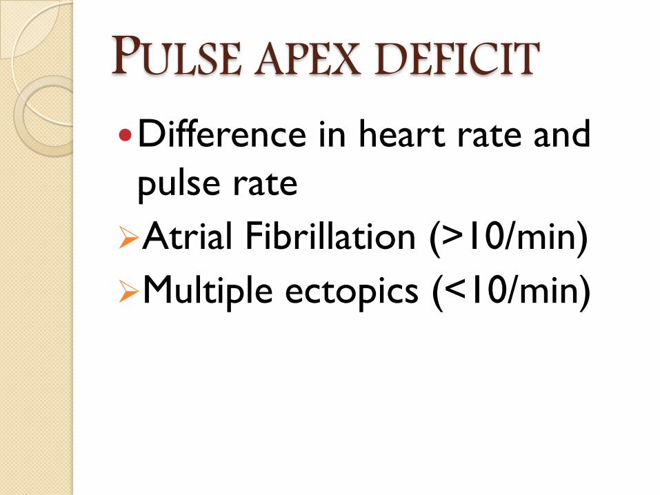
PULSE APEX DEFICITDifference in heart rate and
pulse rate
Atrial Fibrillation (>10/min)
Multiple ectopics (<10/min)
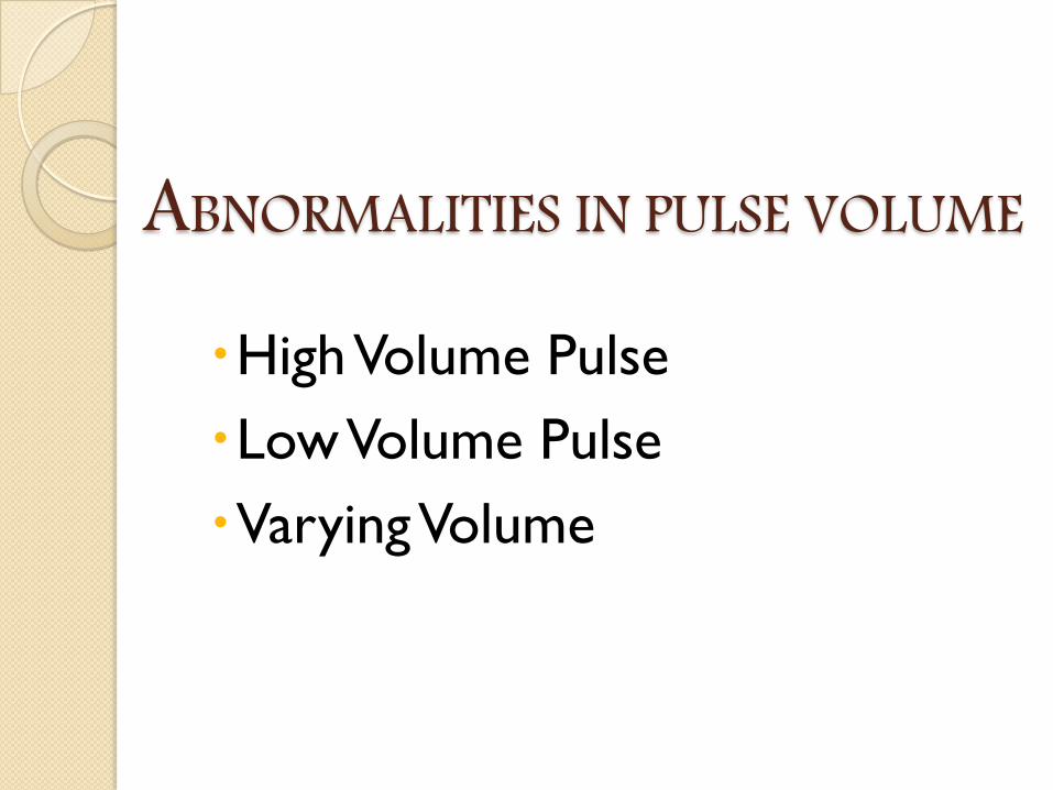
ABNORMALITIES IN PULSE VOLUME
High Volume Pulse
Low Volume Pulse
Varying Volume
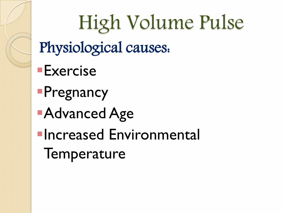
High Volume PulsePhysiological causes:Exercise
Pregnancy
Advanced Age
Increased Environmental
Temperature
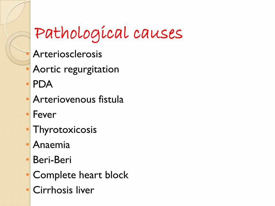
Pathological causes• Arteriosclerosis
• Aortic regurgitation
• PDA
• Arteriovenous fistula
• Fever
• Thyrotoxicosis
• Anaemia
• Beri-Beri
• Complete heart block
• Cirrhosis liver
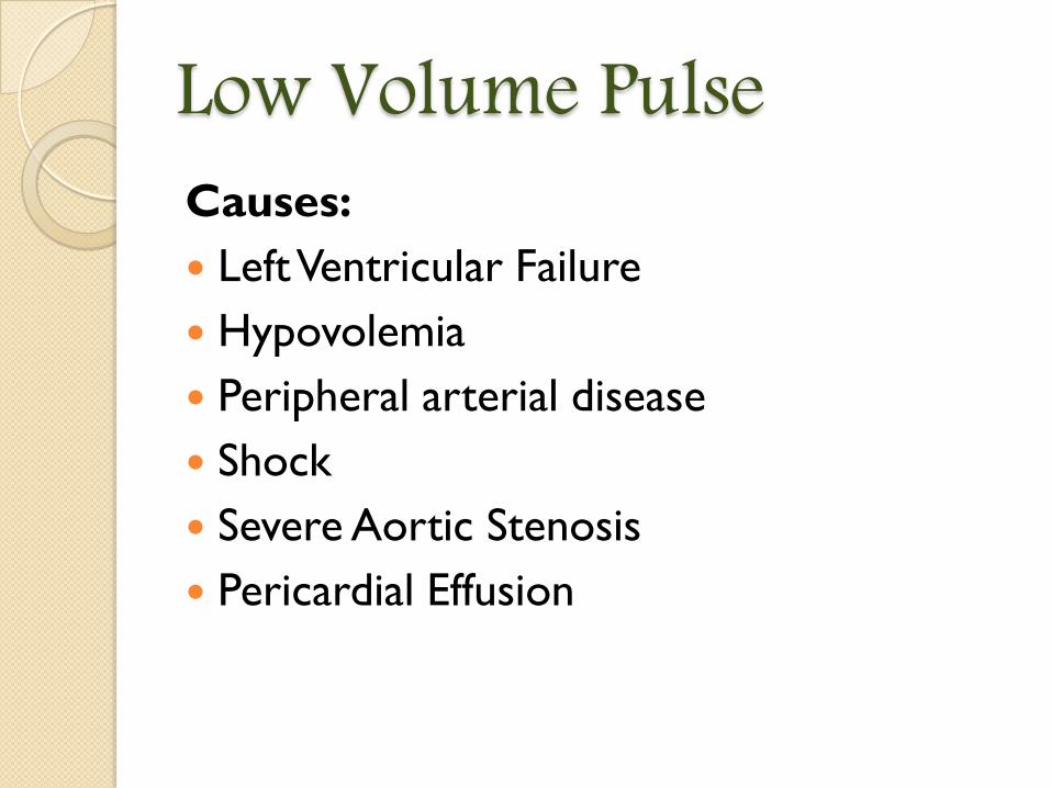
Low Volume PulseCauses:
Left Ventricular Failure
Hypovolemia
Peripheral arterial disease
Shock
Severe Aortic Stenosis
Pericardial Effusion
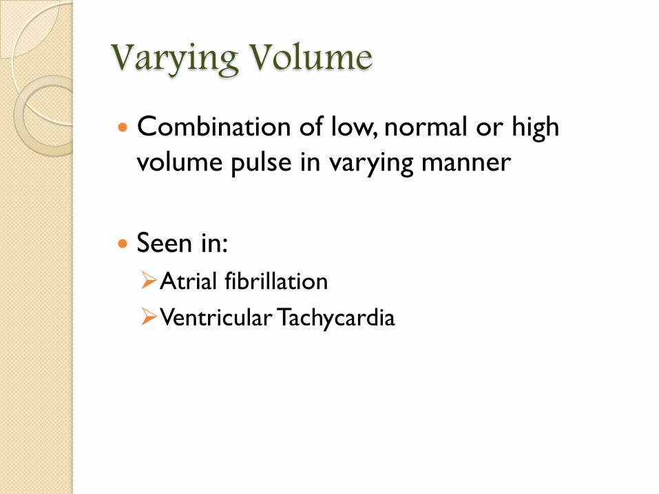
Varying Volume Combination of low, normal or high
volume pulse in varying manner
Seen in:
Atrial fibrillation
Ventricular Tachycardia
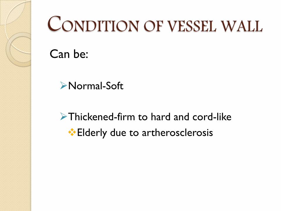
CONDITION OF VESSEL WALLCan be:
Normal-Soft
Thickened-firm to hard and cord-like
Elderly due to artherosclerosis
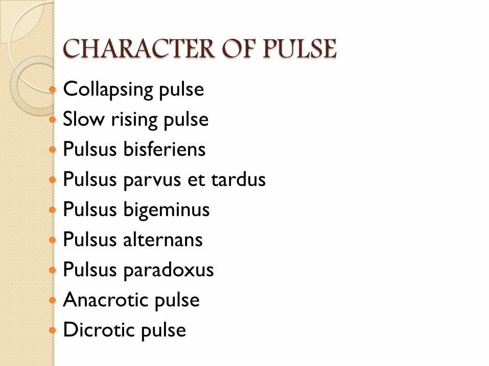
CHARACTER OF PULSE Collapsing pulse
Slow rising pulse
Pulsus bisferiens
Pulsus parvus et tardus
Pulsus bigeminus
Pulsus alternans
Pulsus paradoxus
Anacrotic pulse
Dicrotic pulse

Collapsing pulse/Water-hammer pulse/Corrigan’s pulse
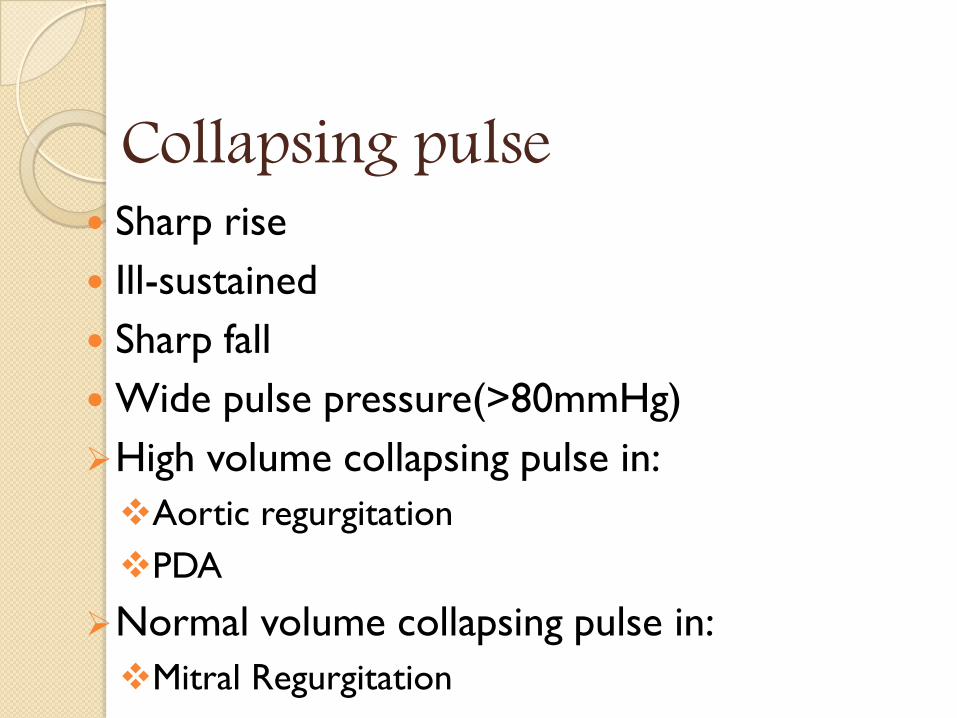
Collapsing pulse Sharp rise
Ill-sustained
Sharp fall
Wide pulse pressure(>80mmHg)
High volume collapsing pulse in:
Aortic regurgitation
PDA
Normal volume collapsing pulse in:
Mitral Regurgitation
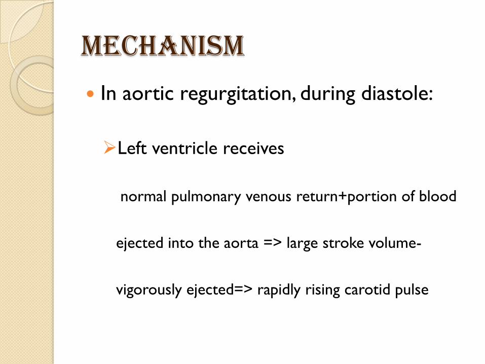
Mechanism
In aortic regurgitation, during diastole:
Left ventricle receives
normal pulmonary venous return+portion of blood
ejected into the aorta => large stroke volume-
vigorously ejected=> rapidly rising carotid pulse
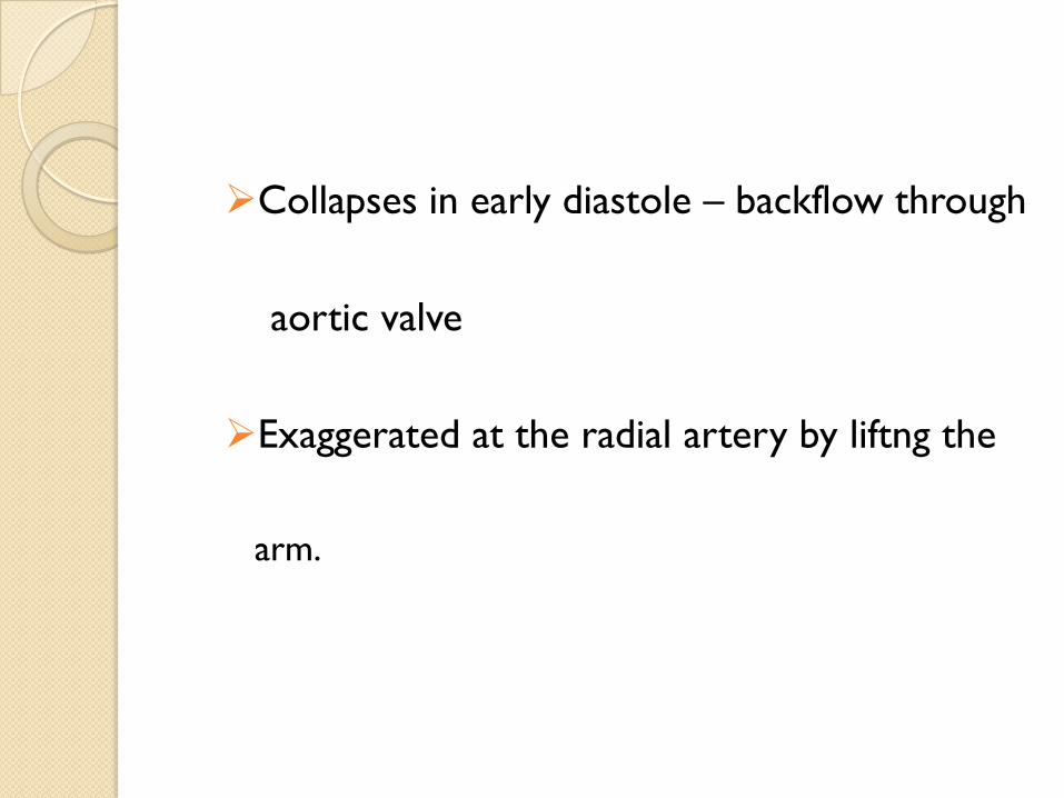
Collapses in early diastole – backflow through
aortic valve
Exaggerated at the radial artery by liftng the
arm.
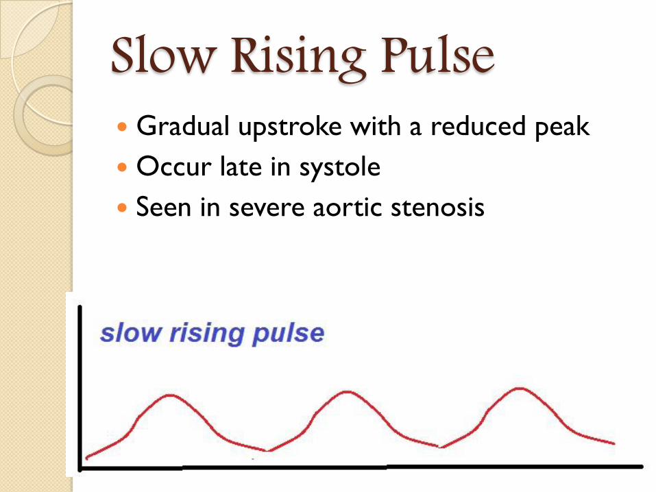
Slow Rising Pulse Gradual upstroke with a reduced peak
Occur late in systole
Seen in severe aortic stenosis
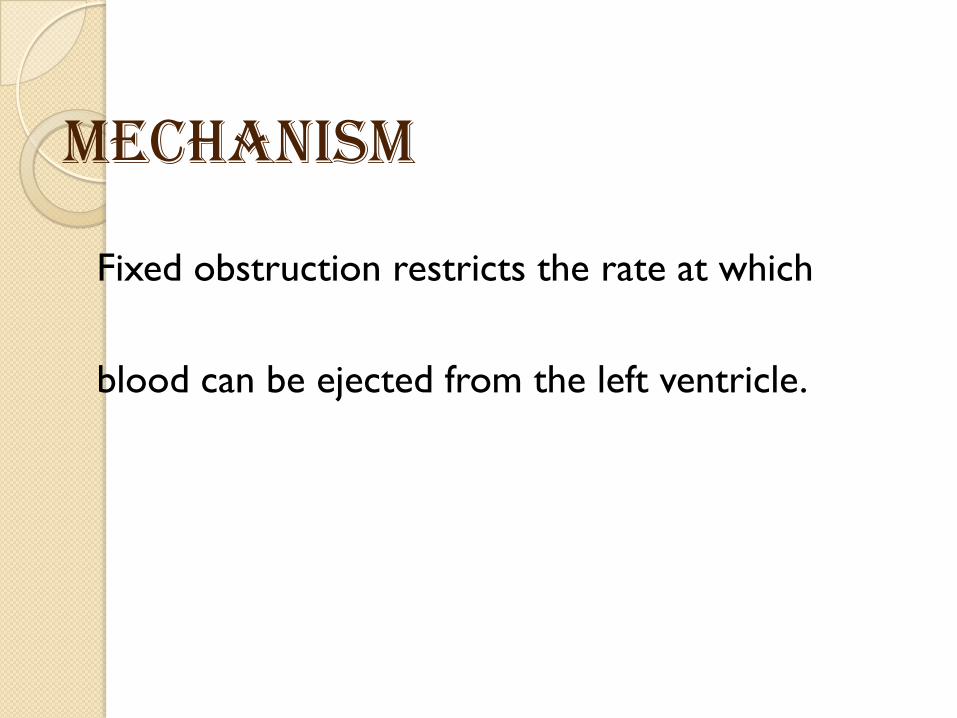
Mechanism
Fixed obstruction restricts the rate at which
blood can be ejected from the left ventricle.
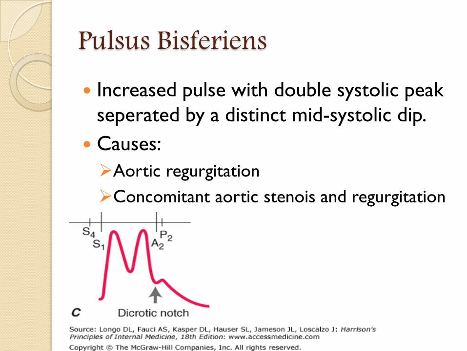
Pulsus Bisferiens Increased pulse with double systolic peak
seperated by a distinct mid-systolic dip.
Causes:
Aortic regurgitation
Concomitant aortic stenois and regurgitation

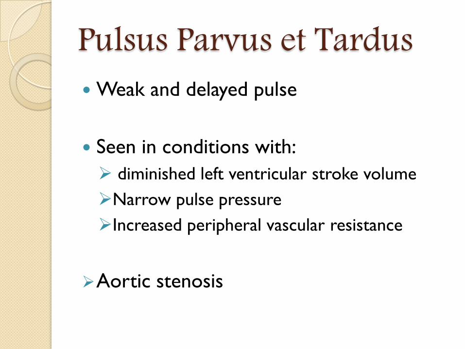
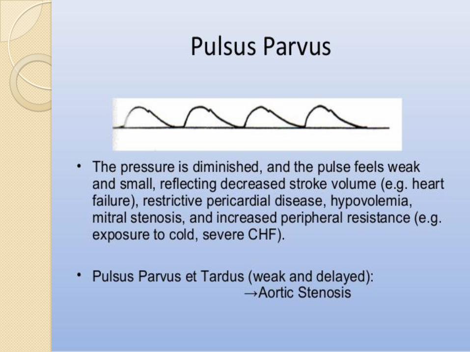
Pulsus Parvus et Tardus Weak and delayed pulse
Seen in conditions with:
diminished left ventricular stroke volume
Narrow pulse pressure
Increased peripheral vascular resistance
Aortic stenosis
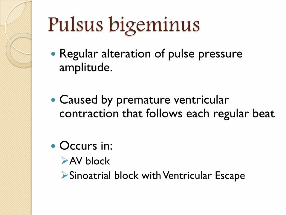
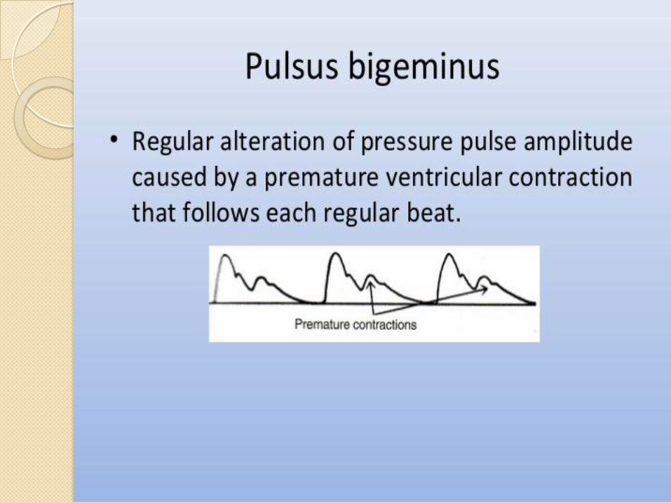
Pulsus bigeminus Regular alteration of pulse pressure
amplitude.
Caused by premature ventricular contraction that follows each regular beat
Occurs in:
AV block
Sinoatrial block with Ventricular Escape
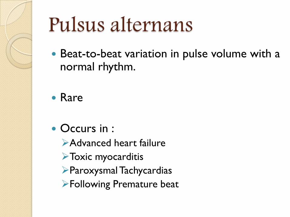
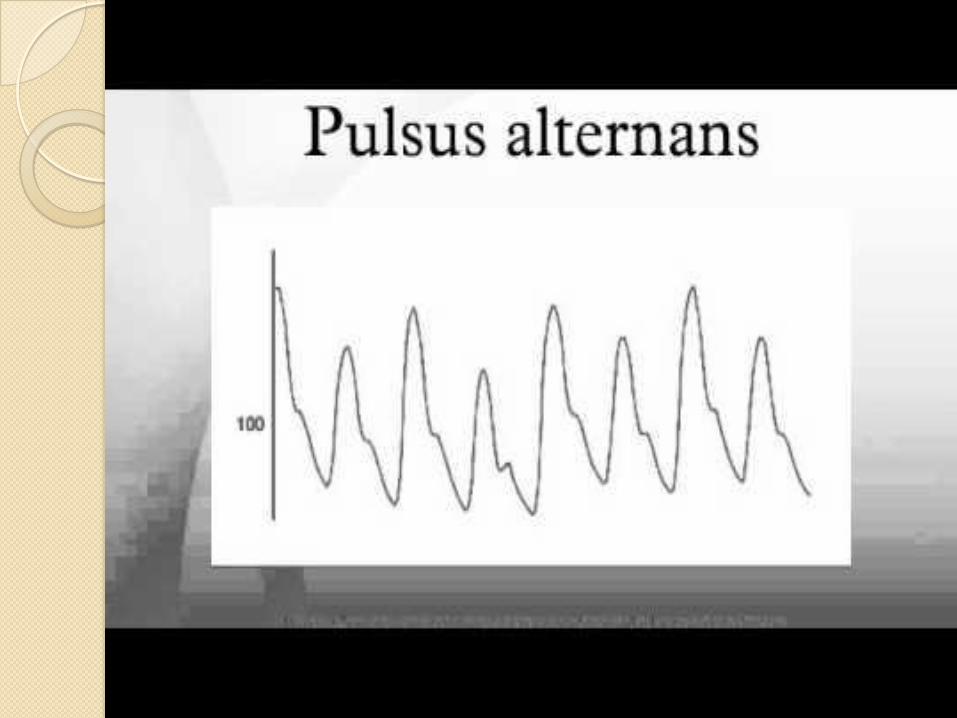
Pulsus alternans Beat-to-beat variation in pulse volume with a
normal rhythm.
Rare
Occurs in :
Advanced heart failure
Toxic myocarditis
Paroxysmal Tachycardias
Following Premature beat
Pulsus Paradoxus Exaggeration of the normal variability of
pulse volume with breathing.
Inspiratory decline in systolic pressure greater than 10mm Hg.
Occurs in:Cardiac tamponade
Constrictive pericarditis
Percardial effusion

Anacrotic Pulse Slow rising
Double beating pulse
Both waves felt in systole
Seen in Aortic Stenosis
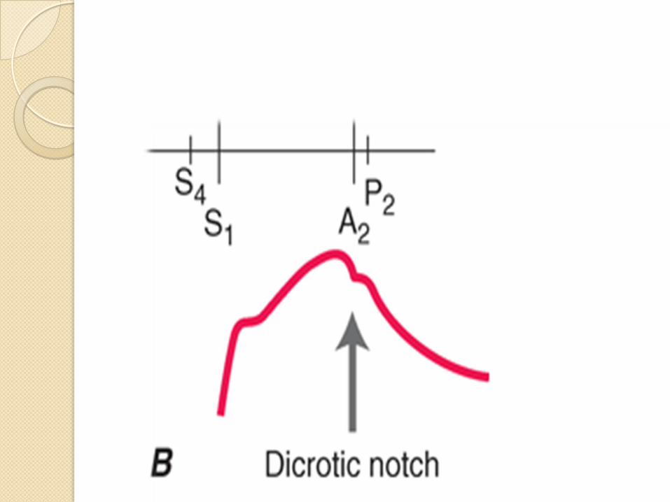
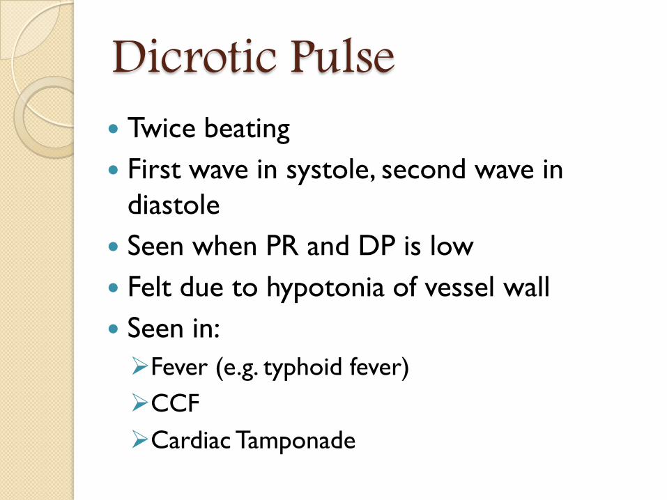
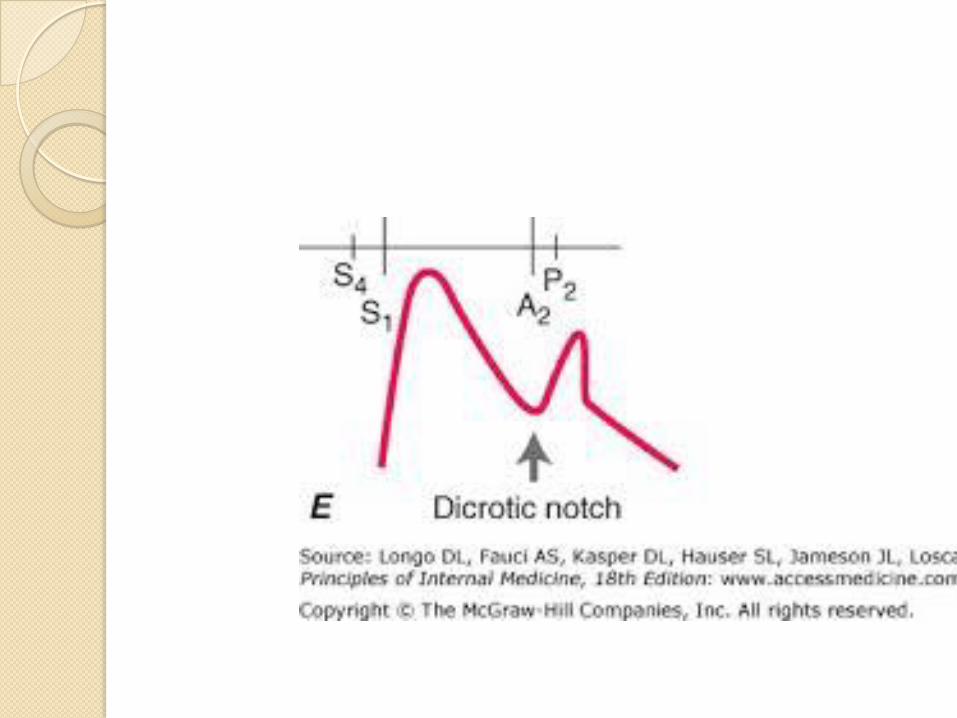
Dicrotic Pulse Twice beating
First wave in systole, second wave in
diastole
Seen when PR and DP is low
Felt due to hypotonia of vessel wall
Seen in:
Fever (e.g. typhoid fever)
CCF
Cardiac Tamponade
RADIO-FEMORAL DELAY Most common cause: Coarctation of aorta
Children:
Upperlimb pulses are usually normal
Reduced volume lowerlimb pulses
Adults:
Usually presents hypertension and heart failure
Other causes:
• Atherosclerosis of aorta
• Thrombosis or embolism of aorta
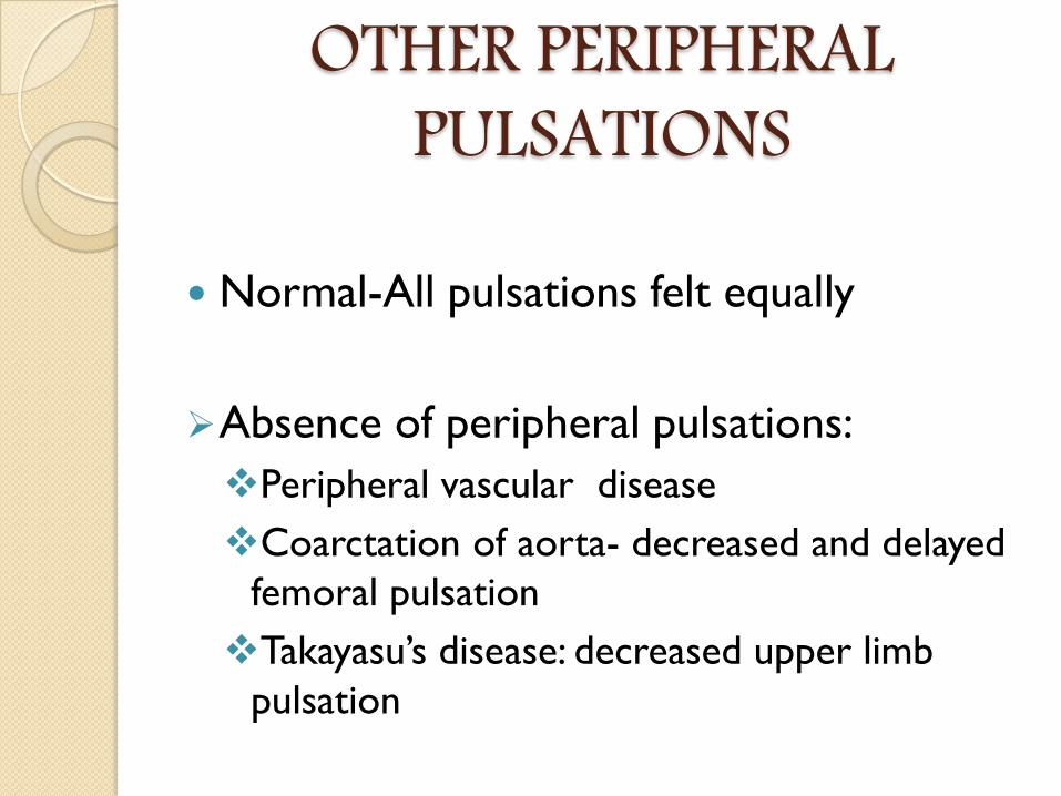
OTHER PERIPHERAL PULSATIONS
Normal-All pulsations felt equally
Absence of peripheral pulsations:
Peripheral vascular disease
Coarctation of aorta- decreased and delayed
femoral pulsation
Takayasu’s disease: decreased upper limb
pulsation
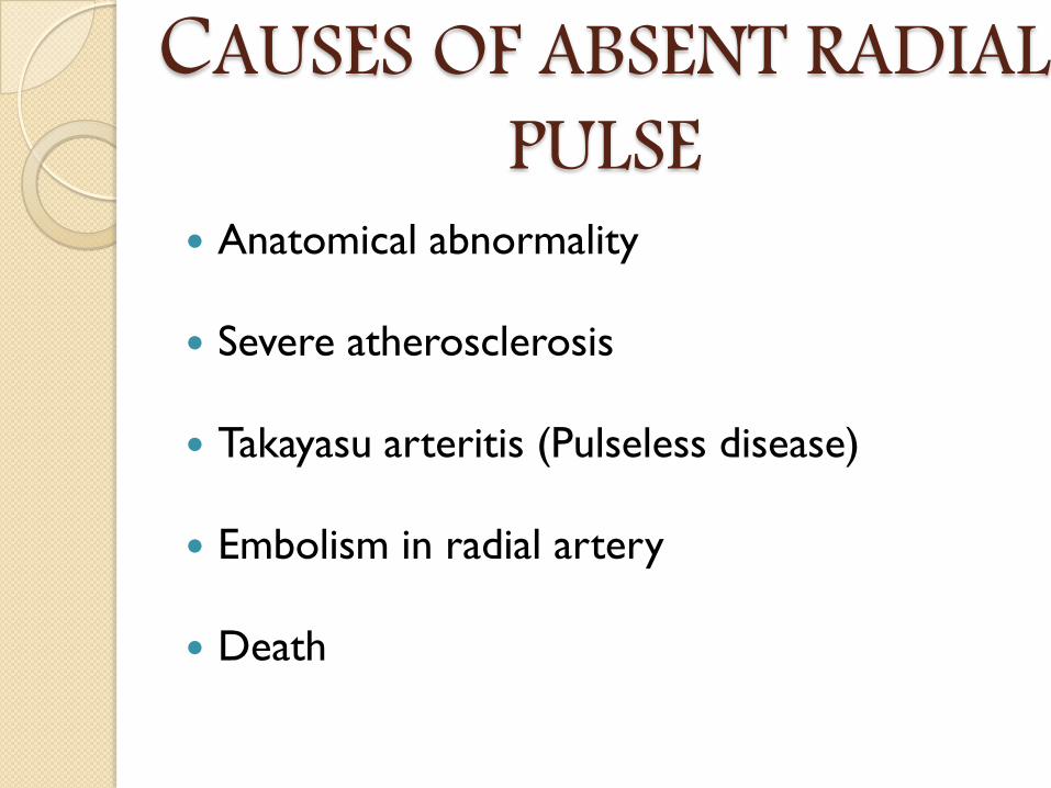
CAUSES OF ABSENT RADIAL PULSE
Anatomical abnormality
Severe atherosclerosis
Takayasu arteritis (Pulseless disease)
Embolism in radial artery
Death
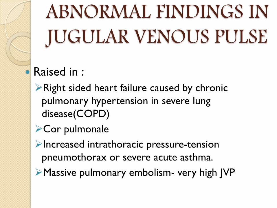
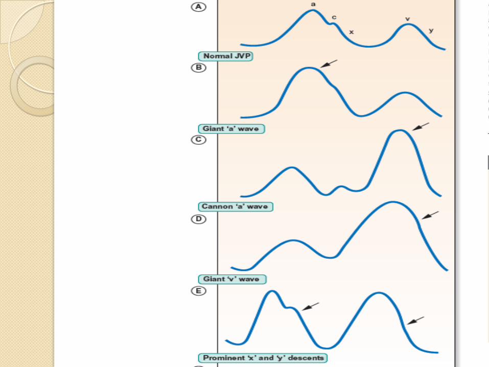
ABNORMAL FINDINGS IN JUGULAR VENOUS PULSE
Raised in :
Right sided heart failure caused by chronic
pulmonary hypertension in severe lung
disease(COPD)
Cor pulmonale
Increased intrathoracic pressure-tension
pneumothorax or severe acute asthma.
Massive pulmonary embolism- very high JVP
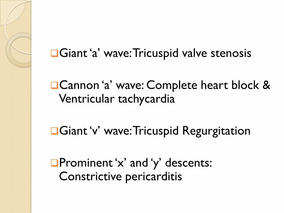
Giant ‘a’ wave: Tricuspid valve stenosis
Cannon ‘a’ wave: Complete heart block & Ventricular tachycardia
Giant ‘v’ wave: Tricuspid Regurgitation
Prominent ‘x’ and ‘y’ descents: Constrictive pericarditis

THANK YOU


![Chest Radiology Interpretation: Findings of Tuberculosisnid]/6a_printable...3 Reading the TB CXR Be systematic! Start centrally and work outwards Normal or abnormal If abnormal, consider](https://img.pdfslide.us/doc/110x75/5e62b1b9322283283b745f66/chest-radiology-interpretation-findings-of-tuberculosis-nid6aprintable-3.jpg)


![ACHS-OfficeCopier-20200529140846 · EXAMINATION (reference gender/iaelghr/oge chart) NORMAL NORMAL ) Pulse ABNORMAL FINDINGS ABNORMAL FINDINGS R MEDICAL Weight Male C] Female C] Corrected](https://img.pdfslide.us/doc/110x75/5fd276c26772de5118123855/achs-officecopier-20200529140846-examination-reference-genderiaelghroge-chart.jpg)