Embed Size (px)
Citation preview
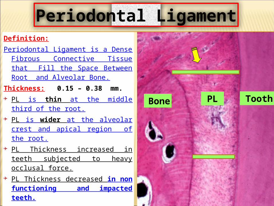
Definition:Periodontal Ligament is a Dense
Fibrous Connective Tissue that Fill the Space Between Root and Alveolar Bone.
Thickness: 0.15 – 0.38 mm. PL is thin at the middle third
of the root. PL is wider at the alveolar
crest and apical region of the root.
PL Thickness increased in teeth subjected to heavy occlusal force.
PL Thickness decreased in non functioning and impacted teeth.
Tooth Bone
PL
Periodontal Ligament
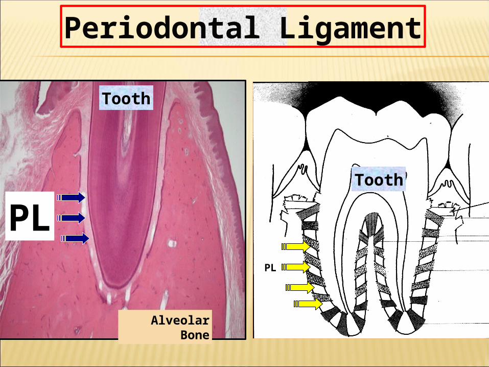
Alveolar Bone
Tooth
PLTooth
Periodontal Ligament
PL
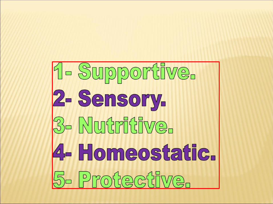
1. Supportive Functions:a. PL support the tooth within the jaw.b. Permits the tooth to withstand the suitable forces of mastication.c. PL is non elastic which prevent the tooth from being moved too far.d. PL act as cushion to distribute the masticatory force and transmit it to the
alveolar bone.2. Sensory Functions:a. PL contain small nerve fibers for pain sensation.b. PL contain large nerve fibers for touch and pressure.c. PL contain special nerve ending termed mechanoreceptors
Functions of periodontal ligament
3- Nutritive:Blood supply of the periodontal ligament provide a nutrition for teeth, PL, and gingiva.4- Formative: Fibroblasts are responsible for the formation of new periodontal ligament fibers and dissolution of the old fibers. Cementoblasts are responsible for the formation of cementum. Osteoblasts are responsible for the formation of bone. 4- protective:By the action of: a- Collagen fibrous groups. b- Blood vessels and inflammatory cells. c- Nerve fibers.
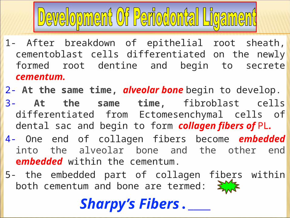
1- After breakdown of epithelial root sheath, cementoblast cells differentiated on the newly formed root dentine and begin to secrete cementum.
2- At the same time, alveolar bone begin to develop.3- At the same time, fibroblast cells differentiated from
Ectomesenchymal cells of dental sac and begin to form collagen fibers of PL.
4- One end of collagen fibers become embedded into the alveolar bone and the other end embedded within the cementum.
5- the embedded part of collagen fibers within both cementum and bone are termed:
Sharpy’s Fibers.
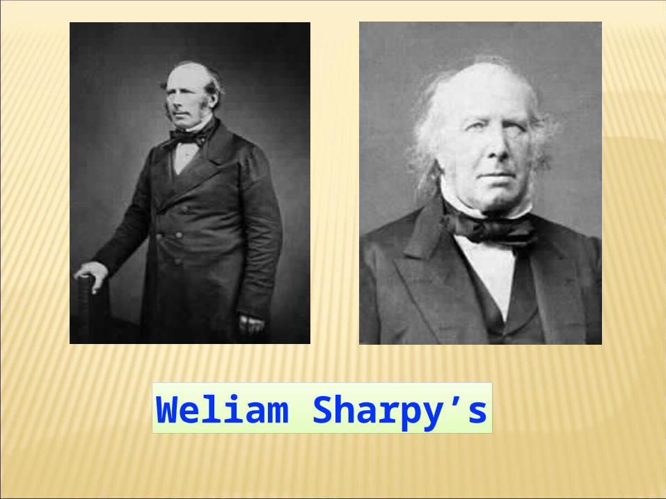
Weliam Sharpy’s
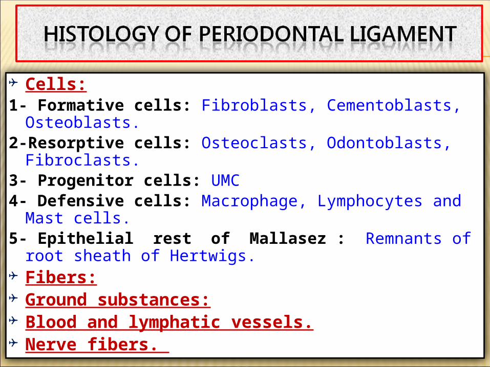
Cells:1- Formative cells: Fibroblasts, Cementoblasts,
Osteoblasts.2-Resorptive cells: Osteoclasts, Odontoblasts,
Fibroclasts.3- Progenitor cells: UMC4- Defensive cells: Macrophage, Lymphocytes and
Mast cells.5- Epithelial rest of Mallasez : Remnants of root
sheath of Hertwigs. Fibers: Ground substances: Blood and lymphatic vessels. Nerve fibers.
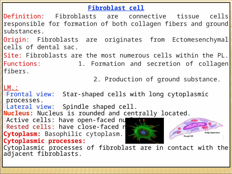
Fibroblast cellDefinition: Fibroblasts are connective tissue cells responsible for formation of both collagen fibers and ground substances. Origin: Fibroblasts are originates from Ectomesenchymal cells of dental sac.Site: Fibroblasts are the most numerous cells within the PL.Functions: 1. Formation and secretion of collagen fibers. 2. Production of ground substance.LM.:Frontal view: Star-shaped cells with long cytoplasmic processes.
Lateral view: Spindle shaped cell.Nucleus: Nucleus is rounded and centrally located. Active cells: have open-faced nuclei.Rested cells: have close-faced nuclei.Cytoplasm: Basophilic cytoplasm.Cytoplasmic processes:Cytoplasmic processes of fibroblast are in contact with the adjacent fibroblasts.
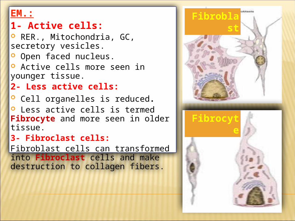
EM.:1- Active cells: RER., Mitochondria, GC, secretory vesicles. Open faced nucleus. Active cells more seen in younger tissue.2- Less active cells: Cell organelles is reduced. Less active cells is termed Fibrocyte and more seen in older tissue.3- Fibroclast cells:Fibroblast cells can transformed into Fibroclast cells and make destruction to collagen fibers.
Fibroblast
Fibrocyte
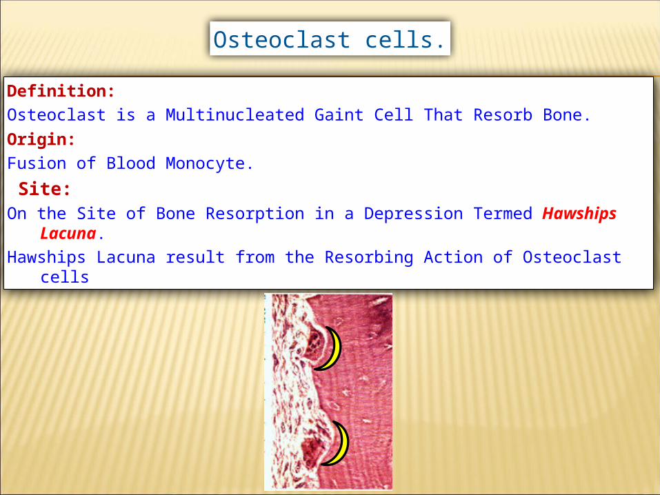
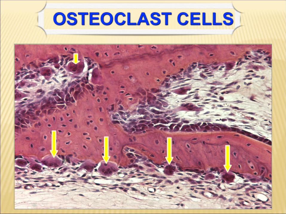
Definition:Osteoclast is a Multinucleated Gaint Cell That Resorb Bone.Origin:Fusion of Blood Monocyte. Site:On the Site of Bone Resorption in a Depression Termed Hawships
Lacuna.Hawships Lacuna result from the Resorbing Action of Osteoclast cells
Osteoclast cells.
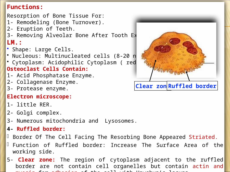
Functions: Resorption of Bone Tissue For:1- Remodeling (Bone Turnover).2- Eruption of Teeth.3- Removing Alveolar Bone After Tooth Extraction. LM.:• Shape: Large Cells.• Nucleous: Multinucleated cells (8-20 nuclei).• Cytoplasm: Acidophilic Cytoplasm ( red).Osteoclast Cells Contain: 1- Acid Phosphatase Enzyme.2- Collagenase Enzyme.3- Protease enzyme.Electron microscope:1- little RER. 2- Golgi complex.3- Numerous mitochondria and Lysosomes.4- Ruffled border: Border Of The Cell Facing The Resorbing Bone Appeared Striated. Function of Ruffled border: Increase The Surface Area of the working side.5- Clear zone: The region of cytoplasm adjacent to the ruffled border are not contain cell
organelles but contain actin and myosin for adhesion of the cell with Hawshyp’s lacuna.
Clear zoneRuffled border
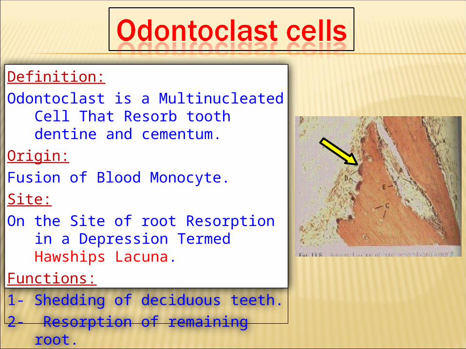
Definition:Odontoclast is a Multinucleated Cell That
Resorb tooth dentine and cementum.Origin:Fusion of Blood Monocyte.Site:On the Site of root Resorption in a Depression
Termed Hawships Lacuna.Functions:1- Shedding of deciduous teeth.2- Resorption of remaining root.
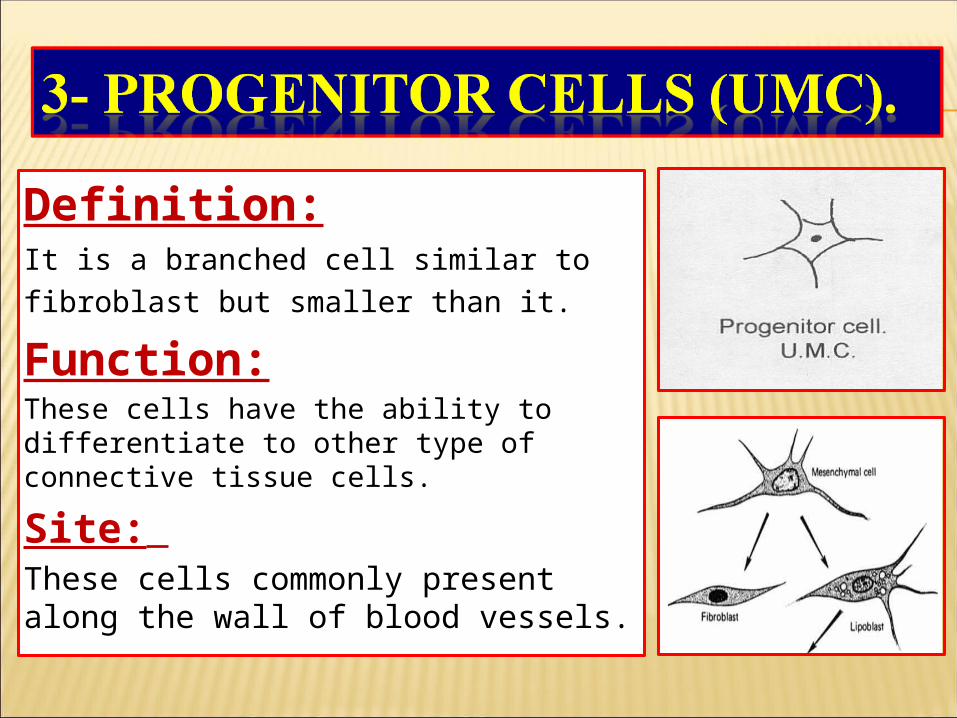
Definition:It is a branched cell similar to fibroblast but smaller than it. Function:These cells have the ability to differentiate to other type of connective tissue cells.
Site: These cells commonly present along the wall of blood vessels.
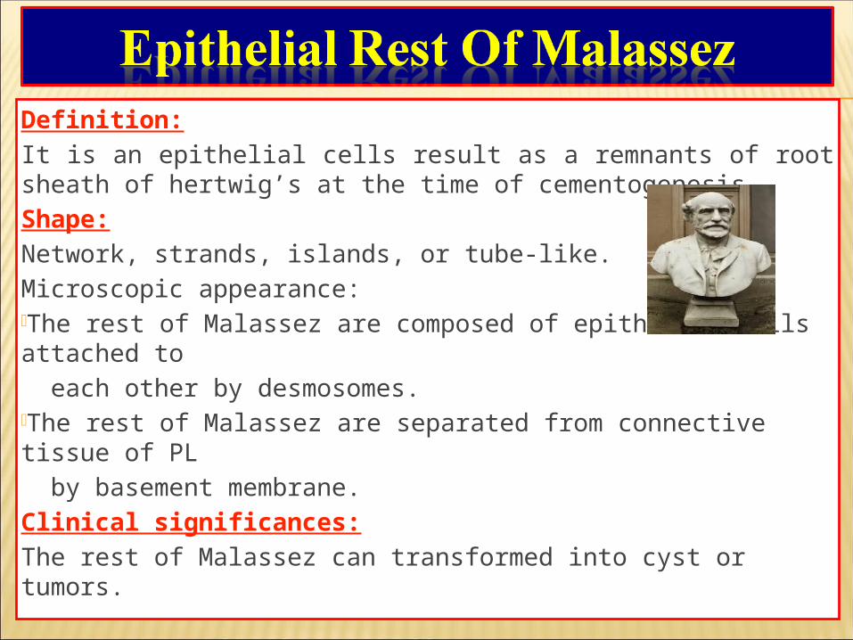
Definition:It is an epithelial cells result as a remnants of root sheath of hertwig’s at the time of cementogenesis.Shape:Network, strands, islands, or tube-like.Microscopic appearance:The rest of Malassez are composed of epithelial cells attached to each other by desmosomes.The rest of Malassez are separated from connective tissue of PL by basement membrane.Clinical significances:The rest of Malassez can transformed into cyst or tumors.
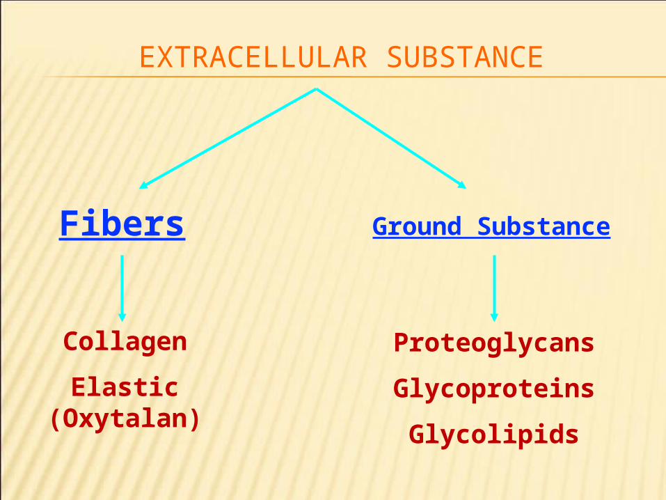
EXTRACELLULAR SUBSTANCE
Fibers Ground Substance
Collagen
Elastic (Oxytalan)
Proteoglycans
Glycoproteins
Glycolipids
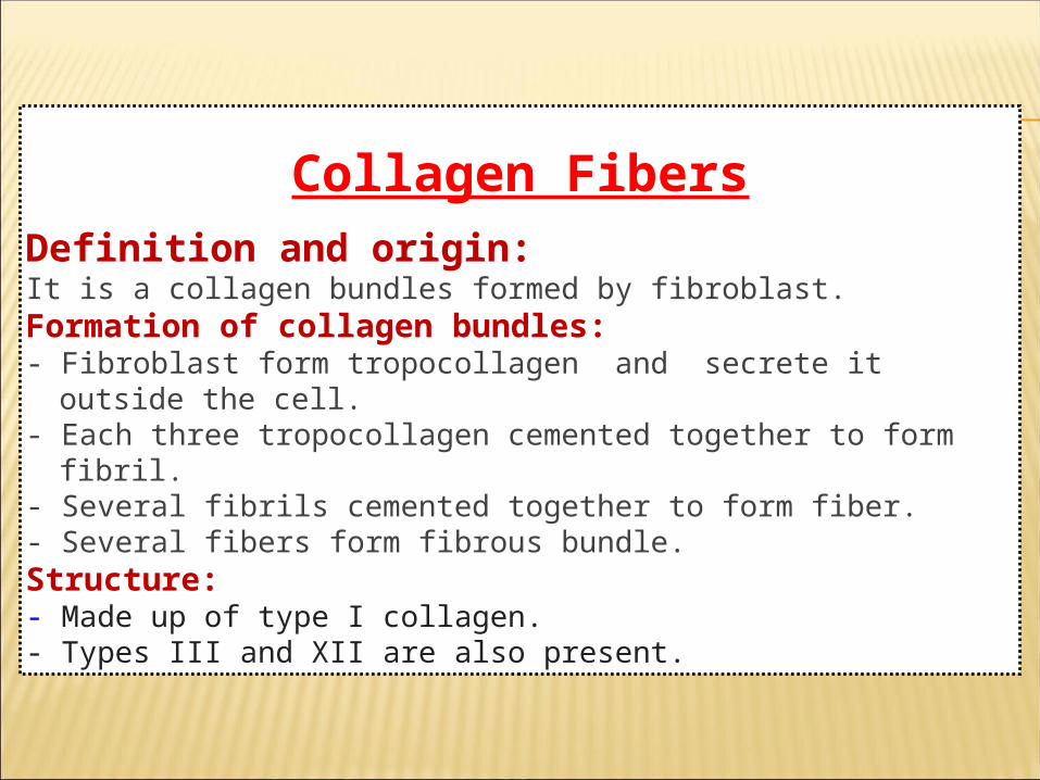
Collagen FibersDefinition and origin:It is a collagen bundles formed by fibroblast.Formation of collagen bundles:- Fibroblast form tropocollagen and secrete it outside the cell.- Each three tropocollagen cemented together to form fibril.- Several fibrils cemented together to form fiber.- Several fibers form fibrous bundle.Structure:- Made up of type I collagen.- Types III and XII are also present.
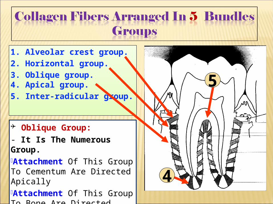
1. Alveolar crest group.2. Horizontal group.3. Oblique group.4. Apical group.5. Inter-radicular group.
5
4
Oblique Group:- It Is The Numerous Group.Attachment Of This Group To Cementum Are Directed ApicallyAttachment Of This Group To Bone Are Directed Coronally.
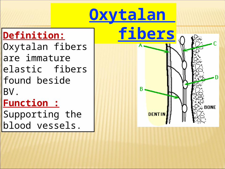
Oxytalan fibersDefinition:
Oxytalan fibers are immature elastic fibers found beside BV.Function :Supporting the blood vessels.
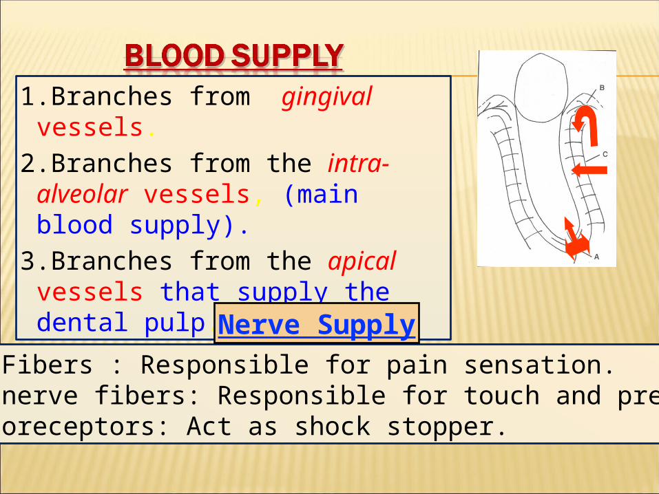
1.Branches from gingival vessels.
2.Branches from the intra-alveolar vessels, (main blood supply).
3.Branches from the apical vessels that supply the dental pulp.
1.Small Fibers : Responsible for pain sensation.2.Large nerve fibers: Responsible for touch and pressure.3.Mechanoreceptors: Act as shock stopper.
Nerve Supply
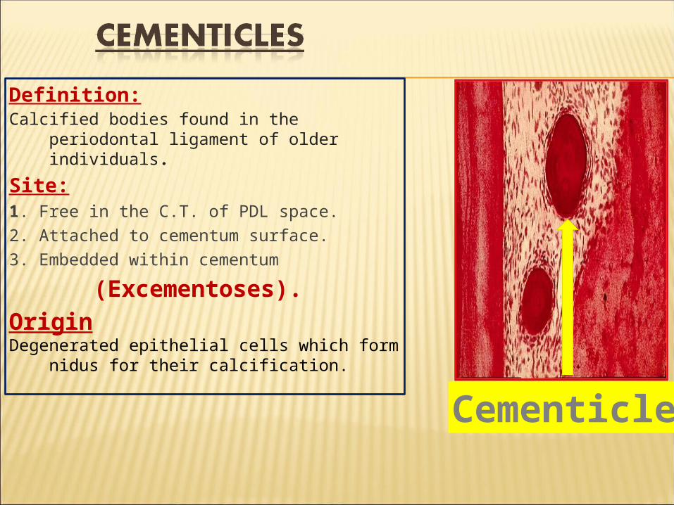
Definition:Calcified bodies found in the periodontal ligament
of older individuals.
Site:1. Free in the C.T. of PDL space.2. Attached to cementum surface.3. Embedded within cementum
(Excementoses). OriginDegenerated epithelial cells which form nidus for
their calcification.
Cementicle
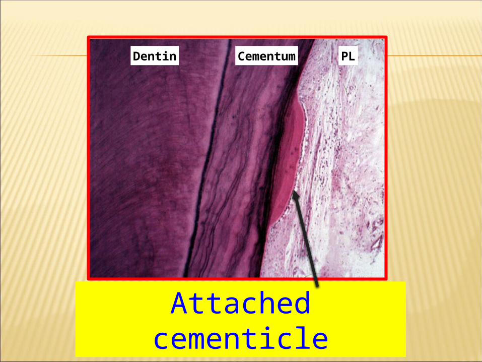
Attached cementicle
Cementum PLDentin