Embed Size (px)
Citation preview

PDL
Upload By : Ahmed Ali Abbas
Babylon University College of Dentistry
download this file from Website on Google
TheOptimalSmile.wix.com
Then choose Lectures
Then Second Stage
Then choose the lecture you
need

The Periodontal Ligament
By :Ahmed Ali Abbas

Periodontium:Is the investing and supporting attachment system of the
teeth. It consists of:
Two soft tissues Two hard tissues
1- Gingiva
2- Periodontal Ligament
1- Cementum
2- Alveolar Bone :

Definition: The periodontal
ligament is the dense fibrous
connective tissue that occupies
periodontal space between the
of the tooth and the alveolus.
Inflammation from the
dental pulp and gingiva
could spread to involve the
PDL and other apical
supporting tissues.

Width of the periodontal ligament ranges from 0.15-0.21 mm.The narrowest area is at the mid-root ( Fulcrum ). The region at thealveolar crest is the widest area followed by the apical region.
DentinBone
The width generally reduced in:• Non-functional teeth. • Un-erupted teeth.
While increased in:• Teeth subjected to an occlusal stress within the physiological limits .• Deciduous teeth


Dental follicle consists of1-Dense fibrous and cellulartissues immediatelyencapsulate the developingtooth
(Cementum)
2-Perifollicular mesenchyme
(PDL ,Alveolar bone)
DevelopmentOf PDL:

cells
Histological structureThe periodontal ligament is formed of :
Fibers,
Intercellular
substances
Synthetic
Resorptive
Progenitor
Defensive
ground substances
blood vessels,
nerves & lymphatics.

a) Gingival fibers.
b) Transseptal or interdental ligament.
c) Alveolodental ligament which is
subdivided into the following five groups:
1- Alveolar crest group.
2- Horizontal group.
3- Oblique group.
4-Apical group.
5- Interradicular group.
A- The principal fibers:They are formed of collagen bundles, which are wavy
in course and are arranged in three groups.
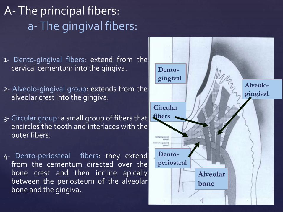
1- Dento-gingival fibers: extend from thecervical cementum into the gingiva.
2- Alveolo-gingival group: extends from thealveolar crest into the gingiva.
3- Circular group: a small group of fibers thatencircles the tooth and interlaces with theouter fibers.
4- Dento-periosteal fibers: they extendfrom the cementum directed over thebone crest and then incline apicallybetween the periosteum of the alveolarbone and the gingiva.
A- The principal fibers:a- The gingival fibers:
Alveolo-
gingival
Dento-
gingival
Dento-
periosteal
Circular
fibers
Alveolar
bone

Function of gingival fibers:They form a rigid cuff around the tooth that can add stability and resist gingival displacement.

*It connects two adjacent teeth.
*The ligament runs from the cementum of one tooth over the crest of the alveolus to the cementum of the adjacent tooth.
*Function:
Resists mesial and distal
tooth separation.
b- The transseptal ligament:
Dentin
Dentin
Bone

1-Alveolar crest group:
radiate from the crest of the alveolar process and attach themselves to the cervical part of the cementum.
Function: resists vertical and
intrusive forces.
2-Horizontal group:
The fiber bundles run from the
cementum to the bone at right
angle to the long axis of the
tooth.
Function: resists horizontaland tipping forces.
c- The alveolodental ligament:
Bone Dentin

3- Oblique group: The fiber bundles run obliquely.
Their attachment in the bone issomewhat coronal (higher) than the attachment in the cementum.
The greatest number of fiber bundles are found in this group.
Function:*Performs the main supportof the tooth against masticatoryforces.*Resists vertical and intrusive
forces.
bone
dentin

4- Apical group:
The bundles radiate from the
apical region of the root to
the surrounding bone
Function: resists vertical
force.
5- Interradicular group:
The bundles radiate from the
interradicular septum to the
furcation of the multirooted
tooth.
Function: resists vertical and lateral forces.
dentin
bone
dentin
bone


They are collagenous in nature and run from bone
to cementum in different planes, more tangentially to
prevent rotation of the tooth and found in the
region of the horizontal group.
B- Accessory fibers:

These are immature elastic (pre-elastic) fibers.
They need special stains to be demonstrated.
They tend to run in an axialdirection, one end being embedded in bone or cementum and the other in the wall of blood vessels.
At the apical region they form a complex network.
2- Oxytalan fibers

The functions of oxytalan fibers:
1-Support nerves.
2- Support blood vessels.
3- Help fibroblasts migration.


Epithelial cells
remnants of the epithelial
root sheath of Hertwig
Synthetic
cells
Resorptive
cells
Progenitor
cells
Defensive
cells
fibroblasts, osteoblasts and cementoblasts.
cementoclasts , osteoclasts and fibroclasts.
Note that fibroblasts perform both synthetic and resorptive activities.
undifferentiated mesenchymal
cells
macrophage, lymphocytes
and mast cells

Fibroblasts
Cementoblasts
Osteoblasts
Synthetic Cells of PDL
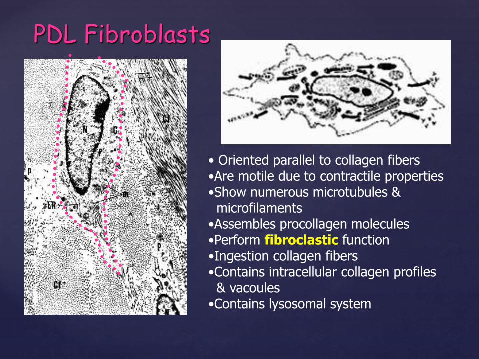
PDL Fibroblasts
• Oriented parallel to collagen fibers•Are motile due to contractile properties•Show numerous microtubules & microfilaments
•Assembles procollagen molecules•Perform fibroclastic function•Ingestion collagen fibers•Contains intracellular collagen profiles& vacoules
•Contains lysosomal system

Cementoblasts Osteoblasts
Both are rich in alkaline phosphatase activity.

Resorptive Cells of PDL
• osteoclasts• cementoclastsBoth are rich in acid phosphataseactivity.

Progenitor cellsU. M. C.
• Can undergo mitotic devision
• Can differentiate to different types of cells
• Have small, closed-face nucleus
• Little amount of cytoplasm
• Found close to blood vessels

Defensive Cells of PDL
Macrophages
lymphocytes
Mast cells
Note: There are no plasma cells.

Epithelial Cells: remnants of the epithelialroot sheath of Hertwig.
•They are separated from the
surrounding connective tissue by a
basal lamina.
•They show low cells turnover,
although they may proliferate to
form cysts or tumors.


Interstitial tissues

*They are found between the
fibers of the periodontal
ligament.
*They are areas that
contain some of the blood
vessels, lymphatic and
nerves.
*They are surrounded
by loose connective tissue.
Interstitial tissues

The arterial blood supply of the periodontal ligament is derived from 3 sources:
Blood supply
3- Branches from the apical vessels that supply the dental pulp.
2- Branches from the intra-alveolarvessels, these branches run
horizontally and these constitute the
main blood supply.
1- Branches from the gingival vessels.

The nerve supply of periodontal ligament comes from either the inferior or superior dental nerves.
1- Bundles of nerve fibers run from the apical region of the root towards the gingival margin.
2- Nerves enter the ligament horizontally through multiple foramina in the bone.
Nerve supply:

(mechanoreceptors)
Large nerve fibers
Small nerve fibers pain sensation
touch & pressure
Are responsible for
Are responsible for
•Stimulation of the mechanoreceptors initiates a reflex
jaw opening.
•This reflex is a protective mechanism to prevent forces
applied to the teeth from reaching damaging levels.

Function of PDL
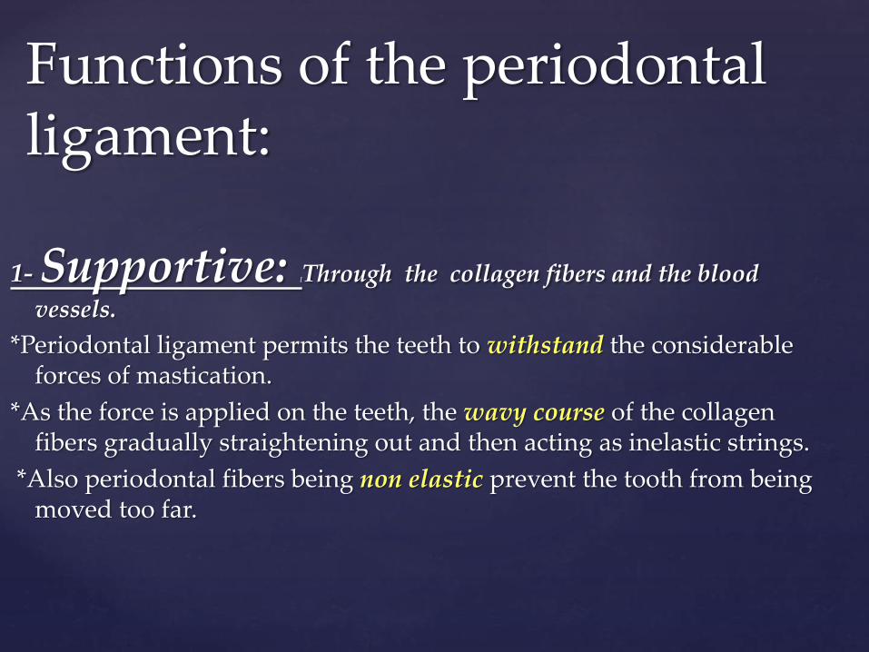
1- Supportive: [Through the collagen fibers and the blood
vessels.
*Periodontal ligament permits the teeth to withstand the considerable forces of mastication.
*As the force is applied on the teeth, the wavy course of the collagen fibers gradually straightening out and then acting as inelastic strings.
*Also periodontal fibers being non elastic prevent the tooth from being moved too far.
Functions of the periodontal ligament:

*Blood vessels and all the components of the ligament act together as a hydraulic damper or shock absorber with the ground substance and the tissue fluid.

Dental implants lack periodontal ligament fibers
and they have a rigid connection to bone
(Osseointegration).

Periimplant tissues
Titanium implant
Sulcular epithelium
Juunctional epithelium
Connective tissue
Bone

The periodontal ligament having the mechanoreceptors
contributes to the sensation of touch and pressure on
the teeth.
2- Sensory:
sudden overloadproprioceptive reflex
inhibition of the activity
of the masticatory muscles
Opening the mouth

The blood vessels in the periodontal ligament provide nutrient supply required by the cells of the ligament
and to the cementocytes and the most superficial
osteocytes.
4- Formative:The fibroblasts are responsible for the formation of new periodontal ligament fibers and dissolution of the old fibers Cementoblasts and osteoblasts are essential in building
up cementum and bone.
3- Nutritive:

The protective function of the periodontal ligament is achieved by:
a- The principal fibers.
b- The blood vessels.
c- The nerves.
a- The principal fibers:
The arrangement of the fiber bundles in the different groups is well adapted to fulfill the functions of the periodontal ligament.
The alveolodental ligament transforms the masticatory
pressure exerted on the tooth into tension or traction on the cementum and bone.
If the exerted force on a tooth is transmitted as pressure this will lead to differentiation of osteoclasts in the pressure area and resorption of bone.
5- Protective

b- The blood vessels:The capillaries form a rich network, they are
arranged in form of a coil and attached to bone and cementum through the oxytalan fibers.
This arrangement makes it possible when pressure is exerted on the tooth, the blood does not escape immediately from the capillaries and thus buffering the pressure action before it reaches the bone.
The behavior of the blood in the capillaries may be simulated to a hydraulic brake.
c- The nerves:By its mechanoreceptors nerves.

*The periodontal ligament through aging shows
Vasuclarity
Cellularity
Thickness *It may contain cementicles.
The Age Changes of periodontal ligament

The cementicles appear near the surface of cementum may be free , attached or embeddedin the cementum.
They have nidus favoring the deposition of concentric layers of calcosphrite as degenerated cells, area of hemorrhage and epithelial rest's of Malassez.
Cementicles are usually seen in periodontal ligament by aging but in some cases they may be seen in a younger person after local trauma.