Embed Size (px)
Citation preview

251
Journal of Hard Tissue Biology 28[3] (2019) 251-2582019 The Hard Tissue Biology Network AssociationPrinted in Japan, All rights reserved.CODEN-JHTBFF, ISSN 1341-7649
OriginalA Human Amelogenin-Derived Oligopeptide Enhances Osteogenic Differentiation
of Human Periodontal Ligament Stem Cells
Tomoki Takeuchi1), Kazuya Masuno2), Hirohito Kato3), Yoichiro Taguchi3), Makoto Umeda3), Nobutaka Okusa4), Akio Tanaka5) and Kazuya Tominaga1)
Correspondence to: Dr. Tomoki Takeuchi, Department of Oral Pathology, Osaka Dental University, 8-1, Kuzuha-hanazono-cho, Hirakata, Osaka 573-1121, Japan; Tel: +81-72-864-3057; Fax: +81-72-864-3157; E-mail: [email protected]
IntroductionEmdogain® is the predominant protein constituent of enamel matrix
derivative (EMD), which is extracted from the tooth germs of 6-month-old pigs. EMD induces the formation of acellular cementum, periodon-tal ligament, and alveolar bone1,2). EMD is commonly used in periodon-tal tissue regeneration therapies. Although one study showed the clinical safety of EMD in the treatment of periodontal defects3), the possibility that EMD may contain unknown pathogens cannot be ruled out since it is derived from animal tissue. Furthermore, EMD is antigenic and in-duces the production of anti-EMD antibodies in patients exposed to it4), Therefore, a completely synthetic material for periodontal tissue regen-eration should be developed.
In our previous study, EMD injected subcutaneously into the backs of rats induced the formation of eosinophilic round bodies (ERBs) and cartilage tissue5). Biochemical analysis of these ERBs was conducted using matrix-assisted laser desorption ionization time-of-flight mass
spectrometry (MALDI-TOF MS). The ERBs consisted of various lengths of peptide fragments, all of which contained a seven amino acid sequence (WYQNMIR). This amino acid sequence corresponds to a portion of porcine amelogenin exon 5, and a porcine amelogenin-de-rived peptide (PAP) was constructed based on this sequence.
We found that bone and cartilage tissue were formed subcutaneously in the backs of rats injected with PAP6), and that PAP induced the forma-tion of hard tissue and promoted early wound healing in artificial perio-dontal tissue defects in rats7,8). In our previous in vitro studies, PAP pro-moted the proliferation and osteogenic differentiation of rat bone marrow stromal cells9), human mesenchymal stem cells (MSCs)10), hu-man periodontal ligament (PDL) fibroblasts11,12), and human periodontal ligament stem cells (PDLSCs)13). These findings suggest that PAP is useful for periodontal tissue regeneration, and exon 5 in the sequence of amelogenin may have an important role in the process of regeneration.
The amino acid sequence of exon 5 in human amelogenin is partly different from porcine amelogenin14). Although human full-length amelogenin enhances the osteogenic differentiation of human bone mar-row MSCs15), it has not been clarified whether human amelogenin exon 5 has any effect on the process of osteogenic differentiation. In this
1) Department of Oral Pathology, Osaka Dental University, Osaka, Japan2) Department of Innovations in Dental Education, Osaka Dental University, Osaka, Japan3) Department of Periodontology, Osaka Dental University, Osaka, Japan4) Department of Forensic Dentistry, Osaka Dental University, Osaka, Japan5) Department of Pathology, Osaka Dental University, Osaka, Japan(Accepted for publication, June 6, 2019)
Abstract: We have found newly synthesized peptide derived from commercially available Emdogain® for periodontal tissue regeneration therapies in our previous study. That synthesized peptide consisted of seven amino acid sequence, WYQN-MIR, which is exon 5 of porcine amelogenin and had the same effects as Emdogain®. The amino acid sequence of human amelogenin exon 5 is WYQSIR which both N (asparagine) and M (methionine) in porcine amelogenin are replaced by S (serine). In the present study, we investigated the effect of a synthetic human amelogenin-derived peptide (HAP) consisting of WYQSIR on human periodontal ligament stem cells (PDLSCs) to be useful or not for the tissue regeneration. PDLSCs were isolated from third molars of adult donors. The effect of HAP on PDLSCs was investigated by culturing them in nor-mal or osteogenic medium with and without HAP. Proliferation of the PDLSCs was evaluated using a cell proliferation as-say after they had been treated with HAP (ranging from 1 ng/ml to 10,000 ng/ml) for 1, 3, 5, and 7 days. Osteogenic differ-entiation was evaluated by measuring alkaline phosphatase (ALP) activity, calcium deposition, osteocalcin production, and mRNA expression of Runx2 and osteonectin after the PDLSCs had been treated with HAP (1,000 ng/ml) for 1, 7, 14, and 21 days. The number of viable cells was significantly increased in the presence of HAP in normal medium. Compared to cells cultured without HAP in osteogenic medium, cells cultured with HAP showed significantly increased ALP activity, calcium deposition, osteocalcin production, and Runx2 mRNA expression. The results show that HAP enhances prolifera-tion and osteogenic differentiation of PDLSCs. The present study suggests that HAP may be a useful material for periodon-tal tissue regeneration.
Key words: Amelogenin, Regeneration, Osteogenesis, Periodontal ligament, Mesenchymal stem cells

252
J.Hard Tissue Biology Vol. 28(3): 251-258, 2019
study, we synthesized a human amelogenin-derived peptide (HAP). This HAP consists of a six amino acid sequence (WYQSIR), which corre-sponds to a portion of human amelogenin exon 5. In general, only pep-tides of greater than approximately 10 residues or over 5 kDa can func-tion as antigens16,17). Because the molecular mass of HAP is 852 Da, there is very little risk of eliciting an immunologic response.
PDLSCs are a type of mesenchymal stem cell that exist in the perio-dontal ligament18). PDLSCs have the potential to differentiate into oste-oblasts19,20) and enhance the periodontal tissue regeneration capacity21-23). PDLSCs exhibit greater potential in the regeneration of periodontal tis-sues than other mesenchymal stem cells, such as bone marrow stromal cells24). Therefore, PDLSCs play a particularly important role in perio-dontal tissue regeneration. The aim of this study was to elucidate the ef-fect of HAP on the proliferation and osteogenic differentiation of PDLSCs.
Materials and MethodsIsolation and culture of PDLSCs
Impacted and non-carious third molars were extracted from three patients (two females and a male, aged 21 to 29 years). After extraction, the teeth were rinsed in Dulbecco’s Modified Eagle’s Medium (DMEM) with 500 U/ml penicillin, 500 µg/ml streptomycin, and 1.25 µg/ml am-photericin B (all from Nacalai Tesque, Kyoto, Japan). After rinsing, the PDL tissues were separated from the middle one-third of the root sur-face and minced into 1-mm3 pieces. The minced tissues were digested for 1 h at 37 °C in a solution of 3 mg/ml collagenase type I (Wako Pure Chemical Industrials Ltd, Tokyo, Japan) and 4 mg/ml dispase (Gibco, Thermo Fisher Scientific, Grand Island, NY, USA). The digested tissue samples were pooled, and single-cell suspensions were obtained by passing the pooled tissues through a 70-µm strainer (Falcon BD, Frank-lin Lakes, NJ, USA). The cells were centrifuged at 1,000 rpm for 5 min and resuspended in normal culture medium containing 10% fetal bovine serum (Biowest, Nuaillé, France), 500 U/ml penicillin (Nacalai Tesque), 500 µg/ml streptomycin (Nacalai Tesque), and 1.25 µg/ml amphotericin B (Nacalai Tesque). The cells were then seeded onto T75 culture dishes (AGC Techno Glass, Shizuoka, Japan), and incubated at 37 °C in 5% CO2. The presence of single-cell colonies was confirmed after 5 to 10 days. PDLSCs at passage zero (P0) were seeded, and cells at P3 to P5 were used for the experiments in the present study. PDLSCs were ob-tained in accordance with the medical ethics guidelines of Osaka Dental University, and all experiments were approved by the Osaka Dental University Medical Ethics Committee (approval no. 110897). All partic-ipants provided written informed consent to participate in the present study, and the study design was approved by the appropriate ethics re-view board.
Characterization of PDLSCsThe PDLSCs were identified by immunocytochemistry, as previous-
ly described19,20). The PDLSCs were plated at a density of 2 × 10⁴ cells/ml in 300 µl of normal culture medium on Lab-Tek® Chamber Slides (Thermo Fisher Scientific Inc, Waltham, MA, USA) and incubated for 3 days. The cells were fixed in cold 70% ethanol for 20 min at -20 °C, and blocked with 3% bovine serum albumin in phosphate-buffered saline (PBS) for 30 min at room temperature. The cells were then incubated with anti-STRO-1 antibody (Thermo Fisher Scientific Inc.), or an-ti-SSEA-4 antibody (Thermo Fisher Scientific Inc.) overnight at 4 °C. After washing with PBS, the cells were incubated for 60 min at room temperature with a fluorescently labeled secondary anti-mouse poly-clonal immunoglobulin G antibody (Thermo Fisher Scientific Inc.). The
samples were washed with PBS and mounted in Vectashield® Mounting Medium with DAPI (Vector Laboratories, Burlingame, CA, USA). Im-ages were obtained with a LSM 700 fluorescence microscope (Carl Zeiss, Jena, Germany).
Preparation of synthetic oligopeptideThe synthesis of HAP was based on the amino acid sequence, WYQ-
SIR. HAP was prepared by traditional solid-phase peptide synthesis in conjunction with the “tea-bag” methodology using Boc/benzyl-based chemistry.
Cell proliferation assayPDLSCs were plated into 96-well microplates at a density of 2 × 10⁴
cells/ml in normal culture medium (100 µl/well) with and without HAP (1, 10, 100, 1,000, or 10,000 ng/ml), and the cells were cultured for 1, 3, 5, and 7 days. The number of viable cells at each time point was deter-mined by measuring the amount of formazan generated in 6 wells per group with a formazan detection kit (Nacalai Tesque). The absorbance of formazan was measured at a wavelength of 450 nm, and the data were analyzed with the Soft Max® Pro Microplate Data Acquisition and Analysis software (Molecular Devices, Sunnyvale, CA, USA).
Measurement of alkaline phosphatase (ALP) activityPDLSCs were plated into 24-well microplates at a density of 4 × 10⁴
cells/ml and cultured to confluence in normal culture medium. The me-dium was replaced with osteogenic medium containing 50 µM L-ascor-bic acid 2-phosphate (Nacalai Tesque), 10 mM β-glycerophosphate (Wako), and 10 nM dexamethasone (MP Biomedicals, LLC, Santa Ana, CA, USA) with and without HAP (1,000 ng/ml), and the cells were cul-tured for 7 and 14 days. The cells were washed with PBS and lysed with 0.2% Triton X-100 (Sigma, St. Louis, MO, USA). The cell lysates were treated with a 1-Step PNPP substrate (Pierce Biotechnology, Inc., Pock-ford, IL, USA), and the absorbance was measured at a wavelength of 405 nm. The DNA content was measured using the PicoGreen dsDNA Assay Kit (Invitrogen, Paisley, UK). To normalize the ALP activity, the amount of ALP was normalized to the amount of DNA in the cell lysate. The data were analyzed with the Soft Max® Pro software (Molecular Devices).
Measurement of calcium depositionPDLSCs were plated into 24-well microplates at a density of 4 × 10⁴
cells/ml and cultured to confluence in normal culture medium. The me-dium was replaced with osteogenic medium with and without HAP (1,000 ng/ml), and the cells were cultured for 21 days. Extracellular cal-cium deposition was measured after dissolving with 10% formic acid. The amount of calcium was quantified using a Calcium E-test Kit (Wako) according to the manufacturer’s protocol. The absorbance was measured at a wavelength of 610 nm, and the data were analyzed with the Soft Max® Pro software (Molecular Devices).
Alizarin red stainingPDLSCs were plated into 24-well microplates at a density of 4 × 10⁴
cells/ml and cultured to confluence in normal culture medium. The me-dium was replaced with osteogenic medium with and without HAP (1,000 ng/ml), and the cells were cultured for 21 days. The cells were washed with PBS and fixed in 70% ethanol for 10 min at −20 °C. PDLSCs were stained with a solution of 1% alizarin red S (Wako) for 3 min at room temperature and washed 3 times with distilled water. Pic-tures of the calcified nodules were processed using a microscope (Olym-

253
Tomoki Takeuchi et al.: Human Amelogenin-derived Peptide
pus, Tokyo, Japan).
Measurement of osteocalcinPDLSCs were plated into 24-well microplates at a density of 4 × 10⁴
cells/ml and cultured to confluence in normal culture medium. The me-dium was replaced with osteogenic medium with and without HAP (1,000 ng/ml), and the cells were cultured for 21 days. The culture su-pernatant was collected, and the osteocalcin levels were measured with an osteocalcin detection kit (Takara Bio, Shiga, Japan).
Quantitative real-time polymerase chain reaction (PCR)The mRNA expression levels of Runx2 and osteonectin were deter-
mined by quantitative real-time PCR analysis. PDLSCs were plated into 24-well microplates at a density of 4 × 10⁴ cells/ml and cultured to con-fluence in normal culture medium. The medium was replaced with oste-ogenic medium with and without HAP (1,000 ng/ml), and the cells were cultured for 24 h. The total cellular RNA was extracted using an RNeasy Mini Kit (Qiagen, Venlo, the Netherlands), and 10 µl RNA from each sample was reverse transcribed into complementary DNA using a Pri-meScript RT Reagent Kit (Takara Bio). All real-time PCR assays were performed according to the manufacturer’s protocol. Gene expression was calculated using a StepOnePlus Real-time PCR System (Thermo Fisher Scientific Inc.) and normalized to GAPDH expression.
Statistical analysisIn this study, 6 wells were prepared for the cell proliferation assay,
and 3 wells were prepared for the remaining experiments; each experi-ment was repeated 3 times. Data are presented as mean ± SD, and were analyzed using SPSS Statistics Ver. 17 (IBM, Chicago, IL, USA). One-way analysis of variance followed by Tukey’s post hoc test was used to determine significance in the cell proliferation assay, and Student’s t-test was used in the remaining experiments. P values <0.05 were considered significant.
ResultsIsolation and characterization of PDLSCs
The PDL-derived cells formed clonogenic clusters of fibroblast-like cells. The PDLSCs derived from the PDL cells were positive for STRO-1 (Fig. 1A) which is one of the most well-known markers for MSCs. It has been heavily relied upon for the recognition and isolation of various types of MSC, particularly those in dental tissues25). The PDLSCs were also positive for SSEA-4 (Fig. 1B), which is an embryonic stem cell marker that has also been detected in periodontal ligament-derived MSCs26).
Cell proliferationThe effect of HAP on the proliferation of PDLSCs is shown in Fig.
2. HAP significantly enhanced the proliferation of PDLSCs cultured for 1, 5, and 7 days in normal culture medium. Additionally, HAP induced
Figure 1. Distinction of human PDLSCs as MSCs. A: Immunocytochemical staining for STRO-1. The PDLSCs were positive for STRO-1 (green). B: Immunocytochemical staining for SSEA-4. The PDLSCs were also positive for SSEA-4 (green). Scale bar = 50 µm.
Figure 2. Effect of HAP on cell proliferation of PDLSCs. After the PDLSCs were cultured for 24 h, the medium was replaced with normal culture medium containing HAP (0, 1, 10, 100, 1,000, or 10,000 ng/ml). Cell proliferation un-der these conditions was measured on days 1, 3, 5, and 7. *p < 0.05 vs. HAP (0 ng/ml), at days 1, 5, and 7.

254
J.Hard Tissue Biology Vol. 28(3): 251-258, 2019
the highest cell proliferation at a concentration of 1,000 ng/ml. There-fore, 1,000 ng/ml of HAP was determined to be the optimal concentra-tion for subsequent assays.
ALP activityThe intracellular ALP activity of PDLSCs was significantly en-
hanced in the presence of HAP on 7 and 14 days, compared with the cells cultured without HAP (Fig. 3).
MineralizationCalcium deposition in the extracellular matrix of PDLSCs cultured
in osteogenic medium for 21 days was significantly increased in the presence of HAP compared with the control (Fig. 4A). The number and size of calcified nodules stained with alizarin red S after incubation in osteogenic medium for 21 days were larger in the presence of HAP than in the control (Fig. 4B).
Osteocalcin productionThe production of osteocalcin in the supernatant of PDLSCs after
incubation in osteogenic medium for 21 days was significantly increased in the presence of HAP compared with the control (Fig. 5).
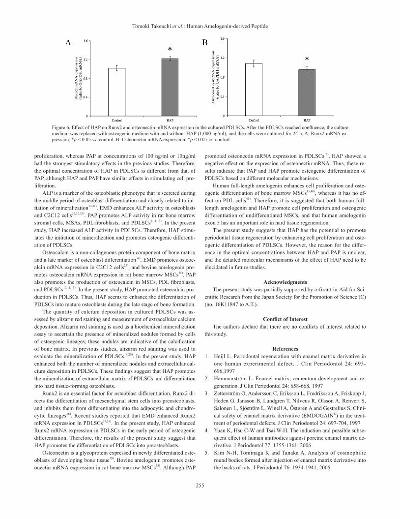
Runx2 and osteonectin mRNA expressionRunx2 mRNA expression after incubation in osteogenic medium for
24 h was significantly enhanced in the presence of HAP compared with the control (Fig. 6A). However, osteonectin mRNA expression after 24 h incubation was significantly lower in the presence of HAP than in the control (Fig. 6B).
DiscussionThe results of this study show that HAP significantly promotes pro-
liferation of PDLSCs in normal medium. In the osteoblast differentiation assays, ALP activity, osteocalcin production, extracellular calcium dep-osition, and mRNA expression of Runx2 were significantly enhanced in osteogenic medium containing HAP.
EMD stimulates proliferation of C2C12 cells (a typical pluripotent mesenchymal cell line), bone marrow stromal cells, and PDL cells27-29). In our previous studies, PAP promoted proliferation of rat bone marrow stromal cells, human MSCs, PDL fibroblasts, and PDLSCs9-11,13). In the present study, HAP promoted proliferation of PDLSCs. In particular, HAP at a concentration of 1,000 ng/ml stimulated the highest rate of cell
Figure 3. Effect of HAP on ALP activity in the cultured PDLSCs. After the PDLSCs reached confluence, the culture medium was replaced with osteogenic medium with and without HAP (1,000 ng/ml). ALP activity was measured at 7 or 14 days. To normalize ALP activity, the amount of ALP was normalized to the amount of DNA. A: Day 7, *p < 0.05 vs. control. B: Day 14, *p < 0.05 vs. control.
Figure 4. Effect of HAP on mineralization in the cultured PDLSCs. A: The ex-tracellular calcium deposition on the PDLSCs was measured after 21 days of cultivation in osteogenic medium with and without HAP (1,000 ng/ml). *p < 0.05 vs. control. B: Confluent PDLSCs were stained with alizarin red at 21 days. Scale bar = 100 µm.
Figure 5. Effect of HAP on osteocalcin production in the cultured PDLSCs. After the PDLSCs reached confluence, the culture medium was replaced with osteogenic medium with and without HAP (1,000 ng/ml), and the cells were cultured for 21 days. *p < 0.05 vs. control.
255
Tomoki Takeuchi et al.: Human Amelogenin-derived Peptide
proliferation, whereas PAP at concentrations of 100 ng/ml or 10ng/ml had the strongest stimulatory effects in the previous studies. Therefore, the optimal concentration of HAP in PDLSCs is different from that of PAP, although HAP and PAP have similar effects in stimulating cell pro-liferation.
ALP is a marker of the osteoblastic phenotype that is secreted during the middle period of osteoblast differentiation and closely related to ini-tiation of mineralization30,31). EMD enhances ALP activity in osteoblasts and C2C12 cells27,32,33). PAP promotes ALP activity in rat bone marrow stromal cells, MSAs, PDL fibroblasts, and PDLSCs9-11,13). In the present study, HAP increased ALP activity in PDLSCs. Therefore, HAP stimu-lates the initiation of mineralization and promotes osteogenic differenti-ation of PDLSCs.
Osteocalcin is a non-collagenous protein component of bone matrix and a late marker of osteoblast differentiation34). EMD promotes osteoc-alcin mRNA expression in C2C12 cells27), and bovine amelogenin pro-motes osteocalcin mRNA expression in rat bone marrow MSCs35). PAP also promotes the production of osteocalcin in MSCs, PDL fibroblasts, and PDLSCs10,11,13). In the present study, HAP promoted osteocalcin pro-duction in PDLSCs. Thus, HAP seems to enhance the differentiation of PDLSCs into mature osteoblasts during the late stage of bone formation.
The quantity of calcium deposition in cultured PDLSCs was as-sessed by alizarin red staining and measurement of extracellular calcium deposition. Alizarin red staining is used as a biochemical mineralization assay to ascertain the presence of mineralized nodules formed by cells of osteogenic lineages; these nodules are indicative of the calcification of bone matrix. In previous studies, alizarin red staining was used to evaluate the mineralization of PDLSCs19,20). In the present study, HAP enhanced both the number of mineralized nodules and extracellular cal-cium deposition in PDLSCs. These findings suggest that HAP promotes the mineralization of extracellular matrix of PDLSCs and differentiation into hard tissue-forming osteoblasts.
Runx2 is an essential factor for osteoblast differentiation. Runx2 di-rects the differentiation of mesenchymal stem cells into preosteoblasts, and inhibits them from differentiating into the adipocytic and chondro-cytic lineages36). Recent studies reported that EMD enhanced Runx2 mRNA expression in PDLSCs37,38). In the present study, HAP enhanced Runx2 mRNA expression in PDLSCs in the early period of osteogenic differentiation. Therefore, the results of the present study suggest that HAP promotes the differentiation of PDLSCs into preosteoblasts.
Osteonectin is a glycoprotein expressed in newly differentiated oste-oblasts of developing bone tissue39). Bovine amelogenin promotes oste-onectin mRNA expression in rat bone marrow MSCs35). Although PAP
promoted osteonectin mRNA expression in PDLSCs13), HAP showed a negative effect on the expression of osteonectin mRNA. Thus, these re-sults indicate that PAP and HAP promote osteogenic differentiation of PDLSCs based on different molecular mechanisms.
Human full-length amelogenin enhances cell proliferation and oste-ogenic differentiation of bone marrow MSCs15,40), whereas it has no ef-fect on PDL cells41). Therefore, it is suggested that both human full-length amelogenin and HAP promote cell proliferation and osteogenic differentiation of undifferentiated MSCs, and that human amelogenin exon 5 has an important role in hard tissue regeneration.
The present study suggests that HAP has the potential to promote periodontal tissue regeneration by enhancing cell proliferation and oste-ogenic differentiation of PDLSCs. However, the reason for the differ-ence in the optimal concentrations between HAP and PAP is unclear, and the detailed molecular mechanisms of the effect of HAP need to be elucidated in future studies.
AcknowledgmentsThe present study was partially supported by a Grant-in-Aid for Sci-
entific Research from the Japan Society for the Promotion of Science (C) (no. 16K11847 to A.T.).
Conflict of InterestThe authors declare that there are no conflicts of interest related to
this study.
References1. Heijl L. Periodontal regeneration with enamel matrix derivative in
one human experimental defect. J Clin Periodontol 24: 693-696,1997
2. Hammarström L. Enamel matrix, cementum development and re-generation. J Clin Periodontol 24: 658-668, 1997
3. Zetterström O, Andersson C, Eriksson L, Fredriksson A, Friskopp J, Heden G, Jansson B, Lundgren T, Nilveus R, Olsson A, Renvert S, Salonen L, Sjöström L, Winell A, Östgren A and Gestrelius S. Clini-cal safety of enamel matrix derivative (EMDOGAIN®) in the treat-ment of periodontal defects. J Clin Periodontol 24: 697-704, 1997
4. Yuan K, Hsu C-W and Tsai W-H. The induction and possible subse-quent effect of human antibodies against porcine enamel matrix de-rivative. J Periodontol 77: 1355-1361, 2006
5. Kim N-H, Tominaga K and Tanaka A. Analysis of eosinophilic round bodies formed after injection of enamel matrix derivative into the backs of rats. J Periodontol 76: 1934-1941, 2005
Figure 6. Effect of HAP on Runx2 and osteonectin mRNA expression in the cultured PDLSCs. After the PDLSCs reached confluence, the culture medium was replaced with osteogenic medium with and without HAP (1,000 ng/ml), and the cells were cultured for 24 h. A: Runx2 mRNA ex-pression, *p < 0.05 vs. control. B: Osteonectin mRNA expression, *p < 0.05 vs. control.

256
J.Hard Tissue Biology Vol. 28(3): 251-258, 2019
6. Hida T, Tominaga K and Tanaka A. Tissue reaction to synthetic oli-gopeptide derived from enamel matrix derivative in rats. Oral Sci Int 7: 26-33, 2010
7. Noguchi M, Tominaga K, Tanaka A and Ueda M. Hard tissue for-mation induced by synthetic oligopeptide derived from an enamel matrix derivative. Oral Med Pathol 16: 75-80, 2012
8. Miki H, Tominaga K, Takahashi T, Tanaka A and Umeda M. Com-parison of the influence of a new synthetic peptide and enamel ma-trix derivative in the early wound healing process of artificial perio-dontal tissue defects in rats. Jpn J Conserv Dent 61: 17-29, 2018
9. Yasui N, Taguchi Y, Tanaka A, Ueda M and Umeda M. Biological effects of emdogain®-derived oligopeptides on rat bone marrow cells in vitro. J Oral Tissue Engin 9: 126-135, 2012.
10. Katayama N, Kato H, Taguchi Y, Tanaka A and Umeda M. The ef-fects of synthetic oligopeptide derived from enamel matrix deriva-tive on cell proliferation and osteoblastic differentiation of human mesenchymal stem cells. Int J Mol Sci 15: 14026-14043, 2014
11. Kawanaka A, Tominaga K and Tanaka A. Effect of peptide derived from Emdogain® on human periodontal ligament fibroblasts. J Osa-ka Dent Univ 43: 111-117, 2009
12. Taguchi Y, Yasui N, Takahashi S, Tominaga K, Kato H, Komasa S, Shida M, Hayashi H, Tanaka A and Umeda M. Hard tissue forma-tion by human periodontal ligament fibroblast cells treated with an emdogain®-derived oligopeptide in vitro. J Hard Tissue Biol 21: 375-384, 2012
13. Kato H, Katayama N, Taguchi Y, Tominaga K, Umeda M and Tana-ka A. A synthetic oligopeptide derived from enamel matrix deriva-tive promotes the differentiation of human periodontal ligament stem cells into osteoblast-like cells with increased mineralization. J Periodontol 84: 1476-1483, 2013
14. Brookes SJ, Robinson C, Kirkham J and Bonass WA. Biochemistry and molecular biology of amelogenin proteins of developing dental enamel. Arch Oral Biol 40: 1-14, 1995
15. Tanimoto K, Huang YC, Tanne Y, Kunimatsu R, Michida M, Yosh-ioka M, Ozaki N, Sasamoto T, Yoshimi Y, Kato Y and Tanne K. Amelogenin enhances the osteogenic differentiation of mesenchy-mal stem cells derived from bone marrow. Cells Tissues Organs 196: 411-419, 2012
16. Shinnick TM, Sutcliffe JG, Green N and Lerner RA. Synthetic pep-tide immunogens as vaccines. Annu Rev Microbiol 37: 425-446, 1983
17. Lerner RA. Tapping the immunological repertoire to produce anti-bodies of predetermined specificity. Nature 299: 592-596, 1982
18. Nagatomo K, Komaki M, Sekiya I, Sakaguchi Y, Noguchi K, Oda S, Muneta T and Ishikawa I. Stem cell properties of human periodontal ligament cells. J Periodontal Res 41: 303-310, 2006
19. Seo BM, Miura M, Gronthos S, Bartold PM, Batouli S, Brahim J, Young M, Robey PG, Wang CY and Shi S. Investigation of multipo-tent postnatal stem cells from human periodontal ligament. Lancet 364: 149-155, 2004
20. Park JC, Kim JM, Jung IH, Kim JC, Choi SH, Cho KS and Kim CS. Isolation and characterization of human periodontal ligament (PDL) stem cells (PDLSCs) from the inflamed PDL tissue: In vitro and in vivo evaluations. J Clin Periodontol 38: 721-731, 2011
21. Liu Y, Zheng Y, Ding G, Fang D, Zhang C, Bartold PM, Gronthos S, Shi S and Wang S. Periodontal ligament stem cell-mediated treat-ment for periodontitis in miniature swine. Stem Cells 26: 1065-1073, 2008
22. Tsumanuma Y, Iwata T, Washio K, Yoshida T, Yamada A, Takagi R,
Ohno T, Lin K, Yamato M, Ishikawa I, Okano T and Izumi Y. Com-parison of different tissue-derived stem cell sheets for periodontal regeneration in a canine 1-wall defect model. Biomaterials 32: 5819-5825, 2011
23. Sonoyama W, Liu Y, Fang D, Yamaza T, Seo BM, Zhang C, Liu H, Gronthos S, Wang CY, Wang S and Shi S. Mesenchymal stem cell-mediated functional tooth regeneration in swine. PLoS One 1: e79, 2006
24. Gronthos S, Mrozik K, Shi S and Bartold PM. Ovine periodontal ligament stem cells: Isolation, characterization, and differentiation potential. Calcif Tissue Int 79: 310-317, 2006
25. Ning H, Lin G, Lue TF and Lin CS. Mesenchymal stem cell marker Stro-1 is a 75kd endothelial antigen. Biochem Biophys Res Com-mun 413: 353-357, 2011
26. Feng-Juan Lv, Tuan RS, Cheung KMC and Leung VYL. Concise Review : The surface markers and identity of human mesenchymal stem cells. Stem cells 32: 1408-1419, 2014
27. Ohyama M, Suzuki N, Yamaguchi Y, Maeno M, Otsuka K and Ito K. Effect of enamel matrix derivative on the differentiation of C2C12 Cells. J Periodontol 73: 543-550, 2002
28. Cattaneo V, Rota C, Silvestri M, Piacentini C, Forlino A, Gallanti A, Rasperini G and Cetta G. Effect of enamel matrix derivative on hu-man periodontal fibroblasts: Proliferation, morphology and root sur-face colonization. An in vitro study. J Periodontal Res 38: 568-574, 2003
29. Guida L, Annunziata M, Carinci F, Di Feo A, Passaro I and Oliva A. In vitro biologic response of human bone marrow stromal cells to enamel matrix derivative. J Periodontol 78: 2190-2196, 2007
30. Weinreb M, Shinar D and Rodan GA. Different pattern of alkaline phosphatase, osteopontin, and osteocalcin expression in developing rat bone visualized by in situ hybridization. J Bone Miner Res 5: 831-842, 1990
31. Aubin JE, Liu F, Malaval L and Gupta AK. Osteoblast and chondro-blast differentiation. Bone 17: 77S-83S, 1995
32. Hägewald S, Pischon N, Jawor P, Bernimoulin JP and Zimmermann B. Effects of enamel matrix derivative on proliferation and differen-tiation of primary osteoblasts. Oral Surg Oral Med Oral Pathol Oral Radiol Endod 98: 243-249, 2004
33. Yoneda S, Itoh D, Kuroda S, Kuroda S, Kondo H, Umezawa A, Ohya K, Ohyama T and Kasugai S. The effects of enamel matrix derivative (EMD) on osteoblastic cells in culture and bone regener-ation in a rat skull defect. J Periodontal Res 38: 333-342, 2003
34. Ikeda T, Nomura S, Yamaguchi A, Suda T and Yoshiki S. In situ hy-bridization of bone matrix proteins in undecalcified adult rat bone sections. J Histochem Cytochem 40: 1079-1088, 1992
35. Izumikawa M, Hayashi K, Polan MAA, Tang J and Saito T. Effects of amelogenin on proliferation, differentiation, and mineralization of rat bone marrow mesenchymal stem cells in vitro. Sci World J 2012; 879731, 2012
36. Komori T. Regulation of osteoblast differentiation by transcription factors. J Cell Biochem 99: 1233-1239, 2006
37. Wang Z, Feng Z, Wu G, Bai S, Dong Y and Zhao Y. Colloids and Surfaces B : Biointerfaces in vitro studies on human periodontal lig-ament stem cell sheets enhanced by enamel matrix derivative. Col-loids Surfaces B Biointerfaces 141: 102-111, 2016
38. Li G, Hu J, Chen H, Chen L, Zhang N, Zhao L, Wen N and Yang Y. Enamel matrix derivative enhances the proliferation and osteogenic differentiation of human periodontal ligament stem cells on the tita-nium implant surface. Organogenesis 13: 103-113, 2017

257
Tomoki Takeuchi et al.: Human Amelogenin-derived Peptide
39. Ishigaki R, Takagi M, Igarashi M and Ito K. Gene expression and immunohistochemical localization of osteonectin in association with early bone formation in the developing mandible. Histocem J 34: 57-66, 2002
40. Huang Y, Tanimoto K, Tanne Y, Kamiya T, Kunimatsu R, Michida M, Yoshioka M, Yoshimi Y, Kato Y and Tanne K. Effects of human full-length amelogenin on the proliferation of human mesenchymal
stem cells derived from bone marrow. Cell Tissue Res 342: 205-212, 2010
41. Tanimoto K, Kunimatsu R, Tanne Y, Huang YC, Michida M, Yoshi-mi Y, Miyauchi M, Takata T and Tanne K. Differential effects of amelogenin on mineralization of cementoblasts and periodontal lig-ament cells. J Periodontol 83: 672-679, 2012

258
J.Hard Tissue Biology Vol. 28(3): 251-258, 2019