Embed Size (px)
Citation preview

MOTOR FUNCTIONS
PRESETED BY
SRILOY MOHANTY
B.N.Y.S

MOTOR CORTEX
• Primary motor cortex ( M1)
• Premotor area (PMA)
• Supplementary motor area (SMA)
Note: All the three projects directly to the spinal cord via
corticospinal tract.
• Premotor and supplementary motor cortex also project to
primary motor cortex and is involved in coordinating &
planning complex sequences of movement (motor learning).

PRIMARY MOTOR CORTEX (M-I)
Location :-
Immediately anterior to the central sulcus and
extends to the medial surface of hemisphere
also known as Broadmann’s area 4 is a
motor homunculus.
Description: Body is represented as up side
down and stretched on the medial surface
where pelvic and leg muscles are
represented.
Hand and mouth has a greater area of
representation and is large because of

- It controls the musculature of the opposite side of the body.
-Face area is bilaterally represented.
Functions:-
Is used in execution of skilled movements also in codes the direction, force and velocity of movements.
Lesions:-
Pure M-I lesions are rare. May have contra lateral weakness in distal muscle (fingers).
Ability to control fine movements is gone.
Ablation of M-I alone cause hypotonia not Spasticity.

SUPPLEMENTARY MOTOR AREA (M-II)
Location: Found on both in lateral and medial aspect of the frontal
lobe. It extends from cingulate sulcus on the medial side to reach premotor cortex on the lateral surface of the brain.
Function:It works together with premotor cortex.Involved in programming of motor sequences. Lesions:
Produces awkwardness in performing complex activity like bimanual coordinated activity.

It function in mental rehearsal of movements before
performing a complex motor functions.
With premotor cortex it translates the desire to
perform a motor task into a series of motor
command that will do the task.

PREMOTOR CORTEX (PMC)
Location:
Broadmann’s area 6. It lies immediately anterior to primary
motor cortex. It is more extensive than primary motor
cortex (about 6 times)
Functions:
It works with the help of basal ganglia, thalamus, primary
motor cortex, posterior parietal cortex. It plays role in
planning and anticipation of a specific motor act.

Premotor cortex – Two-hand Coordination
THE MONKEY HAS LEARNED THE TASK
PUSH THE OBJECT THROUGH THE HOLE AND CATCH IT WITH THE OTHER HAND; With
damage to premotor cortex, cannot coordinate two hands to do the task

Lesion:
It results in re-emergence of suckling and grasp
reflex in adults.
Its lesion do not case paralysis but only slowing of
the complex limb movement.
Lesion may result in loss of short-term or working
memory.
When damaged with supplementary cortex it may
result in APRAXIA.

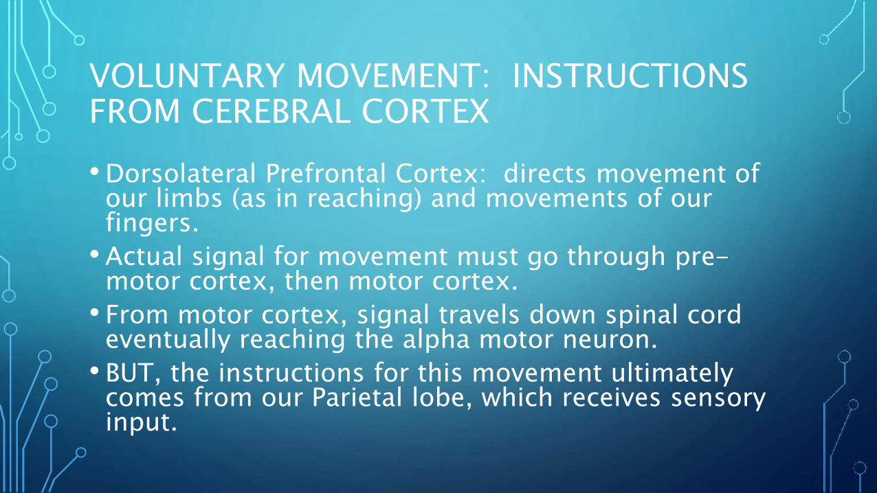
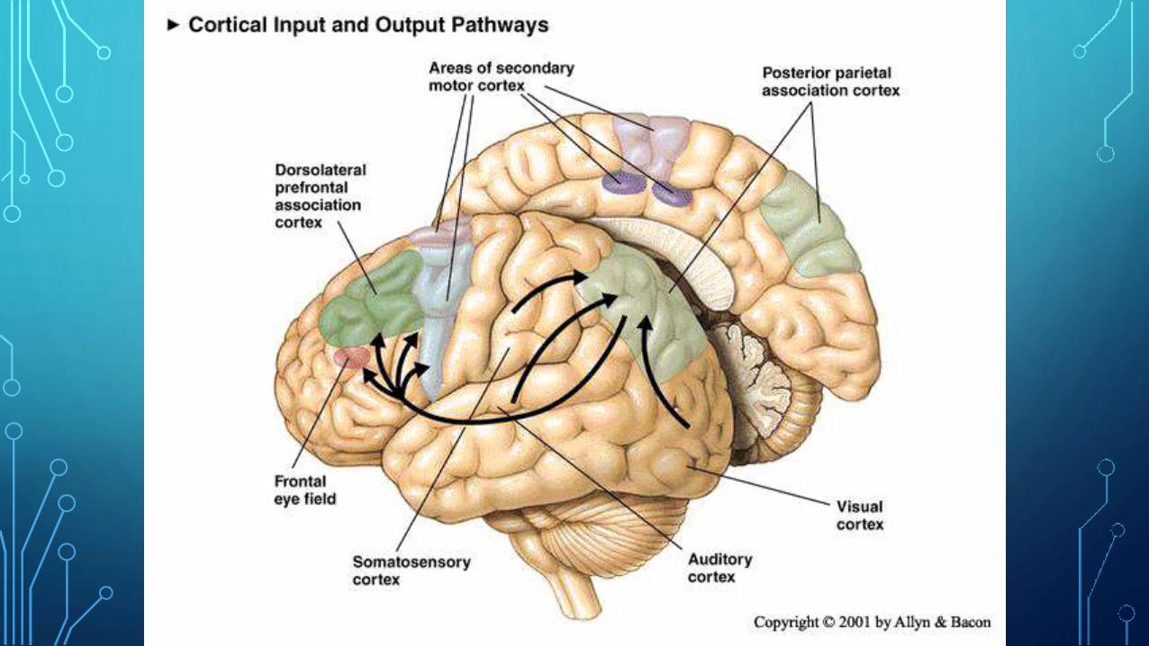
VOLUNTARY MOVEMENT: INSTRUCTIONS FROM CEREBRAL CORTEX
•Dorsolateral Prefrontal Cortex: directs movement of our limbs (as in reaching) and movements of our fingers.
•Actual signal for movement must go through pre-motor cortex, then motor cortex.
• From motor cortex, signal travels down spinal cord eventually reaching the alpha motor neuron.
• BUT, the instructions for this movement ultimately comes from our Parietal lobe, which receives sensory input.


CEREBELLUM• Vermis
• Intermediate zone
• Lateral zone
• Within are deep cerebellar nuclei:
• Fastigial nucleus
• Interpositus nucleus
• Dentate nucleus

VERMIS
Kinesthetic and
somatosensory inputs
from the spinal cord
projections to fastigial
nucleus
• Damage interrupts
posture and walking
• In monkeys, unilateral
lesions of the fastigial
nucleus cause the
monkeys to fall
(ipsilateral side)

INTERMEDIATE ZONE
• Inputs from red nucleus (brain stem & motor cortex) and somatosensory info from the spinal cord
• Projects to interpositus nucleus red nucleus (loop)
• Damage produces rigidity and difficulty in moving limbs
• Action tremor or intention tremor – a tremor causing movement to occur in a staggered manner during motor act.

LATERAL ZONE
• Inputs from motor and association cortices (through pons)
• Projections to dentate nucleus primary motor and premotor cortex
1. Balistic movement –movement that occurs so quickly that it can not be modified by feedback
• E.g., swinging of a batter trying to hit a ball moving 140 km/h

LATERAL ZONE
2. Multijoint movements
3. Learning of new movements
4. Timing of motor movements (and
cognitive functions)

BASAL GANGLIA• Unlike the cerebellum, which
plays a role in rapid balistic movements, the basal ganglia are more important for the accomplishment of movements that may take some time to initiate or stop
• Important for internal guiding (rather then external) of movement
• Dopamine – nigrostriatal pathway

BASAL GANGLIADamage to the basal ganglia:
• Produces either too much activation (hyperkinetic) responses= twitches, movements bursts, jarring, etc.
• Huntington’s Chorea-dominant gene based, increases glutamate in striatum which destroys GABA neurons in BG and loss of inhibition
• No cure
• Tourette’s
OR
• Produces too little force (hypokinetic)=rigidity
• Parkinson’s disease
Pink=inhibitionBlue=excitation

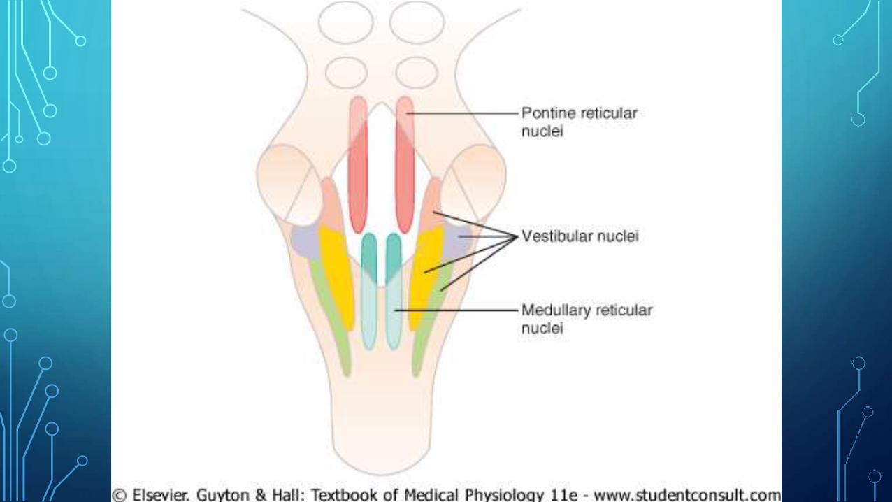
BRAIN STEM MOTOR CENTERS
• Pontine reticular nuclei – excite antigravity
muscles (muscles of the vertebral column and
limb extensor muscles) – pontine reticulospinal
tract.
•Medullary reticular nuclei – inhibit antigravity
muscles – medullary reticulospinal tract.
Pontine & medullary systems balance each other.
• Vestibular nuclei – supplement the excitatory
function of the pontine system by integrating
vestibular information – lateral and medial
vestibulospinal tracts.

Summary of the
major descending
spinal tracts and
their points of
origin corticospinal tract
rubrospinal
tract
reticulospinal tracts
tectospinal,
vestibulospinal
tracts
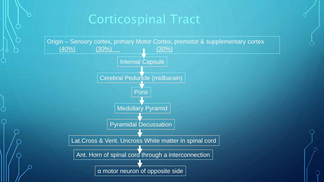
Corticospinal Tract
Origin – Sensory cortex, primary Motor Cortex, premotor & supplementary cortex
(40%) (30%) (30%)
Internal Capsule
Pons
Cerebral Peduncle (midbarain)
Medullary Pyramid
Pyramidal Decussation
Lat.Cross & Vent. Uncross White matter in spinal cord
Ant. Horn of spinal cord through a interconnection
α motor neuron of opposite side

MOTOR UNIT…

•Every striated muscle has encapsulated muscle fibers scattered throughout the muscle called muscle spindles.
•Extrafusal and intrafusalfibers

organization of motor subsystems

Overview - organization of
motor systemsMotor Cortex
Brain Stem
Spinal Cord
Skeletal muscle
-motor
neuron
Final common
pathway

Steps in Motor Action

THANK YOU…