Embed Size (px)
Citation preview
Chapter 4Functions of Myosin Motor Proteinsin the Nervous System
Daniel M. Suter
Abstract The myosin superfamily consists of 24 classes of actin-based molecularmotors that carry out a diverse array of cellular functions ranging from cell motil-ity and morphology to cytokinesis, signal transduction, membrane trafficking, RNAand protein localization. The development and functioning of the nervous systemstrongly depends on the proper establishment of complex networks of neurons withhighly specialized morphologies and molecular composition. Thus, it is not at allsurprising that multiple classes of myosin motors are expressed in the nervoussystem, including classes I, II, III, V, VI, VII, IX, X, XV, and XVI. This reviewdiscusses the current knowledge on myosin functions in both neurons and specificsensory neurons, the hair cells of the inner ear, and the photoreceptors in the eye.The role of myosin II in growth cone motility and neurite outgrowth is the bestcharacterized myosin function in neurons. However, there is increasing evidencethat different myosin motors are also involved in protein and organelle traffickingunderlying synaptic function. Multiple myosin motors have been localized to haircells and photoreceptors and associated with genetic diseases; however, a futurechallenge will be a better characterization of the cellular functions of these variousmotor proteins.
Keywords Motility · Transport · Contractility · Sensory · Stereocilia
4.1 Introduction
Myosins are molecular motors that use the energy derived from ATP hydrolysisto move along actin filaments. Myosins carry out a large variety of cellular func-tions including cell movements, maintenance of cell shape, cytokinesis, organelletransport, signal transduction, phagocytosis, membrane trafficking, RNA and
D.M. Suter (B)Department of Biological Sciences, Purdue University, West Lafayette, IN 47907, USAe-mail: [email protected]
45G. Gallo, L.M. Lanier (eds.), Neurobiology of Actin, Advances in Neurobiology 5,DOI 10.1007/978-1-4419-7368-9_4, C© Springer Science+Business Media, LLC 2011
46 D.M. Suter
protein localization, and transcription (Brown and Bridgman 2004, Gillespie andCyr 2004, Krendel and Mooseker 2005, Sweeney and Houdusse 2007). Over thepast 20 years the number of newly identified myosin molecules and classes hasincreased significantly. The myosin superfamily includes at least 24 classes inspecies ranging from Saccharomyces cerevisiae to Drosophila and humans (Berget al. 2001, Foth et al. 2006). Not all classes are present in each species. The humangenome encodes approximately 40 myosin genes grouped into 12 classes (Berg et al.2001). Since the nomenclature for myosin homologs from different species has beenconfusing in the past, I am following the system suggested for class I myosins; thus,Myo1a will be used as abbreviation for the myosin Ia protein (Gillespie et al. 2001).
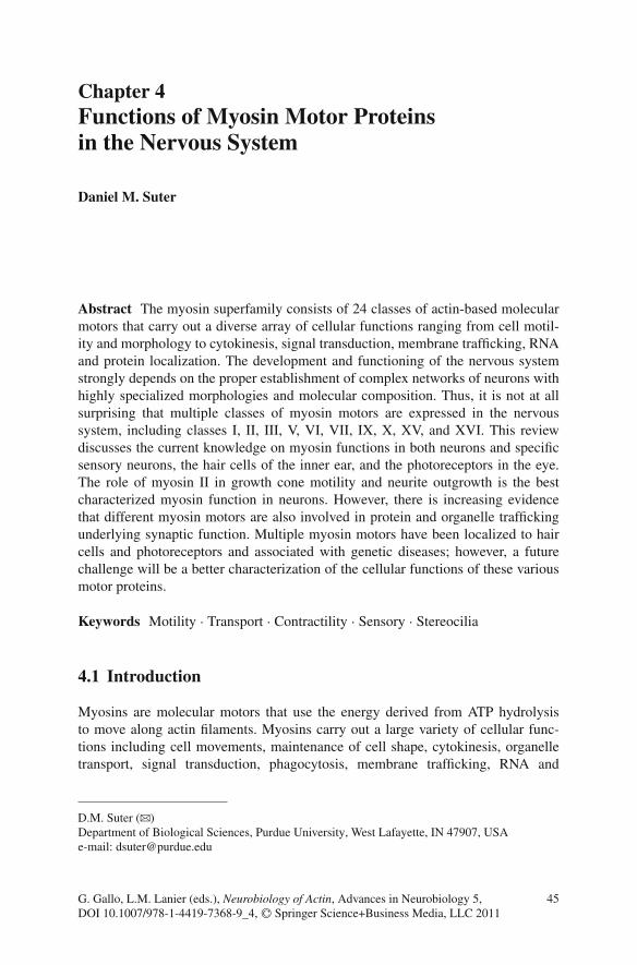
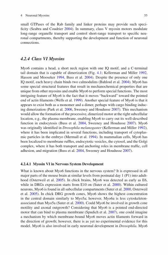
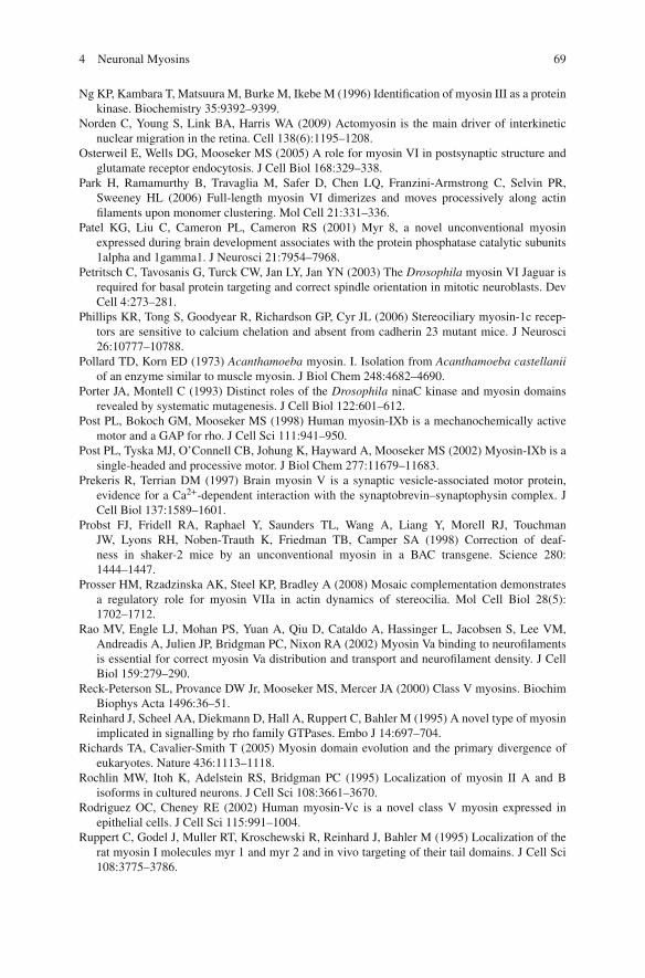
Myosins contain at least one heavy chain with an N-terminal conserved cat-alytic domain of around 80 kDa, followed by a neck region with one or moreputative light chain-binding motifs, sometimes known as the IQ motif (Fig. 4.1).
Fig. 4.1 Structures of myosins in the nervous system. Domain organization of the myosin heavychains represented in the nervous system. Domains are color coded and discussed in the text.The blue coiled-coil tail domains allow dimerization. The right panel shows schematics of thecorresponding myosin structures as either single- or double-headed motors. Myo6 and Myo7a canexist in both monomer and dimer form. Light chains (grey ellipsoids) are indicated based on thenumber of IQ motifs. Figure was modified from Krendel and Mooseker (2005)
4 Neuronal Myosins 47
The most conserved residues of the IQ motif conform to the consensus sequenceIQxxxRGxxxRK (Cheney and Mooseker 1992). The following C-terminal tailcontains specific domains for cargo binding or heavy chain dimerization thatresults in a double-headed motor. Class II myosins have been termed as “con-ventional myosins” since they were discovered first and their role in musclecontraction has been extensively characterized. Beginning with Acanthamoebamyosin I (Myo1) (Pollard and Korn 1973), all newly characterized myosins witha structure different from Myo2 have been classified as “unconventional myosins”(Cheney and Mooseker 1992, Mooseker and Cheney 1995). The classes of themyosin superfamily are mainly defined by differences in the head structure. Morerecently a novel myosin classification scheme has been proposed that is basedon the combinations of various tail domains (Fig. 4.1; Richards and Cavalier-Smith 2005). For a more detailed discussion of the diversity of structural motifsand related functions, I refer to several recent articles and reviews (Berg et al.2001, Krendel and Mooseker 2005, Richards and Cavalier-Smith 2005, Foth et al.2006).
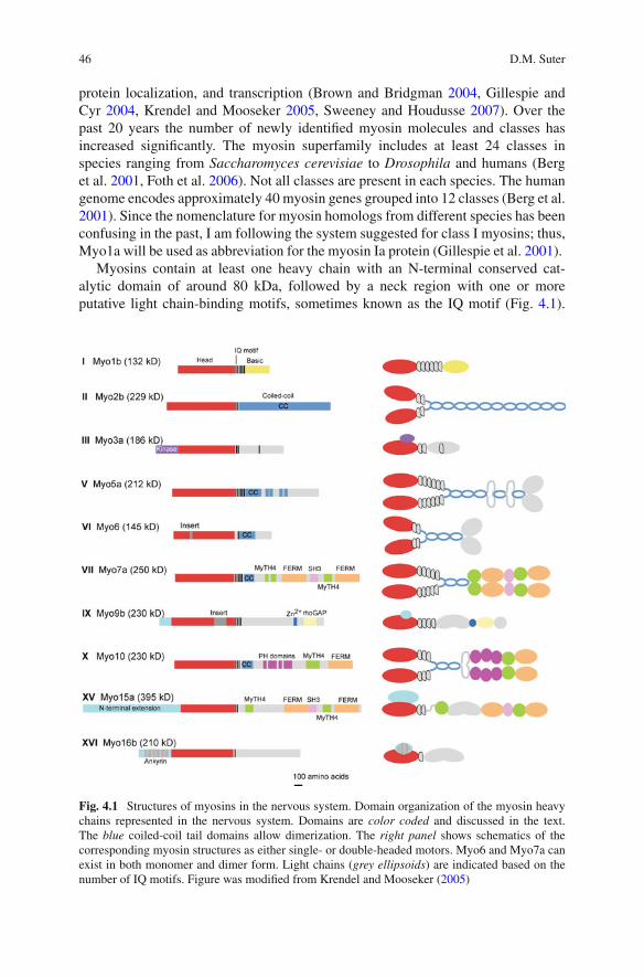
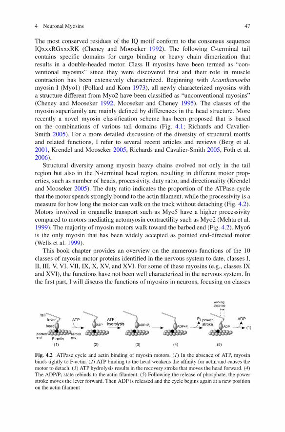
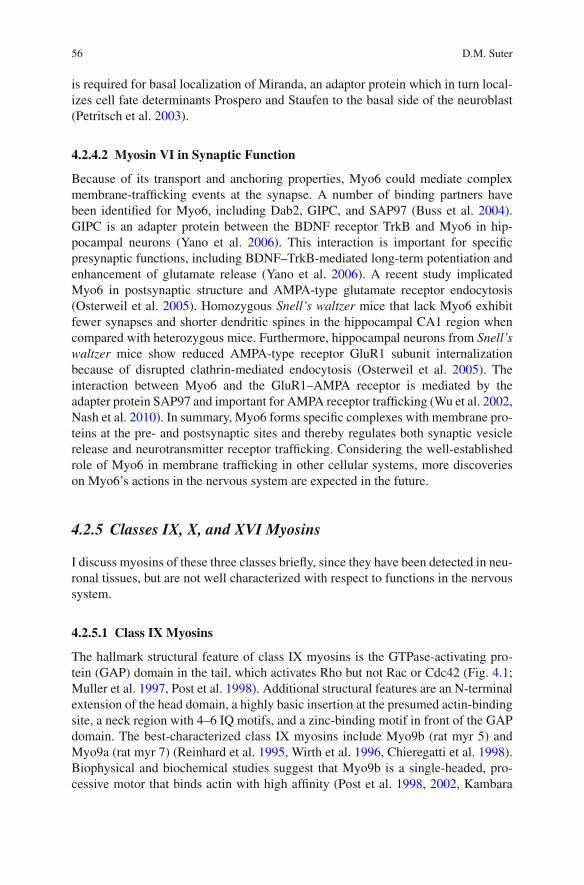
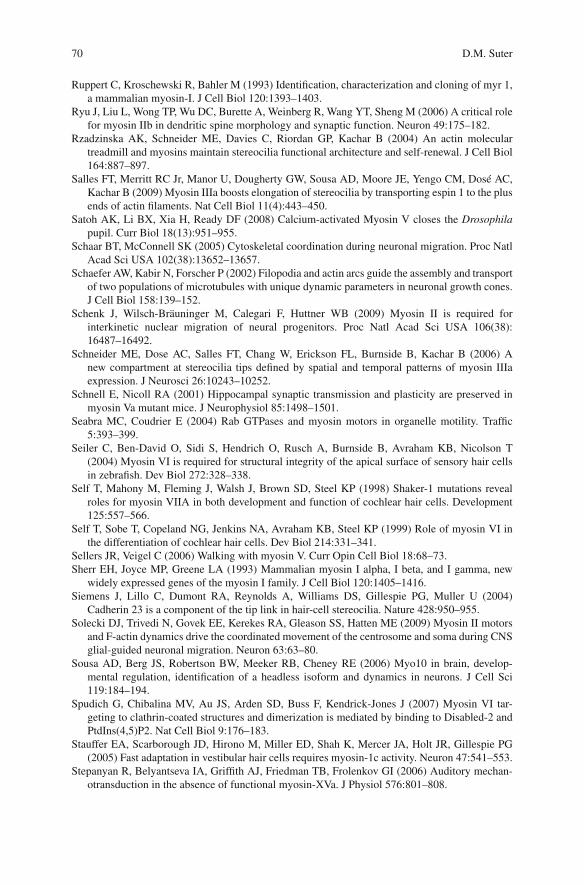
Structural diversity among myosin heavy chains evolved not only in the tailregion but also in the N-terminal head region, resulting in different motor prop-erties, such as number of heads, processivity, duty ratio, and directionality (Krendeland Mooseker 2005). The duty ratio indicates the proportion of the ATPase cyclethat the motor spends strongly bound to the actin filament, while the processivity is ameasure for how long the motor can walk on the track without detaching (Fig. 4.2).Motors involved in organelle transport such as Myo5 have a higher processivitycompared to motors mediating actomyosin contractility such as Myo2 (Mehta et al.1999). The majority of myosin motors walk toward the barbed end (Fig. 4.2). Myo6is the only myosin that has been widely accepted as pointed end-directed motor(Wells et al. 1999).
This book chapter provides an overview on the numerous functions of the 10classes of myosin motor proteins identified in the nervous system to date, classes I,II, III, V, VI, VII, IX, X, XV, and XVI. For some of these myosins (e.g., classes IXand XVI), the functions have not been well characterized in the nervous system. Inthe first part, I will discuss the functions of myosins in neurons, focusing on classes
Fig. 4.2 ATPase cycle and actin binding of myosin motors. (1) In the absence of ATP, myosinbinds tightly to F-actin. (2) ATP binding to the head weakens the affinity for actin and causes themotor to detach. (3) ATP hydrolysis results in the recovery stroke that moves the head forward. (4)The ADP/Pi state rebinds to the actin filament. (5) Following the release of phosphate, the powerstroke moves the lever forward. Then ADP is released and the cycle begins again at a new positionon the actin filament
48 D.M. Suter
I, II, V, and VI. In the second part, I will summarize the different myosin motorfunctions in the best-studied sensory cells, the hair cells of the inner ear, and thephotoreceptors in the eye.
4.2 Myosins in Neurons
Neurons possess a high degree of polarity, and this enables them to effectively trans-mit signals in a directed fashion. The initial establishment and the maintenanceof the axonal and somatodendritic compartments in neurons depend on both theF-actin and microtubule cytoskeleton (Chapter 5). As discussed in Chapter 2, 3,5, and 10, specific neuronal compartments such as peripheral domains of growthcones, presynaptic terminals, and dendritic spines are particularly rich in F-actin.While the polarity of the actin filaments in some of these compartments is not com-pletely understood, different myosins have been implicated in growth cone motility,axonal outgrowth, organelle transport, synaptic function, and plasticity.
4.2.1 Class I Myosins
Myo1 is a single-headed, nonfilament-forming motor with a neck domain and atail. The tail can be short and basic (tail homology 1, TH1 domain) or longer andinclude an SH3 domain (Fig. 4.1; Mooseker and Cheney 1995, Berg et al. 2001). Inhumans and mice, eight different class I myosin heavy chain genes have been iden-tified, Myo1a–Myo1h, two of which (Myo1e and Myo1f) are long tailed and includethe SH3 domain (Berg et al. 2001). Four of the eight Myo1 genes are expressedin the nervous system, Myo1b (rat myr 1), Myo1c (rat myr 2), Myo1d (rat myr 4),and Myo1e (rat myr 3). Myo1b is widely expressed in the rodent brain and spinalcord (Ruppert et al. 1993, Sherr et al. 1993). Its peak levels of expression occurin the late embryonic and early postnatal stages, declining thereafter. In growthcones of cultured rat superior cervical ganglion (SCG) neurons, Myo1b has beenlocalized close to the upper and lower plasma membrane and on both F-actin bun-dles and meshwork (Lewis and Bridgman 1996). Thus, Myo1b could have a role ingrowth cone structure, adhesion, and motility (Fig. 4.3). Although Myo1b is associ-ated with tubulovesicular structures in rat SCG neurons (Lewis and Bridgman 1996)and moves bi-directionally in neurites (Bridgman 1999), there are no functional datato indicate that Myo1b is required for organelle transport in axons.
Myo1c exhibits a wider tissue expression pattern than does Myo1b, but at lowerlevels in the brain (Wagner et al. 1992, Sherr et al. 1993, Ruppert et al. 1995). Verylittle Myo1c staining was reported for growth cones of rat SCG neurons (Brown andBridgman 2003b). However, micro chromophore-assisted laser inactivation (CALI)of Myo1c in chick dorsal root ganglion (DRG) growth cones implicated Myo1c indriving F-actin flow and growth cone motility (Diefenbach et al. 2002). On the otherhand, CALI of Myo2b had little effect on retrograde flow (Diefenbach et al. 2002),
4 Neuronal Myosins 49
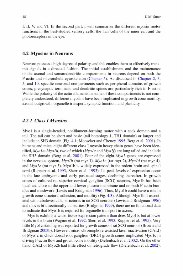
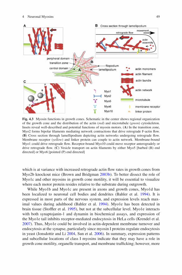
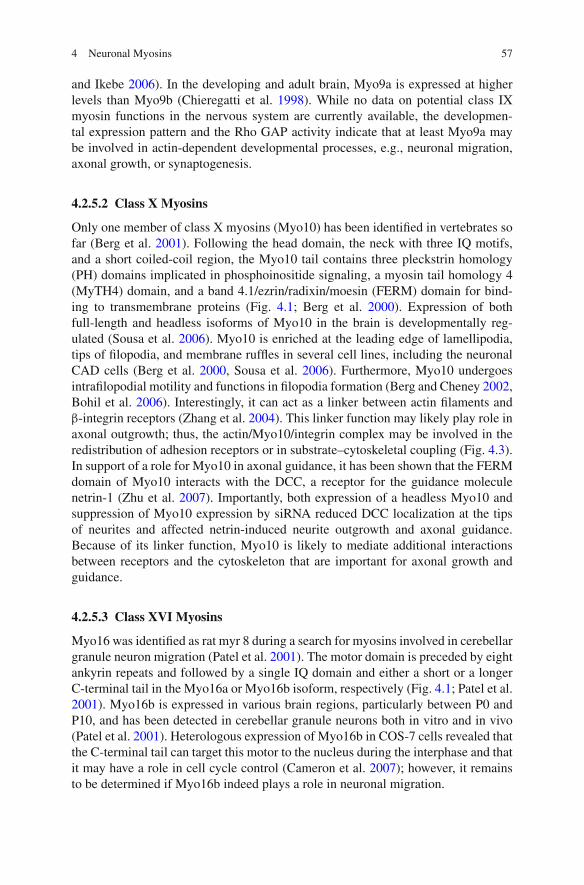
Fig. 4.3 Myosin functions in growth cones. Schematic in the center shows regional organizationof the growth cone and the distribution of the actin (red) and microtubule (green) cytoskeleton.Insets reveal well-described and potential functions of myosin motors. (A) In the transition zone,Myo2 forms bipolar filaments mediating network contractions that drive retrograde F-actin flow.(B) Cross section through lamellipodium depicting actin networks undergoing retrograde flow.Membrane receptor (yellow) and linker protein can couple to actin network. Membrane-boundMyo1 could drive retrograde flow. Receptor-bound Myo10 could move receptor anterogradely ordrive retrograde flow. (C) Vesicle transport on actin filaments by either Myo5 [barbed (B) enddirected] or Myo6 [pointed (P) end directed]
which is at variance with increased retrograde actin flow rates in growth cones fromMyo2b knockout mice (Brown and Bridgman 2003b). To better dissect the role ofMyo1c and other myosins in growth cone motility, it will be essential to visualizewhere each motor protein resides relative to the substrate during outgrowth.
While Myo1b and Myo1c are present in axons and growth cones, Myo1d hasbeen localized to neuronal cell bodies and dendrites (Bahler et al. 1994). It isexpressed in most parts of the nervous system, and expression levels reach max-imal values during adulthood (Bahler et al. 1994). Myo1e has been detected inbrain tissue (Stoffler et al. 1995), but not at the subcellular level. Myo1e interactswith both synaptojanin-1 and dynamin in biochemical assays, and expression ofthe Myo1e tail inhibits receptor-mediated endocytosis in HeLa cells (Krendel et al.2007). Thus, Myo1e could be involved in actin-dependent membrane turnover andendocytosis at the synapse, particularly since myosin I proteins regulate endocytosisin yeast (Jonsdottir and Li 2004, Sun et al. 2006). In summary, expression patternsand subcellular locations of class I myosins indicate that they may have a role ingrowth cone motility, organelle transport, and membrane trafficking; however, more
50 D.M. Suter
isoform-specific knockdown approaches and high-resolution imaging studies areneeded to confirm these potential functions.
4.2.2 Class II Myosins
Myo2 is the conventional double-headed motor protein, comprised of heavy chainsthat contain two IQ motifs followed by a long coiled-coil tail domain, which allowsthe formation of bipolar filaments (Fig. 4.1). Nonmuscle Myo2 is present in alleukaryotic cells and involved in essential cellular functions that require sliding ofadjacent actin filaments against each other, such as cytokinesis, contraction of theactin ring at adherens junctions, and cell migration. Nonmuscle Myo2 is withoutdoubt the best-studied neuronal myosin motor with well-established functions inneuronal migration, axonal growth, and guidance (Brown and Bridgman 2003a,2004). Three different nonmuscle Myo2 heavy chains have been identified in verte-brates: Myo2a, Myo2b, and Myo2c (Kawamoto and Adelstein 1991, Golomb et al.2004). Both Myo2a and Myo2b have overlapping but distinct expression patternsin the nervous system, Myo2b being more abundant than Myo2a (Kawamoto andAdelstein 1991, Itoh and Adelstein 1995). Myo2a and Myo2b are present in cellbodies, axons, and growth cones of cultured rat DRG neurons (Miller et al. 1992).In rat SCG growth cones, Myo2a and Myo2b staining is concentrated in the cen-tral and transition domains (Rochlin et al. 1995). The low Myo2c expression levelsduring development suggest that Myo2c most likely is not involved in neuronalmigration or axonal outgrowth (Golomb et al. 2004).
4.2.2.1 Myosin II in Neuronal Migration and Cell Adhesion
Myo2b knockout mice have major heart and brain defects and die between embry-onic day 14.5 (E14.5) and birth (Tullio et al. 2001). Two recent studies revealedthat Myo2 activity is critical early in CNS development for interkinetic nuclearmigration of neuronal progenitor cells in both cortex and retina (Schenk et al. 2009,Norden et al. 2009). Aberrant neuronal cell migration in Myo2b knockout micecaused accumulation of facial neurons into the fourth ventricle, resulting in hydro-cephalus (Tullio et al. 2001, Ma et al. 2006). Myo2b may have a scaffolding functionindependent of its functional motor domain, affecting cell adhesion of neuroepithe-lial cells facing the spinal canal (Ma et al. 2007). Reduced cell adhesion in theMyo2b mutant causes neuroepithelial cells to invade the spinal canal and obstructcerebrospinal fluid flow. Less is known about in vivo nervous system function ofMyo2a, since knockout mice die much earlier (E6.5), probably due to a decreasedcell–cell adhesion (Conti et al. 2004). More recently, experiments with the chemicalinhibitor blebbistatin have revealed that Myo2 plays a critical role in forward pullingof both the centrosome and the soma during glial-guided neuronal cell migration invitro and in vivo and that this response is regulated by the cell polarity protein Par6α
(Schaar and McConnell 2005, Solecki et al. 2009).
4 Neuronal Myosins 51
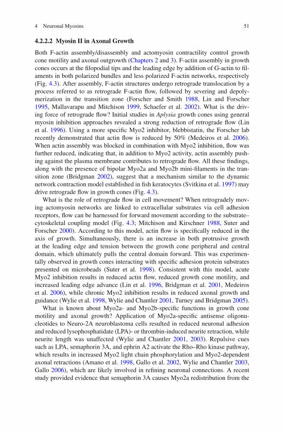
4.2.2.2 Myosin II in Axonal Growth
Both F-actin assembly/disassembly and actomyosin contractility control growthcone motility and axonal outgrowth (Chapters 2 and 3). F-actin assembly in growthcones occurs at the filopodial tips and the leading edge by addition of G-actin to fil-aments in both polarized bundles and less polarized F-actin networks, respectively(Fig. 4.3). After assembly, F-actin structures undergo retrograde translocation by aprocess referred to as retrograde F-actin flow, followed by severing and depoly-merization in the transition zone (Forscher and Smith 1988, Lin and Forscher1995, Mallavarapu and Mitchison 1999, Schaefer et al. 2002). What is the driv-ing force of retrograde flow? Initial studies in Aplysia growth cones using generalmyosin inhibition approaches revealed a strong reduction of retrograde flow (Linet al. 1996). Using a more specific Myo2 inhibitor, blebbistatin, the Forscher labrecently demonstrated that actin flow is reduced by 50% (Medeiros et al. 2006).When actin assembly was blocked in combination with Myo2 inhibition, flow wasfurther reduced, indicating that, in addition to Myo2 activity, actin assembly push-ing against the plasma membrane contributes to retrograde flow. All these findings,along with the presence of bipolar Myo2a and Myo2b mini-filaments in the tran-sition zone (Bridgman 2002), suggest that a mechanism similar to the dynamicnetwork contraction model established in fish keratocytes (Svitkina et al. 1997) maydrive retrograde flow in growth cones (Fig. 4.3).
What is the role of retrograde flow in cell movement? When retrogradely mov-ing actomyosin networks are linked to extracellular substrates via cell adhesionreceptors, flow can be harnessed for forward movement according to the substrate–cytoskeletal coupling model (Fig. 4.3; Mitchison and Kirschner 1988, Suter andForscher 2000). According to this model, actin flow is specifically reduced in theaxis of growth. Simultaneously, there is an increase in both protrusive growthat the leading edge and tension between the growth cone peripheral and centraldomain, which ultimately pulls the central domain forward. This was experimen-tally observed in growth cones interacting with specific adhesion protein substratespresented on microbeads (Suter et al. 1998). Consistent with this model, acuteMyo2 inhibition results in reduced actin flow, reduced growth cone motility, andincreased leading edge advance (Lin et al. 1996, Bridgman et al. 2001, Medeiroset al. 2006), while chronic Myo2 inhibition results in reduced axonal growth andguidance (Wylie et al. 1998, Wylie and Chantler 2001, Turney and Bridgman 2005).
What is known about Myo2a- and Myo2b-specific functions in growth conemotility and axonal growth? Application of Myo2a-specific antisense oligonu-cleotides to Neuro-2A neuroblastoma cells resulted in reduced neuronal adhesionand reduced lysophosphatidate (LPA)- or thrombin-induced neurite retraction, whileneurite length was unaffected (Wylie and Chantler 2001, 2003). Repulsive cuessuch as LPA, semaphorin 3A, and ephrin A2 activate the Rho–Rho kinase pathway,which results in increased Myo2 light chain phosphorylation and Myo2-dependentaxonal retractions (Amano et al. 1998, Gallo et al. 2002, Wylie and Chantler 2003,Gallo 2006), which are likely involved in refining neuronal connections. A recentstudy provided evidence that semaphorin 3A causes Myo2a redistribution from the
52 D.M. Suter
growth cone into the neurite, thereby facilitating collapse, while Myo2b mediatessemaphorin 3A-induced neurite retraction (Brown et al. 2009). On the other hand,Myo2b has also been implicated in neurite outgrowth and turning (Wylie et al. 1998,Tullio et al. 2001, Wylie and Chantler 2001, Turney and Bridgman 2005), tractionforce production, and growth cone motility (Bridgman et al. 2001), as well as ret-rograde flow (Brown and Bridgman 2003b). The mechanism of Myo2-regulatedaxonal growth is dependent on the substrate: on laminin, Myo2 mediates adhe-sion, while on poly-lysine, Myo2 prevents microtubule advance into the growthcone periphery (Ketschek et al. 2007). Furthermore, Myo2 negatively regulates thedevelopment of neuronal polarity (Kollins et al. 2009). In summary, a large bodyof evidence indicates that Myo2 regulates various aspects of neuronal growth conemotility and axonal growth.
4.2.2.3 Myosin II in Synapse Function
Myo2b has been detected in different neurons both pre- and postsynaptically (Milleret al. 1992, Mochida et al. 1994, Takagishi et al. 2005, Ryu et al. 2006). In culturedrat SCG neurons, neurotransmitter release was reduced when a recombinant Myo2bheavy chain tail fragment was injected, but not when a corresponding Myo2a frag-ment was used (Takagishi et al. 2005), confirming earlier studies that implicatedMyo2 in transmitter release (Mochida et al. 1994). In the CNS, dynamic dendriticspine morphology, motility, and synaptic function of hippocampal neurons dependon Myo2b activity (Ryu et al. 2006); however, the details of how Myo2b is involvedin dendritic spine morphology as well as in synaptic vesicle release are not known.A recent ultra-structural localization study by the Svitkina lab identified Myo2 in theneck of dendritic spines of hippocampal neurons, suggesting a role of actomyosincontractility in spine development (Korobova and Svitkina 2010).
4.2.3 Class V Myosins
The polarized morphology of neurons and the extremely long axons require an effi-cient organelle and protein transport system for both neuronal development andfunction. While axonal long-range transport is mainly mediated by the microtubule-based motors kinesins and dyneins, short-range transport at specific locations suchas growth cones, presynaptic terminals, and dendritic spines is achieved by actin-based myosin motors. Myo5 is a barbed end-directed, two-headed motor whoseheavy chains contain six IQ motifs followed by a coiled-coil domain and a globulartail (Fig. 4.1; Cheney et al. 1993). Myo5 has several properties that make it an effec-tive organelle motor (Fig. 4.1): (1) the globular tail binds cargo via adaptor proteins;(2) the coiled-coil domain allows the formation of a double-headed motor; (3) thehigh affinity for F-actin results in attachment of at least one head at all times; (4) thelong neck allows Myo5 to take large (36 nm) steps, making it a highly processivemotor (reviewed in Sellers and Veigel 2006). A variety of biochemical, structural,genetic, and cellular studies largely performed in neurons, melanocytes, and yeast
4 Neuronal Myosins 53
have made Myo5 the best characterized organelle motor within the myosin super-family (Reck-Peterson et al. 2000, Langford 2002, Bridgman 2004). Many studiesaddressing the organelle motor function of Myo5a have used the dilute-lethal mice,which carry a null mutation in the Myo5a gene, exhibit a lightened coat color,seizures and other neurological defects, and die around 3 weeks of age (Merceret al. 1991).
Three different members of class V myosins have been identified in vertebrates:Myo5a, Myo5b, and Myo5c, all exhibiting wide but distinct expression patterns(Rodriguez and Cheney 2002). Myo5a is strongly expressed in the brain and in otherparts of the nervous system (Espindola et al. 1992, Espreafico et al. 1992, Rodriguezand Cheney 2002). Both Myo5b and Myo5c are less abundant than Myo5a in thebrain but exhibit higher expression in kidney, liver, colon, and placenta (Myo5b),and in colon, pancreas, salivary gland, and stomach (Myo5c) (Zhao et al. 1996,Rodriguez and Cheney 2002, Lise et al. 2006).
4.2.3.1 Myosin V in Axonal Transport, Growth Cones, and PresynapticTerminals
Several studies provided evidence for actin-based organelle transport in axonsand growth cones (Kuznetsov et al. 1992, Evans and Bridgman 1995, Morrisand Hollenbeck 1995), and implicated Myo5a as the relevant motor (Prekeris andTerrian 1997, Evans et al. 1998, Tabb et al. 1998, Bridgman 1999). Myo5a wasdetected predominantly in the central domain and transition zone of growth cones(Evans et al. 1997, Suter et al. 2000). Evans et al. (1997) showed that neuriteoutgrowth, growth cone morphology, and cytoskeletal organization of dilute-lethalSCG growth cones are normal. A different growth cone phenotype was reported inanother study, which showed that local Myo5a inactivation by micro-CALI affectsfilopodia extension dynamics (Wang et al. 1996). The filopodia effects in the CALIstudy could be due to reduced membrane traffic to the filopodia. On the otherhand, Myo5a might have a role in axonal extension, since climbing fibers of dilutePurkinje cells exhibit reduced innervation (Takagishi et al. 2007).
With which organelles is Myo5a associated in axons and growth cones? In vitromotility, immunolocalization, and biochemical studies revealed Myo5a associationand Myo5a-mediated transport of large endoplasmic reticulum (ER) vesicles (Tabbet al. 1998), synaptic vesicle precursors (Evans et al. 1998, Miller and Sheetz2000), and potential synaptic vesicles (Prekeris and Terrian 1997). In addition,mitochondria are good candidates for cargo organelles in axons (Hollenbeck andSaxton 2005). Finally, Myo5a associates with neurofilaments, implicating a roleof this interaction for the proper distribution of neurofilaments in axons (Rao et al.2002). This hypothesis was confirmed when green fluorescent protein (GFP)-taggedneurofilaments were observed in axons of dilute-lethal mice (Alami et al. 2009).Fluorescent live cell imaging in both normal and dilute-lethal axons further revealedthat Myo5a-associated vesicles move along microtubules over long distances anduse actin/Myo5a-based transport for local movements, such as in presynaptic ter-minals (Bridgman 1999). Interestingly, the presynaptic terminals in dilute-lethal
54 D.M. Suter
mice have increased cross-sectional area and greater numbers of synaptic vesicles(Bridgman 1999). These findings suggest that Myo5a may act as modulator of vesi-cle transport slowing down microtubule/kinesin-based anterograde transport into thepresynaptic terminal. In conclusion, studies in both neurons and melanocytes haveshown that multiple microtubule- and actin-based motors reside on organelles thatmay become engaged/activated at specific subcellular sites during the journey ofan organelle (Bridgman 1999, Gross et al. 2002, Levi et al. 2006). This model isparticularly intriguing in light of the fact that the cargo-binding domain of Myo5adirectly binds to kinesin, thus forming a hetero-motor complex for fast switching ofthe cytoskeletal track system (Huang et al. 1999).
Based on the studies discussed above, one might expect a potential role forMyo5a in synaptic vesicle release. Both pre- and postsynaptic function of dilutehippocampal CA3-CA1 synapses appeared normal when compared to littermatecontrols (Schnell and Nicoll 2001). In agreement with this study, no effects ofMyo5a on synaptic transmission were reported for SCG neurons from Myo5amutant rats (Takagishi et al. 2005). On the other hand, Myo5a binds to the t-SNAREsyntaxin-1A and mediates Ca2+-induced exocytosis of vesicles in chromaffin cells(Watanabe et al. 2005). Thus, the presynaptic function of Myo5a is still controver-sial and may depend on the specific neuronal connection. In addition, other myosinspresent in presynaptic terminals of Myo5a mutant animals may compensate for thelack of functional Myo5a.
4.2.3.2 Myosin V in Dendrites and Postsynaptic Function
The roles of Myo5a and Myo5b are better defined on the postsynaptic side and indendrites where both myosins have been localized (Espindola et al. 1992, Naisbittet al. 2000, Walikonis et al. 2000, Lise et al. 2006). Indirect binding of Myo5ato postsynaptic density-95 (PSD-95) suggests a potential role for Myo5a in thetransport of the PSD-95 complex (Naisbitt et al. 2000). Myo5a activity is requiredfor smooth ER localization in dendritic spines, local calcium release, and post-synaptic functions such as long-term synaptic depression (Dekker-Ohno et al.1996, Takagishi et al. 1996, Miyata et al. 2000). Furthermore, Myo5a facilitatesthe transport of the mRNA-binding protein TLS and its target RNA Nd1-L in aCa2+-dependent manner into dendritic spines of mouse hippocampal neurons, impli-cating a role for Myo5a in synaptic plasticity (Yoshimura et al. 2006). Myo5a andMyo5b have been implicated in dendritic trafficking and recycling of the AMPA-type glutamate receptor subunit GluR1, the dendritic K+ channel Kv4.2, as wellas of the muscarinic acetylcholine receptor subunit M4, although the functionalinvolvement of these two myosin V isoforms in synaptic plasticity has remainedsomewhat controversial (Volpicelli et al. 2002, Lise et al. 2006, Correia et al. 2008,Lewis et al. 2009). Myo5b appears to be an important Ca sensor in synaptic plas-ticity. Ca influx after NMDA receptor activation causes rapid recruitment of Myo5bto recycling endosomes and thereby exocytosis of AMPA receptors (Wang et al.2008). How is the specificity between motor proteins and cargo receptors achieved?Increasing evidence from both neuronal and melanocyte systems indicates that the
4 Neuronal Myosins 55
small GTPases of the Rab family and linker proteins may provide such speci-ficity (Seabra and Coudrier 2004). In summary, class V myosin motors modulatelong-range organelle transport and control short-range transport to specific neu-ronal compartments, thereby supporting the development and function of neuronalconnections.
4.2.4 Class VI Myosins
Myo6 contains a head, a short neck region with one IQ motif, and a C-terminaltail domain that is capable of dimerization (Fig. 4.1; Kellerman and Miller 1992,Hasson and Mooseker 1994, Buss et al. 2004). Despite the presence of only oneIQ motif, each heavy chain binds two calmodulins (Bahloul et al. 2004). Myo6 hassome special structural features that result in mechanochemical properties that areunique from other myosins and enable Myo6 to perform special functions. The mostintriguing feature of Myo6 is the fact that it moves “backward” toward the pointedend of actin filaments (Wells et al. 1999). Another special feature of Myo6 is that itappears to exist both as a monomer and a dimer, perhaps with cargo binding induc-ing dimerization (Park et al. 2006, Sweeney and Houdusse 2007). This mechanismwould allow the formation of the processive, dimerized motor at the right subcellularlocation, e.g., the plasma membrane, enabling Myo6 to carry out its well-describedfunction in endocytosis (Buss et al. 2004, Sweeney and Houdusse 2007). Myo6was originally identified in Drosophila melanogaster (Kellerman and Miller 1992),where it has been implicated in several functions, including transport of cytoplas-mic particles in the embryo (Mermall et al. 1994). In mammalian cells, Myo6 hasbeen localized to membrane ruffles, endocytotic vesicles, the cytosol, and the Golgicomplex, where it has both transport and anchoring roles in membrane traffic, celladhesion, and migration (Buss et al. 2004, Sweeney and Houdusse 2007).
4.2.4.1 Myosin VI in Nervous System Development
What is known about Myo6 functions in the nervous system? It is expressed in allmajor parts of the mouse brain at similar levels from postnatal day 1 (P1) into adult-hood (Osterweil et al. 2005). In chick brains, Myo6 was detected as early as E6,while in DRGs expression starts from E10 on (Suter et al. 2000). Within culturedneurons, Myo6 is found in all subcellular compartments (Suter et al. 2000, Osterweilet al. 2005). In chick DRG growth cones, Myo6 shows the highest concentrationin the central domain similarly to Myo5a; however, Myo6a is less cytoskeleton-associated than Myo5a (Suter et al. 2000). Could Myo6 be involved in growth conemotility and axonal outgrowth? Considering that Myo6 is a pointed end-directedmotor that can bind to plasma membrane (Spudich et al. 2007), one could imaginea mechanism by which membrane-bound Myo6 moves actin filaments forward inthe direction of growth; however, there is as yet no experimental evidence for thismodel. Myo6 is also involved in early neuronal development in Drosophila. Myo6
56 D.M. Suter
is required for basal localization of Miranda, an adaptor protein which in turn local-izes cell fate determinants Prospero and Staufen to the basal side of the neuroblast(Petritsch et al. 2003).
4.2.4.2 Myosin VI in Synaptic Function
Because of its transport and anchoring properties, Myo6 could mediate complexmembrane-trafficking events at the synapse. A number of binding partners havebeen identified for Myo6, including Dab2, GIPC, and SAP97 (Buss et al. 2004).GIPC is an adapter protein between the BDNF receptor TrkB and Myo6 in hip-pocampal neurons (Yano et al. 2006). This interaction is important for specificpresynaptic functions, including BDNF–TrkB-mediated long-term potentiation andenhancement of glutamate release (Yano et al. 2006). A recent study implicatedMyo6 in postsynaptic structure and AMPA-type glutamate receptor endocytosis(Osterweil et al. 2005). Homozygous Snell’s waltzer mice that lack Myo6 exhibitfewer synapses and shorter dendritic spines in the hippocampal CA1 region whencompared with heterozygous mice. Furthermore, hippocampal neurons from Snell’swaltzer mice show reduced AMPA-type receptor GluR1 subunit internalizationbecause of disrupted clathrin-mediated endocytosis (Osterweil et al. 2005). Theinteraction between Myo6 and the GluR1–AMPA receptor is mediated by theadapter protein SAP97 and important for AMPA receptor trafficking (Wu et al. 2002,Nash et al. 2010). In summary, Myo6 forms specific complexes with membrane pro-teins at the pre- and postsynaptic sites and thereby regulates both synaptic vesiclerelease and neurotransmitter receptor trafficking. Considering the well-establishedrole of Myo6 in membrane trafficking in other cellular systems, more discoverieson Myo6’s actions in the nervous system are expected in the future.
4.2.5 Classes IX, X, and XVI Myosins
I discuss myosins of these three classes briefly, since they have been detected in neu-ronal tissues, but are not well characterized with respect to functions in the nervoussystem.
4.2.5.1 Class IX Myosins
The hallmark structural feature of class IX myosins is the GTPase-activating pro-tein (GAP) domain in the tail, which activates Rho but not Rac or Cdc42 (Fig. 4.1;Muller et al. 1997, Post et al. 1998). Additional structural features are an N-terminalextension of the head domain, a highly basic insertion at the presumed actin-bindingsite, a neck region with 4–6 IQ motifs, and a zinc-binding motif in front of the GAPdomain. The best-characterized class IX myosins include Myo9b (rat myr 5) andMyo9a (rat myr 7) (Reinhard et al. 1995, Wirth et al. 1996, Chieregatti et al. 1998).Biophysical and biochemical studies suggest that Myo9b is a single-headed, pro-cessive motor that binds actin with high affinity (Post et al. 1998, 2002, Kambara
4 Neuronal Myosins 57
and Ikebe 2006). In the developing and adult brain, Myo9a is expressed at higherlevels than Myo9b (Chieregatti et al. 1998). While no data on potential class IXmyosin functions in the nervous system are currently available, the developmen-tal expression pattern and the Rho GAP activity indicate that at least Myo9a maybe involved in actin-dependent developmental processes, e.g., neuronal migration,axonal growth, or synaptogenesis.
4.2.5.2 Class X Myosins
Only one member of class X myosins (Myo10) has been identified in vertebrates sofar (Berg et al. 2001). Following the head domain, the neck with three IQ motifs,and a short coiled-coil region, the Myo10 tail contains three pleckstrin homology(PH) domains implicated in phosphoinositide signaling, a myosin tail homology 4(MyTH4) domain, and a band 4.1/ezrin/radixin/moesin (FERM) domain for bind-ing to transmembrane proteins (Fig. 4.1; Berg et al. 2000). Expression of bothfull-length and headless isoforms of Myo10 in the brain is developmentally reg-ulated (Sousa et al. 2006). Myo10 is enriched at the leading edge of lamellipodia,tips of filopodia, and membrane ruffles in several cell lines, including the neuronalCAD cells (Berg et al. 2000, Sousa et al. 2006). Furthermore, Myo10 undergoesintrafilopodial motility and functions in filopodia formation (Berg and Cheney 2002,Bohil et al. 2006). Interestingly, it can act as a linker between actin filaments andβ-integrin receptors (Zhang et al. 2004). This linker function may likely play role inaxonal outgrowth; thus, the actin/Myo10/integrin complex may be involved in theredistribution of adhesion receptors or in substrate–cytoskeletal coupling (Fig. 4.3).In support of a role for Myo10 in axonal guidance, it has been shown that the FERMdomain of Myo10 interacts with the DCC, a receptor for the guidance moleculenetrin-1 (Zhu et al. 2007). Importantly, both expression of a headless Myo10 andsuppression of Myo10 expression by siRNA reduced DCC localization at the tipsof neurites and affected netrin-induced neurite outgrowth and axonal guidance.Because of its linker function, Myo10 is likely to mediate additional interactionsbetween receptors and the cytoskeleton that are important for axonal growth andguidance.
4.2.5.3 Class XVI Myosins
Myo16 was identified as rat myr 8 during a search for myosins involved in cerebellargranule neuron migration (Patel et al. 2001). The motor domain is preceded by eightankyrin repeats and followed by a single IQ domain and either a short or a longerC-terminal tail in the Myo16a or Myo16b isoform, respectively (Fig. 4.1; Patel et al.2001). Myo16b is expressed in various brain regions, particularly between P0 andP10, and has been detected in cerebellar granule neurons both in vitro and in vivo(Patel et al. 2001). Heterologous expression of Myo16b in COS-7 cells revealed thatthe C-terminal tail can target this motor to the nucleus during the interphase and thatit may have a role in cell cycle control (Cameron et al. 2007); however, it remainsto be determined if Myo16b indeed plays a role in neuronal migration.
58 D.M. Suter
4.3 Myosins in Sensory Cells
Sensory cells have developed a high degree of functional specialization to convertvarious stimuli into electrical signals that are transmitted to the nervous system.In the case of hair cells of the inner ear and photoreceptors of the retina, specialcytoskeletal and membranous compartments support these signal transduction func-tions. The following section summarizes the key functions of several myosins thathave been extensively characterized in these two sensory cells. I refer to a numberof reviews for a more detailed discussion of the experimental evidence for myosinfunctions in hair cells and photoreceptors (Brown and Bridgman 2004, Gillespie andCyr 2004, El-Amraoui and Petit 2005, Lin et al. 2005).
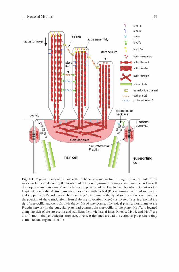
Inner ear hair cells are probably the best-characterized cell type with respect tothe number of different myosins. At least six myosins are essential for hearing andbalance based on mutants characterized in both mice and humans, including Myo1a,Myo2a, Myo3a, Myo6, Myo7a, and Myo15a (Friedman et al. 1999, Gillespie andCyr 2004, El-Amraoui and Petit 2005). To transduce mechanical forces into elec-trical signals, hair cells develop highly organized F-actin-rich protrusions calledstereocilia that are interconnected with cadherin molecules. Classes I, II, III, VI,VII, and XV myosins have been localized to specific subcellular regions in the haircell where they have distinct roles for the development, maintenance, and sensoryfunction of this cell (Fig. 4.4). In the photoreceptor cell, classes II, III, V, VI, and VIImyosins have been detected, of which classes III and VII have been characterizedthe best with respect to a role in vision.
4.3.1 Class I Myosins
Three of the eight class I myosins in higher vertebrates, Myo1b, Myo1c, and Myo1e,are strongly expressed in cochlea and vestibular organs of rodents at birth (Dumontet al. 2002). While mutations in the Myo1a gene have been linked to deafness inhumans but not in mice, there is no information available about the localization ofMyo1a in the inner ear (Donaudy et al. 2003, Tyska et al. 2005). Myo1b is mainlyexpressed in the supporting cells that surround the hair cells and in an apical ringof the hair cell (Dumont et al. 2002). Myo1c is enriched at tips of stereocilia and inthe pericuticular necklace of vestibular hair cells, and uniformly distributed alongthe stereocilia of auditory hair cells (Fig. 4.4; Gillespie et al. 1993, Hasson et al.1997, Dumont et al. 2002, Schneider et al. 2006). Myo1c is an important compo-nent of the hair cell’s adaptation complex that allows closing of the transductionchannel in both the slow and fast adaptation mechanisms by releasing tension (Holtet al. 2002, Gillespie and Cyr 2004, Stauffer et al. 2005). Myo1c is reported to inter-act via its neck domain with PIP2 and cadherin 23, the presumptive tip link, at thetips of stereocilia (Siemens et al. 2004, Hokanson and Ostap 2006, Phillips et al.2006). This interaction enables Myo1c to reposition the tip complex during adapta-tion. Myo1e was detected in the cuticular plate of cochlear and vestibular hair cells(Dumont et al. 2002); however, there are no experimental data on the role of Myo1bor Myo1e in hair cells.
4 Neuronal Myosins 59
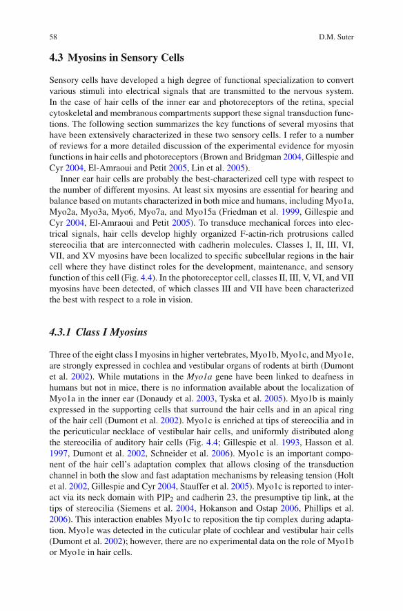
Fig. 4.4 Myosin functions in hair cells. Schematic cross section through the apical side of aninner ear hair cell depicting the location of different myosins with important functions in hair celldevelopment and function. Myo15a forms a cap on top of the F-actin bundles where it controls thelength of stereocilia. Actin filaments are oriented with barbed (B) end toward the tip of stereociliaand the pointed (P) end toward the base. Myo1c is found at the tip of stereocilia where it adjuststhe position of the transduction channel during adaptation. Myo3a is located in a ring around thetip of stereocilia and controls their shape. Myo6 may connect the apical plasma membrane to theF-actin network in the cuticular plate and connect the stereocilia to the plate. Myo7a is locatedalong the side of the stereocilia and stabilizes them via lateral links. Myo1c, Myo6, and Myo7 arealso found in the pericuticular necklace, a vesicle-rich area around the cuticular plate where theycould mediate organelle traffic

60 D.M. Suter
4.3.2 Class II Myosins
A mutation in Myo2a results in the human nonsyndromic deafness DFNA17(Mhatre et al. 2006). Myo2a has been localized along the stereocilia of mousecochlear hair cells and may stabilize the stereocilia (Mhatre et al. 2006). A similarfunction for class II myosins has been found in the retina. In Drosophila photorecep-tors, nonmuscle myosin II zipper forms two stripes of actomyosin extending alongthe sides of each of the R1–6 rhabdomeres, suggesting that this cytoskeletal sys-tem may be important for the alignment of the rhabdomeres with the optical axis(Baumann 2004). This hypothesis was supported by the distorted rhabdomeres inzipper flies carrying a mutation in the motor domain (Baumann 2004).
4.3.3 Class III Myosins
Class III myosins, Myo3a and Myo3b, are single-headed motors that have anN-terminal kinase domain, a neck with variable IQ motifs, and a short tail (Fig. 4.1;Dose and Burnside 2000, Berg et al. 2001). The kinase domain has serine–threoninekinase activity (Ng et al. 1996) and is essential in Drosophila phototransduc-tion (Porter and Montell 1993). Class III myosins were originally identified inDrosophila as two proteins encoded by the ninaC gene (Montell and Rubin 1988).NinaC mutant flies exhibit reduced amounts of rhodopsin in the photoreceptors andabnormal electroretinograms. The exact role of Myo3 in the photoreceptor rhab-domeres is still unclear; it may have structural role (Hicks et al. 1996), a signaling(Porter and Montell 1993, Wes et al. 1999), or transport function for componentsof the phototransduction machinery (Cronin et al. 2004, Lee and Montell 2004, Liuet al. 2008). An essential role for Myo3a in inner ear function is indicated by the factthat point mutations in the motor domain lead to the human nonsyndromic hearingloss DFNB30 (Walsh et al. 2002). Myo3a has recently been localized in a thimble-like pattern around the tips of the hair cell stereocilia (Fig. 4.4; Schneider et al.2006). Deletion of the kinase domain caused lengthening of stereocilia and bulgingof the tips, suggesting that Myo3a might regulate actin bundle maintenance, trans-port of the transduction complex to the stereocilia tip, or be part of the tip complexitself (Schneider et al. 2006). A recent study provided new insights into the roleof Myo3a in hair cells showing that this motor regulates the length of stereociliaby transporting the actin-bundling protein espin 1 to the plus end of actin filaments(Salles et al. 2009).
4.3.4 Class V Myosins
Elegant genetic studies in Drosophila by the Ready lab have recently revealedthat the organelle motor Myo5 with the help of the small GTPase Rab11 andthe linker protein dRip11 transports rhodopsin-containing secretory vesicles to thephotoreceptor rhabdomeres, which are critical for photoreceptor morphogenesis
4 Neuronal Myosins 61
(Li et al. 2007). Furthermore, the same laboratory also reported that calcium-activated Myo5 transports pigment granules to the rhabdomeres in response to brightlight in order to close the fly pupil (Satoh et al. 2008). Thus, Myo5 is important forboth photoreceptor development and physiology.
4.3.5 Class VI Myosins
Myo6 has been localized to the actin-rich cuticular plate of hair cells, indicatingthat it could be important for the anchoring of the stereocilia or membrane–actininteractions (Fig. 4.4; Hasson et al. 1997). A role for Myo6 in hair cell functionwas originally established when a recessive mutation in Myo6 was identified as thecause of deafness observed in the Snell’s Waltzer mouse in which hair cells degen-erate (Avraham et al. 1995). Myo6 mutations have also been linked to deafness inhumans (Melchionda et al. 2001, Ahmed et al. 2003). During hair cell develop-ment in both Snell’s Waltzer mice and zebrafish Myo6b mutants, the apical surfaceprotrudes and stereocilia fuse together, suggesting that Myo6 is critical for anchor-ing the apical plasma membrane to the underlying actin-rich cuticular plate (Selfet al. 1999, Kappler et al. 2004, Seiler et al. 2004). In photoreceptor cells, Myo6has been localized to the inner segments, and Snell’s Waltzer photoreceptors showa decreased responsiveness (Kitamoto et al. 2005). In summary, evidence from sev-eral systems implicates Myo6 in the development and function of inner ear hair andphotoreceptor cells.
4.3.6 Class VII Myosins
Class VII myosins have a head, five IQ motifs, a short coiled-coil domain, andtwo sets of MyTH4 and FERM domains in the C-terminal tail that are separatedby an SH3 domain (Fig. 4.1; Berg et al. 2001). Like Myo6, the short coiled-coildomain could dimerize upon cargo or actin binding and thereby generate a proces-sive motor (Yang et al. 2006). Class VII myosins are expressed in a wide range oforganisms from dictyostelium to humans and are involved in adhesion, phagocyto-sis, and organelle transport (Berg et al. 2001, Krendel and Mooseker 2005). Myo7ais expressed in retina, cochlea, kidney, and testis (Hasson et al. 1995). Mutationsin Myo7a result in the most frequent and severe form of deafness and blindness inhumans, called Usher 1 syndrome (Weil et al. 1995, El-Amraoui and Petit 2005),nonsyndromic deafness in humans (Liu et al. 1997, Weil et al. 1997), as well asdeafness in mice (shaker-1) (Gibson et al. 1995). Stereocilia of shaker-1 mice haircells do not develop normally, implicating Myo7a in the assembly of the actin bun-dles (Self et al. 1998). Myo7a is localized to the pericuticular necklace and along thestereocilia in close association with the lateral connections mediated by cadherin 23and protocadherin 15; thus, Myo7a may be an intracellular anchor for these linkagesin order to maintain the integrity of the hair bundle (Fig. 4.4; Hasson et al. 1997, El-Amraoui and Petit 2005). Recent mosaic complementation experiments provided
62 D.M. Suter
evidence that Myo7a regulates the length of stereocilia by affecting actin dynam-ics (Prosser et al. 2008). Finally, hair cell currents are affected in certain shaker-1mutants, suggesting that Myo7a could also participate in hair cell adaptation (Kroset al. 2002).
In photoreceptors, Myo7a has been localized to the cilium that connects the innerand outer segments, where it transports opsin into the outer segment (Liu et al. 1999,Wolfrum and Schmitt 2000). Myo7a is highly expressed in the pigment epithelialcells, which constantly absorb the shed-off membrane discs of the outer photore-ceptor segments. In shaker-1 mice, these cells exhibit both delayed phagocytosis ofouter segment membranes (Gibbs et al. 2003). Thus, Myo7a has important func-tions in the maintenance of the outer segments, which are severely affected in Usher1 syndrome patients (El-Amraoui and Petit 2005). The lack of retinitis pigmentosain the shaker-1 mouse could be explained by the shorter life span of mice or by thedifferences in the photoreceptor anatomy and physiology between mice and humans.
4.3.7 Class XV Myosins
Vertebrate Myo15a is a 395-kDa motor with a large N-terminal extension ofunknown function and a C-terminal tail similar to Myo7a, which allows interac-tions with membrane and scaffolding proteins (Liang et al. 1999). This motor isexpressed in the cochlea and vestibular systems in the developing mouse inner ear,and mutations result in severe hearing loss both in mice (shaker 2) and humans(DFNB3) (Probst et al. 1998, Wang et al. 1998). In wild-type hair cells, Myo15alocalizes as a cap on the top of stereocilia, and the amount of Myo15a correlateswith the length of the bundles (Fig. 4.4; Belyantseva et al. 2003, Rzadzinska et al.2004, Delprat et al. 2005). Shaker 2 stereocilia are considerably shorter than wildtype and do not exhibit the staircase pattern (Belyantseva et al. 2003, Rzadzinskaet al. 2004). Thus, Myo15a controls the length of stereocilia, for example, by reg-ulating the rate of F-actin assembly at the top of the bundles that slowly turn overby retrograde flow (Rzadzinska et al. 2004, Belyantseva et al. 2005, Delprat et al.2005). On the other hand, the formation and function of the hair cell transductioncomplex does not require Myo15a (Stepanyan et al. 2006). Future biophysical andbiochemical studies will provide important insights regarding if and how this largemotor protein regulates actin dynamics and/or membrane/actin interactions.
4.4 Summary and Perspectives
Neurons and sensory cells of the nervous system have harnessed the wide spectrumof functionality of all the major myosin classes for morphogenesis, cell movement,cell maintenance, and signaling. Probably the best-characterized motor with respectto function is Myo2 in actomyosin contraction and neurite outgrowth. However, it
4 Neuronal Myosins 63
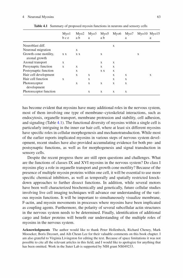
Table 4.1 Summary of proposed myosin functions in neurons and sensory cells
Myo1 Myo2 Myo3 Myo5 Myo6 Myo7 Myo10 Myo15b c e a b a a b a a
Neuroblast diff. xNeuronal migration xGrowth cone motility,
axonal growthx x x x x x
Axonal transport x x xPresynaptic function x x x xPostsynaptic function x x x xHair cell development x x x x xHair cell function x x x xPhotoreceptor
developmentx x x x x
Photoreceptor function x x x x
has become evident that myosins have many additional roles in the nervous system,most of them involving one type of membrane–cytoskeletal interactions, such asendocytosis, organelle transport, membrane protrusion and stability, cell adhesion,and signaling (Table 4.1). The functional diversity of myosins within a single cell isparticularly intriguing in the inner ear hair cell, where at least six different myosinshave specific roles in cellular morphogenesis and mechanotransduction. While mostof the earlier reports implicated myosins in various steps of nervous system devel-opment, recent studies have also provided accumulating evidence for both pre- andpostsynaptic functions, as well as for morphogenesis and signal transduction insensory cells.
Despite the recent progress there are still open questions and challenges. Whatare the functions of classes IX and XVI myosins in the nervous system? Do class Imyosins play a role in organelle transport and growth cone motility? Because of thepresence of multiple myosin proteins within one cell, it will be essential to use morespecific chemical inhibitors, as well as temporally and spatially restricted knock-down approaches to further dissect functions. In addition, while several motorshave been well characterized biochemically and genetically, future cellular studiesinvolving live cell imaging techniques will advance our understanding of the vari-ous myosin functions. It will be important to simultaneously visualize membrane,F-actin, and myosin movements in processes where myosins have been implicatedas coupling agents. Furthermore, the polarity of several subcellular actin structuresin the nervous system needs to be determined. Finally, identification of additionalcargo and linker proteins will benefit our understanding of the multiple roles ofmyosins in the nervous system.
Acknowledgments The author would like to thank Peter Hollenbeck, Richard Cheney, MarkMooseker, Boris Decourt, and Aih Cheun Lee for their valuable comments on this book chapter. Iam also grateful to Virginia Livingston for editing the text. Because of space limitations it was notpossible to cite all the relevant articles in this field, and I would like to apologize for anything thathas been omitted. Work in the Suter Lab is supported by NIH grant NS049233.
64 D.M. Suter
References
Ahmed ZM, Morell RJ, Riazuddin S, Gropman A, Shaukat S, Ahmad MM, Mohiddin SA,Fananapazir L, Caruso RC, Husnain T, Khan SN, Riazuddin S, Griffith AJ, Friedman TB,Wilcox ER (2003) Mutations of MYO6 are associated with recessive deafness, DFNB37. AmJ Hum Genet 72:1315–1322.
Alami NH, Jung P, Brown A (2009) Myosin Va increases the efficiency of neurofilament transportby decreasing the duration of long-term pauses. J Neurosci 29(20):6625–6634.
Amano M, Chihara K, Nakamura N, Fukata Y, Yano T, Shibata M, Ikebe M, Kaibuchi K (1998)Myosin II activation promotes neurite retraction during the action of Rho and Rho-kinase.Genes Cells 3:177–188.
Avraham KB, Hasson T, Steel KP, Kingsley DM, Russell LB, Mooseker MS, Copeland NG,Jenkins NA (1995) The mouse Snell’s waltzer deafness gene encodes an unconventional myosinrequired for structural integrity of inner ear hair cells. Nat Genet 11:369–375.
Bahler M, Kroschewski R, Stoffler HE, Behrmann T (1994) Rat myr 4 defines a novel subclassof myosin I, identification, distribution, localization, and mapping of calmodulin-binding siteswith differential calcium sensitivity. J Cell Biol 126:375–389.
Bahloul A, Chevreux G, Wells AL, Martin D, Nolt J, Yang Z, Chen LQ, Potier N, Van DorsselaerA, Rosenfeld S, Houdusse A, Sweeney HL (2004) The unique insert in myosin VI is a structuralcalcium–calmodulin binding site. Proc Natl Acad Sci USA 101:4787–4792.
Baumann O (2004) Spatial pattern of nonmuscle myosin-II distribution during the develop-ment of the Drosophila compound eye and implications for retinal morphogenesis. Dev Biol269:519–533.
Belyantseva IA, Boger ET, Friedman TB (2003) Myosin XVa localizes to the tips of inner earsensory cell stereocilia and is essential for staircase formation of the hair bundle. Proc NatlAcad Sci USA 100:13958–13963.
Belyantseva IA, Boger ET, Naz S, Frolenkov GI, Sellers JR, Ahmed ZM, Griffith AJ, FriedmanTB (2005) Myosin-XVa is required for tip localization of whirlin and differential elongation ofhair-cell stereocilia. Nat Cell Biol 7:148–156.
Berg JS, Cheney RE (2002) Myosin-X is an unconventional myosin that undergoes intrafilopodialmotility. Nat Cell Biol 4:246–250.
Berg JS, Derfler BH, Pennisi CM, Corey DP, Cheney RE (2000) Myosin-X, a novel myosinwith pleckstrin homology domains, associates with regions of dynamic actin. J Cell Sci113:3439–3451.
Berg JS, Powell BC, Cheney RE (2001) A millennial myosin census. Mol Biol Cell 12:780–794.Bohil AB, Robertson BW, Cheney RE (2006) Myosin-X is a molecular motor that functions in
filopodia formation. Proc Natl Acad Sci USA 103:12411–12416.Bridgman PC (1999) Myosin Va movements in normal and dilute-lethal axons provide support for
a dual filament motor complex. J Cell Biol 146:1045–1060.Bridgman PC (2002) Growth cones contain myosin II bipolar filament arrays. Cell Motil
Cytoskeleton 52:91–96.Bridgman PC (2004) Myosin-dependent transport in neurons. J Neurobiol 58:164–174.Bridgman PC, Dave S, Asnes CF, Tullio AN, Adelstein RS (2001) Myosin IIB is required for
growth cone motility. J Neurosci 21:6159–6169.Brown J, Bridgman PC (2003a) Role of myosin II in axon outgrowth. J Histochem Cytochem
51:421–428.Brown ME, Bridgman PC (2003b) Retrograde flow rate is increased in growth cones from myosin
IIB knockout mice. J Cell Sci 116:1087–1094.Brown ME, Bridgman PC (2004) Myosin function in nervous and sensory systems. J Neurobiol
58:118–130.Brown JA, Wysolmerski RB, Bridgman PC (2009) Dorsal root ganglion neurons react to
semaphorin 3A application through a biphasic response that requires multiple myosin IIisoforms. Mol Biol Cell 20(4):1167–1179.

4 Neuronal Myosins 65
Buss F, Spudich G, Kendrick-Jones J (2004) Myosin VI, cellular functions and motor properties.Annu Rev Cell Dev Biol 20:649–676.
Cameron RS, Liu C, Mixon AS, Pihkala JP, Rahn RJ, Cameron PL (2007) Myosin16b: the COOH-tail region directs localization to the nucleus and overexpression delays S-phase progression.Cell Motil Cytoskeleton 64:19–48.
Cheney RE, Mooseker MS (1992) Unconventional myosins. Curr Opin Cell Biol 4:27–35.Cheney RE, O’Shea MK, Heuser JE, Coelho MV, Wolenski JS, Espreafico EM, Forscher P, Larson
RE, Mooseker MS (1993) Brain myosin-V is a two-headed unconventional myosin with motoractivity. Cell 75:13–23.
Chieregatti E, Gartner A, Stoffler HE, Bahler M (1998) Myr 7 is a novel myosin IX-RhoGAPexpressed in rat brain. J Cell Sci 111:3597–3608.
Conti MA, Even-Ram S, Liu C, Yamada KM, Adelstein RS (2004) Defects in cell adhesion and thevisceral endoderm following ablation of nonmuscle myosin heavy chain II-A in mice. J BiolChem 279:41263–41266.
Correia SS, Bassani S, Brown TC, Lisé MF, Backos DS, El-Husseini A, Passafaro M, EstebanJA (2008) Motor protein-dependent transport of AMPA receptors into spines during long-termpotentiation. Nat Neurosci 11(4):457–466.
Cronin MA, Diao F, Tsunoda S (2004) Light-dependent subcellular translocation of Gqalpha inDrosophila photoreceptors is facilitated by the photoreceptor-specific myosin III NINAC. JCell Sci 117:4797–4806.
Dekker-Ohno K, Hayasaka S, Takagishi Y, Oda S, Wakasugi N, Mikoshiba K, Inouye M,Yamamura H (1996) Endoplasmic reticulum is missing in dendritic spines of Purkinje cellsof the ataxic mutant rat. Brain Res 714:226–230.
Delprat B, Michel V, Goodyear R, Yamasaki Y, Michalski N, El-Amraoui A, Perfettini I, LegrainP, Richardson G, Hardelin JP, Petit C (2005) Myosin XVa and whirlin, two deafness geneproducts required for hair bundle growth, are located at the stereocilia tips and interact directly.Hum Mol Genet 14:401–410.
Diefenbach TJ, Latham VM, Yimlamai D, Liu CA, Herman IM, Jay DG (2002) Myosin 1c andmyosin IIB serve opposing roles in lamellipodial dynamics of the neuronal growth cone. J CellBiol 158:1207–1217.
Donaudy F, Ferrara A, Esposito L, Hertzano R, Ben-David O, Bell RE, Melchionda S, Zelante L,Avraham KB, Gasparini P (2003) Multiple mutations of MYO1A, a cochlear-expressed gene,in sensorineural hearing loss. Am J Hum Genet 72:1571–1577.
Dose AC, Burnside B (2000) Cloning and chromosomal localization of a human class III myosin.Genomics 67:333–342.
Dumont RA, Zhao YD, Holt JR, Bahler M, Gillespie PG (2002) Myosin-I isozymes in neonatalrodent auditory and vestibular epithelia. J Assoc Res Otolaryngol 3:375–389.
El-Amraoui A, Petit C (2005) Usher I syndrome, unravelling the mechanisms that underlie thecohesion of the growing hair bundle in inner ear sensory cells. J Cell Sci 118:4593–4603.
Espindola FS, Espreafico EM, Coelho MV, Martins AR, Costa FR, Mooseker MS, Larson RE(1992) Biochemical and immunological characterization of p190–calmodulin complex fromvertebrate brain, a novel calmodulin-binding myosin. J Cell Biol 118:359–368.
Espreafico EM, Cheney RE, Matteoli M, Nascimento AA, De Camilli PV, Larson RE, MoosekerMS (1992) Primary structure and cellular localization of chicken brain myosin-V (p190), anunconventional myosin with calmodulin light chains. J Cell Biol 119:1541–1557.
Evans LL, Bridgman PC (1995) Particles move along actin filament bundles in nerve growth cones.Proc Natl Acad Sci USA 92:10954–10958.
Evans LL, Hammer J, Bridgman PC (1997) Subcellular localization of myosin V in nerve growthcones and outgrowth from dilute-lethal neurons. J Cell Sci 110:439–449.
Evans LL, Lee AJ, Bridgman PC, Mooseker MS (1998) Vesicle-associated brain myosin-V can beactivated to catalyze actin-based transport. J Cell Sci 111:2055–2066.
Forscher P, Smith SJ (1988) Actions of cytochalasins on the organization of actin filaments andmicrotubules in a neuronal growth cone. J Cell Biol 107:1505–1516.
66 D.M. Suter
Foth BJ, Goedecke MC, Soldati D (2006) New insights into myosin evolution and classification.Proc Natl Acad Sci USA 103:3681–3686.
Friedman TB, Sellers JR, Avraham KB (1999) Unconventional myosins and the genetics of hearingloss. Am J Med Genet 89:147–157.
Gallo G (2006) RhoA-kinase coordinates F-actin organization and myosin II activity duringsemaphorin-3A-induced axon retraction. J Cell Sci 119:3413–3423.
Gallo G, Yee HF Jr, Letourneau PC (2002) Actin turnover is required to prevent axon retractiondriven by endogenous actomyosin contractility. J Cell Biol 158:1219–1228.
Gibbs D, Kitamoto J, Williams DS (2003) Abnormal phagocytosis by retinal pigmented epithe-lium that lacks myosin VIIa, the Usher syndrome 1B protein. Proc Natl Acad Sci USA 100:6481–6486.
Gibson F, Walsh J, Mburu P, Varela A, Brown KA, Antonio M, Beisel KW, Steel KP, Brown SD(1995) A type VII myosin encoded by the mouse deafness gene shaker-1. Nature 374:62–64.
Gillespie PG, Albanesi JP, Bahler M, Bement WM, Berg JS, Burgess DR, Burnside B, Cheney RE,Corey DP, Coudrier E, de Lanerolle P, Hammer JA, Hasson T, Holt JR, Hudspeth AJ, Ikebe M,Kendrick-Jones J, Korn ED, Li R, Mercer JA, Milligan RA, Mooseker MS, Ostap EM, PetitC, Pollard TD, Sellers JR, Soldati T, Titus MA (2001) Myosin-I nomenclature. J Cell Biol155:703–704.
Gillespie PG, Cyr JL (2004) Myosin-1c, the hair cell’s adaptation motor. Annu Rev Physiol66:521–545.
Gillespie PG, Wagner MC, Hudspeth AJ (1993) Identification of a 120 kD hair-bundle myosinlocated near stereociliary tips. Neuron 11:581–594.
Golomb E, Ma X, Jana SS, Preston YA, Kawamoto S, Shoham NG, Goldin E, Conti MA, SellersJR, Adelstein RS (2004) Identification and characterization of nonmuscle myosin II-C, a newmember of the myosin II family. J Biol Chem 279:2800–2808.
Gross SP, Tuma MC, Deacon SW, Serpinskaya AS, Reilein AR, Gelfand VI (2002) Interactionsand regulation of molecular motors in Xenopus melanophores. J Cell Biol 156:855–865.
Hasson T, Gillespie PG, Garcia JA, MacDonald RB, Zhao Y, Yee AG, Mooseker MS,Corey DP (1997) Unconventional myosins in inner-ear sensory epithelia. J Cell Biol 137:1287–1307.
Hasson T, Heintzelman MB, Santos-Sacchi J, Corey DP, Mooseker MS (1995) Expression incochlea and retina of myosin VIIa, the gene product defective in Usher syndrome type 1B.Proc Natl Acad Sci USA 92:9815–9819.
Hasson T, Mooseker MS (1994) Porcine myosin-VI, characterization of a new mammalianunconventional myosin. J Cell Biol 127:425–440.
Hicks JL, Liu X, Williams DS (1996) Role of the ninaC proteins in photoreceptor cell structure,ultrastructure of ninaC deletion mutants and binding to actin filaments. Cell Motil Cytoskeleton35:367–379.
Hokanson DE, Ostap EM (2006) Myo1c binds tightly and specifically to phosphatidylinositol 4,5-bisphosphate and inositol 1,4,5-trisphosphate. Proc Natl Acad Sci USA 103:3118–3123.
Hollenbeck PJ, Saxton WM (2005) The axonal transport of mitochondria. J Cell Sci 118:5411–5419.
Holt JR, Gillespie SK, Provance DW, Shah K, Shokat KM, Corey DP, Mercer JA, Gillespie PG(2002) A chemical–genetic strategy implicates myosin-1c in adaptation by hair cells. Cell108:371–381.
Huang JD, Brady ST, Richards BW, Stenolen D, Resau JH, Copeland NG, Jenkins NA (1999)Direct interaction of microtubule- and actin-based transport motors. Nature 397:267–270.
Itoh K, Adelstein RS (1995) Neuronal cell expression of inserted isoforms of vertebrate nonmusclemyosin heavy chain II-B. J Biol Chem 270:14533–14540.
Jonsdottir GA, Li R (2004) Dynamics of yeast Myosin I, evidence for a possible role in scission ofendocytic vesicles. Curr Biol 14:1604–1609.
Kambara T, Ikebe M (2006) A unique ATP hydrolysis mechanism of single-headed processivemyosin, myosin IX. J Biol Chem 281:4949–4957.
4 Neuronal Myosins 67
Kappler JA, Starr CJ, Chan DK, Kollmar R, Hudspeth AJ (2004) A nonsense mutation in the geneencoding a zebrafish myosin VI isoform causes defects in hair-cell mechanotransduction. ProcNatl Acad Sci USA 101:13056–13061.
Kawamoto S, Adelstein RS (1991) Chicken nonmuscle myosin heavy chains, differential expres-sion of two mRNAs and evidence for two different polypeptides. J Cell Biol 112:915–924.
Kellerman KA, Miller KG (1992) An unconventional myosin heavy chain gene from Drosophilamelanogaster. J Cell Biol 119:823–834.
Ketschek AR, Jones SL, Gallo G (2007) Axon extension in the fast and slow lanes: substratum-dependent engagement of myosin II functions. Dev Neurobiol 67(10):1305–1320.
Kitamoto J, Libby RT, Gibbs D, Steel KP, Williams DS (2005) Myosin VI is required for normalretinal function. Exp Eye Res 81:116–120.
Kollins KM, Hu J, Bridgman PC, Huang YQ, Gallo G (2009) Myosin-II negatively regulatesminor process extension and the temporal development of neuronal polarity. Dev Neurobiol69(5):279–298.
Korobova F, Svitkina T (2010) Molecular architecture of synaptic actin cytoskeleton in hip-pocampal neurons reveals a mechanism of dendritic spine morphogenesis. Mol Biol Cell21(1):165–176.
Krendel M, Mooseker MS (2005) Myosins, tails (and heads) of functional diversity. Physiology(Bethesda) 20:239–251.
Krendel M, Osterweil EK, Mooseker MS (2007) Myosin 1E interacts with synaptojanin-1 anddynamin and is involved in endocytosis. FEBS Lett 581:644–650.
Kros CJ, Marcotti W, van Netten SM, Self TJ, Libby RT, Brown SD, Richardson GP, Steel KP(2002) Reduced climbing and increased slipping adaptation in cochlear hair cells of mice withMyo7a mutations. Nat Neurosci 5:41–47.
Kuznetsov SA, Langford GM, Weiss DG (1992) Actin-dependent organelle movement in squidaxoplasm. Nature 356:722–725.
Langford GM (2002) Myosin-V, a versatile motor for short-range vesicle transport. Traffic 3:859–865.
Lee SJ, Montell C (2004) Light-dependent translocation of visual arrestin regulated by the NINACmyosin III. Neuron 43:95–103.
Levi V, Serpinskaya AS, Gratton E, Gelfand V (2006) Organelle transport along microtubulesin Xenopus melanophores, evidence for cooperation between multiple motors. Biophys J 90:318–327.
Lewis AK, Bridgman PC (1996) Mammalian myosin I alpha is concentrated near the plasmamembrane in nerve growth cones. Cell Motil Cytoskeleton 33:130–150.
Lewis TL Jr, Mao T, Svoboda K, Arnold DB (2009) Myosin-dependent targeting of transmembraneproteins to neuronal dendrites. Nat Neurosci 12(5):568–576.
Li BX, Satoh AK, Ready DF (2007) Myosin V, Rab11, and dRip11 direct apical secre-tion and cellular morphogenesis in developing Drosophila photoreceptors. J Cell Biol 177:659–669.
Liang Y, Wang A, Belyantseva IA, Anderson DW, Probst FJ, Barber TD, Miller W, TouchmanJW, Jin L, Sullivan SL, Sellers JR, Camper SA, Lloyd RV, Kachar B, Friedman TB, Fridell RA(1999) Characterization of the human and mouse unconventional myosin XV genes responsiblefor hereditary deafness DFNB3 and shaker 2. Genomics 61:243–258.
Lin CH, Espreafico EM, Mooseker MS, Forscher P (1996) Myosin drives retrograde F-actin flowin neuronal growth cones. Neuron 16:769–782.
Lin CH, Forscher P (1995) Growth cone advance is inversely proportional to retrograde F-actinflow. Neuron 14:763–771.
Lin HW, Schneider ME, Kachar B (2005) When size matters, the dynamic regulation of stereocilialengths. Curr Opin Cell Biol 17:55–61.
Lise MF, Wong TP, Trinh A, Hines RM, Liu L, Kang R, Hines DJ, Lu J, Goldenring JR, WangYT, El-Husseini A (2006) Involvement of myosin Vb in glutamate receptor trafficking. J BiolChem 281:3669–3678.
68 D.M. Suter
Liu CH, Satoh AK, Postma M, Huang J, Ready DF, Hardie RC (2008) Ca2+-dependentmetarhodopsin inactivation mediated by calmodulin and NINAC myosin III. Neuron59(5):778–789.
Liu X, Udovichenko IP, Brown SD, Steel KP, Williams DS (1999) Myosin VIIa participates inopsin transport through the photoreceptor cilium. J Neurosci 19:6267–6274.
Liu XZ, Walsh J, Mburu P, Kendrick-Jones J, Cope MJ, Steel KP, Brown SD (1997) Mutations inthe myosin VIIA gene cause non-syndromic recessive deafness. Nat Genet 16:188–190.
Ma X, Bao J, Adelstein RS (2007) Loss of cell adhesion causes hydrocephalus in nonmusclemyosin II-B ablated and mutated mice. Mol Biol Cell 18:2305–2312.
Ma X, Kawamoto S, Uribe J, Adelstein RS (2006) Function of the neuron-specific alternativelyspliced isoforms of nonmuscle myosin II-B during mouse brain development. Mol Biol Cell17:2138–2149.
Mallavarapu A, Mitchison T (1999) Regulated actin cytoskeleton assembly at filopodium tipscontrols their extension and retraction. J Cell Biol 146:1097–1106.
Medeiros NA, Burnette DT, Forscher P (2006) Myosin II functions in actin-bundle turnover inneuronal growth cones. Nat Cell Biol 8:215–226.
Mehta AD, Rock RS, Rief M, Spudich JA, Mooseker MS, Cheney RE (1999) Myosin-V is aprocessive actin-based motor. Nature 400:590–593.
Melchionda S, Ahituv N, Bisceglia L, Sobe T, Glaser F, Rabionet R, Arbones ML, NotarangeloA, Di Iorio E, Carella M, Zelante L, Estivill X, Avraham KB, Gasparini P (2001) MYO6, thehuman homologue of the gene responsible for deafness in Snell’s waltzer mice, is mutated inautosomal dominant nonsyndromic hearing loss. Am J Hum Genet 69:635–640.
Mercer JA, Seperack PK, Strobel MC, Copeland NG, Jenkins NA (1991) Novel myosin heavychain encoded by murine dilute coat colour locus. Nature 349:709–713.
Mermall V, McNally JG, Miller KG (1994) Transport of cytoplasmic particles catalysed by anunconventional myosin in living Drosophila embryos. Nature 369:560–562.
Mhatre AN, Li Y, Atkin G, Maghnouj A, Lalwani AK (2006) Expression of Myh9 in themammalian cochlea, localization within the stereocilia. J Neurosci Res 84:809–818.
Miller M, Bower E, Levitt P, Li D, Chantler PD (1992) Myosin II distribution in neurons isconsistent with a role in growth cone motility but not synaptic vesicle mobilization. Neuron8:25–44.
Miller KE, Sheetz MP (2000) Characterization of myosin V binding to brain vesicles. J Biol Chem275:2598–2606.
Mitchison T, Kirschner M (1988) Cytoskeletal dynamics and nerve growth. Neuron 1:761–772.Miyata M, Finch EA, Khiroug L, Hashimoto K, Hayasaka S, Oda SI, Inouye M, Takagishi Y,
Augustine GJ, Kano M (2000) Local calcium release in dendritic spines required for long-termsynaptic depression. Neuron 28:233–244.
Mochida S, Kobayashi H, Matsuda Y, Yuda Y, Muramoto K, Nonomura Y (1994) Myosin II isinvolved in transmitter release at synapses formed between rat sympathetic neurons in culture.Neuron 13:1131–1142.
Montell C, Rubin GM (1988) The Drosophila ninaC locus encodes two photoreceptor cell specificproteins with domains homologous to protein kinases and the myosin heavy chain head. Cell52:757–772.
Mooseker MS, Cheney RE (1995) Unconventional myosins. Annu Rev Cell Dev Biol 11:633–675.Morris RL, Hollenbeck PJ (1995) Axonal transport of mitochondria along microtubules and F-actin
in living vertebrate neurons. J Cell Biol 131:1315–1326.Muller RT, Honnert U, Reinhard J, Bahler M (1997) The rat myosin myr 5 is a GTPase-activating
protein for Rho in vivo, essential role of arginine 1695. Mol Biol Cell 8:2039–2053.Naisbitt S, Valtschanoff J, Allison DW, Sala C, Kim E, Craig AM, Weinberg RJ, Sheng M (2000)
Interaction of the postsynaptic density-95/guanylate kinase domain-associated protein complexwith a light chain of myosin-V and dynein. J Neurosci 20:4524–4534.
Nash JE, Appleby VJ, Corrêa SA, Wu H, Fitzjohn SM, Garner CC, Collingridge GL, MolnárE (2010) Disruption of the interaction between myosin VI and SAP97 is associated with areduction in the number of AMPARs at hippocampal synapses. J Neurochem 112(3):677–690.
4 Neuronal Myosins 69
Ng KP, Kambara T, Matsuura M, Burke M, Ikebe M (1996) Identification of myosin III as a proteinkinase. Biochemistry 35:9392–9399.
Norden C, Young S, Link BA, Harris WA (2009) Actomyosin is the main driver of interkineticnuclear migration in the retina. Cell 138(6):1195–1208.
Osterweil E, Wells DG, Mooseker MS (2005) A role for myosin VI in postsynaptic structure andglutamate receptor endocytosis. J Cell Biol 168:329–338.
Park H, Ramamurthy B, Travaglia M, Safer D, Chen LQ, Franzini-Armstrong C, Selvin PR,Sweeney HL (2006) Full-length myosin VI dimerizes and moves processively along actinfilaments upon monomer clustering. Mol Cell 21:331–336.
Patel KG, Liu C, Cameron PL, Cameron RS (2001) Myr 8, a novel unconventional myosinexpressed during brain development associates with the protein phosphatase catalytic subunits1alpha and 1gamma1. J Neurosci 21:7954–7968.
Petritsch C, Tavosanis G, Turck CW, Jan LY, Jan YN (2003) The Drosophila myosin VI Jaguar isrequired for basal protein targeting and correct spindle orientation in mitotic neuroblasts. DevCell 4:273–281.
Phillips KR, Tong S, Goodyear R, Richardson GP, Cyr JL (2006) Stereociliary myosin-1c recep-tors are sensitive to calcium chelation and absent from cadherin 23 mutant mice. J Neurosci26:10777–10788.
Pollard TD, Korn ED (1973) Acanthamoeba myosin. I. Isolation from Acanthamoeba castellaniiof an enzyme similar to muscle myosin. J Biol Chem 248:4682–4690.
Porter JA, Montell C (1993) Distinct roles of the Drosophila ninaC kinase and myosin domainsrevealed by systematic mutagenesis. J Cell Biol 122:601–612.
Post PL, Bokoch GM, Mooseker MS (1998) Human myosin-IXb is a mechanochemically activemotor and a GAP for rho. J Cell Sci 111:941–950.
Post PL, Tyska MJ, O’Connell CB, Johung K, Hayward A, Mooseker MS (2002) Myosin-IXb is asingle-headed and processive motor. J Biol Chem 277:11679–11683.
Prekeris R, Terrian DM (1997) Brain myosin V is a synaptic vesicle-associated motor protein,evidence for a Ca2+-dependent interaction with the synaptobrevin–synaptophysin complex. JCell Biol 137:1589–1601.
Probst FJ, Fridell RA, Raphael Y, Saunders TL, Wang A, Liang Y, Morell RJ, TouchmanJW, Lyons RH, Noben-Trauth K, Friedman TB, Camper SA (1998) Correction of deaf-ness in shaker-2 mice by an unconventional myosin in a BAC transgene. Science 280:1444–1447.
Prosser HM, Rzadzinska AK, Steel KP, Bradley A (2008) Mosaic complementation demonstratesa regulatory role for myosin VIIa in actin dynamics of stereocilia. Mol Cell Biol 28(5):1702–1712.
Rao MV, Engle LJ, Mohan PS, Yuan A, Qiu D, Cataldo A, Hassinger L, Jacobsen S, Lee VM,Andreadis A, Julien JP, Bridgman PC, Nixon RA (2002) Myosin Va binding to neurofilamentsis essential for correct myosin Va distribution and transport and neurofilament density. J CellBiol 159:279–290.
Reck-Peterson SL, Provance DW Jr, Mooseker MS, Mercer JA (2000) Class V myosins. BiochimBiophys Acta 1496:36–51.
Reinhard J, Scheel AA, Diekmann D, Hall A, Ruppert C, Bahler M (1995) A novel type of myosinimplicated in signalling by rho family GTPases. Embo J 14:697–704.
Richards TA, Cavalier-Smith T (2005) Myosin domain evolution and the primary divergence ofeukaryotes. Nature 436:1113–1118.
Rochlin MW, Itoh K, Adelstein RS, Bridgman PC (1995) Localization of myosin II A and Bisoforms in cultured neurons. J Cell Sci 108:3661–3670.
Rodriguez OC, Cheney RE (2002) Human myosin-Vc is a novel class V myosin expressed inepithelial cells. J Cell Sci 115:991–1004.
Ruppert C, Godel J, Muller RT, Kroschewski R, Reinhard J, Bahler M (1995) Localization of therat myosin I molecules myr 1 and myr 2 and in vivo targeting of their tail domains. J Cell Sci108:3775–3786.
70 D.M. Suter
Ruppert C, Kroschewski R, Bahler M (1993) Identification, characterization and cloning of myr 1,a mammalian myosin-I. J Cell Biol 120:1393–1403.
Ryu J, Liu L, Wong TP, Wu DC, Burette A, Weinberg R, Wang YT, Sheng M (2006) A critical rolefor myosin IIb in dendritic spine morphology and synaptic function. Neuron 49:175–182.
Rzadzinska AK, Schneider ME, Davies C, Riordan GP, Kachar B (2004) An actin moleculartreadmill and myosins maintain stereocilia functional architecture and self-renewal. J Cell Biol164:887–897.
Salles FT, Merritt RC Jr, Manor U, Dougherty GW, Sousa AD, Moore JE, Yengo CM, Dosé AC,Kachar B (2009) Myosin IIIa boosts elongation of stereocilia by transporting espin 1 to the plusends of actin filaments. Nat Cell Biol 11(4):443–450.
Satoh AK, Li BX, Xia H, Ready DF (2008) Calcium-activated Myosin V closes the Drosophilapupil. Curr Biol 18(13):951–955.
Schaar BT, McConnell SK (2005) Cytoskeletal coordination during neuronal migration. Proc NatlAcad Sci USA 102(38):13652–13657.
Schaefer AW, Kabir N, Forscher P (2002) Filopodia and actin arcs guide the assembly and transportof two populations of microtubules with unique dynamic parameters in neuronal growth cones.J Cell Biol 158:139–152.
Schenk J, Wilsch-Bräuninger M, Calegari F, Huttner WB (2009) Myosin II is required forinterkinetic nuclear migration of neural progenitors. Proc Natl Acad Sci USA 106(38):16487–16492.
Schneider ME, Dose AC, Salles FT, Chang W, Erickson FL, Burnside B, Kachar B (2006) Anew compartment at stereocilia tips defined by spatial and temporal patterns of myosin IIIaexpression. J Neurosci 26:10243–10252.
Schnell E, Nicoll RA (2001) Hippocampal synaptic transmission and plasticity are preserved inmyosin Va mutant mice. J Neurophysiol 85:1498–1501.
Seabra MC, Coudrier E (2004) Rab GTPases and myosin motors in organelle motility. Traffic5:393–399.
Seiler C, Ben-David O, Sidi S, Hendrich O, Rusch A, Burnside B, Avraham KB, Nicolson T(2004) Myosin VI is required for structural integrity of the apical surface of sensory hair cellsin zebrafish. Dev Biol 272:328–338.
Self T, Mahony M, Fleming J, Walsh J, Brown SD, Steel KP (1998) Shaker-1 mutations revealroles for myosin VIIA in both development and function of cochlear hair cells. Development125:557–566.
Self T, Sobe T, Copeland NG, Jenkins NA, Avraham KB, Steel KP (1999) Role of myosin VI inthe differentiation of cochlear hair cells. Dev Biol 214:331–341.
Sellers JR, Veigel C (2006) Walking with myosin V. Curr Opin Cell Biol 18:68–73.Sherr EH, Joyce MP, Greene LA (1993) Mammalian myosin I alpha, I beta, and I gamma, new
widely expressed genes of the myosin I family. J Cell Biol 120:1405–1416.Siemens J, Lillo C, Dumont RA, Reynolds A, Williams DS, Gillespie PG, Muller U (2004)
Cadherin 23 is a component of the tip link in hair-cell stereocilia. Nature 428:950–955.Solecki DJ, Trivedi N, Govek EE, Kerekes RA, Gleason SS, Hatten ME (2009) Myosin II motors
and F-actin dynamics drive the coordinated movement of the centrosome and soma during CNSglial-guided neuronal migration. Neuron 63:63–80.
Sousa AD, Berg JS, Robertson BW, Meeker RB, Cheney RE (2006) Myo10 in brain, develop-mental regulation, identification of a headless isoform and dynamics in neurons. J Cell Sci119:184–194.
Spudich G, Chibalina MV, Au JS, Arden SD, Buss F, Kendrick-Jones J (2007) Myosin VI tar-geting to clathrin-coated structures and dimerization is mediated by binding to Disabled-2 andPtdIns(4,5)P2. Nat Cell Biol 9:176–183.
Stauffer EA, Scarborough JD, Hirono M, Miller ED, Shah K, Mercer JA, Holt JR, Gillespie PG(2005) Fast adaptation in vestibular hair cells requires myosin-1c activity. Neuron 47:541–553.
Stepanyan R, Belyantseva IA, Griffith AJ, Friedman TB, Frolenkov GI (2006) Auditory mechan-otransduction in the absence of functional myosin-XVa. J Physiol 576:801–808.
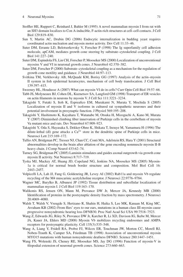
4 Neuronal Myosins 71
Stoffler HE, Ruppert C, Reinhard J, Bahler M (1995) A novel mammalian myosin I from rat withan SH3 domain localizes to Con A-inducible, F-actin-rich structures at cell–cell contacts. J CellBiol 129:819–830.
Sun Y, Martin AC, Drubin DG (2006) Endocytic internalization in budding yeast requirescoordinated actin nucleation and myosin motor activity. Dev Cell 11:33–46.
Suter DM, Errante LD, Belotserkovsky V, Forscher P (1998) The Ig superfamily cell adhesionmolecule, apCAM, mediates growth cone steering by substrate–cytoskeletal coupling. J CellBiol 141:227–240.
Suter DM, Espindola FS, Lin CH, Forscher P, Mooseker MS (2000) Localization of unconventionalmyosins V and VI in neuronal growth cones. J Neurobiol 42:370–382.
Suter DM, Forscher P (2000) Substrate–cytoskeletal coupling as a mechanism for the regulation ofgrowth cone motility and guidance. J Neurobiol 44:97–113.
Svitkina TM, Verkhovsky AB, McQuade KM, Borisy GG (1997) Analysis of the actin–myosinII system in fish epidermal keratocytes, mechanism of cell body translocation. J Cell Biol139:397–415.
Sweeney HL, Houdusse A (2007) What can myosin VI do in cells? Curr Opin Cell Biol 19:57–66.Tabb JS, Molyneaux BJ, Cohen DL, Kuznetsov SA, Langford GM (1998) Transport of ER vesicles
on actin filaments in neurons by myosin V. J Cell Sci 111:3221–3234.Takagishi Y, Futaki S, Itoh K, Espreafico EM, Murakami N, Murata Y, Mochida S (2005)
Localization of myosin II and V isoforms in cultured rat sympathetic neurones and theirpotential involvement in presynaptic function. J Physiol 569:195–208.
Takagishi Y, Hashimoto K, Kayahara T, Watanabe M, Otsuka H, Mizoguchi A, Kano M, MurataY (2007) Diminished climbing fiber innervation of Purkinje cells in the cerebellum of myosinVa mutant mice and rats. Dev Neurobiol 67:909–923.
Takagishi Y, Oda S, Hayasaka S, Dekker-Ohno K, Shikata T, Inouye M, Yamamura H (1996) Thedilute-lethal (dl) gene attacks a Ca2+ store in the dendritic spine of Purkinje cells in mice.Neurosci Lett 215:169–172.
Tullio AN, Bridgman PC, Tresser NJ, Chan CC, Conti MA, Adelstein RS, Hara Y (2001) Structuralabnormalities develop in the brain after ablation of the gene encoding nonmuscle myosin II-Bheavy chain. J Comp Neurol 433:62–74.
Turney SG, Bridgman PC (2005) Laminin stimulates and guides axonal outgrowth via growth conemyosin II activity. Nat Neurosci 8:717–719.
Tyska MJ, Mackey AT, Huang JD, Copeland NG, Jenkins NA, Mooseker MS (2005) Myosin-1a is critical for normal brush border structure and composition. Mol Biol Cell 16:2443–2457.
Volpicelli LA, Lah JJ, Fang G, Goldenring JR, Levey AI (2002) Rab11a and myosin Vb regulaterecycling of the M4 muscarinic acetylcholine receptor. J Neurosci 22:9776–9784.
Wagner MC, Barylko B, Albanesi JP (1992) Tissue distribution and subcellular localization ofmammalian myosin I. J Cell Biol 119:163–170.
Walikonis RS, Jensen ON, Mann M, Provance DW Jr, Mercer JA, Kennedy MB (2000)Identification of proteins in the postsynaptic density fraction by mass spectrometry. J Neurosci20:4069–4080.
Walsh T, Walsh V, Vreugde S, Hertzano R, Shahin H, Haika S, Lee MK, Kanaan M, King MC,Avraham KB (2002) From flies’ eyes to our ears, mutations in a human class III myosin causeprogressive nonsyndromic hearing loss DFNB30. Proc Natl Acad Sci USA 99:7518–7523.
Wang Z, Edwards JG, Riley N, Provance DW Jr, Karcher R, Li XD, Davison IG, Ikebe M, MercerJA, Kauer JA, Ehlers MD (2008) Myosin Vb mobilizes recycling endosomes and AMPAreceptors for postsynaptic plasticity. Cell 135(3):535–548.
Wang A, Liang Y, Fridell RA, Probst FJ, Wilcox ER, Touchman JW, Morton CC, Morell RJ,Noben-Trauth K, Camper SA, Friedman TB (1998) Association of unconventional myosinMYO15 mutations with human nonsyndromic deafness DFNB3. Science 280:1447–1451.
Wang FS, Wolenski JS, Cheney RE, Mooseker MS, Jay DG (1996) Function of myosin-V infilopodial extension of neuronal growth cones. Science 273:660–663.
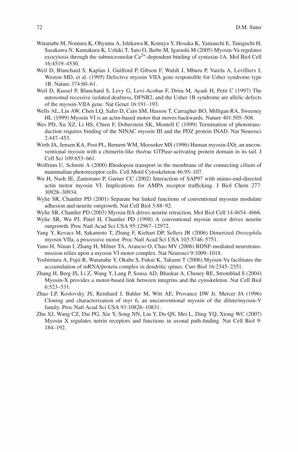
72 D.M. Suter
Watanabe M, Nomura K, Ohyama A, Ishikawa R, Komiya Y, Hosaka K, Yamauchi E, Taniguchi H,Sasakawa N, Kumakura K, Ushiki T, Sato O, Ikebe M, Igarashi M (2005) Myosin-Va regulatesexocytosis through the submicromolar Ca2+-dependent binding of syntaxin-1A. Mol Biol Cell16:4519–4530.
Weil D, Blanchard S, Kaplan J, Guilford P, Gibson F, Walsh J, Mburu P, Varela A, Levilliers J,Weston MD, et al. (1995) Defective myosin VIIA gene responsible for Usher syndrome type1B. Nature 374:60–61.
Weil D, Kussel P, Blanchard S, Levy G, Levi-Acobas F, Drira M, Ayadi H, Petit C (1997) Theautosomal recessive isolated deafness, DFNB2, and the Usher 1B syndrome are allelic defectsof the myosin-VIIA gene. Nat Genet 16:191–193.
Wells AL, Lin AW, Chen LQ, Safer D, Cain SM, Hasson T, Carragher BO, Milligan RA, SweeneyHL (1999) Myosin VI is an actin-based motor that moves backwards. Nature 401:505–508.
Wes PD, Xu XZ, Li HS, Chien F, Doberstein SK, Montell C (1999) Termination of phototrans-duction requires binding of the NINAC myosin III and the PDZ protein INAD. Nat Neurosci2:447–453.
Wirth JA, Jensen KA, Post PL, Bement WM, Mooseker MS (1996) Human myosin-IXb, an uncon-ventional myosin with a chimerin-like rho/rac GTPase-activating protein domain in its tail. JCell Sci 109:653–661.
Wolfrum U, Schmitt A (2000) Rhodopsin transport in the membrane of the connecting cilium ofmammalian photoreceptor cells. Cell Motil Cytoskeleton 46:95–107.
Wu H, Nash JE, Zamorano P, Garner CC (2002) Interaction of SAP97 with minus-end-directedactin motor myosin VI. Implications for AMPA receptor trafficking. J Biol Chem 277:30928–30934.
Wylie SR, Chantler PD (2001) Separate but linked functions of conventional myosins modulateadhesion and neurite outgrowth. Nat Cell Biol 3:88–92.
Wylie SR, Chantler PD (2003) Myosin IIA drives neurite retraction. Mol Biol Cell 14:4654–4666.Wylie SR, Wu PJ, Patel H, Chantler PD (1998) A conventional myosin motor drives neurite
outgrowth. Proc Natl Acad Sci USA 95:12967–12972.Yang Y, Kovacs M, Sakamoto T, Zhang F, Kiehart DP, Sellers JR (2006) Dimerized Drosophila
myosin VIIa, a processive motor. Proc Natl Acad Sci USA 103:5746–5751.Yano H, Ninan I, Zhang H, Milner TA, Arancio O, Chao MV (2006) BDNF-mediated neurotrans-
mission relies upon a myosin VI motor complex. Nat Neurosci 9:1009–1018.Yoshimura A, Fujii R, Watanabe Y, Okabe S, Fukui K, Takumi T (2006) Myosin-Va facilitates the
accumulation of mRNA/protein complex in dendritic spines. Curr Biol 16:2345–2351.Zhang H, Berg JS, Li Z, Wang Y, Lang P, Sousa AD, Bhaskar A, Cheney RE, Stromblad S (2004)
Myosin-X provides a motor-based link between integrins and the cytoskeleton. Nat Cell Biol6:523–531.
Zhao LP, Koslovsky JS, Reinhard J, Bahler M, Witt AE, Provance DW Jr, Mercer JA (1996)Cloning and characterization of myr 6, an unconventional myosin of the dilute/myosin-Vfamily. Proc Natl Acad Sci USA 93:10826–10831.
Zhu XJ, Wang CZ, Dai PG, Xie Y, Song NN, Liu Y, Du QS, Mei L, Ding YQ, Xiong WC (2007)Myosin X regulates netrin receptors and functions in axonal path-finding. Nat Cell Biol 9:184–192.